báo cáo hóa học:"Effect of ethanol on innate antiviral pathways and HCV replication in human liver cells" potx
Bạn đang xem bản rút gọn của tài liệu. Xem và tải ngay bản đầy đủ của tài liệu tại đây (460.71 KB, 12 trang )
BioMed Central
Page 1 of 12
(page number not for citation purposes)
Virology Journal
Open Access
Research
Effect of ethanol on innate antiviral pathways and HCV replication
in human liver cells
Courtney R Plumlee
1,2
, Catherine A Lazaro
3
, Nelson Fausto
4
and
Stephen J Polyak*
5
Address:
1
Department of Laboratory Medicine, University of Washington, Seattle, USA,
2
Department of Biological Sciences, Columbia University,
New York, NY,
3
Department of Pathology, University of Washington, Seattle, USA,
4
Department of Pathology, University of Washington, Seattle,
USA and
5
Departments of Laboratory Medicine, Microbiology and Pathobiology, University of Washington, Seattle, USA
Email: Courtney R Plumlee - ; Catherine A Lazaro - ;
Nelson Fausto - ; Stephen J Polyak* -
* Corresponding author
HCVIFNvirus-host interactionssignal transductionalcohol
Abstract
Alcohol abuse reduces response rates to IFN therapy in patients with chronic hepatitis C. To
model the molecular mechanisms behind this phenotype, we characterized the effects of ethanol
on Jak-Stat and MAPK pathways in Huh7 human hepatoma cells, in HCV replicon cell lines, and in
primary human hepatocytes. High physiological concentrations of acute ethanol activated the Jak-
Stat and p38 MAPK pathways and inhibited HCV replication in several independent replicon cell
lines. Moreover, acute ethanol induced Stat1 serine phosphorylation, which was partially mediated
by the p38 MAPK pathway. In contrast, when combined with exogenously applied IFN-α, ethanol
inhibited the antiviral actions of IFN against HCV replication, involving inhibition of IFN-induced
Stat1 tyrosine phosphorylation. These effects of alcohol occurred independently of i) alcohol
metabolism via ADH and CYP2E1, and ii) cytotoxic or cytostatic effects of ethanol. In this model
system, ethanol directly perturbs the Jak-Stat pathway, and HCV replication.
Infection with Hepatitis C virus is a significant cause of morbidity and mortality throughout the
world. With a propensity to progress to chronic infection, approximately 70% of patients with
chronic viremia develop histological evidence of chronic liver diseases including chronic hepatitis,
cirrhosis, and hepatocellular carcinoma. The situation is even more dire for patients who abuse
ethanol, where the risk of developing end stage liver disease is significantly higher as compared to
HCV patients who do not drink [1,2].
Recombinant interferon alpha (IFN-α) therapy produces sustained responses (ie clearance of
viremia) in 8–12% of patients with chronic hepatitis C [3]. Significant improvements in response
rates can be achieved with IFN plus ribavirin combination [4-6] and pegylated IFN plus ribavirin
[7,8] therapies. However, over 50% of chronically infected patients still do not clear viremia.
Moreover, HCV-infected patients who abuse alcohol have extremely low response rates to IFN
therapy [9], but the mechanisms involved have not been clarified.
Published: 02 December 2005
Virology Journal 2005, 2:89 doi:10.1186/1743-422X-2-89
Received: 06 September 2005
Accepted: 02 December 2005
This article is available from: />© 2005 Plumlee et al; licensee BioMed Central Ltd.
This is an Open Access article distributed under the terms of the Creative Commons Attribution License ( />),
which permits unrestricted use, distribution, and reproduction in any medium, provided the original work is properly cited.
Virology Journal 2005, 2:89 />Page 2 of 12
(page number not for citation purposes)
MAPKs play essential roles in regulation of differentiation, cell growth, and responses to cytokines,
chemokines and stress. The core element in MAPK signaling consists of a module of 3 kinases,
named MKKK, MKK, and MAPK, which sequentially phosphorylate each other [10]. Currently, four
MAPK modules have been characterized in mammalian cells: Extracellular Regulated Kinases (ERK1
and 2), Stress activated/c-Jun N terminal kinase (SAPK/JNK), p38 MAP kinases, and ERK5 [11].
Interestingly, ethanol modulates MAPKs [12]. However, information on how ethanol affects
MAPKs in the context of innate antiviral pathways such as the Jak-Stat pathway in human cells is
extremely limited.
When IFN-α binds its receptor, two receptor associated tyrosine kinases, Tyk2 and Jak1 become
activated by phosphorylation, and phosphorylate Stat1 and Stat2 on conserved tyrosine residues
[13]. Stat1 and Stat2 combine with the IRF-9 protein to form the transcription factor interferon
stimulated gene factor 3 (ISGF-3), which binds to the interferon stimulated response element
(ISRE), and induces transcription of IFN-α-induced genes (ISG). The ISGs mediate the antiviral
effects of IFN. The transcriptional activities of Stats 1, 3, 4, 5a, and 5b are also regulated by serine
phosphorylation [14]. Phosphorylation of Stat1 on a conserved serine amino acid at position 727
(S727), results in maximal transcriptional activity of the ISGF-3 transcription factor complex [15].
Although cross-talk between p38 MAPK and the Jak-Stat pathway is essential for IFN-induced ISRE
transcription, p38 does not participate in IFN induction of Stat1 serine phosphorylation [14,16-19].
However, cellular stress responses induced by stimuli such as ultraviolet light do induce p38 MAPK
mediated Stat1 S727 phosphorylation [18].
In the current report, we postulated that alcohol and HCV proteins modulate MAPK and Jak-Stat
pathways in human liver cells. To begin to address these issues, we characterized the interaction
of acute ethanol on Jak-Stat and MAPK pathways in Huh7 cells, HCV replicon cells lines, and
primary human hepatocytes.
Materials and methods
Cells and chemicals
Human hepatoma Huh7 cells were grown in DMEM con-
taining 10% FBS, 1× penicillin, streptomycin, fungizone,
10mM L-glutamine, and 1× non-essential amino acids (all
reagents were from Invitrogen; Carlsbad, CA). BB7 cells
are derived from Huh7 cells and support the replication of
a subgenomic HCV replicon containing a S2204I adaptive
mutation in the NS5A gene [20]. FL-Neo cells are a stable
Huh7 derived cell line containing a genomic length HCV
replicon with the S2204I mutation in NS5A and a P1496L
mutation in NS3. BB7 and FL-Neo cells were obtained
from Apath, LLC. Subgenomic replicon cell lines 9–13
and 5-15-9-2-3 (referred to as 5–15 in this paper) contain-
ing different adaptive mutations [21-23] were kindly pro-
vided by Dr. Ralf Bartenschalger. Replicon cell lines were
maintained in Huh7 media containing 400 µg/ml of
G418 (Calbiochem; San Diego, CA). Primary human fetal
hepatocytes were isolated and grown in chemically
defined serum free medium as described [24]. Primary
hepatocyte cultures were analyzed within 2 days of isola-
tion. Cells were maintained in humidified incubators at
37°C with 5% CO
2
. Ethanol (AAPER; Shelbyville, KY) at
concentrations of 0–200 mM, was added to cells at the
same time as IFN-α (Sigma, St. Louis, MO). Relative to
untreated cells, ethanol did not induce any cytotoxic or
growth inhibitory effects on any of the cell types at any of
the doses tested (see Additional File 1). MAPK inhibitors
UO126, PD98059, and SB203508, used to inhibit p42/
44, MEK1, and p38 MAPK pathways, respectively, were
solubilized in DMSO and obtained from Calbiochem.
ADH and CYP2E1 inhibitors 4-methylpyrazole (4-MP)
and diallylsulfide (DAS) [25], were obtained from Sigma
and solubilized in DMSO. In all experiments, the final
concentration of DMSO was below 0.2%, so as to prevent
DMSO inhibition of CYP2E1 [26].
Transfection
The day prior to transfection, 2 × 10
5
cells were plated in
12-well tissue culture plates. Endotoxin free plasmid DNA
was purified (Endofree kit, Qiagen; Valencia, CA), and
was introduced into cells with lipofectamine 2000 accord-
ing to manufacturer's recommendations (Invitrogen).
Transfection efficiency was monitored by including 0.5 µg
of plasmid pQ150 (provided by Dr. Jeffery Vieira), which
expresses GFP under control of the constitutive EF-1α pro-
moter. Prior to harvesting protein lysates, cells expressing
GFP were visualized by fluorescence microscopy and the
transfection efficiency calculated by determining the per-
centage of green cells to total cells. For reporter gene stud-
ies, 0.5 µg of the luciferase gene under control of the
interferon stimulated response element (ISRE) in plasmid
pISRE-luc (ISRE promoter; Stratagene; La Jolla, CA), was
transfected into cells in duplicate or triplicate. In certain
Virology Journal 2005, 2:89 />Page 3 of 12
(page number not for citation purposes)
experiments a dominant negative p38 (p38 AGF) express-
ing plasmid [27], provided by Dr. Michael Kracht, was
transfected into cells. Twenty-four hours post-transfec-
tion, ethanol, either alone or in combination with IFN
was added directly to cells. Six hours later, luciferase activ-
ity was measured on cell lysates and normalized for trans-
fection efficiency and total protein content.
Western blot analysis
Protein lysates were quantitated by BCA Protein Assay
(Pierce; Rockford, IL) and equal amounts (typically 20–30
µg) of total protein was separated on 4–20% SDS-PAGE
gels. For detection of phosphorylated Stat1 proteins, Stat1
phosphotyrosine (Y701) and phosphoserine (S727) spe-
cific antibodies were used (Zymed-Invitrogen). Total Stat1
was detected using a polyclonal antibody (Zymed or
Santa Cruz Biotechnology; Santa Cruz, CA). Total and
phosphorylated forms of p42/44 (ERK2/1), and p38
MAPK were detected with specific antisera (Cell Signaling;
Beverly, MA). Cytochrome P4502E1 (CYP2E1) was
detected using polyclonal rabbit antiserum (provided by
Arthur Cederbaum), while alcohol dehydrogenase (ADH)
was detected with a mouse monoclonal antibody
(AbCam; Cambridge, MA). HCV proteins were detected
using random, de-identified HCV infected patient serum,
as described [28]. Infected serum was inactivated by add-
ing triton X-100 to 1% prior to use.
Kinase assays
The activity of p38 MAPK in Huh7 cells was assessed via
kinase assay using a kit (Cell Signaling). Briefly, cell
lysates were immunoprecipitated with antibodies that rec-
ognize the phosphorylated form of p38 MAPK. After strin-
gent washing, recombinant ATF-2 protein, a substrate for
p38, was added to immunoprecipitates and incubated for
30 minutes according to manufacturer's specifications.
Phosphorylated protein ATF-2 was detected by western
blot.
HCV RNA quantitation
HCV RNA was quantitated by real time RT-PCR, using a
modified version of a recent procedure [29]. Total cellular
RNA was isolated from replicon cells using a commercial
kit (Qiagen). Ten nanograms of RNA was added to wells
of a 384 well plate containing the EZ RT-PCR master mix
(Perkin Elmer; Wellesley, MA). Samples were run on an
ABI HT7900 real time RT-PCR machine. The RT reaction
consisted of 50°C for 2 minutes followed by 60°C for 30
minutes. The PCR consisted of an initial denaturation of
2 minutes at 95°C, then 45 cycles of 95°C for 15 seconds
followed by simultaneous annealing/extension at 60°C
for 1 minute. For each run, dilutions of BB7 plasmid DNA
(precisely quantitated using the PicoGreen DNA quantita-
tion kit (Invitrogen)) ranging from 0–10
7
copies per tube
were run in triplicate to generate a standard curve, which
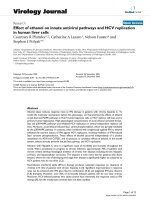
Effect of acute ethanol on the Jak-Stat pathwayFigure 1
Effect of acute ethanol on the Jak-Stat pathway. A, high physiological doses of ethanol activate the ISRE. Huh7 cells were trans-
fected with 0.7 µg of pISRE-luc, and 24 hours later, cells were stimulated with ethanol at the indicated concentrations. Protein
lysates were assayed for luciferase activity 6 hours later. Error bars represent standard deviations. The experiment was
repeated 6 times with similar results. B, acute ethanol induces Stat1 serine phosphorylation. Huh7 cells were left as untreated
controls (C) or treated with 1,000 U/ml of IFN-α (IFN), or 100 mM ethanol (EtOH). Twenty minutes later, equal amounts of
whole cell protein extracts were separated by SDS-PAGE, and blotted for phosphorylated Stat1 S727 (toppanel), Stat1 Y701
(second panel), Stat2 Y690 (third panel), and total forms of the Stat1 protein (lower panel). The figure is representative of 3
independent experiments, which yielded similar results.
0
500
1000
1500
2000
2500
3000
3500
0 25 50 100 200
RLU
EtOH (mM):
A.
B.
Stat1 S727
Stat2 Y690
Stat1 Y701
C IFN EtOH
Stat1 total
Virology Journal 2005, 2:89 />Page 4 of 12
(page number not for citation purposes)
served as a reference to calculate HCV RNA copy number
based on the cycle threshold (C
t
). The HCV RNA copy
number is reported as copies per 10 ng total cellular RNA.
Additional controls included reactions lacking template
as well as RNA from Huh7 cells. For both negative con-
trols, these samples were always negative for HCV RNA.
ADH enzyme assay
Cells were harvested in PBS and whole cell extracts pre-
pared via sonication. Aliquots of protein extracts were
mixed with 0.1 M glycine pH 10.0 buffer, 2.4 mM β-nico-
tinamide adenine dinucleotide, and 33 mM ethanol, and
conversion of NAD to NADH+ was monitored with a
spectrophotometer at a wavelength of 340 nm. All rea-
gents for the assay were from Sigma. As a positive control,
purified human ADH (provided by Dr. Carol Stone) was
also run in the assay.
Statistics
Differences between means of luciferase readings were
compared using a Student's T-test. A p-value of <0.05 was
considered significant. For western blots, data were ana-
lyzed with Image J, a software version of NIH Image for
the Macintosh OS × operating system. Changes in protein
levels were normalized to control western blots and
expressed as fold or percent change relative to controls.
Results
Effect of acute ethanol on Jak-Stat pathway
Figure 1A depicts the effects of acute ethanol on the ISRE
promoter in Huh7 cells. Ethanol did not appear to have
significant effects on ISRE activity at 25 and 50 mM con-
centrations. However, at concentrations of 100 and 200
mM, ethanol caused statistically significant 3.0 (p = 0.03)
and 5.0 (p < 0.001) fold increases in ISRE reporter gene
activity, as compared to cells not treated with ethanol. The
data suggest that high physiological doses of acute etha-
nol activate the ISRE, an IFN responsive promoter.
To investigate this regulation further, we analyzed levels
of phosphorylated Stat1 and Stat2, which are obligatory
steps for ISRE activation. Stat1 and Stat2 activation
involves phosphorylation on conserved tyrosines at
amino acid positions 701 and 690, respectively, while
phosphorylation of Stat1 also occurs on conserved serine
amino acid at position 727 and provides maximal tran-
scriptional activation [15]. Figure 1B depicts the levels of
Stat1 S727 (top panel), Stat1 Y701 (second panel), and
Stat2 Y690 (third panel), and the total levels of Stat1 pro-
tein (fourth panel) in Huh7 cells. Phosphorylation of
Stat1 on S727 was induced by IFN-α or 100 mM ethanol.
Stat1 Y701 and Stat2 Y690 phosphorylation occurred
with IFN treatment, whereas no effect was observed with
100 mM ethanol. The differences in Stat phosphorylation
were not due to differences in the amount of Stat1 pro-
tein, since total Stat1 protein levels were equivalent (Fig-
ure 1B, lower panel). Similar results were also observed
for primary human fetal hepatocytes (see Additional File
2) and HCV replicon cells (data not shown).
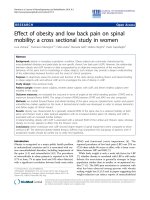
Effect of acute ethanol on the p38 MAPK pathway
Since MAPKs are modulated by ethanol [12] and p38
MAPK is important in ISRE transcription [16,19], we next
examined the effect of acute alcohol on the p38 MAPK
pathway. Figure 2 depicts the effects of acute ethanol on
the p38 MAPK pathway in Huh7 cells and primary human
fetal hepatocytes. In these experiments, p38 kinase assays
were performed. As shown in the upper panel of Figure 2,
acute exposure of Huh7 cells to 25, 50, and 100 mM eth-
anol resulted in 61, 27, and 150-fold activation of p38
kinase activity, respectively, detected as an increase in
recombinant ATF-2 phosphorylation, a natural substrate
for p38 MAPK. The middle panel depicts the amounts of
total p38 protein added to each immunoprecipitate. Acute
ethanol at 25, 50, and 100 mM doses also activated p38
MAPK to levels 2.1, 2.2, and 5.2-fold in primary fetal
human hepatocytes, relative to untreated cells (Figure 2,
lower panel), although basal levels of MAPK were higher
in these cells. The data suggest that acute ethanol activates
p38 MAPK pathways in primary human fetal hepatocytes
and Huh7 cells. Acute ethanol also activated p42/44
MAPK and SAPK in Huh7 (see Additional File 3) and BB7
replicon cells (data not shown).
Ethanol activates p38 MAPK in human liver cell culturesFigure 2
Ethanol activates p38 MAPK in human liver cell cultures.
Huh7 cells or primary human fetal hepatocytes (HFH) were
left as untreated controls (C) or were treated with anisomy-
cin (A) as a positive control, or with 25, 50, or 100 mM etha-
nol for 30 minutes. Active p38 MAPK was
immunoprecipitated from cell lysates and kinase activity
measured by phosphorylation of ATF-2. The figure is repre-
sentative of 2 experiments that produced similar results.
ATF-2 PO4
p38 total
C A 25 50 100
EtOH
Huh7
HFH
ATF-2 PO4
Virology Journal 2005, 2:89 />Page 5 of 12
(page number not for citation purposes)
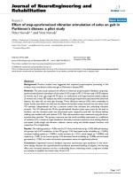
Acute alcohol stimulation of the Jak-Stat pathway involves
MAPKs
Since the p38 MAPK pathway cross-talks to the Jak-Stat
pathway [16,19], we investigated the effect of acute alco-
hol on ISRE transcriptional activity and Stat1 phosphor-
ylation in the presence of MAPK inhibitors and dominant
negative mutants. We performed ISRE reporter gene
experiments with IFN-α and alcohol treatments in the
presence of the p38 MAPK inhibitor, SB203508. As shown
in Figure 3A, SB203508 inhibited ethanol stimulation of
the ISRE by up to 40%. Figure 3B presents related experi-
ments examining the effect of small molecule inhibitors
on ethanol induction of Stat1 serine phosphorylation.
Huh7 cells were treated for 2 hours in the presence of
DMSO carrier, UO126 (a p42/44 MAPK inhibitor),
PD98059 (a MEK1 inhibitor) and SB203508 (a p38
inhibitor). Cells were then stimulated with 100 mM etha-
nol for 20 minutes. Ethanol induction of Stat1 serine
phosphorylation was 90% inhibited by SB203508. Huh7
cells were also transfected with a vector expressing a dom-
inant negative p38 protein (p38 AGF) [27], and the effect
on ethanol induction of Stat1 serine phosphorylation was
investigated. As shown in Figure 3C, expression of the p38
AGF dominant negative mutant abrogated both basal and
ethanol induced Stat1 serine phosphorylation. Together,
the data suggest that acute ethanol activation of p38
MAPK is partially involved in induction of ISRE transcrip-
tion and Stat1 serine phosphorylation.
Effect Of acute alcohol on HCV replicons
Figure 4 depicts the effects of acute ethanol on HCV repli-
cation. BB7 cells were treated once with 0, 25, 50, or 100
mM of alcohol, or 20 U/ml of IFN-α. HCV RNA was quan-
titated using real time RT-PCR on equal amounts (10 ηg)
of total cellular RNA isolated 72 hours after drug treat-
ment (Figure 4A; left panel). As expected, IFN induced a
significant 66-fold inhibition of HCV RNA at this time
point. A single administration of 25 mM ethanol had no
significant effect on HCV RNA replication, although a
slight increase was noted. In contrast, 50 mM and 100
mM ethanol doses induced statistically significant inhibi-
tion of HCV RNA synthesis (p = 0.02 and p = 0.001,
Involvement of p38 MAPK in ethanol induction of ISRE transcription and Stat1 serine phosphorylationFigure 3
Involvement of p38 MAPK in ethanol induction of ISRE transcription and Stat1 serine phosphorylation. Panel A, Huh7 cells
were transfected with 0.7 µg pISRE-luc, and 22 hours later, cells were incubated for 2 hours at the indicated µM concentra-
tions of SB203508 (a p38 inhibitor), followed by 100 mM ethanol. Cell lysates were harvested 6 hours later and luciferase
results were normalized to amounts of total cellular protein. Error bars represent standard deviations. The experiment was
repeated 4 times with identical results. B, Huh7 cells were treated for 2 hours in the presence of 50 µM of various MAPK
inhibitors, and stimulated with 100 mM alcohol for 20 minutes. Whole cell protein extracts were blotted for the serine phos-
phorylated form (S727) or total form of Stat1. The experiment was repeated twice, yielding similar results. C, Huh7 cells were
transfected with control vector plasmid (Vec) or a plasmid expressing a dominant negative mutant p38 protein (p38 AGF).
Twenty-four hours later, cells were not treated or treated for 20 minutes with 100 mM ethanol. Levels of S727 and total Stat1
and transfected p38 proteins were determined by western blot. The figure is representative of 2 independent experiments that
produced similar results.
Stat1 S727
DMSO
UO126
PD980
SB203
100 mM EtOH+
Stat1 Total
B.
Stat1 S727
Stat1 Total
p38
EtOH: - + - +
Vec p38 AGF
C.
B.
SB203: 0 50 100 0 50 100
0
200
400
600
800
1000
1200
1400
+++
RLU
EtOH:
A.
Ctrl
Virology Journal 2005, 2:89 />Page 6 of 12
(page number not for citation purposes)
respectively). The doses of alcohol used did not affect BB7
cell growth, viability, or morphology (data not shown).
HCV NS3 and NS5A protein expression was also inhibited
in a dose-dependent fashion (1.7–5.4 fold) by ethanol
(Figure 4B, right panel).
Since replicons acquire adaptive mutations [20,23] and it
is also possible that the cells acquire genetic or epigenetic
mutations during the G418 selection process and contin-
uous culturing [30,31], we questioned whether the previ-
ous data derived from a single replicon cell line was
typical of other replicons. We therefore examined the
effect of acute ethanol on HCV RNA and protein synthesis
Effect of acute ethanol on HCV replicationFigure 4
Effect of acute ethanol on HCV replication. BB7 replicon cells were treated once with 0, 25, 50, or 100 mM ethanol or 20 U/
ml of IFN, and RNA and protein was harvested 72 hours later. A, HCV RNA copy number was determined by quantitative real
time RT-PCR. The HCV RNA copy number is reported as copies per 10 ng total cellular RNA. Error bars represent standard
deviations. B, HCV protein expression in BB7 cells treated with 0, 25, 50, and 100 mM ethanol, and control Huh7 cells. The
positions of Stat1, HCV NS3 and NS5A proteins are indicated. The experiment was repeated twice with similar results.
0
50000
100000
150000
200000
250000
300000
350000
0 25 50 100 IFN
HCV RNA (copies/10ng RNA)
EtOH (mM):
A.
0 25 50 100 Huh7
BB7
NS3
NS5A
Stat1
B.
Acute ethanol inhibits the replication of other HCV replicon linesFigure 5
Acute ethanol inhibits the replication of other HCV replicon lines. 9–13, and 5–15-replicon cell lines were treated with 0, 100,
or 200 mM of ethanol, and HCV RNA (panel A) and protein (panel B) was quantitated by real time RT-PCR and western blot
analysis as described above. B., quantitation of changes in HCV NS3 and NS5A protein expression. Scanned blots were ana-
lyzed with Image J. For each lane, pixel intensities of NS3 and NS5A bands were normalized to the total Stat1 pixel intensity,
and the percent change relative to untreated cells was calculated.
0
10000
20000
30000
40000
50000
60000
70000
80000
90000
0 100 200
EtOH (mM)
HCV RNA (Copies/10ng RNA)
5-15
9-13
B.
A.
0
10
20
30
40
50
60
70
80
90
100
0 100 200
EtOH (mM)
Relative Pixel Intensity (%)
NS3 (5-15)
NS3 (9-13)
NS5A (5-15)
NS5A (9-13)
Virology Journal 2005, 2:89 />Page 7 of 12
(page number not for citation purposes)
in 2 additional replicon lines, 9–13 and 5–15, obtained
independently from BB7 cells [21-23]. Cells were treated
with 100 or 200 mM of ethanol, and HCV RNA and pro-
tein was assessed 72 hours later. As shown in Figure 5,
although the basal level of HCV RNA differed considera-
bly between 5–15 and 9–13 replicon cells, both doses of
ethanol inhibited HCV RNA and protein production by
up to 50%. Together the data suggest that acute ethanol
inhibits the replication of several independent cell lines
that support robust HCV replication. The replication of a
genomic length replicon cell line, FL-Neo, was also inhib-
ited by acute ethanol (see Additional File 4).
Acute alcohol inhibits the IFN-
α
induced antiviral response
towards HCV
We examined the combined effects of alcohol and IFN-α
treatment on the Jak-Stat pathway. Huh7 cells were left
untreated, or treated with IFN, or IFN plus ethanol. Figure
6A demonstrates that ethanol treatment inhibited IFN-α
induction of Stat1 tyrosine phosphorylation. To investi-
gate the effect of ethanol on the IFN induced antiviral
response, BB7 replicon cells were treated with or without
100 mM ethanol in the presence of varying doses of IFN-
α. HCV protein levels were analyzed by western blot 48
hours later. As shown in Figure 6B, in the absence of eth-
anol, IFN-α inhibited HCV protein in a dose dependent
fashion, and this coincided with a dose-dependent
increase in total Stat1 protein, a known ISG. When cells
were treated with a single dose of 100 mM ethanol,
increases in HCV NS3 and NS5A proteins were detected at
IFN doses of 10, 20 and 100 U/ml relative to cells treated
with IFN alone. Alcohol also inhibited IFN induced up-
regulation of Stat1 at these concentrations. However, at
IFN concentrations of 0, 0.1 and 1 U/ml, ethanol
appeared to decrease the amount of HCV NS3 and NS5A
protein expression, consistent with ethanol's IFN stimula-
tory and anti-HCV effects presented above. Figure 6C
presents a quantitative summary of the protein data based
on pixel intensity, and clearly demonstrates that IFN dose-
dependently inhibits NS3 and NS5A protein expression
Acute alcohol inhibits the antiviral actions of IFNFigure 6
Acute alcohol inhibits the antiviral actions of IFN. A, Huh7 cells were left untreated (Ctrl), or treated with 1,000 U/ml of IFN-
α alone (IFN) or with IFN-α plus 100 mM ethanol (IFN+EtOH). Proteins were probed for Stat1 Y701 (top panel), and total
Stat1 proteins (lower panel). B, BB7 replicon cells were treated with or without 100 mM ethanol, followed immediately by 0,
0.1, 1, 10, 20, 100 IU/ml of IFN-α, and whole cell protein extracts were harvested 48 hours later. Equal amounts of total cellu-
lar protein were analyzed for the presence of Stat1, HCV NS5A and NS3 proteins, and p42/44 MAPK by western blot analysis.
C, quantitation of HCV protein expression shown in panel B. For each lane, pixel intensities of NS3 and NS5A bands were nor-
malized to the total p42/44 pixel intensity, and the fold decrease relative to untreated cells was calculated. The figure is repre-
sentative of 2 independent experiments that produced identical results.
A.
0.01
0.1
1
10
100
1000
0 0.1 1 10 20 100
IFN
(
U
/
ml
)
Fold Decrease
NS3 (IFN)
NS3 (IFN+EtOH)
NS5A (IFN)
NS5A (IFN+EtOH)
B.
C.
Stat1 Y701
Stat1 total
Ctrl
Stat1 total
NS3
NS5A
p42/44
IFN- (U/ml): 0 0.1 1 10 20 100 0 0.1 1 10 20 100
Control +EtOH
IFN
IFN+EtOH
Virology Journal 2005, 2:89 />Page 8 of 12
(page number not for citation purposes)
by 10–100 fold. In the absence of IFN, acute ethanol
inhibits NS3 and NS5A protein expression by 10-fold.
However, acute ethanol prevents IFN-α-mediated clear-
ance of HCV proteins. Similar effects were observed for
HCV RNA production (data not shown). The data indicate
that ethanol inhibits the antiviral actions of exogenously
added IFN.
Expression of alcohol metabolizing enzymes in human liver
cell cultures
Since ethanol can exert differential effects on cells depend-
ing on whether it is metabolized or not [32], the expres-
sion and activity of ADH and CYP2E1, the major ethanol-
metabolizing enzymes, was examined in Huh7 and repli-
con cells. Figure 7A depicts western blot analysis of ADH
(top panel), CYP2E1 (middle panel), and Stat1 (lower
panel) protein expression in Huh7, BB7, 9–13, 5–15, and
FL-Neo cells. Immortalized human hepatocytes (HH2),
primary human fetal hepatocytes (HFH), and purified
human ADH served as positive controls for ADH, while
lysate from cells that were infected with a baculovirus
expressing human CYP2E1 [33], as well as purified
CYP2E1, served as controls for CYP2E1. All replicon and
Huh7 cultures expressed very low to undetectable levels of
ADH and CYP2E1 protein. To determine if Huh7 cells
expressed a functional ADH enzyme, ADH enzyme assays
were performed using purified human ADH as a positive
control. Figure 7B demonstrates that purified ADH
Characterization of ethanol metabolizing enzymes in human liver cell culturesFigure 7
Characterization of ethanol metabolizing enzymes in human liver cell cultures. A, western blot analysis of ADH1 and CYP2E1
expression levels in Huh7, BB7, 9–13, 5–15, and FL-Neo cells. Positive controls for ADH included primary human fetal hepato-
cytes (HFH) [24], and a well differentiated immortalized human liver cell line, HH2 (developed in NF's lab), while controls for
CYP2E1 expression included baculovirus expressed CYP2E1 and purified CYP2E1. Western blots were probed with a mono-
clonal antibody against human ADH, and polyclonal rabbit antiserum against CYP2E1 and Stat1. B, ADH enzyme activity. Huh7
cells were harvested in PBS and whole cell protein extracts prepared via sonication. Conversion of NAD to NADH+ was mon-
itored at a wavelength of 340 nm as described in the Materials and Methods. Purified ADH served as a positive control for
ADH activity. C, effect of CYP2E1 and ADH inhibition on ethanol activation of the ISRE. Huh7 cells in 96 well plates were
transfected in triplicate with 50 ηg of ISRE-luc and 12 hours later, were treated with 5 mM of the ADH inhibitor 4-MP and 10
mM of the CYP2E1 inhibitor DAS for an additional 12 hours. Cells were also separately exposed to 0.1% DMSO, as an addi-
tional control for possible solvent effects. Cells were then treated with 0, 100, or 200 mM ethanol, before luciferase activity
was measured by BriteLite assay. Error bars represent standard deviations. The experiments were repeated twice with identi-
cal results.
ADH
0
0.01
0.02
0.03
0.04
0.05
0.06
0.07
0.08
0 20 40 60 80 100 120 140 160 180 200 220 240
Time (seconds)
A340
ADH
Buffer
Huh7
A.
Huh7
BB7
9-13
5-15
FL-Neo
HH2
HFH
rhADH
Bac
-CYP2E1
rhCYP2E1
CYP2E1
Stat1
B.
C.
0
1000
2000
3000
4000
5000
6000
7000
8000
9000
10000
0 100 200 0 100 200 0 100 200 0 100 200
RLU
Control DMSO 4-MP DAS
Virology Journal 2005, 2:89 />Page 9 of 12
(page number not for citation purposes)
showed a linear increase in absorbance over time, while
buffer alone remained at background levels. In contrast,
Huh7 cells expressed minimal ADH enzymatic activity,
with only slight increases over background detected after
3 minutes. Figure 7C demonstrates that ethanol induction
of ISRE transcription was not affected in the presence of
the ADH and CYP2E1 inhibitors, 4-MP and DAS. Note
that the concentrations of 4-MP (5 mM) and DAS (10 µM)
used in this assay were derived from a previous study [25].
At these concentrations, 4-MP and DAS had no effects on
cell viability or proliferation (data not shown). The data
indicate that Huh7 and replicon cells express low to unde-
tectable levels of ADH and CYP2E1 proteins, and further
suggest that the effects of ethanol on innate antiviral path-
ways is not due to ethanol metabolism via ADH or
CYP2E1 in this model system.
Discussion
In the current study, it was demonstrated that high physi-
ological doses of acute ethanol induces Stat1 serine phos-
phorylation and ISRE transcription. Given alone, ethanol
appears to inhibit HCV replication in several independent
replicon cell lines, and this is in part mediated by a Jak-
Stat transduced antiviral response. In contrast, in the pres-
ence of exogenously added IFN-α, ethanol partially inhib-
its the antiviral actions of IFN-α, involving inhibition of
IFN-α induced Stat1 tyrosine phosphorylation. Analysis
of the effects of chronic ethanol administration on basal
and IFN-α induced signaling responses is currently in
progress.
We also found that acute exposure of human liver cells to
physiological doses of ethanol activates the IFN system via
Summary of effects of acute ethanol on HCV replicationFigure 8
Summary of effects of acute ethanol on HCV replication. Ethanol effects in this system are independent of ethanol metabolism
and as such may involve ethanol-induced perturbations in cell membranes, such as membrane fluidity. Left side, acute ethanol
activates p38 MAPK which leads to Stat1 serine phosphorylation, Jak-Stat signaling and inhibition of HCV replication. Activated
Stat1 may be involved in ISRE transcription but it is possible that other ISRE binding transcription factors such as Stat3 are
involved in this process. Right side, ethanol inhibits the antiviral actions of exogenously applied IFN and this involves inhibition
of IFN-induced Stat1 tyrosine phosphorylation, decreased Jak-Stat signaling and increased HCV replication in the presence of
IFN. Inhibition of Jak-Stat signaling may involve ethanol perturbation of IFN-α induced changes in membrane fluidity, inhibition
of IFN binding to its receptor, direct inhibition of Jak kinases, and/or induction of negative regulators of the Jak-Statpathway
such as SOCS proteins.
p38 MAPK Stat1 S727 Stat1 Y701
Antiviral Signaling
HCV Replication
Antiviral Signaling
HCV Replication
IFNAR Inhibition?
Jak Inhibition?
SOCS Induction?
IFN
ETHANOL
-membrane proteins?
-fluidity?
Other Signaling Proteins?
Virology Journal 2005, 2:89 />Page 10 of 12
(page number not for citation purposes)
the MAPK pathway. The data suggest that ethanol induces
cross talk between the p38 MAPK and Jak-Stat pathway
(Figure 8). Additional evidence for cross talk between
these pathways derives from a study indicating that ERK2
binds to the α-chain of the IFN α/β receptor and STAT1
[34], and JAK2 may be required for MAP kinase pathway
activation [35]. Furthermore, HCV proteins such as NS5A
interact with and modulate MAPK and related pathways
such as Grb2, Ras-ERK, and phosphoinositol 3 kinase
(PI3K) [36-40]. However, p38 kinase activity, which is
important in IFN-α and IFN-γ induced transcription, is
not involved in IFN induced Stat1 serine phosphorylation
[16,19]. Thus, induction of Stat1 serine phosphorylation
by ethanol described in the current report may be mecha-
nistically similar to UV-stress induced activation of Stat1
by p38 MAPK [18].
Recent studies have demonstrated that alcohol abuse may
be associated with increased HCV RNA titers in patients
[9]. This could be due to an increase in release of HCV
RNA from alcohol-damaged hepatocytes, a direct stimula-
tory effect of alcohol on HCV replication, or modulation
of innate and acquired immune responses to HCV. A sin-
gle published report by Zhang and colleagues found that
ethanol stimulates HCV replication in the replicon system
[41], while our data indicate that acute ethanol inhibits
HCV replication. There are several explanations for the
divergent results. First, different stable replicon cell lines
were used in our study as compared to the published
study, so it is very likely that both the replicons [21] and
Huh7 cells [30,31] are genetically different. Second, in
Zhang's study, alcohol was added to replicon cells daily,
so 48 and 72-hour time points actually received 2 and 3
daily doses of ethanol. This is in direct contrast to our
experimental design where a single "shot" of alcohol was
given. Nonetheless, chronic ethanol treatment of cells for
3 consecutive days further inhibited HCV replication in
our system (data not shown). Third, in our study, the
observed effects on the IFN system and HCV replication
appeared to be due to the direct action of ethanol, rather
than via ethanol metabolism, as reported in the Zhang
study [41]. However, the dose of the ADH inhibitor 4-
methypyrazole used in Zhang's study was 0.1 µM, 50,000
fold lower than the 5 mM dose used in our study, and the
dose used in a seminal study demonstrating the effect of
various inhibitors of alcohol metabolism [25]. Further
evidence for a direct effect of ethanol for the observed
results in our study stems from the observation that all
replicon and Huh7 cells expressed low to undetectable
levels of ADH and CYP2E1 protein, and ethanol still
induced ISRE transcription in the presence of ADH and
CYP2E1 inhibitors. The Zhang study did not measure
ADH and CYP2E1 protein expression. Furthermore, in
our studies, the effects of ethanol on the Jak-Stat pathway
occurred at an ethanol concentration of 100 mM, well
above that of the K
m
for ADH (1–5 mM) and CYP2E1 (16
mM) [42]. Moreover, high-dose ethanol has been previ-
ously shown to activate IFN-β-dependent antiviral activi-
ties [43], reminiscent of the data reported in the present
study. Collectively, our data suggest that ethanol acts
directly on cells to modulate hepatocyte signaling path-
ways.
Exactly how ethanol induces these signaling responses is
currently under investigation. Ethanol is known to act on
lipids in cell membranes as well as interact directly with
membrane proteins [44-47], so it is possible that changes
in membrane fluidity (defined as the physical state of the
phospholipids in terms of rate and angular motion)
induce downstream signal transduction events (Figure 8).
In terms of the activation of the Jak-Stat pathway by acute
ethanol, it is possible that besides Stat1, other proteins
with the capacity to bind ISRE-like sequences are involved
in ethanol induced ISRE transcription. A possible candi-
date is Stat3, since Stat3 is modulated by ethanol [48].
Indeed, preliminary data suggest that Stat3 is also modu-
lated by acute ethanol in our system (data not shown). As
for ethanol inhibition of IFN-induced Stat1 tyrosine phos-
phorylation and antiviral actions, several mechanisms
might be operative (Figure 8). Since IFN-β has been
shown to modulate plasma membrane fluidity [49], etha-
nol might inhibit IFN-α induced changes in membrane
fluidity. Other possible mechanisms include ethanol inhi-
bition of IFN-receptor interactions, or induction of nega-
tive regulators of the Jak-Stat pathway such as suppressors
of cytokine signaling (SOCS) proteins. For example,
SOCS-1 inhibits IFN signaling by binding Jaks to prevent
Stat phosphorylation [50]. Also of note is the observation
that ethanol doses of 1–20 mM did not affect HCV repli-
cation (Figure 4A), so it is possible that ethanol-induced
blockade of IFN antiviral activity is more relevant in vivo.
The data presented herein highlight the complexity, and
emphasize the need for further study of the cellular
response to acute and chronic alcohol, on innate antiviral
signaling pathways and HCV replication.
In conclusion, acute ethanol treatment of Huh7
hepatoma, HCV subgenomic and genomic-length repli-
con cells, and primary human fetal hepatocytes has mul-
tiple effects on innate cellular defense pathways. In
particular, high physiological doses of ethanol can acti-
vate antiviral responses and inhibit HCV replication,
whereas it can also inhibit the IFN-α induced antiviral
response against HCV replication. The data suggest that
the effects of alcohol on the IFN system are not simply a
stimulation or inhibition, but rather reflect highly com-
plex processes involving cross-talk of a number of signal-
ing pathways. The net effect of ethanol likely depends on
whether ethanol is given acutely or chronically, the dose
of ethanol, and whether alcohol is metabolized or not.
Virology Journal 2005, 2:89 />Page 11 of 12
(page number not for citation purposes)
Abbreviations
ADH: alcohol dehydrogenase
CYP2E1: cytochrome P450 2E1
DAS: diallysulfide
ERK: extracellular regulated kinase
HCV: hepatitis C virus
IFN: interferon
IFN-α: interferon alpha
ISG: interferon-stimulated gene
ISGF-3: interferon stimulated gene factor 3
ISRE: interferon stimulated response element
Jak: janus associated kinase
MAPK: mitogen activated protein kinase
RLU: relative light units
Stat: signal transducer and activator of transcription
4-MP: 4-methypyrazole
Additional material
Acknowledgements
We thank Michael Austin, John Gallegos, Jacob Glaspey, Jamison Green,
Amanda Heitzke, Kristen Miller, Paula McPoland, and Jessica Wagoner for
technical assistance, Jeffery Vieira for pQ150, Arthur Cederbaum for
CYP2E1 antiserum and helpful advice, Dennis Rasmussen for advice, Carol
Stone for recombinant human ADH, Sidney Nelson for recombinant
CYP2E1, Apath LLC and Ralf Bartenschlager for HCV replicon cell lines,
and Michael Kracht for p38 plasmids. CL and NF are partially supported by
NIH grant AI048214. SJP is partially supported by NIH grants AA13301 and
DK62187, and the University of Washington Royalty Research Fund.
References
1. Schiff ER: Hepatitis C and alcohol. Hepatology 1997,
26(S1):39S-42S.
2. Thomas DL, Astemborski J, Rai RM, Anania FA, Schaeffer M, Galai N,
Nolt K, Nelson KE, Strathdee SA, Johnson L, et al.: The natural his-
tory of hepatitis C virus infection - Host, viral, and environ-
mental factors. JAMA 2000, 284(4):450-456.
3. Lindsay KL: Therapy of hepatitis C: overview. Hepatology 1997,
26(3):71S-77S.
4. Davis GL, Esteban-Mur R, Rustgi V, Hoefs J, Gordon SC, Trepo C,
Shiffman ML, Zeuzem S, Craxi A, Ling MH, et al.: Interferon alfa-2b
alone or in combination with ribavirin for the treatment of
relapse of chronic hepatitis C. New Engl J Med 1998,
339(21):1493-1499.
5. McHutchison JG, Gordon SC, Schiff ER, Shiffman ML, Lee WM, Rustgi
VK, Goodman ZD, Ling MH, Cort S, Albrecht JK: Interferon alfa-
2b alone or in combination with ribavirin as initial treatment
for chronic hepatitis C. N Engl J Med 1998, 339(21):1485-1492.
6. Poynard T, Marcellin P, Lee SS, Niederau C, Minuk GS, Ideo G, Bain
V, Heathcote J, Zeuzem S, Trepo C, et al.: Randomised trial of
interferon alpha2b plus ribavirin for 48 weeks or for 24
weeks versus interferon alpha2b plus placebo for 48 weeks
for treatment of chronic infection with hepatitis C virus.
International Hepatitis Interventional Therapy Group. Lan-
cet 1998, 352(9138):1426-1432.
7. Manns MP, McHutchison JG, Gordon S, Beaumont W, Rustgi V, Shiff-
man ML, Lee WM, Ling ML, Albrecht JK: Peginterferon alfa-2b
plus ribavirin compared to interferon alfa-2b plus ribavirin
for the treatment of chronic hepatitis C: 24 week treatment
analysis of a multicenter, multinational phase III randomized
control trial. Hepatology 2000, 32(4):A552.
Additional File 1
Effect of acute ethanol on Huh7 (panel A) FL-Neo (panel B) genomic
length replicon cell viability and proliferation. Cells were treated with once
0, 50, or 100 mM ethanol, and incubated at 37°C humidified incubator
with 5% CO
2
for 72 hours. Cells were lysed and total cellular ATP content
measured by luciferase assay using the ATPlite system (Perkin Elmer).
Error bars represent standard deviations of quadruplicate cultures. The
experiment was repeated three times with identical results.
Click here for file
[ />422X-2-89-S1.pdf]
Additional File 2
Ethanol activation of Stat1 serine phosphorylation in primary human fetal
hepatocytes. Cells were treated with 0, 25, and 50 mM ethanol or sepa-
rately with 100 U/ml of IFN-
α
for 30 minutes, and blots were probed for
Stat1 S727, Y701 and total proteins. The experiment was repeated twice
with identical results.
Click here for file
[ />422X-2-89-S2.pdf]
Additional File 3
Ethanol activates p42/44 MAPK in Huh7 cells. Huh7 cells were grown in
0.5% serum-containing media for 48 hours, and stimulated with ethanol
alone at the indicated concentrations, or with ethanol and 20% serum-
containing medium. Thirty minutes later, equal amounts of whole cell
protein extracts were separated by SDS-PAGE and blotted for phosphor-
ylated forms of p42/44 (panel 1) or JNK (panels 2), or total forms of p42/
44 (panel 3).
Click here for file
[ />422X-2-89-S3.pdf]
Additional File 4
Acute ethanol inhibits HCV replication in a genomic length replicon cell
line. FL-Neo replicon cells were treated with 0, 100, or 200 mM of etha-
nol, and HCV RNA was quantitated by real time RT-PCR. The HCV
RNA copy number is reported as copies per 10 ng total cellular RNA. Error
bars represent standard deviations.
Click here for file
[ />422X-2-89-S4.pdf]
Virology Journal 2005, 2:89 />Page 12 of 12
(page number not for citation purposes)
8. Fried MW, Shiffman ML, Reddy KR, Smith C, Marinos G, Goncales FL
Jr, Haussinger D, Diago M, Carosi G, Dhumeaux D, et al.: Peginter-
feron alfa-2a plus ribavirin for chronic hepatitis C virus infec-
tion. N Engl J Med 2002, 347(13):975-982.
9. Bhattacharya R, Shuhart MC: Hepatitis C and alcohol: interac-
tions, outcomes, and implications. J Clin Gastroenterol 2003,
36(3):242-252.
10. Widmann C, Gibson S, Jarpe MB, Johnson GL: Mitogen-activate
protein kinase: conservation of a three-kinase module from
yeast to humans. Physiological Reviews 1999, 79(1):143-180.
11. Yang SH, Sharrocks AD, Whitmarsh AJ: Transcriptional regula-
tion by the MAP kinase signaling cascades. Gene 2003,
320:3-21.
12. Aroor AR, Shukla SD: MAP kinase signaling in diverse effects of
ethanol. Life Sci 2004, 74(19):2339-2364.
13. Darnell JE, Kerr IM, Stark GR: Jak-STAT pathways and transcrip-
tional activation in response to IFNs and other extracellular
signaling proteins. Science 1994, 264(3):1415-1421.
14. Decker T, Kovarik P: Serine phosphorylation of STATs. Onco-
gene 2000, 19(21):2628-2637.
15. Wen Z, Zhong Z, Darnell JEJ: Maximal activation of transcrip-
tion by Stat1 and Stat3 requires both tyrosine and serine
phosphorylation. Cell 1995:241-250.
16. Li Y, Sassano A, Majchrzak B, Deb DK, Levy DE, Gaestel M, Nebreda
AR, Fish EN, Platanias LC: Role of p38alpha Map kinase in Type
I interferon signaling. J Biol Chem 2004, 279(2):970-979.
17. Platanias LC: The p38 mitogen-activated protein kinase path-
way and its role in interferon signaling. Pharmacol Ther 2003,
98(2):129-142.
18. Kovarik P, Mangold M, Ramsauer K, Heidari H, Steinborn R, Zotter
A, Levy DE, Muller M, Decker T: Specificity of signaling by
STAT1 depends on SH2 and C-terminal domains that regu-
late Ser727 phosphorylation, differentially affecting specific
target gene expression. Embo J 2001, 20(1–2):91-100.
19. Ramsauer K, Sadzak I, Porras A, Pilz A, Nebreda AR, Decker T, Kova-
rik P: p38 MAPK enhances STAT1-dependent transcription
independently of Ser-727 phosphorylation. Proc Natl Acad Sci U
S A 2002, 99(20):12859-12864.
20. Blight KJ, Kolykhalov AA, Rice CM: Efficient initiation of HCV
RNA replication in cell culture. Science 2000,
290(5498):1972-1974.
21. Krieger N, Lohmann V, Bartenschlager R: Enhancement of hepa-
titis c virus RNA replication by cell culture- adaptive muta-
tions. J Virol 2001, 75(10):4614-4624.
22. Lohmann V, Korner F, Koch J, Herian U, Theilmann L, Bartenschlager
R: Replication of subgenomic hepatitis C virus RNAs in a
hepatoma cell line. Science 1999, 285(5424):110-113.
23. Lohmann V, Korner F, Dobierzewska A, Bartenschlager R: Muta-
tions in hepatitis C virus RNAs conferring cell culture adap-
tation. Journal Of Virology 2001, 75(3):1437-1449.
24. Lazaro CA, Croager EJ, Mitchell C, Campbell JS, Yu C, Foraker J, Rhim
JA, Yeoh GC, Fausto N: Establishment, characterization, and
long-term maintenance of cultures of human fetal hepato-
cytes. Hepatology 2003, 38(5):1095-1106.
25. Wu D, Cederbaum AI: Ethanol cytotoxicity to a transfected
HepG2 cell line expressing human cytochrome P4502E1. J
Biol Chem 1996, 271(39):23914-23919.
26. Easterbrook J, Lu C, Sakai Y, Li AP: Effects of organic solvents on
the activities of cytochrome P450 isoforms, UDP-dependent
glucuronyl transferase, and phenol sulfotransferase in
human hepatocytes. Drug Metab Dispos 2001, 29(2):141-144.
27. Winzen R, Kracht M, Ritter B, Wilhelm A, Chen CY, Shyu AB, Muller
M, Gaestel M, Resch K, Holtmann H: The p38 MAP kinase path-
way signals for cytokine-induced mRNA stabilization via
MAP kinase-activated protein kinase 2 and an AU-rich
region-targeted mechanism. Embo J 1999, 18(18):4969-4980.
28. Polyak SJ, Paschal D, McArdle S, Gale M, Moradpour D, Gretch DR:
Characterization of the Effects of Hepatitis C Virus Non-
Structural 5A Protein Expression In Human Cell Lines and
on Interferon-Sensitive Virus Replication. Hepatology 1999,
29:1262-1271.
29. Cook L, Ng K-W, Bagabag A, Corey L, Jerome KR: Ultrasensitive
quantitation of hepatitis C virus RNA using the MagNA Pure
automated nucleic acid extraction system followed by real-
time RT-PCR. Journal of Clinical Microbiology 2004 in press.
30. Blight KJ, McKeating JA, Marcotrigiano J, Rice CM: Efficient replica-
tion of hepatitis C virus genotype 1a RNAs in cell culture. J
Virol 2003, 77(5):3181-3190.
31. Sumpter R Jr, Loo YM, Foy E, Li K, Yoneyama M, Fujita T, Lemon SM,
Gale M Jr: Regulating intracellular antiviral defense and per-
missiveness to hepatitis C virus RNA replication through a
cellular RNA helicase, RIG-I. J Virol 2005, 79(5):2689-2699.
32. Nagy LE: Molecular aspects of alcohol metabolism: transcrip-
tion factors involved in early ethanol-induced liver injury.
Annu Rev Nutr 2004, 24:55-78.
33. Chen W, Peter RM, McArdle S, Thummel KE, Sigle RO, Nelson SD:
Baculovirus expression and purification of human and rat
cytochrome P450 2E1. Arch Biochem Biophys 1996,
335(1):123-130.
34. David M, Petricoin E 3rd, Benjamin C, Pine R, Weber MJ, Larner AC:
Requirement for MAP kinase (ERK2) activity in interferon
alpha- and interferon beta-stimulated gene expression
through STAT proteins. Science 1995, 269(5231):1721-1723.
35. Winston LA, Hunter T: JAK2, Ras, and Raf are required for acti-
vation of extracellular signal- regulated kinase/mitogen-acti-
vated protein kinase by growth hormone. J Biol Chem 1995,
270(52):30837-30840.
36. Macdonald A, Crowder K, Street A, McCormick C, Harris M: The
hepatitis C virus NS5A protein binds to members of the Src
family of tyrosine kinases and regulates kinase activity. J Gen
Virol 2004, 85(Pt 3):721-729.
37. Street A, Macdonald A, Crowder K, Harris M: The Hepatitis C
virus NS5A protein activates a phosphoinositide 3-kinase-
dependent survival signaling cascade. J Biol Chem 2004,
279(13):12232-12241.
38. Macdonald A, Crowder K, Street A, McCormick C, Saksela K, Harris
M: The hepatitis C virus non-structural NS5A protein inhibits
activating protein-1 function by perturbing ras-ERK pathway
signaling. J Biol Chem 2003, 278(20):17775-17784.
39. Tan SL, Nakao H, He YP, Vijaysri S, Neddermann P, Jacobs BL, Mayer
BJ, Katze MG: NS5A, a nonstructural protein of hepatitis C
virus, binds growth factor receptor-bound protein 2 adaptor
protein in a Src homology 3 domain/ligand-dependent man-
ner and perturbs mitogenic signaling. Proc Nat Acad Sci USA
1999, 96(10):5533-5538.
40. He Y, Nakao H, Tan SL, Polyak SJ, Neddermann P, Vijaysri S, Jacobs
BL, Katze MG: Subversion of cell signaling pathways by hepati-
tis C virus nonstructural 5A protein via interaction with
Grb2 and P85 phosphatidylinositol 3-kinase. J Virol 2002,
76(18):9207-9217.
41. Zhang T, Li Y, Lai JP, Douglas SD, Metzger DS, O'Brien CP, Ho WZ:
Alcohol potentiates hepatitis C virus replicon expression.
Hepatology 2003, 38(1):57-65.
42. Lands WE: A review of alcohol clearance in humans. Alcohol
1998, 15(2):147-160.
43. Chelbi-Alix MK, Chousterman S: Ethanol induces 2',5'-oligoade-
nylate synthetase and antiviral activities through interferon-
beta production. J Biol Chem 1992, 267(3):1741-1745.
44. Franks NP, Lieb WR: Molecular and cellular mechanisms of
general anaesthesia. Nature 1994, 367(6464):607-614.
45. Peoples RW, Li C, Weight FF: Lipid vs protein theories of alcohol
action in the nervous system. Annu Rev Pharmacol Toxicol 1996,
36:185-201.
46. Wilkemeyer MF, Pajerski M, Charness ME: Alcohol inhibition of
cell adhesion in BMP-treated NG108-15 cells. Alcohol Clin Exp
Res 1999, 23(11):1711-1720.
47. Wilkemeyer MF, Sebastian AB, Smith SA, Charness ME: Antagonists
of alcohol inhibition of cell adhesion. Proc Natl Acad Sci U S A
2000, 97(7):3690-3695.
48. Chen J, Kunos G, Gao B: Ethanol rapidly inhibits IL-6-activated
STAT3 and C/EBP mRNA expression in freshly isolated rat
hepatocytes. FEBS Lett 1999, 457(1):162-168.
49. Pfeffer LM, Landsberger FR, Tamm I: Beta-interferon-induced
time-dependent changes in the plasma membrane lipid
bilayer of cultured cells. J Interferon Res 1981, 1(4):613-620.
50. Kubo M, Hanada T, Yoshimura A: Suppressors of cytokine sign-
aling and immunity. Nat Immunol 2003, 4(12):1169-1176.