báo cáo hóa học:" Establishment of an animal model of a pasteurized bone graft, with a preliminary analysis of muscle coverage or FGF-2 administration to the graft" potx
Bạn đang xem bản rút gọn của tài liệu. Xem và tải ngay bản đầy đủ của tài liệu tại đây (3.62 MB, 10 trang )
BioMed Central
Page 1 of 10
(page number not for citation purposes)
Journal of Orthopaedic Surgery and
Research
Open Access
Research article
Establishment of an animal model of a pasteurized bone graft, with
a preliminary analysis of muscle coverage or FGF-2 administration
to the graft
Tatsuya Yoshida, Akio Sakamoto*, Nobuaki Tsukamoto, Koichi Nakayama
and Yukihide Iwamoto
Address: Department of Orthopaedic Surgery, Graduate School of Medical Sciences, Kyushu University, Fukuoka, Japan
Email: Tatsuya Yoshida - ; Akio Sakamoto* - ;
Nobuaki Tsukamoto - ; Koichi Nakayama - ;
Yukihide Iwamoto -
* Corresponding author
Abstract
Background: Pasteurized bone grafting is used following the excision of a bone tumor for the purpose of
eliminating neoplastic cells while preserving bone-inducing ability. In the hopes of guaranteeing the most
favourable results, the establishment of an animal model has been urgently awaited. In the course of establishing
such a model, we made a preliminary examination of the effect of muscle coverage or fibroblast growth factor 2
(FGF-2) administration radiographically.
Methods: Forty pasteurized intercalary bone grafts of the Wistar rat femur treated at 60°C for 30 min were
reimplanted and stabilized with an intramedullary nail (1.1 mm in diameter). Some grafts were not covered by
muscle after the implantation, so that they could act as a clinical model for wide resection, and/or these were
soaked with FGF-2 solution prior to implantation. The grafts were then divided into 3 groups, comprising 12 grafts
with muscle-covering but without FGF-2 (MC+; FGF2-), 12 grafts without muscle-covering and without FGF-2
(MC-; FGF2-) and 16 grafts without muscle covering but with FGF-2 (MC-; FGF2+).
Results: At 2 weeks after grafting, the pasteurized bone model seemed to be successful in terms of eliminating
living cells, including osteocytes. At 4 weeks after grafting, partial bone incorporation was observed in half the
(MC+; FGF2-) cases and in half the (MC-; FGF2+) cases, but not in any of the (MC-; FGF2-) cases. At 12 weeks
after grafting, bone incorporation was seen in 3 out of 4 in the (MC+; FGF2-) group (3/4: 75%) and in 3 out of 8
in the (MC-; FGF2+) group (3/8: 38%). However, most of the grafted bones without FGF-2 were absorbed in all
the cases, massively, regardless of whether there had been muscle-covering (MC+; FGF2-; 4/4: 100%) or no
muscle-covering (MC-; FGF2-; 4/4: 100%), while bone absorption was noted at a lower frequency (2/8: 25%) and
to a lower degree in the (MC-; FGF2+) group.
Conclusion: In conclusion, we have established an animal pasteurized bone graft model in rats. Pasteurized bone
was able to maintain bone induction ability. Despite the low number of cases in each group, the results of each
group suggest that muscle-covering has an effect on bone incorporation, but that it is not able to prevent bone
absorption to the pasteurized bone. However, an application of FGF-2 may have a positive effect on bone
incorporation and may be able to prevent bone absorption of the graft in cases of pasteurized bone graft.
Published: 4 August 2009
Journal of Orthopaedic Surgery and Research 2009, 4:31 doi:10.1186/1749-799X-4-31
Received: 22 December 2008
Accepted: 4 August 2009
This article is available from: />© 2009 Yoshida et al; licensee BioMed Central Ltd.
This is an Open Access article distributed under the terms of the Creative Commons Attribution License ( />),
which permits unrestricted use, distribution, and reproduction in any medium, provided the original work is properly cited.
Journal of Orthopaedic Surgery and Research 2009, 4:31 />Page 2 of 10
(page number not for citation purposes)
Background
Pasteurized bone grafting is a method of heating an
excised bone at a low temperature [1], such as at 60°C for
30 min [2], for the purpose of eliminating neoplastic cells.
This method can be used for reconstruction after the resec-
tion of bone and soft-tissue tumors [3,4]. Pasteurized
bone is reported to preserve bone induction ability, and to
act as scaffolding for invasion by viable bone tissue with
progressive substitution from peripheral adjacent bone,
resulting in deposition of new bone on the graft matrix
[5]. Other advantages of the method include a precise ana-
tomical fit, and no risk of disease transmission or immu-
nological reaction [4-10]. Regardless of such advantages,
clinical problems, such as over-absorption of the grafted
bone or infection, may be due to the prolonged existence
of pasteurized bone without remodeling. In the hopes of
guaranteeing the most favorable results, the establish-
ment of an animal model has been urgently awaited.
Bone clinically affected by a malignant bone tumor is usu-
ally resected accompanied by the surrounding muscle tis-
sue, namely wide-resection. In the current study, as a basic
priority, we established a model of pasteurized bone graft
in rats, in which the graft was accompanied by resection
of the surrounding muscle. Some surgeons utilize a
method of covering the grafted bone with surrounding
muscle in the expectation of a profitable clinical result.
The benefit of muscle coverage seems to be supported by
previous research showing the positive role of muscle
stem cells in the bone repair process [11] and bone revas-
cularization in musculocutaneous flaps [12].
Fibroblast growth factor (FGF) is a family of growth fac-
tors that control the proliferation and differentiation of
various types of cells. FGF-2, or basic FGF, is a potent
mitogen for osteoprogenitor cells, and it plays an impor-
tant role in bone metabolism and in the regulation of
osteoblastic cell proliferation and differentiation [13-16].
Furthermore, FGF-2 also plays an important role in osteo-
clastogenesis and angiogenesis [17].
In the current study, during the course of the establish-
ment of a pasteurized bone model in rats, a preliminary
analysis of the effect of the presence of muscle-covering to
the pasteurized bone graft or the application of FGF-2 to
pasteurized bone was carried out in terms of bone incor-
poration and bone absorption.
Materials and methods
Animals
Nine-week-old male Wistar rats (Kyudo Co. Ltd., Saga,
Japan), ranging in weight from 300 g to 350 g, were used.
The rats were kept at 22°C with free access to standard rat
chow and water on a twelve-hour light-and-dark cycle.
The current research was approved by the Ethical Animal
Committee within Kyushu University (18-001-0).
Surgical technique
We used an intramedullary fixation method to stabilize
the grafted bone [18]. The rats were anesthetized with an
intraperitoneal injection of Nenbutal (50 mg/kg; pento-
barbital sodium). The rear leg was shaved and disinfected
with povidone-iodine. After anesthetization was con-
firmed, a median parapatellar skin incision extending to
the medial thigh was made. The femur was reached
through an incision into the knee joint capsule and
through the vastus medialis muscle. The patella was
retracted laterally with the proximal muscle over the
femur, then the surface of the femur was revealed.
The distal cut-line of the intercalary metaphyseal bone of
the femur was designed above the epicondylar line. The
length of the graft was sized to between 8 mm and 10 mm
using an electronic bone saw, while protecting the poste-
rior vessels. The graft was pasteurized in a sterile test-tube
at 60°C for 30 min [2] in a Heat Block. In the groups
receiving FGF-2 application, the pasteurized bone was
soaked with human recombinant FGF-2 solution (250 μg/
2.5 ml; Kaken Pharmaceutical Co., Ltd., Tokyo, Japan) for
30 min prior to reimplantation. The grafts were divided
into 3 groups. The retracted anterior thigh muscle was
repaired and used to cover the pasteurized bone without
the application of FGF-2 (muscle covered [MC] +; FGF2-;
12 grafts), or the retracted anterior thigh muscle was
removed, and sutured with pylorine to the residual mus-
cle so as not to cover the graft, and either FGF-2 was not
applied to the graft (MC-; FGF2-; 12 grafts) or FGF-2 was
applied to the graft (MC-; FGF2+; 16 grafts).
Kirschner wire of 1.1 mm in diameter was inserted from
an intercondylar area of the knee joint into the medullary
space with a hand-held drill [18]. The wire was inserted
until the wire penetrated as far as the proximal end of the
femur, and stability was gained without disturbing the hip
movement. The distal end of the Kirschner wire was cut,
so as not to interfere with knee movement. After being
washed with saline, the skin was sutured with pylorine.
Radiographical evaluation of the bone formation, bone
incorporation and bone absorption
Rats of the 3 groups were sacrificed at 2, 4 or 12 weeks
under the same procedure as for anesthetization, but with
massive dosage. These time points were chosen according
to previous studies dealing with pasteurized bone grafts
[1,3]. Each group included 4 grafts, except for the (MC-;
FGF2+) group, which included 8 grafts. The femur with
the reimplanted pasteurized graft was sampled, together
with the surrounding soft tissue. Bone formation, bone
Journal of Orthopaedic Surgery and Research 2009, 4:31 />Page 3 of 10
(page number not for citation purposes)
incorporation and bone absorption were analyzed on the
anterior portion of the proximal interface between the
host and graft bone of the harvested samples radiograph-
ically.
Bone formation on the host bone was assessed. When the
new bone formation was larger than the nearby cortex, the
bone formation was classified as positive. In accordance
with a previous study [18], the size of the bone formation
was also quantitatively measured in the lateral view using
Alpha Ease FC software (Alpha Innotech, San Leandro,
CA, USA). The area was calculated in relation with that in
the (MC-; FGF2-) group at 2 weeks in ratio. Bone incorpo-
ration, continuity between the graft and host bone, was
assessed on either plain radiographs or histologically.
Bone absorption and formation on the graft were assessed
with plain radiographs. When the bone was absorbed
within the cortex, the result was classified as mild absorp-
tion, but when the cortex disappeared because of the
absorption, the result was classified as severe absorption.
In accordance with a previous study, we also used a score
system regarding the status of the grafted bone in a modi-
fied way [1]. The appearance of the graft was scaled as fol-
lows: severe bone absorption (-2), mild bone absorption
(-1), no change (0), single nodules of bone formation (1)
and bridging or lamellar bone formation (2). An assess-
ment of these results was made and agreed upon by AS, TY
and NT.
Tartrate-resistant acid phosphatase (TRAP) staining
After radiographical examination, the femurs with the
graft were decalcified with EDTA (ethylenediamine-
tetraacetic acid), and cut sagittally, then stained with
hematoxylin and eosin and tartrate-resistant acid phos-
phatase (TRAP) staining in order to demonstrate the oste-
oclasts. Deparaffinized sections were incubated at 37°C in
0.1 M acetate buffer (pH 5) (Sigma, St Louis, MO, USA)
containing 220 μM naphthol AS-MX phosphate/dimethyl
formaldehyde solution (Sigma), 2 mM fast red violet LB
salt (Sigma), 50 mM L-(+)-sodium tartrate (Sigma), and 1
M MgCl
2
for 30 min. Sections were then counterstained
with hematoxylin.
Statistical analysis
The results were compared using the Chi-square test (Wil-
liams's correction) for qualitative data and the Mann-
Whitney U-test for quantitative data. A p value of < 0.05
was considered to indicate statistical significance.
Results
Representative radiographs (Fig. 1) are shown. The sum-
mary results of bone formation, incorporation and
absorption are shown in Tables 1 and 2, and in the graph
of Figure 2. Representative histological appearance (Figs.
3, 4, 5 and 6) is also shown.
Two weeks after bone grafting
Plain radiographs at 2 weeks after grafting showed no
prominent bone formation or bone absorption on the
pasteurized bone, although prominent bone formation
was observed at the host-bone edge (Figs. 1A, B, C). Prom-
inent bone formation was seen in all 4 cases of the (MC+;
FGF2-) group (4/4; 100%), in 3 out of 4 cases of the (MC-
; FGF2-) group (3/4; 75%) and in 3 out of 4 cases of the
(MC-; FGF2+) group (3/4; 75%) (Table 1). The average
area of bone formation was 1.03, 1.0 and 0.44 in the
(MC+; FGF2-), (MC-; FGF2-) and (MC-; FGF2+) groups,
respectively (Table 2) (Fig. 2, top). On plain radiographs,
neither bone incorporation nor bone absorption was
observed in the series of 3 groups [(MC+; FGF2-) (0/4;
0%), (MC-; FGF2-) (0/4; 0%) and (MC-; FGF2+) (0/4;
0%)] (Table 1). No bone formation or absorption was
seen on the grafted bone in any of the three groups. The
average score of bone formation and bone absorption on
the grafted bone was 0.0 (no change, 4 cases), 0.0 (no
change, 4 cases) and 0.0 (no change, 4 cases) in the (MC+;
FGF2-), (MC-; FGF2-) and (MC-; FGF2+) groups, respec-
tively (Table 2) (Fig. 2, bottom). Histologically, protuber-
ant bone formation with irregular bone trabeculae was
seen at the edge of the host bone (Fig. 3A). Osteoclasts
were not observed on the surface of the grafted bone (Fig.
3B), whereas osteoclasts were observed on the surface of
the bone formation (Fig. 3C). The pasteurized grafts had
empty lacunae lacking osteocytes throughout the entire
area, suggesting a successful model of pasteurized bone
graft (Fig. 3D). Pasteurized bone was surrounded by
fibrous tissue (Fig. 3E). These findings were the same
among the 3 groups, regardless of whether there had been
muscle-covering or the application of FGF-2.
Four weeks after bone grafting
On plain radiographs at 4 weeks after grafting, bone for-
mation at the edge of the host bone was still frequently
seen in all 3 groups [(MC+; FGF2-) (4/4; 100%), (MC-;
FGF2-) (2/4; 50%), and (MC-; FGF2+) (4/4; 100%)]
(Table 1) (Figs. 1D, E, F). The average area of bone forma-
tion was 0.85, 0.27 and 0.82 in the (MC+; FGF2-), (MC-;
FGF2-) and (MC-; FGF2+) groups, respectively. The size of
the bone formation was decreased in the (MC+; FGF2-)
and (MC-; FGF2-) groups at 4 weeks compared with that
at 2 weeks, with a prominent decrease in the (MC-; FGF2-
) group (Table 2) (Fig. 2, top). Histologically, the bone
formation was composed of rather regular bone trabecu-
lae (Figs. 4A, B). Osteoclasts were also placed on the sur-
faces of the bone trabeculae (Figs. 4C, D). These
histological features were consistent in all 3 groups. Bone
incorporation was observed on either plain radiographs
Journal of Orthopaedic Surgery and Research 2009, 4:31 />Page 4 of 10
(page number not for citation purposes)
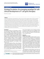
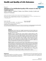
Representative plain radiographs of pasteurized bone grafts at 2 weeks (A-C), 4 weeks (D-F) and 12 weeks (G-I) after grafting are shownFigure 1
Representative plain radiographs of pasteurized bone grafts at 2 weeks (A-C), 4 weeks (D-F) and 12 weeks (G-
I) after grafting are shown. Pasteurized bone with (MC+; FGF2-) (A, D, G), (MC-; FGF2-) (B, E, H) and (MC-; FGF2+) (C, F,
I) is shown. Arrows show the anterior portion of the proximal interface between grafted bone and host bone for observation.
Bone formations at the edge of the host bone can be seen in all 3 groups (A-C). Mild bone absorption can be observed on the
(MC+; FGF2-) graft at 4 weeks (D). Massive bone absorption can be observed on the (MC+; FGF2-) graft (G, right) and on the
(MC-; FGF2-) graft (H) at 12 weeks. Bone incorporation with a bridge of bone formation from the host bone can be seen on
the (MC-; FGF2+) graft (I).
Journal of Orthopaedic Surgery and Research 2009, 4:31 />Page 5 of 10
(page number not for citation purposes)
or histological specimens in half the cases in the (MC+;
FGF2-) group (2/4; 50%) and in half the cases in the (MC-
; FGF2+) group (2/4; 50%). Bone incorporation was not
observed in any of the (MC-; FGF2-) cases (0/4; 0%). Bone
absorption was seen in 3 out of 4 of the (MC+; FGF2-)
cases (3/4; 75%). On the other hand, bone absorption
was not observed in any of the (MC-; FGF2-) cases (0/4;
0%) or the (MC-; FGF2+) cases (0/4; 0%) (Table 1) (P <
0.05). These degrees of absorption on the (MC+; FGF2-)
cases were within the cortex and were classified as mild
(Table 1). Histologically, the absorbed pasteurized bone
was replaced by fibrous or granulation tissue (Fig. 4E)
associated with an accumulation of osteoclasts (Fig. 4F).
The average score of bone formation and bone absorption
on the grafted bone was -0.75 (mild bone absorption, 3
cases; no change, 1 case), 0.25 (no change, 3 cases; single
nodules of bone formation, 1 case) and 0.0 (no change, 4
cases) in the (MC+; FGF2-), (MC-; FGF2-) and (MC-;
FGF2+) groups, respectively (Table 2) (Fig. 2, bottom).
Twelve weeks after bone grafting
On plain radiographs at 12 weeks after grafting, the
number of cases with bone formation at the host bone
became small in comparison to that at 2 or 4 weeks after
grafting (MC+; FGF2-) (2/4; 50%), (MC-; FGF2-) (0/4,
0%), and (MC-; FGF2+) (5/8; 63%)] (Table 1) (Figs. 1G,
H, I). Bone incorporation of the pasteurized bone to the
host bone was seen in 3 out of 4 cases in the (MC+; FGF2-
) group (3/4; 75%), but in only 3 out of 8 cases in the
(MC-; FGF2+) group (3/8; 38%). On the other hand, bone
incorporation was not observed in any of the (MC-; FGF2-
) cases (0/4; 0%) with a significant difference to the (MC+;
FGF2-) group (3/4; 75%) (Table 1) (P < 0.05). The average
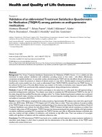
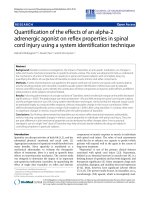
The size of the bone formation of the host bone was also quantitatively measured in the lateral viewFigure 2
The size of the bone formation of the host bone was
also quantitatively measured in the lateral view. The
area was calculated in relation with that in the (MC-; FGF2-)
group at 2 weeks in ratio (top). The status of the grafted
bone is scaled and the average is given. The scale is as fol-
lows: severe bone absorption (-2), mild bone absorption (-1),
no change (0), single nodules of bone formation (1) and
bridging or lamellar bone formation (2) (bottom).
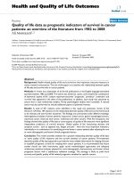
Pasteurized bone with (MC-; FGF2-) at 2 weeks after grafting shows the grafted bone (left part) and the host bone (right part)Figure 3
Pasteurized bone with (MC-; FGF2-) at 2 weeks after
grafting shows the grafted bone (left part) and the
host bone (right part). Protuberant bone formation from
the end surface of the host bone can be seen (A). Osteo-
clasts can not be observed on the surface of the grafted bone
(B), whereas osteoclasts can be observed on the surface of
the bone formation with numerous osteoclasts (C). Pasteur-
ized bone shows empty lacunae without osteocytes (D). Pas-
teurized bone is surrounded by fibrous tissue (E). (Original
magnification, H&E staining; A; ×70, D; E; ×250, TRAP stain-
ing; B; C; ×150).
Journal of Orthopaedic Surgery and Research 2009, 4:31 />Page 6 of 10
(page number not for citation purposes)
area of bone formation was 0.51, 0.11 and 0.77 in the
(MC+; FGF2-), (MC-; FGF2-) and (MC-; FGF2+) groups,
respectively (Table 2) (Fig. 2, top). The bone formation
was particularly decreased in the (MC-; FGF2-) group at
12 weeks compared with the same group at 2 weeks. How-
ever, in the (MC+; FGF2-) cases, bone absorption was
prominent (4/4; 100%), with the degree of absorption
being classified as severe in 3 cases and mild in 1 case,
whereas in the (MC-; FGF2+) cases, bone absorption was
less prominent, in 2 out of the 8 cases (2/8; 25%) (P <
0.01), with the degree of absorption being classified as
severe in 1 case and as mild in 1 case (Table 1). In the
(MC-; FGF2-) cases, most of the pasteurized bone was
almost completely absorbed (4/4; 100%) (Table 1) (Figs.
1G, H, I). Histologically, completely absorbed pasteurized
bone was replaced by fibrous or granulation tissue (Figs.
5A, B, C). Osteoclasts were seen on the residual pasteur-
ized bone which had empty lacunae without osteocytes
(Figs. 5D, E) and on the surface of the host bone (Figs. 5F,
G). On the other hand, pasteurized bone which had been
completely incorporated to the host bone in one of the
(MC-; FGF2+) cases showed an unclear interface between
the pasteurized bone and the host bone (Fig. 6A). Bone
matrix had been remodeled in an irregular fashion (Figs.
6B–D), and osteocytes could be observed on pasteurized
bone (Fig. 6C) and on the host bone (Fig. 6D). Bone mar-
row formation was also observed (Figs. 6A, C). Osteo-
clasts were not observed on the surface of the pasteurized
bone (Fig. 6E) or on the host bone (Fig. 6F). The average
score of bone formation and bone absorption on the
grafted bone was -1.75 (severe bone absorption, 3 cases;
mild bone absorption, 1 case), -2.0 (severe bone absorp-
tion, 4 cases) and 0.13 (severe bone absorption, 1 case;
mild bone absorption, 1 case; no change, 3 cases; single
nodules of bone formation, 2 cases; bridging or lamellar
bone formation, 1 case) in the (MC+; FGF2-), (MC-;
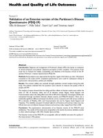
Pasteurized bone with (MC+; FGF2-) at 4 weeks after graft-ing shows the grafted bone (left part) and the host bone (right part)Figure 4
Pasteurized bone with (MC+; FGF2-) at 4 weeks after
grafting shows the grafted bone (left part) and the
host bone (right part). Bone formation at the end of the
host bone is rather mature (A, B) with osteoclasts on the
surface of the bone trabeculae (C, D). Grafted bone charac-
terized by empty lacunae is absorbed and replaced by fibrous
tissue (E) associated with osteoclasts on the surface of the
pasteurized bone (F). (Original magnification, H&E staining;
A; ×70, B; E; ×150, TRAP staining; C; ×70, D; F; ×150).
Pasteurized bone with (MC+; FGF2-) at 12 weeks after graft-ing shows the grafted bone (left part) and the host bone (right part)Figure 5
Pasteurized bone with (MC+; FGF2-) at 12 weeks
after grafting shows the grafted bone (left part) and
the host bone (right part). Completely absorbed pasteur-
ized bone has been replaced by fibrous tissue (A, B, C). The
residual pasteurized bone with empty lacunae is embedded in
the fibrous tissue (D). Osteoclasts can be seen on the resid-
ual bone (E) and the surface of the host bone (F, G). (Original
magnification, H&E staining; A; ×70, B; C; D; ×100, TRAP
staining; E; F; G; ×150).
Journal of Orthopaedic Surgery and Research 2009, 4:31 />Page 7 of 10
(page number not for citation purposes)
FGF2-) and (MC-; FGF2+) groups, respectively. There was
a significant difference between the (MC-; FGF2+) group
and the other (MC+; FGF2-) and (MC-; FGF2-) groups (P
< 0.05) (Table 2) (Fig. 2, bottom).
Discussion
Heating of a resected bone segment at a low temperature,
such as at 60°C for 30 min has been used as a method of
pasteurization [3,4,19]. In the current study, pasteurized
bone had empty lacunae at 2 weeks after grafting. For this
reason, the pasteurized bone model seemed to be success-
ful in terms of eliminating living cells, including osteo-
cytes. Bone incorporation was seen in about half the cases
of muscle-covering without FGF-2 at 4 weeks after the pro-
cedure. This result suggests that pasteurized bone after
treatment at 60°C for 30 min helps to maintain bone
induction ability.
Pasteurized bone without muscle-covering was examined
as a model for wide resection of bone tumors. In a com-
parison between muscle-covering without FGF-2 and no
muscle-covering without FGF-2, plain radiographs
showed that after 2 weeks, bone was well formed at the
edge of the hosted bone, and after 4 weeks, the size was
decreased, especially when there was no muscle covering
without FGF-2. Bone incorporation was seen in about half
the (MC+; FGF2-) cases at 4 weeks after the procedure,
whereas bone incorporation was seen in none of the 4
(MC-; FGF2-) cases. Therefore, muscle-covering of the pas-
teurized bone seemed to provide a positive effect on bone
incorporation. Some surgeons utilize a method of cover-
ing a pasteurized bone graft using nearby muscle after
resection of the affected bone together with the surround-
ing muscle. The current results showing an increased abil-
ity of bone incorporation with muscle-covering on the
pasteurized bone seem to support the effectiveness of
such clinical experience. The benefit of muscle coverage
seems to be supported by previous research showing the
positive role of muscle stem cells in the bone repair proc-
ess [11]. Furthermore, it has been reported that the first
step in bone formation in pasteurized bone might be the
migration of mesenchymal stem cells from the contiguous
normal medullary cavity [1]. The current study suggests
that the circumstances outside the medullary cavity are
also important for bone induction.
At 4 weeks after grafting, bone absorption of the pasteur-
ized bone was only seen in the muscle-covering cases, and
was not seen in cases without muscle-covering, with/with-
out FGF-2. Bone absorption was replaced by granulation
or fibrous tissue and was associated with osteoclast accu-
mulation. At 12 weeks after grafting, in the series of (MC+;
FGF2-) cases, even after bone incorporation in part, bone
absorption of the pasteurized bone continued. Therefore,
muscle-covering to pasteurized bone not only has a posi-
tive effect on bone incorporation to the host bone, but
also on bone absorption associated with osteoclastic
activity. In a previous model, a muscle flap was found to
be superior to a cutaneous flap in revascularizing isolated
bone segments, and furthermore, muscle flaps showed
osteoblasts and osteoclasts, whereas neither were seen in
the cutaneous flap [12]. In the current study, the increased
positive effect on bone incorporation and bone absorp-
tion may be associated with the revascularizing that was
associated with the surrounding muscle.
Mesenchymal stem cells are able to self-replicate and dif-
ferentiate into a variety of cell types [20,21]. It has been
suggested that FGF-2 increases the osteogenic and chon-
drogenic differentiation potentials of human mesenchy-
mal stem cells [17]. Moreover, FGF-2 is a potent mitogen
for osteoprogenitor cells, and it plays an important role in
bone metabolism and in the regulation of osteoblastic cell
proliferation and differentiation [13-16]. On the other
hand, FGF-2 has been reported to stimulate bone resorp-
tion in bone organ cultures [22], as well as osteoclastogen-
esis in a mouse bone marrow culture [23]. FGF-2 plays a
pivotal role in osteoclastogenesis through the up-regula-
Pasteurized bone with (MC-; FGF2+) at 12 weeks after graft-ing shows the grafted bone (left part) and the host bone (right part)Figure 6
Pasteurized bone with (MC-; FGF2+) at 12 weeks
after grafting shows the grafted bone (left part) and
the host bone (right part). Completely incorporated pas-
teurized bone to the host bone can be seen (A). Pasteurized
bone has been remodeled with irregular bone matrix and
osteocytes (B-D). Bone marrow formation can be seen (A,
C). Osteoclasts can not be observed on the surface of the
pasteurized bone (E) or the host bone (F). (Original magnifi-
cation, H&E staining; A; ×70, B; ×150, C; D; ×190, TRAP
staining; E; F; ×150).
Journal of Orthopaedic Surgery and Research 2009, 4:31 />Page 8 of 10
(page number not for citation purposes)
tion of RANKL (receptor activator of nuclear factor-kappa
B ligand) [24]. In the case of FGF-2 application in the cur-
rent study, achievement of bone incorporation was seen
in 3 out of 8 (MC-; FGF2+) cases, while bone absorption
was seen in only 2 out of these 8 cases. Considering that
bone absorption was seen in all of the (MC-; FGF2-) cases,
FGF-2 would seem to have a positive role to play in bone
incorporation, and a negative role to play in bone absorp-
tion in the current model.
The lasting time of FGF-2 and its concentration from the
grafted bone soaked in FGF-2 solution has been
unknown. The possible releasing mechanism seemed to
be a manner of diffusion. Since some research has
reported that more than 80% of FGF-2 in solution form
was cleared from the injected site of subcutaneous tissue
of the mouse back within 1 day [25], it would seem that
the effect of FGF-2 may be only short-term, even in the
case of the current study. In a previous report on pasteur-
ized bone, revascularization was thought to be important
for bone remodeling [1]. FGF-2 also has angiogenic activ-
ity [26]. Therefore, it would seem that not only the initial
induction of osteoblastic progenitor cells, but also the ini-
tial vascularization might play an important role in the
process of bone incorporation of the pasteurized bone.
In this study, pasteurized bone grafts were soaked in FGF-
2 solution and re-implanted. Results showing the poten-
tial usefulness of FGF-2 in the current study are encourag-
ing with regard to pasteurized bone. A study including
long-term use such as local delivery or controlled release
of FGF-2 would be interesting, since the prolonged effect
of FGF-2 may provide greater effectiveness in terms of
increasing osteoblastic activity and decreasing bone
absorption. In order to control the release of biologically-
active growth factors, such as FGF-2, biodegradable
hydrogels have been developed [25]. The effectiveness of
the controled release of growth factors has been con-
firmed for the induction of angiogenesis in regenerated
skin [26].
As for the limitations of this study, the current study did
not include the group of muscle-covering pasteurized
bone with the application of FGF-2 (MC+; FGF2+). Dur-
ing the establishment of a pasteurized bone model, we
carried out a preliminary examination of the effect of the
presence of muscle-covering or the application of FGF-2
to pasteurized bone as an independent concept. The syn-
ergetic effect of muscle-covering and FGF-2 administra-
tion is worth further examination. Due to the preliminary
concept, the number of cases in each group was small and
varied, yet the results seemed to be consistent in the cur-
rent study. In any future project, a large number of cases
with independent assessors would be preferable.
In the current study, we have assessed bone formation and
bone absorption with plain radiographs. Histomorphom-
etry analysis of the pasteurized bone grafts and the host
Table 1: Summary of bone formation, incorporation and absorption
2 weeks 4 weeks 12 weeks
Bone formation on the host bone
Muscle cover (+); FGF2 (-) 4/4 (100%) 4/4 (100%) 2/4 (50%)
Muscle cover (-); FGF2 (-) 3/4 (75%) 2/4 (50%) 0/4 (0%)
a
Muscle cover (-); FGF2 (+) 3/4 (75%) 4/4 (100%) 5/8 (63%)
a
Bone incorporation
Muscle cover (+); FGF2 (-) 0/4 (0%) 2/4 (50%) 3/4 (75%)
b
Muscle cover (-); FGF2 (-) 0/4 (0%) 0/4 (0%) 0/4 (0%)
b
Muscle cover (-); FGF2 (+) 0/4 (0%) 2/4 (50%) 3/8 (38%)
Bone absorption on grafted bone
Muscle cover (+); FGF2 (-) 0/4 (0%) 3/4 (75%)* 4/4 (100%)
Mild 3 Mild 1
Severe 0 Severe 3
Muscle cover (-); FGF2 (-) 0/4 (0%) 0/4 (0%) 4/4 (100%)
Mild 0
Severe 4
Muscle cover (-); FGF2 (+) 0/4 (0%) 0/4 (0%) 2/8 (25%)**
Mild 1
Severe 1
FGF; fibroblast growth factor.
a
; p < 0.05,
b
; p < 0.05
*; p < 0.05, **; p < 0.01 (compared to the other groups).
Journal of Orthopaedic Surgery and Research 2009, 4:31 />Page 9 of 10
(page number not for citation purposes)
bone to quantify the numbers of osteocytes, osteoclasts,
osteoblasts and newly formed osteoid would be necessary
to analyze bone remodeling. Moreover, proper markers
would be helpful for visualizing blood vessel invasion or
inflammatory cells within the granulation tissue sur-
rounding the pasteurized bone, in order to analyze angio-
genesis.
Conclusion
In conclusion, we have established an animal pasteurized
bone graft model in rats. Despite the small number of
cases in each group, the results of each group suggest that
muscle-covering without FGF-2 has an effect on bone
incorporation, but is not able to prevent bone absorption
to pasteurized bone. FGF-2 application seems to be useful
in bone, in that it increases bone incorporation and pre-
vents muscle absorption.
Abbreviations
FGF: fibroblast growth factor; MC: muscle covered; TRAP:
tartrate-resistant acid phosphatase
Competing interests
The authors declare that they have no competing interests.
Authors' contributions
AS drafted the manuscript. TY, AS, NT and KN performed
the experiment. TY and AS participated in the design of
the study. YI conceived of the study, and participated in its
design and coordination and helped to draft the manu-
script. All authors read and approved the final manu-
script.
Acknowledgements
The English used in this manuscript was revised by Miss K. Miller (Royal
English Language Centre, Fukuoka, Japan).
References
1. Zoricic S, Bobinac D, Lah B, Maric I, Cvijanovic O, Bajek S, Golubovic
V, Mihelic R: Study of the healing process after transplantation
of pasteurized bone grafts in rabbits. Acta Med Okayama 2002,
56:121-128.
2. Liebergall M, Simkin A, Mendelson S, Rosenthal A, Amir G, Segal D:
Effect of moderate bone hyperthermia on cell viability and
mechanical function. Clin Orthop Relat Res 1998:242-248.
Table 2: Size and scores of bone formation and absorption
2 weeks 4 weeks 12 weeks
a
Relative size of bone formation on the host bone
Muscle cover (+); FGF2 (-) 1.03 0.85 0.51
Muscle cover (-); FGF2 (-) 1 0.27 0.11
Muscle cover (-); FGF2 (+) 0.44 0.82 0.77
Status of grafted bone based on average score
Muscle cover (+); FGF2 (-) 0.0 -0.75 -1.75
Bridging or lamellar bone (2) 0 0 0
Single nodules of bone (1) 0 0 0
No change (0) 4 1 0
Mild bone absorption (-1) 0 3 1
Severe bone absorption (-2) 0 0 3
Muscle cover (-); FGF2 (-) 0.0 0.25 -2.0
Bridging or lamellar bone (2) 0 0 0
Single nodules of bone (1) 0 1 0
No change (0) 4 3 0
Mild bone absorption (-1) 0 0 0
Severe bone absorption (-2) 0 0 4
Muscle cover (-); FGF2 (+) 0.0 0.0 0.13*
Bridging or lamellar bone (2) 0 0 1
Single nodules of bone (1) 0 0 2
No change (0) 4 4 3
Mild bone absorption (-1) 0 0 1
Severe bone absorption (-2) 0 0 1
FGF; fibroblast growth factor. The scores are shown in parentheses.
a
; Relative size of bone formation to [muscle cover (-); FGF2 (-)] at 2 weeks on the host bone in ratio.
*; p < 0.05 (compared to the other groups).
Publish with BioMed Central and every
scientist can read your work free of charge
"BioMed Central will be the most significant development for
disseminating the results of biomedical research in our lifetime."
Sir Paul Nurse, Cancer Research UK
Your research papers will be:
available free of charge to the entire biomedical community
peer reviewed and published immediately upon acceptance
cited in PubMed and archived on PubMed Central
yours — you keep the copyright
Submit your manuscript here:
/>BioMedcentral
Journal of Orthopaedic Surgery and Research 2009, 4:31 />Page 10 of 10
(page number not for citation purposes)
3. Ahmed AR, Manabe J, Kawaguchi N, Matsumoto S, Matsushita Y:
Radiographic analysis of pasteurized autologous bone graft.
Skeletal Radiol 2003, 32:454-461.
4. Rong Y, Sato K, Sugiura H, Ito T, Sakano S, Iwata H, Kimata K: Effect
of elevated temperature on experimental swarm rat chond-
rosarcoma. Clin Orthop Relat Res 1995:227-231.
5. Inokuchi T, Ninomiya H, Hironaka R, Yoshida S, Araki M, Sano K:
Studies on heat treatment for immediate reimplantation of
resected bone. J Craniomaxillofac Surg 1991, 19:31-39.
6. Ehara S, Nishida J, Shiraishi H, Tamakawa Y: Pasteurized interca-
lary autogenous bone graft: radiographic and scintigraphic
features. Skeletal Radiol 2000, 29:335-339.
7. Ortiz-Cruz E, Gebhardt MC, Jennings LC, Springfield DS, Mankin HJ:
The results of transplantation of intercalary allografts after
resection of tumors. A long-term follow-up study. J Bone Joint
Surg Am 1997, 79:97-106.
8. Sabo D, Brocai DR, Eble M, Wannenmacher M, Ewerbeck V: Influ-
ence of extracorporeal irradiation on the reintegration of
autologous grafts of bone and joint. Study in a canine model.
J Bone Joint Surg Br 2000, 82:276-282.
9. Kattapuram SV, Phillips WC, Mankin HJ: Intercalary bone allo-
grafts: radiographic evaluation. Radiology 1989, 170:137-141.
10. Bonnarens F, Einhorn TA: Production of a standard closed frac-
ture in laboratory animal bone. J Orthop Res 1984, 2:97-101.
11. Schindeler A, Liu R, Little DG: The contribution of different cell
lineages to bone repair: exploring a role for muscle stem
cells. Differentiation 2009, 77:12-18.
12. Fisher J, Wood MB: Experimental comparison of bone revascu-
larization by musculocutaneous and cutaneous flaps. Plast
Reconstr Surg 1987, 79:81-90.
13. Jaye M, Schlessinger J, Dionne CA: Fibroblast growth factor
receptor tyrosine kinases: molecular analysis and signal
transduction. Biochim Biophys Acta 1992, 1135:185-199.
14. Basilico C, Moscatelli D: The FGF family of growth factors and
oncogenes.
Adv Cancer Res 1992, 59:115-165.
15. Robinson D, Bab I, Nevo Z: Osteogenic growth peptide regu-
lates proliferation and osteogenic maturation of human and
rabbit bone marrow stromal cells. J Bone Miner Res 1995,
10:690-696.
16. Chaudhary LR, Avioli LV: Extracellular-signal regulated kinase
signaling pathway mediates downregulation of type I procol-
lagen gene expression by FGF-2, PDGF-BB, and okadaic acid
in osteoblastic cells. J Cell Biochem 2000, 76:354-359.
17. Ito T, Sawada R, Fujiwara Y, Tsuchiya T: FGF-2 increases osteo-
genic and chondrogenic differentiation potentials of human
mesenchymal stem cells by inactivation of TGF-beta signal-
ing. Cytotechnology 2008, 56:1-7.
18. Boes M, Kain M, Kakar S, Nicholls F, Cullinane D, Gerstenfeld L, Ein-
horn TA, Tornetta P 3rd: Osteogenic effects of traumatic brain
injury on experimental fracture-healing. J Bone Joint Surg Am
2006, 88:738-743.
19. Manabe J, Kawaguchi N, Matsumoto S: Pasteurized autogenous
bone graft for reconstruction after resection of malignant
bone and soft tissue tumors: imaging features. Semin Muscu-
loskelet Radiol 2001, 5:195-201.
20. Caplan AI, Bruder SP: Mesenchymal stem cells: building blocks
for molecular medicine in the 21st century. Trends Mol Med
2001, 7:259-264.
21. Pittenger MF, Mackay AM, Beck SC, Jaiswal RK, Douglas R, Mosca JD,
Moorman MA, Simonetti DW, Craig S, Marshak DR: Multilineage
potential of adult human mesenchymal stem cells. Science
1999, 284:143-147.
22. Simmons HA, Raisz LG: Effects of acid and basic fibroblast
growth factor and heparin on resorption of cultured fetal rat
long bones. J Bone Miner Res 1991, 6:1301-1305.
23. Hurley MM, Lee SK, Raisz LG, Bernecker P, Lorenzo J: Basic fibrob-
last growth factor induces osteoclast formation in murine
bone marrow cultures. Bone 1998, 22:309-316.
24. Nakano K, Okada Y, Saito K, Tanaka Y:
Induction of RANKL
expression and osteoclast maturation by the binding of
fibroblast growth factor 2 to heparan sulfate proteoglycan
on rheumatoid synovial fibroblasts. Arthritis Rheum 2004,
50:2450-2458.
25. Yamamoto M, Ikada Y, Tabata Y: Controlled release of growth
factors based on biodegradation of gelatin hydrogel. J Bio-
mater Sci Polym Ed 2001, 12:77-88.
26. Tanihara M, Suzuki Y, Yamamoto E, Noguchi A, Mizushima Y: Sus-
tained release of basic fibroblast growth factor and angio-
genesis in a novel covalently crosslinked gel of heparin and
alginate. J Biomed Mater Res 2001, 56:216-221.