báo cáo hóa học:" Anatomical, Clinical and Electrical Observations in Piriformis Syndrome" docx
Bạn đang xem bản rút gọn của tài liệu. Xem và tải ngay bản đầy đủ của tài liệu tại đây (3.83 MB, 7 trang )
RESEA R C H ARTIC L E Open Access
Anatomical, Clinical and Electrical Observations in
Piriformis Syndrome
Roger M Jawish
1,2*
, Hani A Assoum
2
, Chaker F Khamis
3
Abstract
Background: We provided clinical and electrical descriptions of the piriformis syndrome, contributing to better
understanding of the pathogenesis and further diagnostic criteria.
Methods: Between 3550 patients complaining of sciatica, we concluded 26 cases of piriformis syndrome, 15
females, 11 males, mean age 35.37 year-old. We operated 9 patients, 2 to 19 years after the onset of symptoms, 5
had piriformis steroids injection. A dorsolumbar MRI were performed in all cases and a pelvic MRI in 7 patients. The
electro-diagnostic test was performed in 13 cases, between them the H reflex of the peroneal nerve was tested 7
times.
Results: After a followup 1 to 11 years, for the 17 non operated patients, 3 patients responded to conservative
treatment. 6 of the operated had an excellent result, 2 residual minor pain and one failed. 3 new anatomical
observations were described with atypical compression of the sciatic nerve by the piriformis muscle.
Conclusion: While the H reflex test of the tibial nerve did not give common satisfaction in the literature for
diagnosis, the H reflex of the peroneal nerve should be given more importance, because it demonstrated in our
study more specific sign, with six clinical criteria it contributed to improve the method of diagnosis. The cause of
this particular syndrome does not only depend on the relation sciatic nerve-piriformis muscle, but the
environmental conditions should be considered with the series of the anatomical anomalies to explain the real
cause of this pain.
Background
Sincemanyyears,wehadaparticularinterestforthe
intractable sciatica with failure of long term treatment
of lumbar pain. In such cases, our investigation was
focused on a suspected piriformis syndrome missing
from many decades specific signs for diagnosis.
Yeoman [1] 1928, re ported that the sciatica m ay be
caused by a periarthritis involving the anterior sacroiliac
ligament, the piriformis muscle and the adjacent
branches of the sciatic nerve. Freiberg and Vinke [2]
1934 , considered that the inflammat ion of the sacroiliac
joint may primarily cause reaction of the piriformis
muscle and its fascia, and secondarly, the irritation of
the overlying lumbosacral plexus.
Based on cadaver dissections, Beaton and Anson [3]
1938, gave the hypothesis that the spasm of the pirifor-
mis muscle could be responsible for the irritation of the
nerve. Robinson [4] 1947, has introduced the term “piri-
formis syndrome” and applied it to sciatica related to
abnormal muscle, which is usually traumatic i n origin,
with emphasis on the necessity to rule out all other
causes of sciatica.
Even though it is commonly accepted that no consen -
sus was defined about the clinical and the laboratory
studies, we have tried to describe further clinical criteria
that we concluded from the physical examination of
patients complaining of sciatica. The electro-diagnostic
test is also considered as an important method of diag-
nosis,whiletestingofthesciaticnervehascontributed
in many studies [5-7] to expect the presence of a pirifor-
mis impingement, with a particular interest for the H-
reflex of the tibial nerve [7]. We, however, believe that
more impor tance should be given to the H-reflex of the
peroneal nerve which has demonstrated more specific
signs in our study.
The lack of reliable objective test to identify the piri-
formis muscle syndrome leads in many cases to great
* Correspondence:
1
Medical School, St Joseph University, Beirut, Lebanon
Jawish et al. Journal of Orthopaedic Surgery and Research 2010, 5:3
/>© 2010 Jawish et al; license e BioMed Central Ltd. This is an Open Access article distributed under the ter ms of the Creative Commons
Attribution Lic ense (http://creativec ommons.org/licenses/by/2.0), which permits unrestricted use, distri bution, and reproduction in
any medium, provided the original work is properly cited.
expenses in repetitive imaging studies and to time loss
in searching for the origin of the intractable sciatica
among the lumbar pathologies. Our clinical criteria con-
cluded from the epidemiologic study and anatomical
observations, added to the electri cal testing of the pe ro-
neal nerve, could improve the method of diagnosis and
avoid the delays in unnecessary suffering.
Materials and methods
Between 1997 and 2007, about 3550 patients complain-
ing of low back pain and sciatica were examined by the
first author and not referred by any other physician. We
retained 26 cases of piriformis syndrome, 15 women
and 11 men, aged between 15 and 66 years (average:
35.37), 14 left and 12 right. 9 patients have accep ted the
surgery after either, failure of conservative treatment or
presence of neuro-muscular deficiencies.
The 17 non operated patie nts were 10 women and 7
men, aged between 18 and 66, 10 left and 7 right, none
had a previous history of trauma to the gluteal region; 4
were athletics (one gymnastics, 2 walkers and one bas-
ketballer). The time average from the beginning of the
pain to the treatment was 3.14 years (range: 1 month to
11 years). One patient had a failed previous lumbar disc
surgery for sciatica. Five of them have benefited from
intrapiriformis muscle steroids injection.
The 9 operated patients (table 1) were 5 women and 4
men, aged between 15 and 65 (average: 35.88), 4 left
and 5 right. The weight average was 73.88 Kg (range: 55
to 110). Six athletics distributed between 3 walkers, 2
footballers and 1 swimmer, only one patient had a pre-
vious history of a fall onto a buttock, 3 months before
the onset of the symptoms. All patients had followed a
preoperative medical treatment including painkillers and
muscle relaxants; three have also had intrapiriformis
muscle steroids injection. The time average from the
beginning of the pain to surgery was: 5.44 years (range,
2 to 19 years).
The neurological preoperative examination showed
one complete right drop foot, and one patient was
obliged to stand up in a triple flexion position, in pro-
longed standing; 5 patients had dysesthesia and altered
reflexes; 4 patients had gluteal atrophy at the affected
side and one patient had posterior leg atrophy.
All patients of the study benefited of a dorsolumbar
MRI, none of them has revealed nerve root compression
or any spinal pathology responsible of the sciatica. A
pelvic MRI has been performed in 7 patients and has
demonstrated an obvious hypertrophy of the homolat-
eral pir iformis muscle in two cases, and in 4 cases, there
were mild congestion of the venous plexus around the
sciatic nerve.
The EMG was performed on 13 patients. Only three
of them have shown alteration of the H reflex of the
tibial nerve. For the last seven patients, we started to
explore the H reflex of the common peroneal nerve. We
observed during the EMG recording, a complete disap-
pearance of the peroneal’s H reflex when the affected
lower limb was put in the pain position (internal rota-
tion and adduction); the H reflex reappeared when the
limb was returned to the relieved s traight position (Fig.
1). When this test was performed at the unaffected
opposite site, the H r eflex remained no rmal in all
positions.
The various tests p erformed in our series have
revealed constancy of the following signs in all our
patients: 1)Absence of any spinal pathology at the dor-
solumbar MRI. 2) Tenderness with digital pressure of
the sciatic spine and absence of pain complaint at the
lower back and the sacroiliac joint. 3) Intolerance to sit-
ting on the involved side with the body inclined over
the thigh. 4) Sciatica in the sitting position when the
homolateral leg is crossed over the unaffected side. 5)
Exacerbated sciatica by the maneuver o f internal rota-
tion and maximal addu ction of the hip. 6) The H refl ex
tested for the common peroneal nerve (EMG) has disap-
peared in pain position with internal rotation and forced
adduction.
Results
Clinical outcome
Considering the 17 none operated patients and after a
follow up ranging from one to 11 years, we have
obtained the following results: one patient has
responded to medical treatment, one was operated by
another sur geon for piriformis muscle syndrome with a
good result, two have responded to infiltration, seven
have not responded to conservative measures and six
patients were missed.
After a follow up between 1 and 11 years, the 9 oper-
ated patients have b een interrogated and reexamined by
the senior author and noted a relief of pain in 2 weeks
to 12 months after the operation (mean 5.61 months).
Six patients have obtain ed an excellent result with a
complete relief of pain even in prolonged periods of sit-
ting. Two patients have reported minor residual pain in
the buttock precipitated by strenuous activities. One
patient has considered that the operation was not bene-
ficial to her knowing that we were not able to examine
her (table 1).
The five patients with preoperative sensory problem s
have had a transient tinnel s ign for a maximum of five
months, a nd one of them has demonstrated a paresthe-
sia in the territory of deep peroneal nerve. The patient
with a drop foot has recovered within six months. None
of the patients had used walkers or crutches postopera-
tively. We have observed one posto perative transitory
limp and one superficial cutaneous infection.
Jawish et al. Journal of Orthopaedic Surgery and Research 2010, 5:3
/>Page 2 of 7
Operative findings
In a prone position using Kocher-langenbeck incision,
the pirifor mis muscle was reached through the fibers of
the gluteus maximus and s ectioned after dissection of
the nerve. A neurolysis of the sciatic nerve was per-
formed in all the cases. The intra operati ve observations
of the 9 cases were as following:
The sciatic nerve was bifid passing under the hyper-
trophied piriformis muscle, 1 case (fig. 2). A bifid piri-
formis muscle and a bifid sciatic nerve, one branch of
the nerve was passing proximal to the muscle and the
other one through the split, 1 case (fig. 3). A sciatic
impingement by the piriformis muscle and the sacros-
ciatic ligament, 1 case (fig. 4). The piriformis muscle
was hypertrophied, squeezing the sciatic nerve which
passed directly below it, 2 case s. A transverse fibrous
band compressed the sciatic nerve, 1 case (fig. 5). A ner-
vous connection existed between the sciatic nerve and
the inferior gluteal nerve, 1 case. There was no evidence
of anatomical impingement of the sciatic nerve in three
cases. Congested tortuous veins around the sciatic nerve
sight were present in almost all the patients.
Discussion
It is well known among the authors who studied the pir-
iformis syndrome that many patients treated for low
back pain could have sciatic nerve impingement at the
buttock. Since the extended use of MRI to evaluate
spinal disorders, the piriformis muscle syndrome has
becomeamoreseparateentityeventhoughtherelated
specific signs were not completely defined and the
mechanism is still obscure.
Although the incidence of this affection remains con-
troversial, it was increasing progressively with the
improvement of investigations. Most of the reported
cases were spora dic, but the latest series described more
cases with variable incidence, from 0.33% [8] to 6% [9]
depending on the nature of the referral system to the
investigators. However, in patients referred for spinal
disorders after failure of the treatment, the maximal rate
was 5% for Parziale [10] and 14/93 for Benson [5];
although in 1997, Goldner [11] has criticized this high
rate and considered that the prevalence in a referral
orthopaedic surgery should not exceed 1%, whi ch is
close to our value (0.7%) but in a none referral practice.
Regardless of the physiopathologic origin of the com-
plex disorder (muscular or nervous), symptoms and
imaging should be combined to confirm the diagnosis.
Contrary to many authors [1,2,4], we agree with Bernard
and Kirkaldy-Willis [8] that there is no relation between
the sacroiliac joint syndrome and the piriformis syn-
drome, and we also consider that the absence of sacroi-
liac pain is an essential sign for a positive diagnosis.
Based on two observations, Robinson [4] described the
cardinal features of the syndrome with six criteria: (I) a
history of trauma to the sacroiliac and gluteal regions;
Table 1 Clinical Data on 9 operated patients
Patient 1 2 3 4 5 6 7 8 9
Sex M f m f f m m f f
Age(years) 32 32 58 23 44 15 39 15 65
Weight(kg) 70 60 99 58 57 110 76 55 80
Side L L L R L R R R R
Sport - - Football Walker Swim football Walker Walker -
Gluteal trauma - - - - - yes - - -
Preop. Steroid injection 0 1 3 0 2 0 0 0 0
Delay to surgery (years) 3 7 3 4 2 3 6 2 19
Sciatica yes yes yes Drop foot yes yes yes yes yes
Pain on sitting position + + + + + + + + +
Gluteal atrophy - - - + - - + + +
Pain on digital pressure + + + + + + + + +
H-reflex peroneal nerve + + + + + + +
Preop.MRI (spine) 1 1 4 7 3 1 3 2 1
Preop.MRI (pelvis) Veinous
sign
Piriformis
hypertrophy
Veinous
sign
Veinous
sign
Piriformis
hypertrophy
Veinous
sign
Normal
From surgery to pain
relief
One year 6 months 3
months
2 weeks No relief 1 year 1 year 4
months
1
month
Residual gluteal pain - + - - + - - - +
Functional result Excellent Good Excellent Excellent Bad Excellent Excellent Excellent good
The preoperative and last followup evaluation concerning the clinical status and the results of the MRI images and the H-reflex of the peroneal nerve.
Jawish et al. Journal of Orthopaedic Surgery and Research 2010, 5:3
/>Page 3 of 7
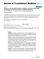
Figure 1 Electro-diagnostic test of a 22 year-old female patient complaining of right sided piriformis muscle syndrome since 6 years.
(A-1) The H reflex of the tibial nerve, the leg in a straight position, was normal, (A-2) showed slight disturbance of the H wave, during the stress
maneuver of flexion and internal rotation of the lower limb. (B-1) the H-reflex of the common peroneal nerve, the leg in a straight position, was
normal, (B-2) noted the complete extinction of the H wave, during the painful maneuver of forced adduction-internal rotation, (B-3) the H reflex
reappeared when the leg was returned in the relieved straight position.
Jawish et al. Journal of Orthopaedic Surgery and Research 2010, 5:3
/>Page 4 of 7
(II) pain in the region of the sacroiliac joint, greater
sciatic notch, and pirifo rmis muscle that usually extends
down the limb and causes difficulty with walking; (III)
acut e exacer bation of pain caused by stooping or lifting;
(IV) a palpable sausage-shaped mass, tender to palpa-
tion, over the piriformis muscle on the affected side; (V)
a positive Lasègue sign; and (VI) gluteal atrophy,
depending on the duration of the condition.
Many authors [4-6,12,13] have considered trauma in
the gluteal area as the major cause of piriformis syn-
drome, which was not the rule in our series where
trauma was evocated in one case only. We, however,
believe that piriformis syndrome could be related to exa-
cerbated rotators activity as it was observed in patients
with hard physical activity, walkers, athletic s and foot-
baller or with repetitive trauma of nerve in patients with
prolonged sitting position.
Among all the signs reported in the literature, we have
accepted the pain induced by passive internal rotation
and a dduction of the hip described by Freiberg [2], but
the pain induced by resisted abduction and external
rotation of the affected thigh, as described by Pace [12],
was not in our series a specific sign of this syndrome.
However, we have considered pathognomonic the signs
which were constantly observed in all the patients of
our study, and we have excluded all others that were
uncommon as impressive gluteal atrophy, or a palpable
sausage-shaped mass [13].
While the cases reported in the past have suffered
from none contribution of the modern imaging, the use
of MRI has become esse ntial to rule out any spinal dis-
orders or pelvic disorders as mentioned by Pecina [14]
who found an MRI abnormality for the piriformis mus-
cle syndrome in 7 out of his 10 patients; it is in practice
Figure 2 A 23-yea r-old female complaining of right sided piriformis muscle syndrome since 4 years. We noted intraoperati vely a bifid
sciatic nerve passing under the hypertrophied piriformis muscle.
Figure 3 32-year-old female complaining of left sided pi riformis muscle sy ndrome since 7 years. We noted intraoperatively a bifid
piriformis muscle and a bifid sciatic nerve, one branch of the nerve passing proximal to the muscle and the other one through the split
Jawish et al. Journal of Orthopaedic Surgery and Research 2010, 5:3
/>Page 5 of 7
the first exam that evokes the piriformis muscle, parti-
cularly in patient with chronic sciatica. However, and
apart from the MR neurography or piriformis blocks
[15,16] in which we have no experience, the MRI of pel-
vis remains unable to define a criteria for diagnosis,
since the asymmetrical size of the Piriformis muscle
observed in our cases, is common in normal people and
identified in T1-weighted MRI of the pelvis performed
for 100 persons [17].
The electromyographic is another test for diagnosis,
but nerve conduction results reported in the literature
were not conclusive and their methods were ve ry con-
troversial. However, it is well admitted that the tibial
divisio n of the nerve is usually spared [6] and the infer-
ior gluteal nerve that supplies the gluteus maximus may
be affected and the muscle atrophied as observed in
four cases of our series. It is well accepted that the
impingement of the sciatic nerve should delay the H-
reflex as described by Fishman [7], whereas many
authors [5,6] have obtained variable results concerning
the tibial nerve.
We, however, have demonstrated that the H reflex of
the peroneal nerve was more reliable than testing of the
tibi al nerve, and we have constantly observ ed extinction
of the H wave, during the painful maneuver of forced
adduction-interna l rotation of the affected leg. In the
same condition of stress test, the H reflex of the tibial
nerve remained normal for 10 of 13 patients. We believe
that fibers of the peroneal nerve could be more vulner-
able because they are anatomically more exposed to
injury at the buttock in case of trauma or impingement.
This electrical testing of peroneal’sH-reflexandthe
clinical criteria constantly observe d in all the patients
suffering from a nondisk scia tica, could help to prove
Figure 4 A 65-year-old female compla ining of r ight sided piriformis muscle syndrome since 19 years. Note the impingement of the
sciatic nerve in contact with the sacrospinous ligament.
Figure 5 A 58-year-old male complaining of left sided piriformis muscle syndrome since 3 years .Notethetransversefibrousband
squeezing the sciatic nerve.
Jawish et al. Journal of Orthopaedic Surgery and Research 2010, 5:3
/>Page 6 of 7
the d iagnosis or reveal more clearly the presence of the
entrapment.
The anatomical studies of the piriformis muscle
reported in the literature did not contribute to make a
real correlation between the clinical signs and the anat-
omy a nd to describe the different anatomical forms for
the same syndrome. A study [3] involving 240 cadaver
dissections has revealed that in 90 percent of cases the
sciatic nerve emerges from below the piriformis muscle,
in 7 percent the piriformis and the sciatic are divided,
one branch of the scia tic nerve passing through the split
and the other branch passing distal to the muscle, in 2
percent only the sciatic nerve is divided and in 1 percent
the piriformis is divided by the sciatic nerve. Pecina M.
found that in 6.15% of cases, the nervous peroneus com-
muni s passes between the tend inous parts of m. pirifor-
mis, and he considers this variation of practical
significance for the development of the Piriformis Syn-
drome [18]. After review ing the cadave ric anatomical
variants of the literature [3,19] and surgical anatomical
descriptions [5,20-22], we demonstrated three anatomi-
cal observations in our series (Fig. 2,3,4), but they did
not add further information on the a natomical variants
and their clinical expressions.
Considering the different anatomical findings, we
think that the real cause of this particular syndrome
does not only depend on the relation sciatic nerve-piri-
formis muscle, because the incidence of the anatomical
anomalies of these entities is definitely superior to those
treated in the reported cases. We, however, lay emphasis
on the environmental aspect of this affection, consider-
ing the physical activity and lifestyle of the patient
which could be an essential factor in revealing an under-
lying inadaptable anatomy.
Conclusion
The observations added to those of the literature have
contributed to prove the diversity of the anatomical
forms of this syndrome which remains very controver-
sial to many surgeons.
We have defined a group of clinical signs, imaging
findings and EMG testing which could contribute to
avoid diagnostic mistakes and the confusion with the
multiple spinal disorders. The environmental conditions
should be considered w ith the anatomical anomalies to
explain the real cause of this pain.
Author details
1
Medical School, St Joseph University, Beirut, Lebanon.
2
Department of
Orthopaedic, Sacré Coeur Hospital, BP 116 Hazmieh, Lebanon.
3
Department
of Electrodiagnostic, Sacré Coeur Hospital, BP 116 Hazmieh, Lebanon.
Authors’ contributions
RJ carried out the surgery, defined the different anatomical descriptions and
conceived the H-reflex of the peroneal nerve. HA tested the clinical follow-
up and helped to draft the manuscript. CK performed the electro-diagnostic
test. All authors read and approved the final manuscript.
Competing interests
The authors declare that they have no competing interests.
Received: 15 June 2009
Accepted: 21 January 2010 Published: 21 January 2010
References
1. Yeoman W: The relation of arthritis of the sacro-iliac joint to sciatica,
with an analysis of 100 cases. Lancet 1928, 2:1119-1122.
2. Freiburg AH, Vinke TA: Sciatica and the sacroiliac joint. J Bone and Joint
Surg 1934, 16:126-36.
3. Beaton LE, Anson BJ: The sciatic nerve and the piriformis muscle. Their
interrelation and possible cause of coccygodynia. J Bone Joint Surg Am
1938, 20:686-688.
4. Robinson D: Piriformis syndrome in relation to sciatic pain. Am J Surg
1947, 73:356-358.
5. Benson ER, Schutzer SF: Posttraumatic piriformis syndrome: diagnosis and
results of operative treatment. J Bone Joint Surg Am 1999, 81:941-9.
6. Hugues SS, Goldstein MN, Hicks DG, Pelligrini VD Jr: Extrapelvic
compression of the sciatic nerve. An unusual cause of pain about the
hip: Report of five cases. J Bone and Joint Surg 1992, 74-A:1553-1559.
7. Fishman LM, Zybert PA: Electrophysiologic evidence of piriformis
syndrome. Arch Phys Med Rehabil 1992, 73(4):359-64.
8. Bernard TN Jr, Kirkaldy-Willis WH: Recognizing specific characteristics of
nonspecdific low back pain. Clinical orthop 1987, 217:266-80.
9. Papadopoulos EC, Khan SN: Piriformis syndrome and low back pain: a
new classification and review of the literature. Orthopedic Clinics of North
America 2004, 35:65-71.
10. Parziale JR, Hudgins TH, Fishman IM: The piriformis syndrome. Am J orthop
1996, 25:819-23.
11. Goldner JL: Piriformis compression causing low back and lower extremity
pain. Am J orthop 1997, 26:316-318.
12. Pace JB, Nagle D: Piriformis syndrome. Western J Med 1976, 124:435-439.
13. Kouvalchouk JF, Bonnet JM, de Mondenard JP: Le syndrome du pyramidal.
A propos de 4 cas traités chirurgicalement et revue de la littérature. Rev
Chir Orthop 1996, 82:647-57.
14. Pecina HI, Boric I, Smoljanovic T, Duvancic D, Pecina M: Surgical evaluation
of magnetic resonance imaging findings in piriformis muscle syndrome.
Skeletal Radiol 2008, 37(11):1019-23.
15. Filler AG: Piriformis and related entrapment syndromes: Diagnosis &
Management. Neurosurg Clin N Am
2008, 19:609-622.
16. Filler AG, Haynes J, Jordan SE, Prager J, Villablanca JP, Farahani K,
Johnson JP, McBride DQ, Tsuruda JS, Morisoli G, Batzdorf U: Sciatica of
non-disk origin and piriformis syndrome: diagnosis by MR neurography
and interventional MRI with outcome study of resulting treatment. J
Neurosurg Spine 2005, 2:99-115.
17. Russell JM, Kransdorf MJ, Bancroft LW, Peterson JJ, Berquist TH, Bridges MD:
Magnetic resonance imaging of the sacral plexus and piriformis
muscles. Skeletal Radiol 2008, 37:709-713.
18. Pecina M: Contribution to the etiological explanation of the piriformis
syndrome. Acta Anat (Basel) 1979, 105(2):181-7.
19. Windisch G, Braune M, Anderhuber F: Piriformis muscle: clinical anatomy
and consideration of the piriformis syndrome. Surgical and radiologic
anatomy 2007, 29:37-45.
20. Babinski MA, Machado FA, Costa WS: A Rare Variation in the High Division
of the Sciatic Nerve Surrounding the Superior Gemellus Muscle.
European Journal of Morphology 2003, 41:41-42.
21. Meknas k, Christensen A, Johansen O: the internal obturator muscle may
cause sciatic pain. Pain 2003, 104:375-80.
22. Beauchesne RP, Schutzer SF: Myositis ossificans of the piriformis muscle:
an unusual cause of piriformis syndrome. A case report. J Bone Joint Surg
1997, 79A:906-910.
doi:10.1186/1749-799X-5-3
Cite this article as: Jawish et al.: Anatomical, Clinical and Electrical
Observations in Piriformis Syndrome. Journal of Orthopaedic Surgery and
Research 2010 5:3.
Jawish et al. Journal of Orthopaedic Surgery and Research 2010, 5:3
/>Page 7 of 7