báo cáo hóa học:" Revision hip replacement for recurrent Hydatid disease of the pelvis: a case report and review of the literature" doc
Bạn đang xem bản rút gọn của tài liệu. Xem và tải ngay bản đầy đủ của tài liệu tại đây (488.69 KB, 5 trang )
REVIEW Open Access
Revision hip replacement for recurrent Hydatid
disease of the pelvis: a case report and review of
the literature
Venkata SS Neelapala, Coonoor R Chandrasekar
*
, Robert J Grimer
Abstract
A case of a large recurrent hydatid cyst involving the right ilium and right hip treated with excision of the cyst,
Total hip replacement and revision of the acetabular component with a Trip olar articulation for cyst recurrence and
acetabular component loosening is pres ented along with a review of the relevant literature. To our knowledge
there is no reported case of Total Hip replacement and revision for hydatid disease involving the bony pelvis.
Introduction
Hydatid disease commonly involves liver and lung. There
are many reports on Hydatid disease w ith the involve-
ment of the musculoskeletal system [1-11]. Involvement
of the musculoskeletal system occurs in 1% to 4% of all
cases [7]. Hydatid disease is a parasitic infection caused
by tapeworm Echinococcus whic h inhabits in the small
intestine of carnivores. The adult worms produce eggs
that are released with the faeces and spread in various
ways, such as through the wind, water or flies [6]. After
ingestion by the host, the embryos migrate through the
intes tinal wall and are either arrested in the capill ary bed
of the liver developing into liver cysts, or manage to
penetrate into systemic circulation thus ending up in
remote organs. Due to their physiologic role as capillary
filters and their vast capillary volume, the liver and lung
are most often affected. The brain, the muscle s or the
bones are the more frequently involved distant organs.
In this report, we present acaseofalargerecurrent
hydatid cyst involving the right ilium and r ight hip trea-
ted with excision of the cyst and Total hip replacement
which was functiona l for 80 months and revision with a
Tripolar articulation for cyst recurrence and acetabular
component loosening followed for 12 months is pre-
sented. To our knowledge there is no reported case of
Total Hip replacement and revision for hydatid disease
involving the bony pelvis.
Case Report
A 35-year-old female patient who had lived in the Uni-
ted Kingdom all her life was referred with pain in the
right side of pelvis in 1997. She was investigated for
back and hip problems. All her blood results including
inflammatory markers were normal.
Radiographs and bone scan were normal. She was
thought to have hip dysplasia and a MRI revealed abnor-
mal signal changes in the right ilium suggestive of neopla-
sia or infection. She was referred to the Oncology team for
further opinion and management in May1998. She was
afebrile and there wa s no hist ory of infections and expo-
sure to Tuberculosis. She did not have any pets and there
was no history of contact with farm animals. Biopsy of the
pelvic bone was carried out on 11/6/98 and the histology
showed necrotic bone with microsequestra surrounded by
a foreign body reaction of histiocytes and giant cells with a
thin fibrous wall. Special stains for mycobacterium and
fungi were negative. Propionibacterium was grown in cul-
ture sensitive to penicillin, amoxicillin, erythromycin and
ciprofloxacin. She was advised to take of Penicillin-V
500 mg four times a day for six weeks. Nevertheless Pro-
pionibacterium bone infection was thought to be the unli-
kely cause of her hip pain. She was reviewed on 14/12/98
with worsening right h ip pain. Examination showed limita-
tion of right hip movements. Radiographs now showed
abnormality in the right ilium with narrowing of the joint
space. She was advised to have repeat blood tests including
a FBC, ESR and Myeloma screen (all the blood tests were
normal). MRI scan on 7/1/99 showed altered marrow sig-
nal change from inferior part of right sacro-iliac region to
* Correspondence:
The Royal Orthopaedic Hospital, Bristol Road South, Birmingham B31 2AP,
UK
Neelapala et al. Journal of Orthopaedic Surgery and Research 2010, 5:17
/>© 2010 Neelapala et al; licensee BioMed Central Ltd. This is an Open Access arti cle distributed under the terms of the Creative
Commons Attri bution License ( which permits unrestricted use, distribution, and
reproduction in any medium, provided the original work is properly cited.
acetabulum and lobulated cyst in the soft tissue around
the right hip region. Hydatid disease was considered as a
diagnosis and Core biopsy of the abnormal region and
aspiration of the cysts lateral to the ilium were performed
on 28/1/99 and samples were sent for his tology, micro-
biology and hydatid immunotests. Histology suggested
cyst with laminar wall, reaction to non-human tissue and
inflammation not typical of an abscess. Cultures had
grown coagulase negative staphylococci. ELISA test was
positive for hydatid, 1:265.
Based on the MRI findings, histology report and posi-
tive serology a diagnosis of hydatid disease of the pelvis
was made and she was referred to the Infectious disease
unit for further management where she was started on
treatment with Albendazole. Further investigations
showed no evidence of liver or lung disease.
Despite the treatment with albendazole, symptoms
persisted and a MRI on 24/9/99 showed progression of
cysts in the right ilium and thigh with hip joint effusion.
Although the cysts in the thigh region were thought not
be of hydatid origin, due to the pain she was having she
underwent an operation and had removal of two cysts
one along the lateral side of rectus femoris and the
other one deep to gluteus medius on 25/10/99. The
operation was covered with Praziquental and Albenda-
zole and she was discharge d home on Albendazole. His-
tology again confirmed the diagnosis of hydatid disease.
She was reviewed in the clinic on 6/6/00 and had an
MRI on 27/6/00 as she had increasing pain in the right
hip and she stopped taki ng alb endazole as she was hav-
ing hair loss. MRI was compared with the old one and
was reported that the cystic lesion in the right ilium was
getting bigger.
On 8/8/00 she returned with severe right hip pain
with reduced walking distance of only one hundred
yards. She now had limitations of hip flexion and rota-
tions. X-rays showed reduction of right hip joint space
with changes in the right ilium (Figure 1). Chest x-ray
did not reveal any abnormality. It was thought that hip
replacement was too risky and she was advised to co n-
tinue Albendazole.
On 21/6/01 she was noted to have a fixed flexion
deformityofthehipof40degreesandfurtherflexion
and rotations were painful. X-ray showed lytic lesion of
the ilium and 2 centimetre proximal migration of the
femoral head into the infected bone (Figure 2). It was
felt that her symptoms had reached a stage where sur-
gery was the only option. After considering different
surgical options including hindquarter amputation,
internal hemipelvectomy and total hip replacement
along with the risk of an anaphylactic reaction if she
was to have surgery, she underwent a cemented total
hip replacement (Figure 3) and subtotal excision of
hydatid cysts on 5/2/2002 with appropriate precautions.
Postoperative recovery was uneventful. She was pain
free and she was a ble to walk unaided within thre e
months following the total hip replacement. Annual
review with radiographs and MRI showed gradual recur-
renceofthecystsdespitetheongoingAlbendazole
treatment. She returned again on 8/7/08, with increasing
pain in the right hip. X-ray has shown that loosening of
the acetabular component of the total hip replacement
(Figure 4). MRI showed extensive cystic changes all
aroundthehip(Figure5).Shewasgiventheoptionof
Figure 1 Radiograph showing involvement of Right ilium and
hip by the Hydatid disease.
Figure 2 CT scan showing destruction of the hip with superior
migration of the femoral head into the iliac bone affected by
the hydatid disease.
Neelapala et al. Journal of Orthopaedic Surgery and Research 2010, 5:17
/>Page 2 of 5
having a he mi pelvic resection or an acetabular recon-
struction leaving the hydatid cysts. She opted for the
acetabular reconstruction option due to the potential
functional loss associated with hemi pelvic resection. On
24/10/08 she underwent a customised Ice-cream cone
hemi pelvic replacement [Stanmore Implants World-
wide]. During the operation, cysts were seen beneath
the deep fascia and an attempt was made to remove all
the visible cysts. The acetabular component was loose
and it was easily removed. There was a complete loss of
posterior column with discontinuity of ilium from pelvis.
Curettage and excision of all the visible cysts was per-
formed. A small Ice-cream cone prosthesis was carefully
inserted into the remaining ilium and the whole con-
struct was surrounded with bone cement. A tripolar cup
was inserted with a cemented 50 mm Serc liner and
bipolar head articulated with existing femoral compo-
nent. She was given Praziquental for 3 days and Alben-
dazole for 28 days based on the advice from the
infection and tropical medicine team. Post operative
recovery was uneventful. She was reviewed on 13/1/09
and X-rays showed that the ice-cream cone replacement
wasingoodalignment.Sheiswalkingunaidedandshe
was able to do household work.
Her recent review (Figure 6) was on 14/10/09 she was
pain free and she was able to walk unaided and she
could flex her hip to 90 degrees. She has also been
advised to take Albendazole for one month every year.
Discussion
Establishing a diagnosis of bony Hydatid disease can be
difficult especially in countries where the disease is
extremely rare. Bony hydatid disease is rare even in
endemic areas. Symptoms and signs are often mistak en
for bacterial infection and the presence of organisms
Figure 4 Radiograph showing loose acetabular component 80
months after index surgery.
Figure 5 MRI scans showing recurrent Hydatid cysts.
Figure 3 Radiograph after Right total hip replacement.
Neelapala et al. Journal of Orthopaedic Surgery and Research 2010, 5:17
/>Page 3 of 5
like Propionibacterium and coagulase negative staphylo-
coc ci from the biopsy material can mask the underlying
hydatid disease. In our case, we encountered an
extended bone and soft tissue disease with no signs of
systemic infection but a history of multiple recurrences.
Extensive involvement of the ilium and destruction of
the hip joint not responding to Albendazole necessitated
surgical intervention i n the form of right total hip repla-
cement which lasted for 80 months. Due to the recur-
rence of the Hydatid cyst the acetabular component
became loose and symptomatic and it was revised. To
our knowledge, no such case has been reported in the
worldwide literature though there are few reports of
musculoskeletal Hydatid disease. (Table 1).
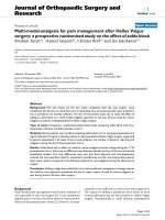
Determining the ideal therapeutic approach for a mus-
culoskeletal hydatid cyst not responding to medical treat-
ment can be quite challenging. Conservative treatment,
complete excision and simple drainage have al l been sug-
gested as treatment options [11]. Hydatid disease pro-
gresses slowly and is rarely life-th reatening, e specially
when located in the soft tissue or muscl es, thus support-
ing a conservative therapeutic approach. However, if the
Hydatid disease causes profound disabilities or mobiliza-
tion problems, complete cystopericystectomy and even
total joint replacement becomes an option. Radical surgi-
cal excision is especially indicated in cases of unilocular
manifestations as only this method offers hope of perma-
nent cure [10]. Therape utic dilemmas could arise in
cases of extended disease with many muscles or muscle
layers in different sites of the body which are communi-
cating via fistulas. Communication between lesions
should always be susp ected and rev ealed, even if primary
and daughter cysts are distant. Complete surgical treat-
ment should include the primary lesion, the d aughter
cysts and the communicating fistulas as a whole speci-
men. Bony pelvis is a difficult location for radical surg ical
excision of the Hydatid cyst and the morbidity of a Hind-
quarter amputation can be considerab le. Subtotal exci-
sion of the cyst and joint replacement is an acceptable
option based on o ur case report. Subtotal excision of the
Hydatidcystofthepelvisandahipreplacementcan
be durable providing adequate function. Patients should
be carefully monitored for cyst recurrence and compo-
nent loosening. Loose components due to recurrent cysts
can be successfully revised to provide good clinical
outcome.
Authors’ contributions
All authors contributed to the article. All authors have read and approved
the final manuscript.
Competing interests
The authors declare that they have no competing interests.
Received: 2 December 2009 Accepted: 11 March 2010
Published: 11 March 2010
References
1. Bal N, Kocer NE, Arpaci R, Ezer A, Kayaselcuk F: Uncommon locations of
hydatid cyst. Saudi Med J 2008, 29(7):1004-8.
2. Bellil S, Limaiem F, Bellil K, Chelly I, Mekni A, Haouet S, Kchir N, Zitouna M:
[Descriptive epidemiology of extrapulmonary hydatid cysts: a report of
265 Tunisian cases]. Tunis Med 2009, 87(2):123-6, French.
3. Dahniya MH, Hanna RM, Ashebu S, Muhtaseb SA, el-Beltagi A, Badr S, el-
Saghir E: The imaging appearances of hydatid disease at some unusual
sites. Br J Radiol 2001, 74:283-289.
4. Duncan GJ, Tooke SM: Echinococcus infestation of the biceps brachii. A
case report. Clin Orthop Relat Res 1990, 261:247-250.
5. Kural C, Ugras AA, Sungur I, Ozturk H, Erturk AH, Unsaldi T: Hydatid bone
disease of the femur. Orthopedics 2008, 31(7):712.
6. Lewall DB: Hydatid disease: biology, pathology, imaging and
classification. Clin Radiol 1988, 53:863-874.
7. Merkle EM, Schulte M, Vogel J, Tomczak R, Rieber A, Kern P, Goerich J,
Brambs HJ, Sokiranski R: Musculoskeletal involvement in cystic
echinococcosis: report of eight cases and review of the literature. AJR
Am J Roentgenol 1997, 168:1531-1534.
Table 1 Reported sites of Hydatid disease of the
musculoskeletal system.
Author No* Site of infection
Bal et al [1] 3 bone
Bellil et al [2] 6 bone
Merkle et al [7] 8 Iliopsoas, left adductor musculature, left
femur, left gluteus medius muscle,
musculature of right upper leg
Metcalf JE [8] 1 bone - humerus
Natarajan MV [9] 3 bone - femur
Torricelli et al [11] 14 bone infection with adjacent soft tissue
involvement in 12 cases
Dahniya et al [3] 7 5 bone infections without soft tissue
involvement, 2 primary intramuscular
(lt. shoulder, rectus femoris and vastus
lateralis)
* Number of patients
Figure 6 Radiograph one year after revision of the acetabular
component.
Neelapala et al. Journal of Orthopaedic Surgery and Research 2010, 5:17
/>Page 4 of 5
8. Metcalfe JE, Grimer RJ: Tackling osseous hydatidosis using orthopaedic
oncology techniques. Ann R Coll Surg Engl 2000, 82(4):287-9.
9. Natarajan MV, Kumar AK, Sivaseelam A, Iyakutty P, Raja M, Rajagopal TS:
Using a custom mega prosthesis to treat hydatidosis of bone: a report
of 3 cases. J Orthop Surg (Hong Kong) 2002, 10(2):203-5.
10. Siwach R, Singh R, Kadian VK, Singh Z, Jain M, Madan H, Singh S: Extensive
hydatidosis of the femur and pelvis with pathological fracture: a case
report. Int J Infect Dis 2009, 13(6):e480-2, Epub 2009 Apr 1.
11. Torricelli P, Martinelli C, Biagini R, Ruggieri P, De Cristofaro R: Radiographic
and computed tomographic findings in hydatid disease of bone. Skeletal
Radiol 1990, 19:435-439.
doi:10.1186/1749-799X-5-17
Cite this article as: Ne elapala et al.: Revis ion hip replacement for
recurrent Hydatid disease of the pelvis: a case report and review of the
literature. Journal of Orthopaedic Surgery and Research 2010 5:17.
Submit your next manuscript to BioMed Central
and take full advantage of:
• Convenient online submission
• Thorough peer review
• No space constraints or color figure charges
• Immediate publication on acceptance
• Inclusion in PubMed, CAS, Scopus and Google Scholar
• Research which is freely available for redistribution
Submit your manuscript at
www.biomedcentral.com/submit
Neelapala et al. Journal of Orthopaedic Surgery and Research 2010, 5:17
/>Page 5 of 5