báo cáo hóa học:" Granuloma debridement and the use of an injectable calcium phosphate bone cement in the treatment of osteolysis in an uncemented total knee replacement" doc
Bạn đang xem bản rút gọn của tài liệu. Xem và tải ngay bản đầy đủ của tài liệu tại đây (659.62 KB, 6 trang )
Atkinson et al. Journal of Orthopaedic Surgery and Research 2010, 5:29
/>Open Access
CASE REPORT
BioMed Central
© 2010 Atkinson et al; licensee BioMed Central Ltd. This is an Open Access article distributed under the terms of the Creative Commons
Attribution License ( which permits unrestricted use, distribution, and reproduction in
any medium, provided the original work is properly cited.
Case report
Granuloma debridement and the use of an
injectable calcium phosphate bone cement in the
treatment of osteolysis in an uncemented total
knee replacement
Henry D Atkinson*
1,2
, Vijai S Ranawat
1,2
and Roger D Oakeshott
2
Abstract
Polyethylene particulate debris-induced periprosthetic osteolysis is a known complication of knee arthroplasty surgery,
and may result in the need for revision surgery. The management of these bony defects can be surgically challenging,
and full revisions of well-fixed total knee components can lead to substantial bone loss. We present the case of a 71
year old man who developed knee pain and osteolysis around an uncemented total knee replacement. Due to
significant medical comorbidies he was treated by percutaneous cyst granuloma debridement and grafting using an
injectable calcium phosphate bone substitute. There were no wound complications, and the patient was allowed to
fully weight-bear post-operatively. Histopathology and microbiology of the cyst material confirmed polyethylene
granulomata without any evidence of infection. At 6 weeks post-operatively the patient's previous knee pain had
resolved, he was able to comfortably fully weight-bear. Preoperative scores (Knee Society Score (KSS) 41, WOMAC score
46.2, and Oxford Knee Score 39) had all improved at the 12-month post-operative review KSS 76, WOMAC 81.7 and
Oxford Knee score 21). This is a safe and effective technique with minimal morbidity and may be an appropriate
treatment modality when more extensive revision surgery is not possible. The case is discussed with reference to the
literature.
Background
Polyethylene particulate debris-induced periprosthetic
osteolysis is a known complication of knee arthroplasty
surgery, and may result in the need for revision surgery.
The management of these bony defects can be surgically
challenging, and full revisions of well-fixed total knee
components can lead to substantial bone loss.
Granuloma debridement and grafting of osteolytic
defects around well-fixed cementless implants has been
successfully performed in total hip arthroplasty [1-7]; and
recovery can be relatively quick as these osseointegrated
implants do not require postoperative activity restriction
[8]. Allograft bone chips are typically used, and though a
variety of bone substitutes have also been utilised, their
efficacy has not been widely documented.
We present the case of a patient with osteolysis occur-
ring around an uncemented total knee replacement,
treated by cyst granuloma debridement and grafting
using an injectable calcium phosphate bone substitute.
Case Report
In May 1996, a 59 year-old moderately obese farmer had
1 week-staged uncemented bilateral total knee replace-
ments without patella resurfacing. He had severe medial
and lateral compartment osteoarthritis with minimal
osteophytes on the patella, and had previously undergone
arthroscopic debridement procedures in both knees 2
years earlier. He made a good post-operative recovery,
with 0-110 degree active flexion by 6 weeks, normal lower
limb alignment and a resolution of knee pain symptoms,
returning to work at 3 months.
In May 1997 he began to develop increasing bilateral
anterior knee pain especially while walking on stairs and
inclines. Radiographs showed progression in the patellar
* Correspondence:
1
Department of Trauma and Orthopaedics and North London Sports
Orthopaedics, North Middlesex University Hospital, Sterling Way, London N18
1QX, UK
Full list of author information is available at the end of the article
Atkinson et al. Journal of Orthopaedic Surgery and Research 2010, 5:29
/>Page 2 of 6
osteoarthritis and a bone scan revealed increased uptake
in both patellae. He underwent bilateral synchronous
patellar resurfacings with cemented components in July
1997. There was no significant wear seen on either tibial
polyethylene spacer, however as there was florid synovitis
in both knees, the tibial spacers were exchanged and the
patient also had total synovectomies. Post-operatively he
recovered well with 0-110 knee flexion having been re-
gained by 3 months post-revision surgery.
However, in August 1998 he began developing some
medial tibial pain and mild swelling in the right knee,
which continued and by August 2000 had developed large
granulomatous cysts around the stem of the tibial com-
ponent. He underwent revision surgery on the right knee
in October 2000. Intraoperatively, he was once again
found to have massive synovitis and so underwent a total
synovectomy. There was a 3 × 3 cm uncontained bony
perforation medial to the tibial tubercle, and the tibial
polyethylene had badly delaminated and was excessively
worn. The tibial component was removed and the granu-
lomatous material removed from the proximal tibia. The
femoral component, which was poorly ingrown, was also
removed leaving an anterior cortical defect. The femoral
and tibial bony cortical defects were reconstructed with
anatomic specific allograft and the cavities impaction
grafted with morcelised bone. The knee was then revised
using stemmed cemented implants. Histolopathology
and microbiological analyses confirmed polyethylene
granulomata, polyethylene particulate debris and synovi-
tis, with no evidence of infection. The bone graft incor-
porated fully by 9 months and the right knee remains
pain-free with good function and a 0-125 degree range of
flexion 8 years later.
In January 2005 the patient began to develop similar
medial pain and swelling in the left knee. A bone scan
showed increased activity in the medial femoral condyle
indicating possible localised osteolysis, and there was
some low-grade uptake in the proximal tibia and suprap-
atellar pouch, though no abnormalities were seen on CT.
The patient underwent a left knee arthroscopy in May
2005, where he was once again found to have florid syno-
vitis though the patellar button and tibial spacer appeared
to be in a good condition. He underwent a full synovec-
tomy and had good post-operative clinical improvement
with a resolution of the knee swelling and pain by 3
months.
He remained symptom-free until 2008, when he began
developing medial pain in the left knee and difficulty
weight-bearing. His Knee Society Score (KSS) was 41,
WOMAC score was 46.2, and Oxford Knee Score 39.
Clinically there was no knee swelling or effusion, and he
did not appear to have active synovitis; the symptoms
appeared to be more characteristic of implant loosening.
Laboratory screening including erythrocyte sedimenta-
tion rate and C-reactive protein was normal. Plain radio-
graphs revealed areas of periprosthetic osteolysis in the
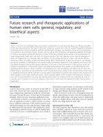
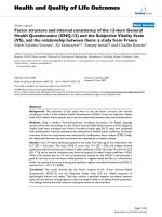
femur and the tibia, and a CT scan demonstrated an
extensive lytic area measuring 16 cm
3
in the medial femo-
ral condyle and a 3.6 cm
3
cyst in the medial tibial condyle;
both consistent with polyethylene particulate disease
(Figures 1 and 2). A further bone scan did not indicate
any clear evidence of component loosening, and succes-
sive knee radiographs had not shown obvious joint-space
narrowing. Nevertheless with this amount of bone loss
major revision knee surgery, similar to that performed on
the right knee, appeared to now be clinically and radio-
logically indicated in the left knee. Unfortunately over
this period the patient had developed renal failure and
had undergone renal transplant surgery in December
2005. He required daily oral steroid and immunosuppres-
sive therapy, and unfortunately still had poor renal func-
tion. His renal physicians advised against any further
major surgery. It was thus decided to perform a curettage
and grafting of these femoral and tibial bony defects
alone, to improve the patient's pain symptoms and to halt
any cyst progression, and subsequent fracture or cata-
strophic implant failure.
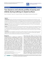
In December 2008, under a short general anaesthetic,
two 3 cm incisions were made over both the medial femo-
ral and medial tibial condyles. Two 4.5 mm drill holes
were made in both defects under image intensification,
and an arthroscopic chondrotome was introduced into
each defect to clear it of its granulomatous content.
Hydroset™ (Stryker Howmedica) calcium phosphate was
then inserted under direct vision into both defects, with
Figure 1 Axial CT of Femur demonstrating large lytic area in the
medial femoral condyle.
Atkinson et al. Journal of Orthopaedic Surgery and Research 2010, 5:29
/>Page 3 of 6
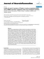
simultaneous venting to allow for free inflow of the mate-
rial (Figures 3 and 4). After 8 minutes the calcium phos-
phate had set, and the wounds were closed routinely
without drains. There were no wound complications, or
blood chemistry derangements, and the patient was
allowed to fully weight-bear post-operatively. Histopa-
thology and microbiology of the cyst material confirmed
polyethylene granulomata, without any evidence of infec-
tion. At 6 week review the patient's knee pain had
resolved, and he was able to comfortably fully weight-
bear. The graft material showed some signs of incorpora-
tion on x-ray by 4 months (Figures 5 and 6), and the
patient remains well at their 12 month review. KSS at
most recent view was 76, WOMAC score 81.7 and
Oxford Knee score 21. The patient will continue to have
regular clinical and radiological implant surveillance.
Discussion
When dealing with massive periprosthetic osteolysis the
aims of surgery are usually to restore bone stock around
the arthroplasty, gain stable implant fixation, restore the
joint mechanics and reduce the particle debris load [8].
This was successfully accomplished in the patient's con-
tralateral knee which underwent a full revision knee
arthroplasty with stemmed implants and extensive bone-
Figure 2 Sagittal CT demonstrating the lytic area in the antero-
medial aspect of the tibia.
Figure 3 Fluoroscopic image of the left knee showing the intro-
ducing cannula and simultaneous venting of the medial tibial
condylar cyst.
Figure 4 Fluoroscopic image of the left knee after cementation of
the medial femoral and medial tibial condyles.
Atkinson et al. Journal of Orthopaedic Surgery and Research 2010, 5:29
/>Page 4 of 6
grafting in 2000, following severe polyethylene delamina-
tion, osteolysis and component loosening. However,
treatment decisions are also very much dependent on
non-joint patient factors, such as the patient's clinical sta-
tus and physiological age, comorbidities, and activity lev-
els [8-11], and our patient was deemed no longer
medically fit to undergo any kind of extensive or invasive
surgery. He thus underwent minimally invasive cyst deb-
ridement and grafting alone.
One can argue that the cyst debridements should have
been performed in conjunction with a further total syn-
ovectomy and tibial polyethylene spacer exchange, in
order to remove any residual particulate material and the
wear particle generator [8]. Indeed modular polyethylene
exchange is a reasonable option when dealing with poly-
ethylene wear and osteolysis in patients with well-fixed
knee components [12,13], without evidence of acceler-
ated wear or severe delamination [10], and has similar
short-term results and failure rates when compared with
patients having full revisions of all the knee components
[8]. However, our patient had an arthroscopic synovec-
tomy almost 4 years earlier during which no visible poly-
ethylene delamination or macroscopic wear was seen,
and successive knee radiographs had not shown progres-
sive joint space narrowing. Additionally, the patient's pre-
senting symptoms were not consistent with active
synovitis, and the authors were keen to avoid the more
extensive surgery required for a bearing exchange that
may not have been necessary.
Traditionally, bony defects in revision knee arthroplasty
have been treated with autologous bone, allograft, polym-
ethylmethacrylate (PMMA) bone cement and implant
augments [9-11,14], however not all these options were
open to our patient. Given the minimally invasive nature
of the intended surgery, we considered using PMMA as a
void filler for our patient's osteolytic cysts. Cementation
would have had the advantage of providing immediate
stability [15,16], though would not have functioned as a
biological scaffold, and might have caused thermal necro-
sis of the surrounding bone possibly leading to further
osteolysis [17,18]. Autologous bone grafting was also
considered because of its biological advantages, as well as
its proven record in dealing with small tibial bony defects
[19], and with impaction grafting in knee and hip revision
arthroplasty [20].
However we decided instead to use Hydroset™, an
injectable osteoconductive calcium phosphate bone
cement, for its versatility and simplicity of use [21], and to
Figure 5 Lateral radiograph of the knee demonstrating the area
of calcium phosphate bone cementage in the medial femoral and
medial tibial condyles.
Figure 6 AP radiograph of the knee demonstrating the area of
calcium phosphate bone cementage in the medial femoral and
medial tibial condyles.
Atkinson et al. Journal of Orthopaedic Surgery and Research 2010, 5:29
/>Page 5 of 6
avoid any additional donor-site morbidity. Hydroset™ is
composed of a mixture of tetracalcium phosphate, dical-
cium phosphate dihydrate and trisodium citrate, which
crystallizes to form hydroxyapatite without an exother-
mic reaction, reaching 75% compressive strength by 4
hours and full strength at 24 hours [21]. It physically
interdigitates with the adjacent bone [21], and though not
designed to provide any structural support, forms a struc-
ture that is more mechanically stable than either cancel-
lous bone graft or bone substitute blocks or pellets [22-
24]. It has improved manual handling and mechanical
properties when compared with other calcium phosphate
and calcium sulphate cements [21,25,26] and creates a
scaffold for osteogenesis which is gradually replaced by
creeping substitution [21].
We were unable to find any data in the literature per-
taining to the use of bone cements in the context of oste-
olytic cysts in knee arthroplasty, however these analogues
have been successfully used in the treatment of benign
bone tumors, and for hardware augmentation in fracture
surgery, and have proven biocompatibility, bioactivity and
osteoconductivity [26-30] One study of 13 patients with
large expansive (mean volume of 38.5 mls) osteolytic
benign bone tumors, (similar in size to our patient's cysts)
used composite bioceramic osteoconductive grafts, com-
bining porous hydroxyapatite with calcium sulphate, fol-
lowing curettage and phenolisation. 11 of the13 lesions
displayed clinical and radiological consolidation at a
mean of 4.6 months, and patients had good return to nor-
mal function [31]. Another study of 23 patients with bone
cysts or benign bone tumors (measuring a mean of 23
mls) treated with calcium-sulphate pellets with or with-
out added demineralised bone matrix, found complete
bone regeneration by 6 months in both treatment groups
[32]. A multicenter trial of 46 patients with benign bone
tumors (ranging in size from 0.15 to 112 mls), found 96%
bony ingrowth and 100% graft resorption at 12 months
when using the same calcium-sulphate pellets, with no
difference between those patients treated with calcium-
sulphate alone or in combination osteoinductive agents
[33]. Additionally, a recent meta-analysis of 14 random-
ized controlled trials using calcium phosphate bone
cement for augmentation of metaphyseal fracture fixation
(in the tibial plateau, femoral neck and calcaneum), found
that patients had a lower prevalence of pain at the frac-
ture site and a decrease in the loss of fracture reduction,
compared with those patients treated with autogenous
bone graft [34].
Conclusion
Our patient had no intraoperative or postoperative com-
plications. He has had a dramatic improvement in KSS,
WOMAC and Oxford Knee scores and remains well and
fully ambulatory at 12 months. We believe that injectable
osteoconductive calcium phosphate bone cements may
be a useful adjunct in treating osteolytic cysts around
well-fixed knee replacement components.
Consent
Written informed consent was obtained from the patient
for publication of this case report and any accompanying
images. A copy of the written consent is available for
review by the Editor-in-Chief of this journal
Author information
Henry D Atkinson, MBChB, BSc, FRCS Tr & Orth
Department of Trauma and Orthopaedics and North
London Sports Orthopaedics, North Middlesex Univer-
sity Hospital, Sterling Way, London N18 1QX, UK.
and: Sportsmed SA, 32 Payneham Road, Stepney 5069,
Adelaide, South Australia.
Vijai S Ranawat, MBBS, FRCS Tr & Orth
Department of Trauma and Orthopaedics, The Whit-
tington Hospital, Highgate Hill, Archway, London N19
5NF, UK.
and: Sportsmed SA, 32 Payneham Road, Stepney 5069,
Adelaide, South Australia.
Roger D Oakeshott, MBBS, FAOrthA, FRACS
Sportsmed SA, 32 Payneham Road, Stepney 5069, Ade-
laide, South Australia.
Abbreviations
WOMAC: Western Ontario and McMaster osteoarthritis index; CT: Computed
Tomography; KSS: Knee Society Score; PMMA: polymethylmethacrylate.
Competing interests
The authors declare that they have no competing interests.
Authors' contributions
RO operated the patient and is the senior author. HA managed the patient and
wrote the manuscript. VR assisted with the literature review and manuscript
preparation.
All authors have read and approved the final manuscript.
Author Details
1
Department of Trauma and Orthopaedics and North London Sports
Orthopaedics, North Middlesex University Hospital, Sterling Way, London N18
1QX, UK and
2
Sportsmed SA, 32 Payneham Road, Stepney, Adelaide 5069,
Australia
References
1. Maloney WJ, Herzwurm P, Paprosky W, Rubash HE, Engh C: Treatment of
pelvic osteolysis associated with a stable acetabular component
inserted without cement as part of a total hip replacement. J Bone Joint
Surg [Am] 1997, 79:1628-34.
2. Maloney WJ, Paprosky W, Engh CA, Rubash H: Surgical treatment of
pelvic osteolysis. Clin Orthop Relat Res 2001, 393:78-84.
3. Rubash HE, Sinha RK, Maloney WJ, Paprosky WG: Osteolysis: Surgical
treatment. Instr Course Lect 1998, 47:321-9.
4. Maloney WJ: The surgical management of femoral osteolysis. J
Arthroplasty 2005, 20(4 suppl 2):75-8.
5. O'Brien JJ, Burnett RS, McCalden RW, MacDonald SJ, Bourne RB, Rorabeck
CH: Isolated liner exchange in revision total hip arthroplasty: Clinical
results using the direct lateral approach. J Arthroplasty 2004, 19:414-23.
Received: 23 November 2009 Accepted: 27 April 2010
Published: 27 April 2010
This article is available from: 2010 Atkinson et al; licensee BioMed Central Ltd. This is an Open Access article distributed under the terms of the Creative Commons Attribution License ( which permits unrestricted use, distribution, and reproduction in any medium, provided the original work is properly cited.Journal of Orthopaedic Surgery and Research 2010, 5:29
Atkinson et al. Journal of Orthopaedic Surgery and Research 2010, 5:29
/>Page 6 of 6
6. Terefenko KM, Sychterz CJ, Orishimo K, Engh CA: Polyethylene liner
exchange for excessive wear and osteolysis. J Arthroplasty 2002,
17:798-804.
7. Benson ER, Christensen CP, Monesmith EA, Gomes SL, Bierbaum BE:
Particulate bone grafting of osteolytic femoral lesions around stable
cementless stems. Clin Orthop Relat Res 2000, 381:58-67.
8. Maloney W, Rosenberg A: What is the outcome of treatment for
osteolysis? J Am Acad Orthop Surg 2008, 16(Suppl 1):S26-32.
9. Backstein D, Safir O, Gross A: Management of bone loss: Structural grafts
in revision total knee arthroplasty. Clin Orthop Relat Res 2006,
446:104-12.
10. Engh GA, Herzwurm PJ, Parks NL: Treatment of major defects of bone
with bulk allografts and stemmed components during total knee
arthroplasty. J Bone Joint Surg [Am] 1997, 79:1030-9.
11. Fehring TK, Odum S, Olekson C, Griffin WL, Mason JB, McCoy TH: Stem
fixation in revision total knee arthroplasty: A comparative analysis. Clin
Orthop Relat Res 2003, 416:217-24.
12. Griffin WL, Scott RD, Dalury DF, Mahoney OM, Chiavetta JB, Odum SM:
Modular insert exchange in knee arthroplasty for treatment of wear
and osteolysis. Clin Orthop Relat Res 2007, 464:132-7.
13. Jensen CL, Petersen MM, Jensen KE, Therbo M, Schrøder HM: Outcome of
isolated tibial polyethylene insert exchange after uncemented total
knee arthroplasty: 27 patients followed for 8-71 months. Acta Orthop
2006, 77:917-20.
14. Gupta SK, Chu A, Ranawat AS, Slamin J, Ranawat CS: Osteolysis after total
knee arthroplasty. J Arthroplasty 2007, 22:787-99.
15. Pals SD, Wilkins RM: Giant cell tumor of bone treated by curettage,
cementation, and bone grafting. Orthopedics 1992, 15:703-8.
16. O'Donnell RJ, Springfield DS, Motwani HK, Ready JE, Gebhardt MC, Mankin
HJ: Recurrence of giant-cell tumors of the long bones after curettage
and packing with cement. J Bone Joint Surg [Am] 1994, 76:1827-33.
17. Persson BM, Wouters HW: Curettage and acrylic cementation in surgery
of giant cell tumors of bone. Clin Orthop Relat Res 1976, 120:125-33.
18. Persson BM, Ekelund L, Lovedahl R, Gunterberg B: Favourable results of
acrylic cementation for giant cell tumors. Acta Orthop Scand 1984,
55:209-14.
19. Ahmed I, Logan M, Alipour F, Dashti H, Hadden WA: Autogenous bone
grafting of uncontained bony defects of tibia during total knee
arthroplasty a 10-year follow up. J Arthroplasty 2008, 23(5):744-50.
20. Steens W, Loehr JF, Wodtke J, Katzer A: Morselized bone grafting in
revision arthroplasty of the knee: a retrospective analysis of 34
reconstructions after 2-9 years. Acta Orthop 2008, 79(5):683-8.
21. Larsson S: Cement augmentation in fracture treatment. Scand J Surg
2006, 95:111-8.
22. Yetkinler DN, Goodman SB, Reindel ES, Carter D, Poser RD, Constantz BR:
Mechanical evaluation of a carbonated apatite cement in the fixation
of unstable intertrochanteric fractures. Acta Orthop Scand 2002,
73(2):157-64.
23. Yetkinler DN, Ladd AL, Poser RD, Constantz BR, Carter D: Biomechanical
evaluation of fixation of intra-articular fractures of the distal part of the
radius in cadavera: Kirschner wires compared with calcium-phosphate
bone cement. J Bone Joint Surg [Am] 1999, 81(3):391-9.
24. Elder S, Frankenburg E, Goulet J, Yetkinler D, Poser R, Goldstein S:
Biomechanical evaluation of calcium phosphate cement-augmented
fixation of unstable intertrochanteric fractures. J Orthop Trauma 2000,
14(6):386-93.
25. Ogose A, Hotta T, Kawashima H, Kondo N, Gu W, Kamura T, Endo N:
Comparison of hydroxyapatite and beta tricalcium phosphate as bone
substitute after excision of bone tumors. J Biomed Mater Res B Appl
Biomater 2005, 72:94-101.
26. Friedman CD, Costantino PD, Takagi S, Chow LC: BoneSource
hydroxyapatite cement: a novel biomaterial for craniofacial skeletal
tissue engineering and reconstruction. J Biomed Mater Res 1998,
43(4):428-32.
27. LeGeros RZ: Properties of osteoconductive biomaterials: calcium
phosphates. Clin Orthop Relat Res 2002, 395:81-98.
28. Vaccaro AR: The role of osteoconductive scaffold in synthetic bone
graft. Orthopedics 2002, 25(5 Suppl):S571-8.
29. Shors E, Holmes R: Bone formation in porous hydroxyapatite obtained
from human biopsies. Bioceramics 1993, 6:375-9.
30. Kurashina K, Kurita H, Kotani A, Takeuchi H, Hirano M: In vivo study of a
calcium phosphate cement consisting of alpha-tricalcium phosphate/
dicalcium phosphate dibasic/tetracalcium phosphate monoxide.
Biomaterials 1997, 18(2):147-51.
31. Schindler OS, Cannon SR, Briggs TW, Blunn GW: Composite ceramic bone
graft substitute in the treatment of locally aggressive benign bone
tumours. J Orthop Surg (Hong Kong) 2008, 16:66-74.
32. Gitelis S, Piasecki P, Turner T, Haggard W, Charters J, Urban R: Use of a
calcium sulfate-based bone graft substitute for benign bone lesions.
Orthopedics 2001, 24:162-6.
33. Kelly CM, Wilkins RM, Gitelis S, Hartjen C, Watson JT, Kim PT: The use of a
surgical grade calcium sulfate as a bone graft substitute: results of a
multicenter trial. Clin Orthop Relat Res 2001, 382:42-50.
34. Bajammal SS, Zlowodzki M, Lelwica A, Tornetta P, Einhorn TA, Buckley R,
Leighton R, Russell TA, Larsson S, Bhandari M: The use of calcium
phosphate bone cement in fracture treatment. A meta-analysis of
randomized trials. J Bone Joint Surg [Am] 2008, 90:1186-96.
doi: 10.1186/1749-799X-5-29
Cite this article as: Atkinson et al., Granuloma debridement and the use of
an injectable calcium phosphate bone cement in the treatment of osteolysis
in an uncemented total knee replacement Journal of Orthopaedic Surgery and
Research 2010, 5:29