báo cáo hóa học:" Treatment of refractory hip pain with sodium hyaluronate (Hyalgan©) in a patient with the Marshall-Smith Syndrome: A case report" pot
Bạn đang xem bản rút gọn của tài liệu. Xem và tải ngay bản đầy đủ của tài liệu tại đây (2.5 MB, 5 trang )
CAS E REP O R T Open Access
Treatment of refractory hip pain with sodium
hyaluronate (Hyalgan
©
) in a patient with the
Marshall-Smith Syndrome: A case report
Matthew Salter
*
, Chandoo Kalmat, Henry Kroll, David Kim
Abstract
The Marshall Smith Syndrome (MSS) is a rare congenital disorder, displaying a constellation of unique symptoms,
including orofacial dysmorphisms, accelerated osseous maturation and dysplasias, mental retardation, and respira-
tory maladies. Few individuals with MSS survive past early childhood. In this case report, we describe a unique
treatment for a 30 year-old patient with MSS who presented to our pain medicine clinic for management of pa in
secondary to uncontrolled bilateral hip dysplasias.
Background
The Marshall-Smith Syndrome was first described in 1971
by Marshall et al as a rare congenital disorder, and to date
there are fewer than 40 reported cases [1-3]. The etiology
is unknown but is presumed to be due to a de novo domi-
nant mutation. It is characterized by a constellation of fea-
tures involving the neurologicandrespiratorysystems,
and accelerated skeletal maturation leading to skeletal dys-
plasias. Patients have retarded intellectual development,
small chins, glossoptosis, prominent eyes, protruding fore-
heads and are small in stature. They generally do not sur-
vive past early childhood mainly due to respiratory
complications, such as aspiration pneumonia. However, if
the respiratory conditions are managed aggressively,
patients have been known to survive longer. To our
knowledge, our patient is one of the oldest living patients
with this rare disorder [3]. Given the natural history of the
syndrome, one would anticipate that older subjects would
suffer a great deal of pain as a result of the accelerated ske-
letal maturation. We report a unique treatment of incapa-
citating bilateral hip pain in a 30 year-old MSS patient
with intra-articular hyaluronate (Hyalgan
©
).
Case Report
L.W. is a 30 year- old woman with MSS. Her medical his-
tory was obtained from her pa rents, who accompanied
her and upon whom she was totally depen dent for care.
The patient had speech and cognitive impairment that
limited the abilit y to obtain a direct history. The parents
described a history of worsening hip pain from progres-
sive, bilateral hip dysplasias. Whereas previously th eir
daughter could ambulate with assistance, she was now
incapacitated by relentless pain. As best as they were able
to determine, the pain radiated from her hips laterally,
down her thighs and provoked regular paroxysms of
screaming, crying and guarding.
Prior to our encounter, this pain had been managed
by an orthopedic surgeon at an outside faci lity, who
performed a series of nine ultrasound-guided intra-
articular hip steroid injections, over a period of several
years. The last one was perf ormed a few months before
presenting. None of the patient’spreviousrecordswere
available to us, but the parents relayed that their daugh-
ter’s response to the injections had begun to wane with
each repeat injection. The most recent recommendation
from the orthopedist was to perform bilat eral hip
arthroplasties. The parents were hesitant to pursue this
option, in light of their daughter’sprevioussurgical
experience, wherein she required an emergency tra-
cheostomy after failed attempts at securing her airway
under anesthesia. Because they were told that future
surgeries would require an “ awake” tracheostomy for
airway protection during surgery, they decided to seek
alternative, non-surgical treatments for their daughter’s
hip pain.
* Correspondence:
Henry Ford Hospital, Department of Anesthesiology, 2799 West Grand
Boulevard, Detroit, MI, USA, 48202
Salter et al. Journal of Orthopaedic Surgery and Research 2010, 5 :61
/>© 2010 Salter et al; licensee BioMed Central Ltd. This is an Open Access article distributed under the terms of the Creative Commons
Attribution License (http: //creati vecommons.org/licenses/by/2.0), which permits unrestricted use, distribution, and reproduction in
any medium, provided the original work is properly cited.
The rest of the review of systems was unremarkable.
Notably, the parents denied any history of bleeding dia-
thesis. Though previous diagnostic imaging was not
available at the time of our initial consultation, they
were reviewed at a later date, prior to treatment, and
revealed dysplasia of bo th acetabula, and severe osteoar-
thritis and subluxation of both hip joints.
On physical examination, the patient was small in sta-
ture (4ft 2in, 65lbs) and had obvious craniofacial
abnormalities. The neuromuscular exam was limited
due to lack of patient cooperation. The greater trochan-
ters were asymmetrical, with the right side about 2 cm
superior compared to the left. Both were easily palpable
and visibly appeared to be grossly out of socket. Though
muscle tone was good with no flaccidity, the patient’s
inability to obey commands prevented us from assessing
motor or sensory function. Reflex testing revealed patel-
lar and Achilles hyporeflexia.
Based on the patient’s history, physical examination,
and radiographic findings, our impression was that the
patient’s symptoms arose directly from the articular sur-
faces of her hips, and possibly from the bilateral impin-
gement of her lateral femoral cutaneous nerves, as a
result of her inadequately developed acetabula and sub-
luxed femurs. We suggested a series of three fluorosco-
pically guided intra-articular hip injections with sodium
hyaluronate (Hyalgan
©
), administered weekly. In the
event of an unsatisfactory result from the injec tions, we
intended to perform bilateral lateral femoral cutaneous
nerveblocks.Thepatient’s parents elected to pursue
injection of sodium hyaluronate, and scheduled an
appointment for the procedure.
Method
After discussing the risks, benefits, and alternatives on
the day of the procedure, informed consent was obtained
from the patient’s parents. Her parents were present dur-
ing the entire procedure, to facilitate cooperation. After
placement on the fluoroscopy table in the supine posi-
tion, the region of the greater trochanters were cleansed
with chlorhexidine (Cholraprep
©
) and draped fully. The
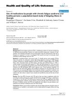
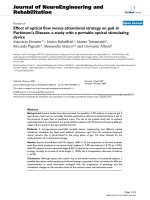
fluoroscopic camera was positioned to visualize the right
greater trochanter, femoral neck and acetabulum in the
AP projection (Figure 1). Using a sterile marker, we
marked the needle insertion site one centime ter cephalad
to the gre ater trochanter. A 25-gauge, 1-1/2 inch needle
was used to infiltrate the skin with 1% lidocaine. Sub se-
quently, a 3-1/2 inch, 22-gauge spinal needle was
advanced to the femoral neck and into the joint capsule
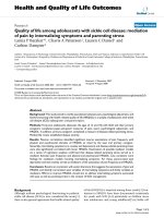
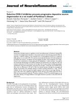
at this level. Following this, after n egativ e aspiration for
blood, 1 milliliter (ml) of iopamidol-300 (Isovue-300
©
)
dye was injected, verifying intra-articular spread of the
dye (Figure 2). This was followed with an injection of 2
mL of 0.5% preservative-free bupivacaine and 2 ml
(20 mg) of Hyalgan
©
intothejointspace.Thesamepro-
cedure was repeated on the left hip. The patient tolerated
the procedure well without complications. We discharged
the patient home after meeting discharge criteria. The
patient returned to our clinic for a total of three injec-
tions of Hyalgan
©
, separated by one week.
Discussion
Marshall et al. first described this r are congenital dis-
order in 1971 as a sporadic entity of unknown etiology
Figure 1 A fluoroscopic image of the patient’s right hip, taken
prior to needle insertion for intra-articular hip injection.
Figure 2 A right hip fluoroscopic image with needle in place,
showing spread of contrast within joint capsule. Contrast spread
is apparent from the base of the femoral neck and around joint.
Salter et al. Journal of Orthopaedic Surgery and Research 2010, 5 :61
/>Page 2 of 5
[4,5]. Patients classically have a normal karyotype and
are born of nonconsanguineous parents [4]. Most signif-
icantly, they display skeletal maturation that is advanced
for age, along with osteopenia and sclerosis, leading
some to describe this kind of ossification as “disharmo-
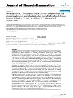
nic” [6-8]. Skeletal abnormalities like broadened pha-
langes with abnormal epiphyses, thinned or bowing long
bones, and dysmorphic vertebrae leading to scoliosis,
kyphosis, or cervica l, thoracic, or lumbar instabili ty are
commonly present [[3-5,7-9], Figure 3, Figure 4, Figure
5, Figure 6]. Specific facial anomalies may include
hypertrichosis, prominent eyes and forehead (frontal
bossing), megalocornea, blue sclerae, a flat nasal bridge,
micrognathia, and anteverted nostrils [5,10] . Neurologic
derangements may consist of an absent corpus callosum,
macrogyria, ventricular dilatation or hydrocephalus,
periventricular leukomalacia, resulting in motor and
mental retardation [5,10]. They might a lso display optic
nerve hypoplasia [11]. It should be noted that not all
patients afflicted with MSS display the same anatomic
findings.
Most infants affected with MSS succumb to death
early in life. This is usually due to pulmonary complica-
tions, such as aspiration and chronic lung obstruction
leading to pulmonary infections, pulmonary hyperten-
sion, and right heart failure [2,4,5,10-13].
The concept of applying viscosupplementation (VS),
using modified hyaluronic acid to form hyaluronan s
(HA) and their cross-linked derivatives, to the treat-
ment of osteoarthritis arose in the mid 1970’s, though
these compounds had also been investigated for use in
various ophthalmologic procedures [14-17]. As joints
affected by osteoarthritis are depleted of their natural
synovial fluid, which contains the glycosaminoglycan
hyaluronic acid, it was postulated that injecting exo-
genous HA would increase the viscosity and elasticity
to the joint, thereby improving joint function and
relieving symptoms [18,19]. While research on, and
FDA-approval of, VS is primarily for use in the treat-
ment of knee osteoarthritis, it has also been used suc-
cessfully in small trials for the treatment of arthritis of
the temporomandibular, sacroiliac, hip, shoulder, foot,
and ankle joints [17,20-23]. In 1997, Hyalgan® (sodium
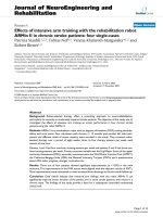
Figure 3 A wrist/hand X-ray of a female with MSS, at
chronological age of 18 weeks displaying wide phalanges &
stippled epiphyses, showing bone age of 1.9 years [9].
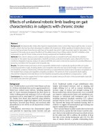
Figure 4 An X-ray showing thinned ribs and dysmorphic
lumbar vertebrae in a 4-week old male with MSS [4].
Salter et al. Journal of Orthopaedic Surgery and Research 2010, 5 :61
/>Page 3 of 5
hyaluronate), a high molecular-w eight HA obtained
from rooster’ s combs, was approved by the FDA
[24,25]. Like o ther HA, it is only approved for osteoar-
thritis of the knee, and should be used with caution in
patients with sensitivities to avian proteins, feathers,
and egg products. It also carries a similar side-effect
profile, including local inflammation, injection site
pain and itching, anaphylaxis/anaphylactoid reactions,
local ecchymosis, nausea/vomiting, diarrhea, anorexia,
and headache [26]. While Hyalgan
©
has a relatively
short half-life of about twenty-four hours, the effect of
the injections lasts for weeks, which suggests that it
augments natural synovium production, mitigates
nociception and inflammation, as well as increases the
rheological properties (viscosity, elasticity, pseudopla s-
ticity) of synovial fluid [27-29].
Our patient ’s survival to adulthood is somewhat
anomalous, compared to the life expectancy of others
afflicted with MSS. Therefore, we c an only assume that
if other carriers of the disease survived the historically
perilous respiratory maladies of childhood, they too
would suffer from the chronic pain of disharmo nic ske-
letal development and ensuing arthralgias, as we
observed in our patient, and as it has been noted in
other carriers [13]. L.W. underwent a series of three
intra-articular bilateral hip injections one week apart,
and gradual improvement in sympto ms over this period
was noted. At the time of the first visit, the patient
arrived in her wheelchair, and passive movement of her
hips caused her great distress. Two months after the last
injection, we reevaluated the patient in our clinic. She
arrived for her appointment walking,semi-indepen-
dently, with her parents on either side of h er. They felt
that the injections successfully decreased the frequency
and intensity of her painful episodes, noting a marked
improvement in her daily functioning.
Conclusion
We describe a unique treatment alternative for a patient
with Marshall Smith Syndrome and deb ilitating, painful
bilat eral hip dysplasias using intra-articular sodium hya-
luronate injections. This management option should be
Figure 5 Femur and Tibia X-ray of 7 year-old female with MSS
demonstrating dysplastic hips with shallow, horizontal
acetabula, post-traumatic bowing, Arrow indicates healing
tibial fracture. Diaphyses are gracile with thin cortices and
obliterated medullae, in contrast with widened metaphyses and
epiphyses, which are relatively spared [13].
Figure 6 X-ray of lumbar spine demonstrating an apex left
45 degree scoliosis in the same 7 year-old female with MSS [13].
Salter et al. Journal of Orthopaedic Surgery and Research 2010, 5 :61
/>Page 4 of 5
considered in one’ s armamentarium, especially in the
high-risk surgical population.
Competing interests
The authors declare that they have no competing interests.
Authors’ contributions
MS and CK conceived the project and conducted the primary literature
review and manuscript composition. HK and DK contributed additional data
to the literature review and manuscript. All authors read and approved the
final manuscript.
Consent
Written informed consent was obtained from the parent/guardian of the
patient for publication of this case report and accompanying images. A
copy of the written consent is available for review by the Editor-in-Chief of
this journal.
Received: 10 April 2010 Accepted: 23 August 2010
Published: 23 August 2010
References
1. Marshall R, Graham B, Scott C, Smith D: Syndrome of Accelerated Skeletal
Maturation and Relative Failure to Thrive: A Newly Recognized Clinical
Growth Disorder. J Ped 1971, 78(1):95-101.
2. Johnson J, Carey J, Glassy F, Paglieroni T, Lipson M: Marshall-Smith
Syndrome: Two Case Reports and a Review of Pulmonary
Manifestations. Pediatrics 1983, 71:219-21.
3. Travan L, Oretti C, Zennaro F, Demarani S: Marshall-Smith Syndrome and
Septo-Optic Dysplasia: An Unreported Association. Am J Med Gen Part A
2008, 146A:2138-2140.
4. Dernedde G, Pendeville P, Veyckemans F, Verellen G, Gillerot Y: Anaesthetic
Management of a Child with Marshall-Smith Syndrome. Can J Anaesth
1998, 45(7):660-663.
5. Yoder C, Wiswell T, Cornish J, Cunningham B, Crumbaker D: Marhsall-Smith
Syndrome: Further Delineation. South Med J 1998, 81(10):1297-300.
6. Sperli D, Concolino D, Barbato C, Strisciuglio P, Andria G: Long Survival of
a Patient with Marshall-Smith Syndrome without Respiratory
Complications. J Med Genet 1993, 30:877-879.
7. Adam M, Hennekam R, Keppen L, Bull M, Clericuzio C, Burke L, Ormond K,
Hoyme H: Natural History and Evidence of an Osteochondrodysplasia
with Connective Tissue Abnormalities. American Journal of Medical
Genetics 2005, 137A:117-124.
8. Eich G, Silver M, Weksberg R, Daneman A, Costa T: Marshall-Smith
Syndrome: New Radiographic, Clinical, and Pathologic Observations.
Radiology 1991, 181(1):183-188.
9. Williams D, Carlton D, Green S, Pearman K, Cole T: Marshall-Smith
Syndrome: The Expanding Phenotype. J Med Genet 1997, 34:842-845.
10. Summers D, Cooper H, Butler M: Marshall-Smith Syndrome: Case Report
of a Newborn Male and Review of the Literature. Clin Dysmorph 1999,
8(3):207-10.
11. Desphande C, Forrest M, Russell-Eggitt I, Hall C, Mehta R, Paterson J: Visual
Impairment and Prolonged Survival in a Girl with Marshall-Smith
Syndrome. Clin Dysmorph 2006, 15:111-113.
12. Cullen A, Clarke T, O’Dwyer T: The Marshall-Smith Syndrome: A Review of
Laryngeal Complications. Eur J Pediatr 1997, 156:463-464.
13. Diab M, Raff M, Gunther D: Osseous Fragility in Marshall-Smith Syndrome.
Amer J Med Gen 2003, 119A:218-222.
14. Wen D: Intra-articular Hylarunonic Acid Injections for Knee Osteoarthritis.
American Family Physician 2001, 62
:565-70.
15. De La Pena E, Sala S, Rovira J, Schmidt R, Belmonte C: Elastoviscous
Substances with Analgesic Effects on Joint Pain Reduce Stretch-
Activated Ion Channel Activity in Vitro. Pain 2002, 99:501-508.
16. Srejic U, Calvillo O, Kabakibou K: Viscosupplementation: A New Concept in
the Treatment of Sacroiliac Joint Syndrome: A Preliminary Report of
Four Cases. Regional Anesthesia and Pain Medicine 1999, 24(1):84-88.
17. Itokazu M, Matsunaga T: Clinical Evaluation of High-Molecular-Weight
Sodium Hyaluronate for the Treatment of Patients with Periarthritis of
the Shoulder. Clinical Therapeutics 1995, 17(5):946.
18. Smith G, Myers S, Brandt K, Mickler E: Effect of Intra-articular Hyaluronan
Injection in Experimental Canine Osteoarthritis. Arthritis and Rheumatism
1998, 41(6):976-985.
19. Bannuru R, Natov N, Obadan I, Price L, Schmid C, McAlindon T: Therapeutic
Trajectory of Hyaluronic Acid Versus Corticosteroids in the Treatment of
Knee Osteoarthritis: A Systematic Review and Meta-analysis. Arthritis Care
& Management 2009, 61(12):1704-1711.
20. Perdikis D: Sustained Effect of Hyaluronate Viscosupplementation in
Chronic Sacroiliac Joint Pain. The Journal of Pain 2006, 7(4):S34.
21. Salk R, Chang T, D’Costa W, Soomekh D, Grogan K: Sodium Hyaluronate in
the Treatment of Osteoarthritis of the Ankle: A Controlled, Randomized,
Double-blind pilot study. The Journal of Bone and Joint Surgery 2006,
88:295-302.
22. Perdikis D: Intra-articular Hyaluronate Improves Chronic
Temporomandibular Joint Pain. The Journal of Pain 2006, 7(4):S62.
23. Van den Bekerom M, Lamme B, Sermon A, Mulier M: What is the Evidence
for Viscosupplementation in the Treatment of Patients with Hip
Osteoarthritis? Systematic Review of the Literature. Archives of
Orthopaedic and Trauma Surgery 2008, 128:815-823.
24. Briem K, Axe M, Snyder-Mackler L: Medical Knee Joint Loading Increases
in Those Who Respond to Hyaluronan Injection for Medical Knee
Osteoarthritis. Journal of Orthopoedic Research 2009, 27:1420-1425.
25. Sanofi Aventis/Hyalgan. [ />Complete_Patient_Information.pdf].
26. Juni P, Reichenbach S, Trelle S, Tschannen B, Wandel S, Jordi B, Zullig M,
Guetg R, Hauselmann H, Schwarz H, Theiler R, Ziswiler H, Dieppe P,
Villiger P, Egger M: Efficacy and Safety of Intraarticular Hylan or
Hyaluronic Acids for Osteoarthritis of the Knee. Arthritis & Rheumatism
2007, 56(11):3610-3619.
27. Jansen E, Emans P, Douw C, Guldemond N, Van Rhijn L, Bulstra S, Kuijer R:
One Intra-Articular Injection of Hyaluronan Prevents Cell Death and
Improves Cell Metabolism in a Model of Injured Articular Cartilage in
the Rabbit. Journal Orthopaedic Research 2008, 26(5):624-630.
28. Gomis A, Miralles A, Schmidt R, Belmonte C: Nociceptive Nerve Activity in
an Experimental Model of Knee Joint Oteoarthritis of the Guinea Pig:
Effect of Intra-articular Hyaluronan Application. Pain 2007, 130:126-136.
29. Jean Y, Wen Z, Chang Y, Lee H, Hsieh S, Wu C, Yeh C, Wong C: Hyaluronic
Acid Attenuates Osteoarthritis Development in the Anterior Cruciate
Ligament-Transected Knee: Association with Excitatory Amino Acid
Release in the Joint Dialysate. Journal of Orthopaedic Research 2006,
24(5):1052-1061.
doi:10.1186/1749-799X-5-61
Cite this article as: Salter et al.: Treatment of refractory hip pain with
sodium hyaluronate (Hyalgan
©
) in a patient with the Marshall-Smith
Syndrome: A case report. Journal of Orthopaedic Surgery and Research
2010 5:61.
Submit your next manuscript to BioMed Central
and take full advantage of:
• Convenient online submission
• Thorough peer review
• No space constraints or color figure charges
• Immediate publication on acceptance
• Inclusion in PubMed, CAS, Scopus and Google Scholar
• Research which is freely available for redistribution
Submit your manuscript at
www.biomedcentral.com/submit
Salter et al. Journal of Orthopaedic Surgery and Research 2010, 5 :61
/>Page 5 of 5