báo cáo hóa học:" What do standard radiography and clinical examination tell about the shoulder with cuff tear arthropathy?" pptx
Bạn đang xem bản rút gọn của tài liệu. Xem và tải ngay bản đầy đủ của tài liệu tại đây (407.11 KB, 7 trang )
RESEARCH ARTICLE Open Access
What do standard radiography and clinical
examination tell about the shoulder with cuff
tear arthropathy?
Bart Middernacht
1*
, Philip Winnock de Grave
1
, Georges Van Maele
1
, Luc Favard
2
, Daniel Molé
3
, Lieven De Wilde
1
Abstract
Background: This study evaluates the preoperative conventional anteroposterior radiography and clinical testing in
non-operated patients with cuff tear arthropathy. It analyses the radiological findings in relation to the status of the
rotator cuff and clinical status as also the clinical testing in relation to the rotator cuff quality. The aim of the study
is to define the usefulness of radiography and clinical examinatio n in cuff tear arthropathy.
Methods: This study analyses the preoperative radiological (AP-view, (Artro-)CT-scan or MRI-scan) and clinical
characteristics (Constant-Murley-score plus active and passive mobility testing) and the peroperative findings in a
cohort of 307 patients. These patients were part of a multicenter, retrospective, consecutive study of the French
Orthopaedic Society (SOFCOT-2006). All patients had no surgical antecedents and were all treated with prosthetic
shoulder surgery for a painful irreparable cuff tear arthropathy (reverse-(84%) or hemi-(8%) or double cup-bipolar
prosthesis (8%)).
Results: A positive significancy could be found for the relationship between clinical testing and the rotator cuff
quality; between acromiohumeral distance and posterior rotator cuff quality; between femoralization and posterior
rotator cuff quality.
Conclusion: A conventional antero-posterior radiograph can not provide any predictive information on the clinical
status of the patient.
The subscapular muscle can be well tested by the press belly test and the teres minor muscle can be well tested
by the hornblower’ sign and by the exorotation lag signs.
The upward migration index and the presence of femoralization are good indicators for the evaluation of the
posterior rotator cuff.
An inferior coracoid tip positioning suggests rotator cuff disease.
Background
Painful cuff tear arthropathy (CTA) affects the i ndepen-
dence of the elderly [1,2] by altering the biomechanics
[3] and bony characteristics of the normal glenohumeral
joint [4,5]. C TA is a progressive disease which presents
a unique therapeutical challenge necessitating an algo-
rithm for treatment based on clinic al and radiological
parameters [6].
The seriousness of the disease is evaluate d clinically
and radiologically.
The Constant and Murley score [7] is a well accepted
clinical method to evaluate pain, activities of daily living,
passive motion, and active motion. Clinical lag signs
seem to have an important predictive value in the assess-
ment of the location and the size of the tear [8]. Plain
radiographs are known, since longtime [9], to be a sensi-
tive diagnostic tool to evaluate rotator cuff disorders.
A conventional antero-posterior radiograph of the
shoulder is the most frequently performed examination
to study structural bony wear in CTA [2,10-18]. These
structural changes include a small or absent acromio-
humeral distance [17,18], an ascension and/or medializa-
tion of the center of rotation of the glenohumeral joint
[6,17], a femoralization of the proximal humerus [6,19],
* Correspondence:
1
Ghent University Hospital, De Pintelaan 185, Ghent B-9000, Belgium
Full list of author information is available at the end of the article
Middernacht et al. Journal of Orthopaedic Surgery and Research 2011, 6:1
/>© 2011 Middernacht et al; licensee BioMed Central Ltd. This is an Open Access article distributed under th e terms of the Creative
Commons Attribution License ( which permits unrestricted use, distribution, and
reproduction in any medium, provided the original work is proper ly cited.
an acetabularization of the acromion [11], an excavation
or thinning of the acromion [11] and medial erosion of
the glenoid [16]. The extent of this bony wear seems to
be related to the seriousness of the disease [20,21]. These
AP-views are also useful to evaluate some morphological
osseous properties of the shoulder predisposing to rota-
tor cuff disease: coracoid tip positioning in the lower half
of the glenoid may suggests an antero-superior rotator
cuff tear [15]; a lateral acromion angle below 70 degrees
suggests a full thickness rotator cuff tear [10]; a glenoid
inclination angle is bigger (98.6°) in patients having full
thickness rotator cuff tears compared to the normal incli-
nation angle (91°) [12] and a large lateral extension of th e
acromion appears to be associated with full thickness
tearing of the rotator cuff [14,22].
Scarce information exists about the relationships
between the radiological findings, the clinical evaluation
[6,8,21,23,24] and the location and extent of the rotator
cuff tear [10,13-15,20]. Never theless all these properties
have therapeutical consequences either conservative or
surgical [6,23,25,26].
To evaluate these relationships the authors hypothe-
sized first that a low Constant score [7] in CTA is an
indicator for important bony structural changes as seen
on conventional antero-posterior radiographs as men-
tioned above. Second, lag signs [8] reflect the location of
the tendinous tear and the muscular quality . Third, the
bony structural changes are a reflection of the location
and size of the rotator cuff tear. Fourth, the morphologi-
cal osseous properties, as mentioned above, are indica-
tive for the location and/or size of the rotator cuff tear.
Methods
Being part of the multicentrical (Lyon; Reims, Zurich,
Lille, Nice, Tours, Ghent, Nancy and Toulouse) group
asked by the “Société Française de Chirurgie Orthopédi-
que et Traumatologique” to evaluate eccentric omar-
throsis, the authors had access to the preoperative
clinical and radiological data and peroperative findings
of a cohort of 307 patients treated with a shoulder pros-
thesis. All of these patients had a standard radiograph in
neutral rotation as used in daily practice, 187 of them
had a CT-scan and 31 had an MRI-scan.
All data was filled in on uniform charts by the responsi-
ble surgeons, collected and turned into one big database.
Not all charts were filled in completely explaining all the
different numbers of patients (n) used in our study.
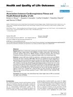
The authors studied eccentric omarthrosis, according
to the classification of Hamada [11] (figure 1), and cen-
tered omarthrosis, with irreparable rotator cuff disease,
in patients without any surgical antecedents.
The data on fatty degeneration was derived from CT or
MRI-scans with or without arthrography, interpreted by
each of the responsible surgeons, taken in the transversal
and sagittal plane of the shoulder. The degree of fatty
degeneration of the rotator cuff was determined accord-
ing to Goutallier [27] and the muscular status of the teres
minor was defined as n ormal, hypotrophic, absent or
hypertrophic. All patients were divided into two groups
for comparison: one with good to acceptable muscular
quality (stade 0, 1 and 2 according to Goutallier and nor-
mal or hypertrophic) and o ne with bad muscular quality
(stade 3 and 4 and absent or hypotrophic).
The state of the tendons of the rotator cuff is obtained
from arthro CT- or MRI-scan and/or peroperative find-
ings, interpreted by the responsible surgeon. The ten-
dons are classified as normal and partially or completely
ruptured. All patients were divided into two groups for
comparison: one with good to acceptable tendon quality
(without rupt ure) and one group with bad tendon qual-
ity (partial or complete rupture).
The clinical evaluati on is done according to Constant-
Murley [7] (for pain, activities of daily living, range of
movement and power); the range of motion of the active
external rotation in adduction and abduction; the pre-
sence of a hornblower’ sig n [21] and the feasibilit y of
the press-belly test [28].
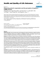
The radiological data, digitally measured by the first
author (Adobe
®
Photoshop
®
7.0; San Jose, California, US),
from patients in a standing position, was obtained on AP-
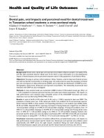
views according to Neer [19] in neutral rotation (Figure 2).
On AP-view the following marking points were placed
(Figure 2)
m: the midpoint of the best fitting circle of the humeral
head; 1: the most lateral point of the humeral head; 2:
the most lateral point of the acromion; 3: the most
inferior point of the acromion; 4: the most superior
point of the humeral head; 5: the most lateral point of
the coracoid basis; 6: the most lateral point of the cora-
coid tip.
On AP-view the following lines were placed and their
angulations to the horizon were measured (Figure 2):
A: a line best fitting the direction of the coracoid pro-
cess; B: a line best fitting the direction of the ac romi on;
C: a line best fitting the direction of the glenoid; D: a
line connecting marking point 2 and 6.
On AP-view the following parameters were measured
humeral head radius: the radius, in mm, of the best fit-
ting circle of the humeral head.
acromiohumeral distance: measured in mm between
two lines drawn through point 3 and 4 parallel to the B-
line [13,17,29];
acromial thickness: measured in mm at the most thin
part;
medialisation and ascention: measured in mm
between marking points m and 5, measured between
Middernacht et al. Journal of Orthopaedic Surgery and Research 2011, 6:1
/>Page 2 of 7
mandalineparallelwithBdrawnthroughpoint5
and measured betwe en m and a line pa rallel with C
drawn through point 5 (Figure 2). The distance
between point m and the D-line was also measured.
The upward migration index [17] was calculated;
coracoid tip positioning: the distance between two par-
allel lines drawn through the most inferior point of the
coracoid tip and the most inferior point of the glenoid,
parallel to the B-line, compared to the supero-inferior
length of the glenoid;
the mean lateral acromion angle [14,22] was deter-
mined by the difference in degrees between the B- and
C-line.
the glenoid inclination angle [12] was here determined
in relation to the horizontal.
the acromial index [14]: the distance from the glenoid
plane t o the lateral border of the acromion was divided
by the distance from the glenoid plane to the lateral
aspect of the humeral head.
On AP-view the following parameters were described
Femoralization of the proximal humerus [6,19] was
defined as absence or presence of erosion of the grea ter
tuberosity.
Acetabularization of the acromion [11] was defined as
absent or present.
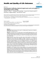
Medial erosion of the glenoid was defined as absent
(E0) or present (E1, E2, E3 and E4) according to Sir-
veaux et al. [16] (figure 3).
The relationships between the different clinical para-
meters as well as the total Constant score and all radi-
ological parameters cited above are analysed.
Statistical analysis was performed with R (a language
and environment for statistical computing) [30].
Figure 1 Hamada’s classification of omarthrosis [11].
Figure 2 Example of the marking points and lines drawn onto each radiograph.
Middernacht et al. Journal of Orthopaedic Surgery and Research 2011, 6:1
/>Page 3 of 7
Univariate comparison was done with the Fisher’ s
Exact test fo r categorical data. The non-pa rametric
Mann-Whitney U-test was used to compare continuous
variables. Also the Spearman correlation was u sed. The
significance level was set at alpha = 0.05.
Five different radiographs were analysed twice by the
first author in order to determine the intra-observer varia-
bility. There was only one observer so an inter-observer
variability was not to be performed. To determine these
variabilities, the intraclass correlation coefficient was used
(ICC), in combination with the Wilcoxon Signed Ranks
test [31].
Results
Descriptive measurements
According to Hamada [11] we defined 25 patients as
type1,53patiensastype2,27patientsastype3,48
patients as type 4a, 94 patients a s type 4b, 27 patients
as type 5 and 33 patients as centered omarthrosis
(Figure 1).
On CT- or MRI- (arthro-)scan the infraspinate muscle
is fat ty degenerated for at least half of its volume in 82%
of described cases; t he subscapular muscle in 49% of
patients and the teres minor muscle was atrophic or
absent in 32% of described patients.
On arthro CT- or MRI-scan and peroperative findings,
the supraspinate tendon is partially or completely rup-
tured in 98% of described cases; the infraspinate tendon
in 69% of cases; the subs capular tendon in 92% of cases
and the teres minor muscle in 37% of described patients.
The mean Constant-Murley score is 24/100 (10)
(mean (SD)) (n = 307).
The mean a cromiohumeral distance is 4 .5 mm ( 3.6).
The mean humeral head radius is 24 mm (5). The mean
acr omial thickness is 6.2 mm (2.5) and the mean lateral
extension of the acromion is 9.8 mm (6.0). The mean
supero-inferior distance of the glenoid is 36 mm (7).
We defined 240/294 (82%) of our patients to be type I
coracoid according to Schulz et al. [15]
The Intraclass Correlation Coefficient [31] was 0,982
(95% confidence interval (CI): 0.875, 0.998).
Relationships between bony structural changes versus
Constant score are summed up in table 1.
Relationships between lag signs versus location of the
tendinous tear and muscular quality can be seen in
table 2.
Relationships between the location of the tendinous
tear and muscular quality of the rotator cuff ve rsus bony
structural changes and morphological osseous properties
are also displayed in table 2.
Discussion
An anteroposterior radiograph is u sed today to docu-
ment patients with rotator cuff tear arthropathy.
Furthermore this basic investigation is applied to distin-
guish various types of the disease with specific therapeu-
tical consequences.
This multicenter database studies preoperative con-
ventional anteroposterior radiographs, in non-operated
patients with cuff tear arthropathy, in relation to the
clinical status and the status of the rotator cuff derived
from peroperative findings, CT- and MRI-scans.
Being multicenter will be the m ajor weakness of this
study because nine different institutes provided the
clinical data and peroperative findings. However, to
our knowledge no such study exists evaluating these
relationships on such an important number of patients
(n = 307).
Another w eakness of this study is that we didn’thave
a CT and/or MRI for each patient. However we did
have a large number of CT’sandMRI’ sandhadpero-
perative findings for each of the patients. The last minor
Figure 3 The classificat ion of Sirveaux et al . [16] was used to
devide the glenoids into two groups.
Table 1 Relationships between bony structural changes versus Constant score
table 1 evaluation of the statistical relationship
between the Constant score and:
statistical test used P-value to evaluate significance
(number of cases)
Acromio-Humeral distance Pearson 0,377 (305)
Medialisation Spearman 0,064 (303)
Femoralisation Mann-Whitney U 0,315 (281)
Acetabularisation Mann-Whitney U 0,966 (303)
Acromial thickness Mann-Whitney U 0,099 (303)
Medial erosion of the glenoid Mann-Whitney U 0,653 (303)
Middernacht et al. Journal of Orthopaedic Surgery and Research 2011, 6:1
/>Page 4 of 7
Table 2 Relationships between lag signs, bony structural changes and morphological osseous properties versus location of the tendinous tear and muscular
quality
P-Values calculated with the Fisher’s exact statistical test
between colum and row (number of cases)
Subscapular
muscular quality
Infraspinatus
muscular quality
teres minor
muscular quality
Subscapular
tendon tear
Supraspinatus
tendon tear
Infraspinatus
tendon tear
teres minor
tendon tear
exorotation in adduction 0,16 (166) 0,113 (167)
<0,001 (137) 0,05 (234) 1 (208) 1 (208) 0,003 (121)
exorotation in abduction 0,367 (88) 1 (89)
<0,001 (76) 0,834 (123) 0,519 (100) 1 (100) 0,052 (66)
hornblower’s sign 0,547 (103) 0,092 (65)
0,004 (45) 0,432 (76) 0,548 (72) 0,548 (72) 0,002 (55)
press belly test
<0,001 (111) 1 (110) 0,82 (100) <0,001 (132) 0,247 (118) 0,503 (119) 0,387 (94)
Upward migration index 0,305 (231)
0,019 (230) 0,029 (190) 0,373 (304) 0,665 (277) 0,012 (278) 0,794 (170)
Medialisation 0,281 (231) 0,59 (230) 0,332 (190) 0,705 (304) 0,253 (278) 0,252 (170)
Femoralisation 0,519 (231)
<0,001 (230) <0,001 (190) 0,042 (304) 0,496 (277) 0,003 (278) <0,001 (170)
Medial erosion of the glenoid 0,293 (231) 0,165 (230)
0,029 (190) 0,024 (303) 0,66 (276) 0,65 (277) 0,428 (170)
Acetabularisation 0,684 (231)
0,018 (230) 0,419 (190) 1 (304) 1 (277) 0,164 (278) 0,141 (170)
lateral acromion angle 0,277 (231) 0,774 (230) 0,796 (190) 0,69 (304) 1 (277) 1 (278) 0,793 (170)
acromial index 0,28 (231) 0,474 (230) 0,339 (190) 0,256 (304) 0,653 (277) 0,647 (278) 0,113 (170)
acromial thickness 0,084 (231) 0,076 (230) 0,756 (190) 0,901 (304) 1 (277) 0,819 (278) 0,526 (170)
Glenoid inclination angle 0,185 (231) 0,052 (230) 0,347 (190) 0,172 (304) 0,665 (277) 0,068 (278) 0,341 (170)
Middernacht et al. Journal of Orthopaedic Surgery and Research 2011, 6:1
/>Page 5 of 7
pointofourworkwillbethelackofacontrolgroup
without cuff tear arthropathy.
The seriousness of clinical impairment of our studied
population is reflected by the low mean Constant score
(24/100). Because this study could not find any relation-
ship between the radiologic extent of the bony structural
changes and the clinical status of the patient, we believe
a conventional antero-posterior radiograph cannot pro-
vide any predictive information on the clinical status of
the patient. This is in contrast with the statement of
Nové-Josserand et al. who demonstrates a stro ng statis-
tical correlation between the Constant score versus
Hamada stage or the severity of the glenohumeral
degradation [2].
We agree with Tokish et al. who found the subscapu-
lar muscle and tendon can be well tested with the press
belly test [28] and with Walch et al. who stated the
teres minor muscle and tendon can be well evaluated
with the hornblower’ sign [21]. Our study also confirms
the statement of Hertel et al. who found clinical testing
for lag signs to be efficient, reproducible, and reliable in
evaluating the teres minor tendon and muscle [8].
We found the upward migration index [18] and the
presence of femoralization [6,19] to be good indicators
for the evaluation of the posterior rotator cuff. There-
fore we can agree with van de Sande et al. [18] who sta-
ted that fatty infiltration of the infraspinatus muscle
shows the strongest correlation with proximal migration.
We could not find any significant relations hip between
the rotator cuff status on the one hand and medialization,
ver tical erosion of the glenoid [16] and acetabularization
[11] on the other hand. This relativates the statement of
Visotsky et al. who suggests that the amount of decentra-
lization depends on the extent of the rotator cuff tear,
the integrity of the coracoacromial arch, and the degree
and direction of the glenoid bone erosion [6].
All our studied patients had rotator cuff disease and
82% of them had an inferior projection to the middle of
the glenoid (type I coracoid tip positioning) [15]. We
could not find a visible difference in coracoid tip posi-
tioning and site of the rotator cuff weakness as proposed
by Schulz et al. [15] who concluded that type I cora-
coids are predominant in shoulders with supraspinatus
tears and type II coracoids in shoulders with subscapu-
laris tears.
Furthermore we could not find any significant rela-
tionship between the location and/or site of the rotator
cuff tear versus the lateral acromion angle [10], the
acromion index [14,22] and the glenoid inclination
angle [12]. These three latter morphological osseous
properties are predictive for the general rotator cuff
quality [10,14,22] but are of less use in localizing the
cuff tears.
Conclusions
A conventional antero-posterior radiograph cannot pro-
vide any predictive information on the clinical status of
the patient.
The subscapular muscle can be well tested by the
press belly test [28].
The teres minor muscle can be well tested by the horn-
blower’ sign [21] and by the exorotation lag signs [8].
The upward migration index [18] and the presence of
femorali zation [6,19] are good indicators for the evalua-
tion of the posterior rotator cuff.
82% of patients with rotator cuff disease present with
an inferior coracoid tip positioning to the glenoid [15].
Author details
1
Ghent University Hospital, De Pintelaan 185, Ghent B-9000, Belgium.
2
University of Tours, Boulevard Tonnellé 10, BP 3223, 37032 Tours Cedex 1,
France.
3
Clinic for Traumatology and Orthopaedics, Rue Hermitte 49, 54000
Nancy, France.
Authors’ contributions
BM: Collecting data; analysing data; writing the article; PWdG: Collecting
data;
GVM: Statistical analyses; LF: Providing data; DM: Providing data;
LDW: Coordinating; providing data; providing study idea; writing the article.
All authors have read and approved the final manuscript.
Competing interests
The authors declare that they have no competing interests.
Received: 6 February 2010 Accepted: 5 January 2011
Published: 5 January 2011
References
1. Noel E: Les ruptures de la coiffe des rotateurs avec tête huméralle
centrée. Résultats du traitement conservateur. A propos de 171 épaules.
Journées Lyonnaises d’epaule 1993, 283-97.
2. Nove-Josserand L, Walch G, Adeleine P, Courpron P: Effect of age on the
natural history of the shoulder: a clinical and radiological study in the
elderly. Rev Chir Orthop Reparatrice Appar Mot 2005, 91(6):508-14.
3. Burkhart SS: Fluoroscopic comparison of kinematic patterns in massive
rotator cuff tears. A suspension bridge model. Clin Orthop Relat Res 1992,
284:144-52.
4. Jensen KL, Williams GR Jr, Russell IJ, Rockwood CA Jr: Rotator Cuff Tear
Arthropathy. J Bone Joint Surg 1999, 81-A:1312-1324.
5. Sher JS, Uribe JW, Posada A, Murphy HJ, Zlatkin MR: Abnormal Findings on
Magnetic Resonance Images of Asymptomatic Shoulders. J Bone Joint
Surg 1995, 77-A:10-15.
6. Visotsky JL, Basamania C, Seebauer L, Rockwood CA, Jensen KL: Cuff tear
arthropathy: pathogenesis, classification, and algorithm for treatment. J
Bone Joint Surg Am 2004, 86-A(Suppl 2):35-40.
7. Constant CR, Murley AH: A clinical method of functional assessment of
the shoulder. Clin Orthop Relat Res 1987, , 214: 160-4.
8. Hertel R, Ballmer FT, Lombert SM, Gerber C: Lag signs in the diagnosis of
rotator cuff rupture. J Shoulder Elbow Surg 1996, 5(4):307-13.
9. Golding FC: The shoulder: the forgotten joint. Br J Radiol 1962, 35:149-58.
10. Banas MP, Miller RJ, Totterman S: Relationship between the lateral
acromion angle and rotator cuff disease. J Shoulder Elbow Surg 1995,
4:454-61.
11. Hamada K, Fukuda H, Mikasa M, Kobayashi Y: Roentgenographic findings
in massive rotator cuff tears. Clin Orthop Relat Res 1990, 254:92-6.
12. Hughes RE, Bryant CR, Hall JM, Wening J, Huston LJ, Kuhn JE, Carpenter JE,
Blasier RB: Glenoid inclination is associated with full-thickness rotator
cuff tears. Clin Orthop Relat Res 2003, , 407: 86-91.
Middernacht et al. Journal of Orthopaedic Surgery and Research 2011, 6:1
/>Page 6 of 7
13. Nove-Josserand L, Edwards TB, O’Connor DP, Walch G: The
acromiohumeral and coracohumeral intervals are abnormal in rotator
cuff tears with muscular fatty degeneration. Clin Orthop Relat Res 2005, ,
433: 90-6.
14. Nyffeler RW, Werner CML, Sukthankar A, Schmid MR, Gerber C: Association
of a large lateral extension of the acromion with rotator cuff tears. J
Bone Joint Surg 2006, 88A4:800-5.
15. Schulz CU, Anetzberger H, Glaser C: Coracoid tip position on frontal
radiographs of the shoulder: a predictor of common shoulder
pathologies? Br J Radiol 2005, 78:1005-8.
16. Sirveaux F, Favard L, Oudet D, Huquet D, Walch G, Mole D: Grammont
inverted total shoulder arthroplasty in the treatment of glenohumeral
osteoarthritis with massive rupture of the cuff. Results of a multicentre
study of 80 shoulders. J Bone Joint Surg Br 2004, 86(3):388-95.
17. van de Sande MA, Rozing PM: Proximal migration can be measured
accurately on standardized anteroposterior shoulder radiographs. Clin
Orthop Relat Res 2006, 443:260-5.
18. van de Sande MA, Stoel BC, Rozing PM: Subacromial space measurement:
a reliable method indicating Fatty infiltration in patients with
rheumatoid arthritis. Clin Orthop Relat Res 2006, 451:73-9.
19. Neer C, Craig E, Fukuda H: Cuff-tear arthropathy. J Bone Joint Surg Am
1983, 65(9):1232-44.
20. Heininger-Biner K, Muller M, Hertel R: Diagnosis of rotator cuff rupture:
correlation of clinical findings and magnetic resonance tomography
with intraoperative findings. Z Orthop Ihre Grenzgeb 2000, 138(6):478-80.
21. Walch G, Boulahia A, Calderone S, Robinson AHN: The ‘dropping’ and
‘hornblower’s’ signs in evaluation of rotator-cuff tears. J Bone Joint Surg
[Br] 1998, 80-B:624-8.
22. Torrens C, López JM, Puente I, Cáceres E: The influence of the acromial
coverage index in rotator cuff tears. J Shoulder Elbow Surg 2007,
16(3):347-51.
23. Blanchard TK, Bearcroft PW, Constant CR, Griffin DR, Dixon AK: Diagnostic
and therapeutic impact of MRI and arthrography in the investigation of
full-thickness rotator cuff tears. Eur Radiol 1999, 9(4):638-42.
24. De Smet AA, Ting YM: Diagnosis of rotator cuff tears on routine
radiographs. J Can Assoc Radiol 1977, 2854.
25. Goutallier D, Postel JM, Gleyze P, Legurilloux P, Van Driessche S: Influence
of cuff muscle fatty degeneration on anatomic and functional outcomes
after simple suture of full-thickness tears.
J Shoulder Elbow Surg 2003,
12(6):550-4.
26. Walch G, Marechal E, Maupas J, Liotard JP: Surgical treatment of rotator
cuff rupture. Prognostic factors. Rev Chir Orthop Reparatrice Appar Mot
1992, 78(6):379-88.
27. Goutallier D, Postel JM, Bernageau J, Lavau L, Voisin MC: Fatty muscle
degeneration in cuff ruptures: Pre- and postoperative evaluation by CT
scan. Clin Orthop 1994, 304:78-83.
28. Tokish JM, Decker MJ, Ellis HB, Torry MR, Hawkins RJ: The belly-press test
for the physical examination of the subscapularis muscle:
electromyographic validation and comparison to the lift-off test. J
Shoulder Elbow Surg 2003, 12(5):427-30.
29. Weiner DS, Macnab I: Superior migration of the humeral head A
radiological aid in the diagnosis of tears of the rotator cuff. J Bone Joint
Surg Br 1970, 52(3):524-7.
30. R Development Core Team: R: A language and environment for statistical
computing. R Foundation for Statistical Computing, Vienna, Austria; 2008,
ISBN 3-900051-07-0, URL .
31. Shrout PE, Fleiss JL: Intraclass correlations: uses in assessing rater
reliability. Psychological Bulletin 1979, 86:420-28.
doi:10.1186/1749-799X-6-1
Cite this article as: Middernacht et al.: What do standard radiography
and clinical examination tell about the shoulder with cuff tear
arthropathy? Journal of Orthopaedic Surgery and Research 2011 6:1.
Submit your next manuscript to BioMed Central
and take full advantage of:
• Convenient online submission
• Thorough peer review
• No space constraints or color figure charges
• Immediate publication on acceptance
• Inclusion in PubMed, CAS, Scopus and Google Scholar
• Research which is freely available for redistribution
Submit your manuscript at
www.biomedcentral.com/submit
Middernacht et al. Journal of Orthopaedic Surgery and Research 2011, 6:1
/>Page 7 of 7