Properties and Applications of Silicon Carbide Part 12 doc
Bạn đang xem bản rút gọn của tài liệu. Xem và tải ngay bản đầy đủ của tài liệu tại đây (1.5 MB, 30 trang )
Properties and Applications of Silicon Carbide322
template is the determination of the chemical nature of the carbonaceous species at the
surface (Cicoira & Rosei, 2006).
Bioceramic nanocomposites were synthesized by sintering compacted bodies of
hydroxyapatite mixed with 5 or 15 wt% nanosilicon carbide at 1100 or 1200 °C in a reducing
atmosphere. The results indicate that the composite of 95 wt% hydroxyapatite and 5 wt%
SiC exhibited better mechanical and biological properties than pure hydroxyapatite and
further addition of SiC failed strength and toughness (Hesaraki et al., 2010). The preparation
of nano-sized silicon carbide has received considerable attention, because it allows the
preparation of bulk materials with increased plasticity (Stobierski & Gubernat, 2003) or
nanocomposites with enhanced mechanical and tribological properties. In conclusion, it
opens up exciting possibilities in the area of template-assisted growth at the nanoscale.
14. Drug delivery
Drug delivery systems (DDS) are an area of study in which researchers from almost every
scientific discipline can make a significant contribution. Understanding the fate of drugs inside
the human body is a high standard classical endeavor, where basic and mathematical analysis
can be used to achieve an important practical end. No doubt the effectiveness of drug therapy
is closely related to biophysics and physiology of drug movement through tissue. Therefore,
DDS requires an understanding of the characteristics of the system, the molecular mechanisms
of drug transport and elimination, particularly at the site of delivery. In the last decade DDS
have received much attention since they can significantly improve the therapeutic effects of the
drug while minimizing its side effects.In recent years, Poly (D,L-Lactide) (PLA) and Poly (D,L-
Lactide-co-Glycolide) (PLGA) have been extensively investigated for use as implantable
biodegradable carriers for controlled release of drugs. Silicon carbide coated stents have been
coated with a layer of PLA or PLGA containing the drug by dip coating or spray coating
techniques. Several drugs have been considered as candidates for stent coatings preventing
instent restenosis. SiC is used as a basis for drug delivery systems or bioactive coatings in
order to modulate vascular cell growth. For a sufficient polymer-drug coating of a silicon
carbide stent and a long-term release of the desired agent, PLA and PLGA are biocompatible
materials useful for a variety of applications, including the design and properties of the
controlled-release systems for pharmaceutical agents.
Despite the phenomenal pace of stent design technology and the improvements in
biocompatibility that have been achieved with the SiC coating, the incidence of in-stent
restenosis remains unacceptably high. To address this problem, intense research is being
conducted in order to find new stent coatings. Coatings with specific polymer-drug
composites or with specific glycosaminoglycans showed promising results in modulating
the proliferation of vascular smooth muscle cells and endothelial cells (Bayer, 2001). Using
an existing technology for dip coating, glycosaminoglycans can be covalently bonded to the
silicon carbide surface via a spacer molecule (Hildebrandt, 2001). Crosslinking the network
of coated glycosaminoglycans should result in a stable bioactive layer with long-term anti-
proliferative effects (Bayer, 2001). Finally, it is suggested that more detailed experiments are
required and would be useful to distinguish and clarify SiC-based materials application in
drug delivery.
15. Surface modification of Ti-6Al-4V alloy
by SiC paper for orthopaedic applications
It is possible to change localized areas of metals in order to obtain both compositions and
microstructures with improved properties. Titanium and titanium alloys are the most
frequently used material for load-bearing orthopaedic implants, due to their specific
properties such as high corrosion resistance, surface oxidation layer, high strength and high-
temperature resistance (Feng et al., 2003). Titanium and its alloys’ application like any other
biomaterials involve the creation of at least one interface between the material and biological
tissues. Biocompatibility and bioactivity of biomaterials rely on the interactions that take
place between the interface of the biomaterials and the biological system (Wang & Zheng,
2009). It is generally believed that proteins adsorbed on implant surface can play an
important role in cell-surface response. Different proteins such as collagen, fibronectin and
vitronectin which are acting as ligands are particularly important in osteoblast interaction
with surface. Ligands are the junctions which facilitate adhesion of bone cells to implant
surface. In another word, more ligand formation implies a better cell-surface interaction
(Tirrell et al., 2002). In vitro studies can be used to study the influence of surface properties
on processes such as cell attachment, cell proliferation and cell differentiation. However, in
vivo studies must be performed to achieve a complete understanding of the healing process
around implants. Previous studies have shown that surface characteristics named above
have a significant influence on adhesion, morphology and maturation of cultured
osteoblasts (Masuda et al., 1998). Also, it has been demonstrated that for primary bovine
osteoblasts, the wettability is one of the key factors. In our studies (Khosroshahi et al., 2007;
Khosroshahi
)
, 2007; Khosroshahi et al., 2008; Khosroshahi et al., 2008; Khosroshahi et al.,
2009), it is shown that the wettability of the surface can provide a better spreading condition
for osteoblast cells due to reduced contact angle. Bearing in mind that the adhesion of bone
cells to implant surface consists of two stages. In primary stage the cells must get close
enough to surface at an appropriate distance known as focal distance over which the cells
can easily be spread over it. In this respect, the wettability can be effective in providing a
preferred accessability to surface and thus reaching the focal distance. The secondary stage
includes cell-cell attachment which obeys the regular biological facts.
Interface reactions between metallic implants and the surrounding tissues play a crucial role
in the success of osseointegration. The titanium and its alloys like some other medical grade
metals are the materials of choice for long-term implants. The effect of implant surface
characteristics on bone reactions has thus attracted much attention and is still considered to
be an important issue (Buchter et al., 2006).
So far as the surface characteristics of the implants are concerned, two main features that can
influence the establishment of the osseointegration are the physico-chemical properties and
the surface morphology. Cell adhesion is involved in various phenomena such as
embryogenesis, wound healing, immune response and metastasis as well as tissue
integration of biomaterial. Thus, attachment, adhesion and spreading will depend on the
cell-material interaction and the cell’s capacity to proliferate and to differentiate itself on
contact with the implant (Bigerelle et al., 2005).
Cell behavior, such as adhesion, morphologic change and functional alteration are greatly
influenced by surface properties including texture, roughness, hydrophilicity and
morphology. In extensive investigations of tissue response to implant surfaces, it has been
shown that surface treatment of implant materials significantly influences the attachment of
Fundamentals of biomedical applications of biomorphic SiC 323
template is the determination of the chemical nature of the carbonaceous species at the
surface (Cicoira & Rosei, 2006).
Bioceramic nanocomposites were synthesized by sintering compacted bodies of
hydroxyapatite mixed with 5 or 15 wt% nanosilicon carbide at 1100 or 1200 °C in a reducing
atmosphere. The results indicate that the composite of 95 wt% hydroxyapatite and 5 wt%
SiC exhibited better mechanical and biological properties than pure hydroxyapatite and
further addition of SiC failed strength and toughness (Hesaraki et al., 2010). The preparation
of nano-sized silicon carbide has received considerable attention, because it allows the
preparation of bulk materials with increased plasticity (Stobierski & Gubernat, 2003) or
nanocomposites with enhanced mechanical and tribological properties. In conclusion, it
opens up exciting possibilities in the area of template-assisted growth at the nanoscale.
14. Drug delivery
Drug delivery systems (DDS) are an area of study in which researchers from almost every
scientific discipline can make a significant contribution. Understanding the fate of drugs inside
the human body is a high standard classical endeavor, where basic and mathematical analysis
can be used to achieve an important practical end. No doubt the effectiveness of drug therapy
is closely related to biophysics and physiology of drug movement through tissue. Therefore,
DDS requires an understanding of the characteristics of the system, the molecular mechanisms
of drug transport and elimination, particularly at the site of delivery. In the last decade DDS
have received much attention since they can significantly improve the therapeutic effects of the
drug while minimizing its side effects.In recent years, Poly (D,L-Lactide) (PLA) and Poly (D,L-
Lactide-co-Glycolide) (PLGA) have been extensively investigated for use as implantable
biodegradable carriers for controlled release of drugs. Silicon carbide coated stents have been
coated with a layer of PLA or PLGA containing the drug by dip coating or spray coating
techniques. Several drugs have been considered as candidates for stent coatings preventing
instent restenosis. SiC is used as a basis for drug delivery systems or bioactive coatings in
order to modulate vascular cell growth. For a sufficient polymer-drug coating of a silicon
carbide stent and a long-term release of the desired agent, PLA and PLGA are biocompatible
materials useful for a variety of applications, including the design and properties of the
controlled-release systems for pharmaceutical agents.
Despite the phenomenal pace of stent design technology and the improvements in
biocompatibility that have been achieved with the SiC coating, the incidence of in-stent
restenosis remains unacceptably high. To address this problem, intense research is being
conducted in order to find new stent coatings. Coatings with specific polymer-drug
composites or with specific glycosaminoglycans showed promising results in modulating
the proliferation of vascular smooth muscle cells and endothelial cells (Bayer, 2001). Using
an existing technology for dip coating, glycosaminoglycans can be covalently bonded to the
silicon carbide surface via a spacer molecule (Hildebrandt, 2001). Crosslinking the network
of coated glycosaminoglycans should result in a stable bioactive layer with long-term anti-
proliferative effects (Bayer, 2001). Finally, it is suggested that more detailed experiments are
required and would be useful to distinguish and clarify SiC-based materials application in
drug delivery.
15. Surface modification of Ti-6Al-4V alloy
by SiC paper for orthopaedic applications
It is possible to change localized areas of metals in order to obtain both compositions and
microstructures with improved properties. Titanium and titanium alloys are the most
frequently used material for load-bearing orthopaedic implants, due to their specific
properties such as high corrosion resistance, surface oxidation layer, high strength and high-
temperature resistance (Feng et al., 2003). Titanium and its alloys’ application like any other
biomaterials involve the creation of at least one interface between the material and biological
tissues. Biocompatibility and bioactivity of biomaterials rely on the interactions that take
place between the interface of the biomaterials and the biological system (Wang & Zheng,
2009). It is generally believed that proteins adsorbed on implant surface can play an
important role in cell-surface response. Different proteins such as collagen, fibronectin and
vitronectin which are acting as ligands are particularly important in osteoblast interaction
with surface. Ligands are the junctions which facilitate adhesion of bone cells to implant
surface. In another word, more ligand formation implies a better cell-surface interaction
(Tirrell et al., 2002). In vitro studies can be used to study the influence of surface properties
on processes such as cell attachment, cell proliferation and cell differentiation. However, in
vivo studies must be performed to achieve a complete understanding of the healing process
around implants. Previous studies have shown that surface characteristics named above
have a significant influence on adhesion, morphology and maturation of cultured
osteoblasts (Masuda et al., 1998). Also, it has been demonstrated that for primary bovine
osteoblasts, the wettability is one of the key factors. In our studies (Khosroshahi et al., 2007;
Khosroshahi
)
, 2007; Khosroshahi et al., 2008; Khosroshahi et al., 2008; Khosroshahi et al.,
2009), it is shown that the wettability of the surface can provide a better spreading condition
for osteoblast cells due to reduced contact angle. Bearing in mind that the adhesion of bone
cells to implant surface consists of two stages. In primary stage the cells must get close
enough to surface at an appropriate distance known as focal distance over which the cells
can easily be spread over it. In this respect, the wettability can be effective in providing a
preferred accessability to surface and thus reaching the focal distance. The secondary stage
includes cell-cell attachment which obeys the regular biological facts.
Interface reactions between metallic implants and the surrounding tissues play a crucial role
in the success of osseointegration. The titanium and its alloys like some other medical grade
metals are the materials of choice for long-term implants. The effect of implant surface
characteristics on bone reactions has thus attracted much attention and is still considered to
be an important issue (Buchter et al., 2006).
So far as the surface characteristics of the implants are concerned, two main features that can
influence the establishment of the osseointegration are the physico-chemical properties and
the surface morphology. Cell adhesion is involved in various phenomena such as
embryogenesis, wound healing, immune response and metastasis as well as tissue
integration of biomaterial. Thus, attachment, adhesion and spreading will depend on the
cell-material interaction and the cell’s capacity to proliferate and to differentiate itself on
contact with the implant (Bigerelle et al., 2005).
Cell behavior, such as adhesion, morphologic change and functional alteration are greatly
influenced by surface properties including texture, roughness, hydrophilicity and
morphology. In extensive investigations of tissue response to implant surfaces, it has been
shown that surface treatment of implant materials significantly influences the attachment of
Properties and Applications of Silicon Carbide324
cells (Heinrich et al., 2008). Additionally, these modified surfaces must resist both the
mechanical wear and the corrosion (Sighvi et al., 1998). It is therefore important to evaluate
systematically the role of different surface properties and to assess the biological
performance of different implant materials.
The surface morphology, as well as manipulation with the physical state and chemical
composition of implant surfaces may be significant for bone-implant integration. Surfaces
are treated to facilitate an intimate contact between bone and implant. So, the tissue
response to an implant involves physical factors, depending on implant design, surface
topography, surface charge density, surface free energy and chemical factors associated with
the composition of the materials. These substrate characteristics may directly influence cell
adhesion, spreading and signaling, events that regulate a wide variety of biological
functions (Ronold et al., 2003). Numerous surface treatments including Ion implantation,
coating, shot blast, machining, plasma spray, plasma nitrid, nitrogen diffusion hardening
are some of the relatively older techniques in the field of material processing which can be
used to change implant’s surface topography. Thus, the main intention of this work is to
extend the earlier research by carrying out some detailed In vitro and In vivo experiments
using a 300 and 800 grit SiC papers on surface physico-chemical changes, surface
wettability, corrosion resistance, microhardness and osteoblast cells adhesivity of Ti6Al4V
with respect to possible orthopaedic applications.
16. Materials and methods
Rectangular–shaped specimens with 20×10 mm dimensions and the thickness of 2 mm, were
made from a medical grade Ti6Al4V (ASTM F136, Friadent, Mannheim- Germany- GmbH)
with chemical formulation Ti(91.63%)Al(5.12% V(3.25%). The samples were divided into
three groups of untreated, 300 and 800 grit SiC paper. Prior to treatment, all samples were
cleaned with 97% ethanol and were subsequently washed twice by distilled water in an
ultrasonic bath (Mattachanna, Barcelona-Spain). A final rinse was done by de-ionized water
at a neutral pH to ensure a clean surface was obtained. They were polished using 300 and
800 grit SiC paper. Finally, an optical microscope with magnification of ×20 was used to
ensure that no particles were left on the sample surface.
Surface roughness
The surface micro roughness (Ra) measurements were carried out using a non-contact laser
profilemeter (NCLP) (Messtechnik, Germany) equipped with a micro focus sensor based on
an auto focusing system. Ra is the arithmetical mean of the absolute values of the profile
deviations from the mean line. Five two-dimensional NCLP profiles were obtained for each
surface over a distance of 3.094 mm with a lateral resolution of 1µm using a Gaussian filter
and an attenuation factor of 60% at a cut-off wavelength of 0.59 mm . The roughness
parameters were calculated with the NCLP software similar to that described by Wieland et
al. (Wieland et al., 2001).
Surface hardness
Surface microhardness test was carried out with 50 gram load in 10 seconds by a diamond
squared pyramid tip (Celemx CMT, Automatic). Each related test was considered at 5 points
and reported as an average. The Vickers diamond pyramid hardness number is the applied
load divided by the surface area of the indentation (mm
2
) which could be calculated from
equation bellow:
VHN = {2FSin (136°/2)}/d
2
(1)
This equation could be re-written approximately as:
VHN = 1.854(F/d
2
)
(2)
Corrosion tests
The standard Tafel photodynamic polarization tests (EG&G, PARC 273) were carried out to
study the corrosion behavior of specimens in Hank’s salt balanced physiological solution at
37ºC. The metal corrosion behavior was studied by measuring the current and plotting the
E-logI (Voltage – Current) diagram. The corrosion rate (milli per year (mpy)) was
determined using equation:
C.R. = 0.129 ( M/n ) ( I
corr
/ρ ) (3)
Where M is the molecular weight, n is the charge, I
corr
is the corrosion current and ρ is the
density.
Surface tension
The surface energy of the samples were determined by measuring the contact angle (θ) of
test liquids (diiodo-Methane and water; Busscher) on the titanium plates using Kruss-G40-
instrument (Germany).The geometric mean equation divides the surface energy in to two
components of dispersive and polar and when combined with Young’s equation it yields:
γ
lv
(1+cosθ)=2(γ
l
d
. γ
s
d
)
0.5
+2(γ
l
p
. γ
s
p
)
0.5
(4)
Equation (4) can be rearranged as by Ownes-Wendt-Kaeble’s equation:
γ
lv
(1+cosθ)/ (γ
l
d
)
0.5
= (γ
s
p
)
0.5
((γ
l
p
)
0.5
/ (γ
l
d
)
0.5
)+ ( γ
s
d
)
0.5
(5)
Where s and l represent solid and liquid surfaces respectively, γ
d
stands for the dispersion
component of the total surface energy (γ) and γ
p
is the polar component.
In vitro test
Mice connective tissue fibroblasts (L-929) with 4×10
5
ml were provided and maintained in
culture medium (RPMI-1640) consisting of 100U/ml Penicillin, 100U/ml Streptomicine, and
10% fetal calf serum (FCS) .The untreated sample, and SiC treated samples along with a
negative control (ie. fibroblast cells only in the cell culture medium) were then placed inside
the culture medium in a polystyrene dish. All the samples were incubated at 37°C in 5% CO
2
atmosphere and 90% humidity for 24h. Then the samples were washed with the de-ionized
water and sterilized by water steam for 20 min at 120 °C. Subsequently, the samples were then
fixed by using 50%, 65%, 75%, 85%, 96% ethanol and stained by Gimsa. Finally, they were
evaluated, without extracting the samples from cell culture dish, with an optical microscope
Fundamentals of biomedical applications of biomorphic SiC 325
cells (Heinrich et al., 2008). Additionally, these modified surfaces must resist both the
mechanical wear and the corrosion (Sighvi et al., 1998). It is therefore important to evaluate
systematically the role of different surface properties and to assess the biological
performance of different implant materials.
The surface morphology, as well as manipulation with the physical state and chemical
composition of implant surfaces may be significant for bone-implant integration. Surfaces
are treated to facilitate an intimate contact between bone and implant. So, the tissue
response to an implant involves physical factors, depending on implant design, surface
topography, surface charge density, surface free energy and chemical factors associated with
the composition of the materials. These substrate characteristics may directly influence cell
adhesion, spreading and signaling, events that regulate a wide variety of biological
functions (Ronold et al., 2003). Numerous surface treatments including Ion implantation,
coating, shot blast, machining, plasma spray, plasma nitrid, nitrogen diffusion hardening
are some of the relatively older techniques in the field of material processing which can be
used to change implant’s surface topography. Thus, the main intention of this work is to
extend the earlier research by carrying out some detailed In vitro and In vivo experiments
using a 300 and 800 grit SiC papers on surface physico-chemical changes, surface
wettability, corrosion resistance, microhardness and osteoblast cells adhesivity of Ti6Al4V
with respect to possible orthopaedic applications.
16. Materials and methods
Rectangular–shaped specimens with 20×10 mm dimensions and the thickness of 2 mm, were
made from a medical grade Ti6Al4V (ASTM F136, Friadent, Mannheim- Germany- GmbH)
with chemical formulation Ti(91.63%)Al(5.12% V(3.25%). The samples were divided into
three groups of untreated, 300 and 800 grit SiC paper. Prior to treatment, all samples were
cleaned with 97% ethanol and were subsequently washed twice by distilled water in an
ultrasonic bath (Mattachanna, Barcelona-Spain). A final rinse was done by de-ionized water
at a neutral pH to ensure a clean surface was obtained. They were polished using 300 and
800 grit SiC paper. Finally, an optical microscope with magnification of ×20 was used to
ensure that no particles were left on the sample surface.
Surface roughness
The surface micro roughness (Ra) measurements were carried out using a non-contact laser
profilemeter (NCLP) (Messtechnik, Germany) equipped with a micro focus sensor based on
an auto focusing system. Ra is the arithmetical mean of the absolute values of the profile
deviations from the mean line. Five two-dimensional NCLP profiles were obtained for each
surface over a distance of 3.094 mm with a lateral resolution of 1µm using a Gaussian filter
and an attenuation factor of 60% at a cut-off wavelength of 0.59 mm . The roughness
parameters were calculated with the NCLP software similar to that described by Wieland et
al. (Wieland et al., 2001).
Surface hardness
Surface microhardness test was carried out with 50 gram load in 10 seconds by a diamond
squared pyramid tip (Celemx CMT, Automatic). Each related test was considered at 5 points
and reported as an average. The Vickers diamond pyramid hardness number is the applied
load divided by the surface area of the indentation (mm
2
) which could be calculated from
equation bellow:
VHN = {2FSin (136°/2)}/d
2
(1)
This equation could be re-written approximately as:
VHN = 1.854(F/d
2
)
(2)
Corrosion tests
The standard Tafel photodynamic polarization tests (EG&G, PARC 273) were carried out to
study the corrosion behavior of specimens in Hank’s salt balanced physiological solution at
37ºC. The metal corrosion behavior was studied by measuring the current and plotting the
E-logI (Voltage – Current) diagram. The corrosion rate (milli per year (mpy)) was
determined using equation:
C.R. = 0.129 ( M/n ) ( I
corr
/ρ ) (3)
Where M is the molecular weight, n is the charge, I
corr
is the corrosion current and ρ is the
density.
Surface tension
The surface energy of the samples were determined by measuring the contact angle (θ) of
test liquids (diiodo-Methane and water; Busscher) on the titanium plates using Kruss-G40-
instrument (Germany).The geometric mean equation divides the surface energy in to two
components of dispersive and polar and when combined with Young’s equation it yields:
γ
lv
(1+cosθ)=2(γ
l
d
. γ
s
d
)
0.5
+2(γ
l
p
. γ
s
p
)
0.5
(4)
Equation (4) can be rearranged as by Ownes-Wendt-Kaeble’s equation:
γ
lv
(1+cosθ)/ (γ
l
d
)
0.5
= (γ
s
p
)
0.5
((γ
l
p
)
0.5
/ (γ
l
d
)
0.5
)+ ( γ
s
d
)
0.5
(5)
Where s and l represent solid and liquid surfaces respectively, γ
d
stands for the dispersion
component of the total surface energy (γ) and γ
p
is the polar component.
In vitro test
Mice connective tissue fibroblasts (L-929) with 4×10
5
ml were provided and maintained in
culture medium (RPMI-1640) consisting of 100U/ml Penicillin, 100U/ml Streptomicine, and
10% fetal calf serum (FCS) .The untreated sample, and SiC treated samples along with a
negative control (ie. fibroblast cells only in the cell culture medium) were then placed inside
the culture medium in a polystyrene dish. All the samples were incubated at 37°C in 5% CO
2
atmosphere and 90% humidity for 24h. Then the samples were washed with the de-ionized
water and sterilized by water steam for 20 min at 120 °C. Subsequently, the samples were then
fixed by using 50%, 65%, 75%, 85%, 96% ethanol and stained by Gimsa. Finally, they were
evaluated, without extracting the samples from cell culture dish, with an optical microscope
Properties and Applications of Silicon Carbide326
(Nikon TE 2000-U) for cell growth and cytotoxicity. It is worth mentioning that the
biocompatibility of the samples was investigated In vitro by L-929 fibroblast cell counting on
samples through methyl thiazole tetrazolium (MTT) assay. For this purpose an enzymic
method ie.1ml of Trypsin/EDTA was used and the cells were then left to trypsinize in the flask
at 37° in the incubator for 3 minutes and were monitored by the same optical microscope.
In vivo test
Anesthetization
Before depilation of the operation site, the animal was completely anesthetized with
midazolam (Dormicum®, Roche, Switzerland) 2.5 mg/Kg intravenously (IV). With any sign
of recovery during operation, diluted fluanisone/fentanyl (Hypnorm®, India) was injected
slowly until adequate effect was achieved, usually 0.2 ml at a time.
Animal implantation
Untreated sample and SiC treated samples were implanted on femur bone of an eight
months male goat weighing 30 Kg. Specimens were steam sterilized before implantation in
an autoclave (Mattachnna, Barcelona-Spain). The steam sterilization was conducted under
132 °C, 2 bar and in 45 minutes. All the specimens were labeled by separate codes for further
studies. The operation site was shaved and depilated with soft soap and ethanol before
surgery; the site was also disinfected with 70% ethanol and was covered with a sterile
blanket. In order to proceed with implantation, cortex bone was scraped by osteotom
(Mattachnna, Barcelona-Spain) after cutting the limb from one-third end in lateral side and
elevating it by a self – retaining retractor. Copious physiological saline solution irrigation
was used during the implantation to prevent from overheating. To ensure a stable passive
fixation of implants during the healing period, they were stabilized by size 4 and 8 titanium
wires (Atila ortoped®, Tehran-Iran) without any external compression forces (Fig.15).
Fig. 15. Placement of implants in the femur bone of the goat
After the operation the animal was protected from infection by proper prescribed uptake of
Penicillin for first four days and Gentamicine for second four days. During the eight days of
recovery, the goat was administrated with multi-vitamins to help to regain its strength.
During this period, the goat was kept in an isolated space under room temperature,
ordinary humidity, lighting and air conditioning, and before it returns to its natural life
environment, X-ray radiographs (Fig. 16) were taken in order to ensure that the implant has
not been displaced during the maintenance period. It was observed that calus bone had
grown in the vicinity of the implant. After five months the animal was sacrificed and the
specimens were removed (Fig. 17).
Fig. 16. The X-ray of implants wired to the bone
Fig. 17. Implant removal from the femur bone of the goat: (a) before detachment of the
wires, (b) after detachmented (c, d) the foot-print of the implants on the bone
The experiments had been approved by the Yazd School of Veterinary Science (Iran) and its
animal research authority and conducted in accordance with the Animal Welfare Act of
December 20th 1974 and the Regulation on Animal Experimentation of January 15th 1996.
The explantation procedure was performed by first cutting the upper and lower section of
femur bone using an electric saw and then the implant together with its surrounding tissues
was placed in 4% formalin solution for pathological assessment and SEM.
Cell analysis
Fundamentals of biomedical applications of biomorphic SiC 327
(Nikon TE 2000-U) for cell growth and cytotoxicity. It is worth mentioning that the
biocompatibility of the samples was investigated In vitro by L-929 fibroblast cell counting on
samples through methyl thiazole tetrazolium (MTT) assay. For this purpose an enzymic
method ie.1ml of Trypsin/EDTA was used and the cells were then left to trypsinize in the flask
at 37° in the incubator for 3 minutes and were monitored by the same optical microscope.
In vivo test
Anesthetization
Before depilation of the operation site, the animal was completely anesthetized with
midazolam (Dormicum®, Roche, Switzerland) 2.5 mg/Kg intravenously (IV). With any sign
of recovery during operation, diluted fluanisone/fentanyl (Hypnorm®, India) was injected
slowly until adequate effect was achieved, usually 0.2 ml at a time.
Animal implantation
Untreated sample and SiC treated samples were implanted on femur bone of an eight
months male goat weighing 30 Kg. Specimens were steam sterilized before implantation in
an autoclave (Mattachnna, Barcelona-Spain). The steam sterilization was conducted under
132 °C, 2 bar and in 45 minutes. All the specimens were labeled by separate codes for further
studies. The operation site was shaved and depilated with soft soap and ethanol before
surgery; the site was also disinfected with 70% ethanol and was covered with a sterile
blanket. In order to proceed with implantation, cortex bone was scraped by osteotom
(Mattachnna, Barcelona-Spain) after cutting the limb from one-third end in lateral side and
elevating it by a self – retaining retractor. Copious physiological saline solution irrigation
was used during the implantation to prevent from overheating. To ensure a stable passive
fixation of implants during the healing period, they were stabilized by size 4 and 8 titanium
wires (Atila ortoped®, Tehran-Iran) without any external compression forces (Fig.15).
Fig. 15. Placement of implants in the femur bone of the goat
After the operation the animal was protected from infection by proper prescribed uptake of
Penicillin for first four days and Gentamicine for second four days. During the eight days of
recovery, the goat was administrated with multi-vitamins to help to regain its strength.
During this period, the goat was kept in an isolated space under room temperature,
ordinary humidity, lighting and air conditioning, and before it returns to its natural life
environment, X-ray radiographs (Fig. 16) were taken in order to ensure that the implant has
not been displaced during the maintenance period. It was observed that calus bone had
grown in the vicinity of the implant. After five months the animal was sacrificed and the
specimens were removed (Fig. 17).
Fig. 16. The X-ray of implants wired to the bone
Fig. 17. Implant removal from the femur bone of the goat: (a) before detachment of the
wires, (b) after detachmented (c, d) the foot-print of the implants on the bone
The experiments had been approved by the Yazd School of Veterinary Science (Iran) and its
animal research authority and conducted in accordance with the Animal Welfare Act of
December 20th 1974 and the Regulation on Animal Experimentation of January 15th 1996.
The explantation procedure was performed by first cutting the upper and lower section of
femur bone using an electric saw and then the implant together with its surrounding tissues
was placed in 4% formalin solution for pathological assessment and SEM.
Cell analysis
Properties and Applications of Silicon Carbide328
Osteoblast cells spreading (ie. lateral growth) on the implants was analyzed after removal
by SEM (stero scan 360-cambridge) and their spreading condition in a specific area was
studied using Image J Program software in three separate regions of each specimen at a
frequency of 10 cells per each region. The number of attached cells in 1 cm
2
area of each
specimen was calculated by a Coulter counter (Eppendorf, Germany) using enzyme
detachment method and Trypsin-EDTA (0.025 V/V) in PBS media at pH = 7.5. The final
amount of attached cell can be studied by plotting cell detachment rate versus time.
Histopathology
Surrounding tissues of specimens were retrieved and prepared for histological evaluation.
They were fixed in 4% formalin solution (pH = 7.3), dehydrated in a graded series of ethanol
(10%, 30%, 50%, 70% and 90%) and embedded in paraffin after decalcification. Then, 10 µm
thick slices were prepared per specimen using sawing microtome technique. A qualitative
evaluation of macrophage, osteoblast, osteoclast, PMN, giant cells, fibroblast, lymphocyte
was carried out by Hematoxylin and Eosin stain and light microscopy (Zeiss, Gottingen-
Germany). The light microscopy assessment consisted of a complete morphological
description of the tissue response to the implants with different surface topography.
Osteoblasts can be in two states; (a) active, forming bone matrix; (b) resting or bone-
maintaining. Those make collagen, glycoproteins and proteoglycans of bone the matrix and
control the deposition of mineral crystals on the fibrils. Osteoblast becomes an osteocyte by
forming a matrix around itself and is buried. Lacunae empty of osteocytes indicate dead
bone. Osteoclast, a large and multinucleated cell, with a pale acidophilic cytoplasm lies on
the surface of bone, often an eaten-out hollow-Howship’s lacuna. Macrophages, are
irregularly shaped cells that participate in phagocytosis.
SEM of adhered cells
After implants removal, all three group implants were rinsed twice with phosphate buffer
saline (PBS) and then fixed with 2.5% glutaraldehyde for 60 minutes. After a final rinse with
PBS, a contrast treatment in 1% osmium tetroxide (Merck) was performed for 1 hour,
followed by an extensive rinsing in PBS and dehydration through a graded series of ethanol
from 30% to 90% as described in histology section. After free air drying, surfaces were thinly
sputter coated with gold (CSD 050, with 40 mA about 7 min). Cell growth on implanted
specimens and their spreading condition in a specific area was analyzed using Image J
Program software in three separate regions of each specimen for 10 cells per each region.
Statistical analysis
All calculated data were analyzed by using a software program SPSS (SPSS Inc., version 9.0).
The results of variance analysis were used to identify the differences between the cells
spread area of the treated and cleaned un-treated samples (p≤0.05).
17. Results and discussion
Characterization of surface topography
SiC paper effect
Figure 18 indicates that SiC treated surfaces have some unevenly distributed microgrooves
with occasional scratch and pitting made on it by SiC paper. More directionally defined
track lines were produced by 800 than 300.
Fig. 18. SEM of SiC paper treated surface by: (a) 300 grit, (b) 800grit
Surface roughness
In order to obtain a quantitative comparison between the original and treated surface, the
arithmetic average of the absolute values of all points of profile (Ra) was calculated for all
samples. The Ra values for untreated, 800, and 300 SiC paper were 12.3±0.03, 16.6±0.15, and
21.8 ±0.05 respectively. All the calculations were performed for n=5 and reported as a mean
value of standard deviation (SD).
Surface hardness
The surface hardness measurements presented in table 1 clearly indicate that micro
hardness of the metal decreases with SiC paper. The surface hardness was found to vary
from 377 VHN for SiC treated to 394 VHN for untreated.
Table 1. Surface hardness tests before and after treatment
EDX analysis
The experimental results of EDX spectroscopy of the untreated and SiC treated samples in
the ambient condition is given in table 2. The analysis exhibited K-α lines for aluminium and
titanium for both samples, though it was expected that carbon would be detected too.
Sample Microhardness (HVN)
Untreated 394
SiC paper ( 300 grit) 377
SiC paper ( 800 grit) 378
Fundamentals of biomedical applications of biomorphic SiC 329
Osteoblast cells spreading (ie. lateral growth) on the implants was analyzed after removal
by SEM (stero scan 360-cambridge) and their spreading condition in a specific area was
studied using Image J Program software in three separate regions of each specimen at a
frequency of 10 cells per each region. The number of attached cells in 1 cm
2
area of each
specimen was calculated by a Coulter counter (Eppendorf, Germany) using enzyme
detachment method and Trypsin-EDTA (0.025 V/V) in PBS media at pH = 7.5. The final
amount of attached cell can be studied by plotting cell detachment rate versus time.
Histopathology
Surrounding tissues of specimens were retrieved and prepared for histological evaluation.
They were fixed in 4% formalin solution (pH = 7.3), dehydrated in a graded series of ethanol
(10%, 30%, 50%, 70% and 90%) and embedded in paraffin after decalcification. Then, 10 µm
thick slices were prepared per specimen using sawing microtome technique. A qualitative
evaluation of macrophage, osteoblast, osteoclast, PMN, giant cells, fibroblast, lymphocyte
was carried out by Hematoxylin and Eosin stain and light microscopy (Zeiss, Gottingen-
Germany). The light microscopy assessment consisted of a complete morphological
description of the tissue response to the implants with different surface topography.
Osteoblasts can be in two states; (a) active, forming bone matrix; (b) resting or bone-
maintaining. Those make collagen, glycoproteins and proteoglycans of bone the matrix and
control the deposition of mineral crystals on the fibrils. Osteoblast becomes an osteocyte by
forming a matrix around itself and is buried. Lacunae empty of osteocytes indicate dead
bone. Osteoclast, a large and multinucleated cell, with a pale acidophilic cytoplasm lies on
the surface of bone, often an eaten-out hollow-Howship’s lacuna. Macrophages, are
irregularly shaped cells that participate in phagocytosis.
SEM of adhered cells
After implants removal, all three group implants were rinsed twice with phosphate buffer
saline (PBS) and then fixed with 2.5% glutaraldehyde for 60 minutes. After a final rinse with
PBS, a contrast treatment in 1% osmium tetroxide (Merck) was performed for 1 hour,
followed by an extensive rinsing in PBS and dehydration through a graded series of ethanol
from 30% to 90% as described in histology section. After free air drying, surfaces were thinly
sputter coated with gold (CSD 050, with 40 mA about 7 min). Cell growth on implanted
specimens and their spreading condition in a specific area was analyzed using Image J
Program software in three separate regions of each specimen for 10 cells per each region.
Statistical analysis
All calculated data were analyzed by using a software program SPSS (SPSS Inc., version 9.0).
The results of variance analysis were used to identify the differences between the cells
spread area of the treated and cleaned un-treated samples (p≤0.05).
17. Results and discussion
Characterization of surface topography
SiC paper effect
Figure 18 indicates that SiC treated surfaces have some unevenly distributed microgrooves
with occasional scratch and pitting made on it by SiC paper. More directionally defined
track lines were produced by 800 than 300.
Fig. 18. SEM of SiC paper treated surface by: (a) 300 grit, (b) 800grit
Surface roughness
In order to obtain a quantitative comparison between the original and treated surface, the
arithmetic average of the absolute values of all points of profile (Ra) was calculated for all
samples. The Ra values for untreated, 800, and 300 SiC paper were 12.3±0.03, 16.6±0.15, and
21.8 ±0.05 respectively. All the calculations were performed for n=5 and reported as a mean
value of standard deviation (SD).
Surface hardness
The surface hardness measurements presented in table 1 clearly indicate that micro
hardness of the metal decreases with SiC paper. The surface hardness was found to vary
from 377 VHN for SiC treated to 394 VHN for untreated.
Table 1. Surface hardness tests before and after treatment
EDX analysis
The experimental results of EDX spectroscopy of the untreated and SiC treated samples in
the ambient condition is given in table 2. The analysis exhibited K-α lines for aluminium and
titanium for both samples, though it was expected that carbon would be detected too.
Sample Microhardness (HVN)
Untreated 394
SiC paper ( 300 grit) 377
SiC paper ( 800 grit) 378
Properties and Applications of Silicon Carbide330
Table 2. Surface elements composition before and after treatment
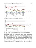
Corrosion test
The comparison of these curves indicates a few important points: 1-a value of 1.77×10
-3
mpy
for untreated sample (Fig. 19a), 2- the corresponding corrosion rates for 300 and 800 grit SiC
paper were measured as 1.8×10
-3
and 1.79×10
-3
mpy respectively (Figs. 19 b,c) 4- E
corr
varied
from -0.36 V to -0.21 V after the treatment at SiC paper 300 grit. This means that the SiC
treated samples are placed at a higher position in the cathodic section of the curve hence
releasing hydrogen easier and acts as an electron donor to the electrolyte. Therefore, by
smoothly reaching the passivation region, a more noble metal is expected to be achieved.
The corrosion current (I
corr
) was decreased from 2.59 μAcm
-2
to 0.66 μAcm
-2
after surface
treatment with SiC paper 300 grit and the corrosion current (I
corr
) for 800 grit was measured
2.51 μAcm
-2
. A better corrosion resistance was achieved by SiC paper.
Fig. 19. Tafel potentiodynamic polarization curves of Ti6Al4V for: (a) untreated, (b) SiC
paper (300 grit), and (c) SiC paper (800 grit)
Surface tension
The change in surface wettability was studied by contact angle measurement for all
specimens treated and untreated (Fig.20). Thus a decrease of contact angle occurred from 70º
to 50º indicating a higher degree of wettability. Following the SiC treatment at 800 grit the
contact angle reduced to 45 º showing still a more acceptable hydrophilic behaviour.
Also, variation of surface tension for all specimens was calculated by measured contact
angle. It is known that as contact angle decreases, the related surface tension will be
increased. Therefore, a value of 46 mN/m was obtained for γ at 300 grit which is
considerably higher than 39mN/m of the untreated sample. The corresponding value of γ
for 800 grit was found as 50mN/m (Fig. 20b).
Element
Sample
% Al %V %Ti
Untreated 5.15 3.25 91.6
SiC paper ( 300 grit) 5.19 3.37 91.4
SiC paper ( 800 grit) 6.05 3.35 90.6
Fig. 20. Variation of contact angle: (a) and surface tension, (b) with sample surface texture
In vitro
Figures 21 a-c illustrate the morphology and the spreading of cells on the negative control,
the untreated and SiC treatment respectively. As it is observed in all cases, some of the
attached cells spread radially from the centre and developed a filopodia type shape. The
surface of cells which are not spread, were convoluted in to micro ridges and the
neighboring cells maintain a physical contact with one another through multiple extensions.
Cell spreading is an essential function of cell adhesivity to any surface and it proceeds the
proliferation until the surface is fully covered by the cellular network. The number of cells
attached to the surface was evaluated by SiC treated samples assay. More cells are attached
to the surface for 300 and 800 grits of SiC paper, 9× 10
5
and 10 × 10
5
respectively, which are
higher than 8×10
5
for untreated sample.
Fig. 21. Light microscopy of cell culture evaluation (a) negative control, (b) untreated
sample, (c) SiC paper ( 800 grit).
In vivo
Cell spreading analysis
The experimental results of bone cell growth are given in table 3. As it can be seen, cells
spreading over the specimen surface are related to surface texture which was measured by
Image J program software (IJP). The highest spreading area (383 µm
2
) belongs to SiC treated
sample (800 grit).
Fundamentals of biomedical applications of biomorphic SiC 331
Table 2. Surface elements composition before and after treatment
Corrosion test
The comparison of these curves indicates a few important points: 1-a value of 1.77×10
-3
mpy
for untreated sample (Fig. 19a), 2- the corresponding corrosion rates for 300 and 800 grit SiC
paper were measured as 1.8×10
-3
and 1.79×10
-3
mpy respectively (Figs. 19 b,c) 4- E
corr
varied
from -0.36 V to -0.21 V after the treatment at SiC paper 300 grit. This means that the SiC
treated samples are placed at a higher position in the cathodic section of the curve hence
releasing hydrogen easier and acts as an electron donor to the electrolyte. Therefore, by
smoothly reaching the passivation region, a more noble metal is expected to be achieved.
The corrosion current (I
corr
) was decreased from 2.59 μAcm
-2
to 0.66 μAcm
-2
after surface
treatment with SiC paper 300 grit and the corrosion current (I
corr
) for 800 grit was measured
2.51 μAcm
-2
. A better corrosion resistance was achieved by SiC paper.
Fig. 19. Tafel potentiodynamic polarization curves of Ti6Al4V for: (a) untreated, (b) SiC
paper (300 grit), and (c) SiC paper (800 grit)
Surface tension
The change in surface wettability was studied by contact angle measurement for all
specimens treated and untreated (Fig.20). Thus a decrease of contact angle occurred from 70º
to 50º indicating a higher degree of wettability. Following the SiC treatment at 800 grit the
contact angle reduced to 45 º showing still a more acceptable hydrophilic behaviour.
Also, variation of surface tension for all specimens was calculated by measured contact
angle. It is known that as contact angle decreases, the related surface tension will be
increased. Therefore, a value of 46 mN/m was obtained for γ at 300 grit which is
considerably higher than 39mN/m of the untreated sample. The corresponding value of γ
for 800 grit was found as 50mN/m (Fig. 20b).
Element
Sample
% Al %V %Ti
Untreated 5.15 3.25 91.6
SiC paper ( 300 grit) 5.19 3.37 91.4
SiC paper ( 800 grit) 6.05 3.35 90.6
Fig. 20. Variation of contact angle: (a) and surface tension, (b) with sample surface texture
In vitro
Figures 21 a-c illustrate the morphology and the spreading of cells on the negative control,
the untreated and SiC treatment respectively. As it is observed in all cases, some of the
attached cells spread radially from the centre and developed a filopodia type shape. The
surface of cells which are not spread, were convoluted in to micro ridges and the
neighboring cells maintain a physical contact with one another through multiple extensions.
Cell spreading is an essential function of cell adhesivity to any surface and it proceeds the
proliferation until the surface is fully covered by the cellular network. The number of cells
attached to the surface was evaluated by SiC treated samples assay. More cells are attached
to the surface for 300 and 800 grits of SiC paper, 9× 10
5
and 10 × 10
5
respectively, which are
higher than 8×10
5
for untreated sample.
Fig. 21. Light microscopy of cell culture evaluation (a) negative control, (b) untreated
sample, (c) SiC paper ( 800 grit).
In vivo
Cell spreading analysis
The experimental results of bone cell growth are given in table 3. As it can be seen, cells
spreading over the specimen surface are related to surface texture which was measured by
Image J program software (IJP). The highest spreading area (383 µm
2
) belongs to SiC treated
sample (800 grit).
Properties and Applications of Silicon Carbide332
Row Specimens Spread cell area (μm
2
)
1 untreated 352 ± 6
2 SiC paper (800 grit) 383 ± 5
3 SiC paper (300 grit) 367± 3
Table 3. Bone cells spread over the surface of the implanted specimens (average of ten
measurements in three separate regions)
The SEM analysis of attached cells morphology (Fig. 22) indicates that the density of cell network
is directly dependent on the surface topography. In SiC treated surfaces, the orientation of cells
was longitudinal and parallel to the lines made by SiC paper. It is observed from Fig. 22 that SiC
treated surfaces have more fibroblast cells compared with the untreated sample.
Fig. 22. SEM micrographs of attached cells on the surface for: (a) untreated, (b) 800 grit, (c)
300 grit
Histopathology
When the implants were retrieved, no inflammatory reaction was observed inside or around the
implants. Mineralized matrix deposition and bone cells were observed on the surface of implants
which are formed during the five months implantation. This deposition was found all around of
SiC treated samples (Fig. 23a) and bone formation was characterized by the occurrence of
osteocyte embedded in the matrix. Also the above samples were surrounded by fibroblast and
osteoblast cells and the untreated sample (Fig. 23b) showed not only fewer number of fibroblast
cells, but it also contained osteoclast and polymorpho nuclear leukocytes (PMN).
Fig. 23. Light microscopy evaluation of bone tissue for: (a) 800 grit, and (b) untreated
In table 4, the symbols indicate the presence of 2-3 cells (+), 3-5 cells (++), more than 5 cells
(+++) and lack of cells (-) respectively. No PMN, giant cells and osteoclast were seen in SiC
treated samples. Also tissue healing was better conducted near mentioned implant.
Fibroblast and osteoblast cells were seen in samples.
The successful incorporation of bone implants strongly depends on a firm longstanding
adhesion of the tissue surrounding the implants. The cellular reaction is influenced by the
properties of the bulk materials as well as the specifications of the surface, that is, the
chemical composition and the topography (Birte et al., 2003, Siikavitsas et al., 2003). When
one is considering materials for application of orthopaedic implants, it is important to
consider a number of factors, such as biocompatibility and surface wettability. The
interaction of living cells with foreign materials is complicated matter, but fundamental for
biology medicine and is a key for understanding the biocompatibility. The initial cellular
events which take place at the biomaterials interface mimic to a certain extent the natural
adhesive interaction of cells with the extra cellular matrix (ECM).
Sample Cell
SiC paper (800 grit) SiC paper (300 grit) untreated
Fibroblast ++ ++ ++
Osteoblast + + +
Giant cell - - -
Osteoclast - - +
PMN - - +
Lymphocyte ++ ++ ++
Macrophage +++ +++ ++
Healing + + +
Table 4. Qualitative evaluation of histology results of bone tissue around the implants
The osteoblasts, which play a principal role in bone formation, readily attach to the material
surfaces via adsorbed protein layer consisting or RGD containing ligands like fibronectin,
vitronectin or fibrinogen. Family of cell surface receptors that provide trans- membrane
links between the ECM and the cytoskeleton. Our study showed that surface micro grooves
can affect the orientation guidance of bone cells i.e. the deeper grooves were more effective
in guiding the cells as it was evaluated by SEM. However, we did not conduct or evaluate
systematically the exact effects of grooves depth and size on cell orientation, but our
preliminary results were similar to those reported by Xiong et.al (Xiong et al., 2003).
This study was focused on the topographic effects of Ti-6Al-4V produced by SiC paper on
goat bone cell adhesion. The results showed a common feature reported in the previous
studies on a variety of cell types and substrates ie, topographic features strongly affects the
cell guidance. Micro grooved surfaces increase of surface tension and reduction of contact
angle. The test confirmed that the highest number of cells is attached to SiC paper modified
surface. It is also concluded from the SEM, contact angle measurements and preliminary in
vitro and in vivo tests that SiC paper can induce a desirable surface modification on Ti-6Al-4V
alloy for cell adhesivity and that a noble and biocompatible. Finally, it is suggested that
more detailed experiments are required and would be useful to distinguish and clarify the
relation between the grooves size and their orientation must be studied more carefully with
respect to cell attachment and their reliability as well as endurance.
Fundamentals of biomedical applications of biomorphic SiC 333
Row Specimens Spread cell area (μm
2
)
1 untreated 352 ± 6
2 SiC paper (800 grit) 383 ± 5
3 SiC paper (300 grit) 367± 3
Table 3. Bone cells spread over the surface of the implanted specimens (average of ten
measurements in three separate regions)
The SEM analysis of attached cells morphology (Fig. 22) indicates that the density of cell network
is directly dependent on the surface topography. In SiC treated surfaces, the orientation of cells
was longitudinal and parallel to the lines made by SiC paper. It is observed from Fig. 22 that SiC
treated surfaces have more fibroblast cells compared with the untreated sample.
Fig. 22. SEM micrographs of attached cells on the surface for: (a) untreated, (b) 800 grit, (c)
300 grit
Histopathology
When the implants were retrieved, no inflammatory reaction was observed inside or around the
implants. Mineralized matrix deposition and bone cells were observed on the surface of implants
which are formed during the five months implantation. This deposition was found all around of
SiC treated samples (Fig. 23a) and bone formation was characterized by the occurrence of
osteocyte embedded in the matrix. Also the above samples were surrounded by fibroblast and
osteoblast cells and the untreated sample (Fig. 23b) showed not only fewer number of fibroblast
cells, but it also contained osteoclast and polymorpho nuclear leukocytes (PMN).
Fig. 23. Light microscopy evaluation of bone tissue for: (a) 800 grit, and (b) untreated
In table 4, the symbols indicate the presence of 2-3 cells (+), 3-5 cells (++), more than 5 cells
(+++) and lack of cells (-) respectively. No PMN, giant cells and osteoclast were seen in SiC
treated samples. Also tissue healing was better conducted near mentioned implant.
Fibroblast and osteoblast cells were seen in samples.
The successful incorporation of bone implants strongly depends on a firm longstanding
adhesion of the tissue surrounding the implants. The cellular reaction is influenced by the
properties of the bulk materials as well as the specifications of the surface, that is, the
chemical composition and the topography (Birte et al., 2003, Siikavitsas et al., 2003). When
one is considering materials for application of orthopaedic implants, it is important to
consider a number of factors, such as biocompatibility and surface wettability. The
interaction of living cells with foreign materials is complicated matter, but fundamental for
biology medicine and is a key for understanding the biocompatibility. The initial cellular
events which take place at the biomaterials interface mimic to a certain extent the natural
adhesive interaction of cells with the extra cellular matrix (ECM).
Sample Cell
SiC paper (800 grit) SiC paper (300 grit) untreated
Fibroblast ++ ++ ++
Osteoblast + + +
Giant cell - - -
Osteoclast - - +
PMN - - +
Lymphocyte ++ ++ ++
Macrophage +++ +++ ++
Healing + + +
Table 4. Qualitative evaluation of histology results of bone tissue around the implants
The osteoblasts, which play a principal role in bone formation, readily attach to the material
surfaces via adsorbed protein layer consisting or RGD containing ligands like fibronectin,
vitronectin or fibrinogen. Family of cell surface receptors that provide trans- membrane
links between the ECM and the cytoskeleton. Our study showed that surface micro grooves
can affect the orientation guidance of bone cells i.e. the deeper grooves were more effective
in guiding the cells as it was evaluated by SEM. However, we did not conduct or evaluate
systematically the exact effects of grooves depth and size on cell orientation, but our
preliminary results were similar to those reported by Xiong et.al (Xiong et al., 2003).
This study was focused on the topographic effects of Ti-6Al-4V produced by SiC paper on
goat bone cell adhesion. The results showed a common feature reported in the previous
studies on a variety of cell types and substrates ie, topographic features strongly affects the
cell guidance. Micro grooved surfaces increase of surface tension and reduction of contact
angle. The test confirmed that the highest number of cells is attached to SiC paper modified
surface. It is also concluded from the SEM, contact angle measurements and preliminary in
vitro and in vivo tests that SiC paper can induce a desirable surface modification on Ti-6Al-4V
alloy for cell adhesivity and that a noble and biocompatible. Finally, it is suggested that
more detailed experiments are required and would be useful to distinguish and clarify the
relation between the grooves size and their orientation must be studied more carefully with
respect to cell attachment and their reliability as well as endurance.
Properties and Applications of Silicon Carbide334
18. Future considerations in biomedical applications of SiC
The next decade will see a great increase in scientific research into the biomedical
applications of SiC. Many analysis techniques may be used to analyze SiC biocompatibility.
In particular, primary cell lines could be cultured on SiC surfaces in the future since their
behavior would be a closer description of the in vivo performance of the material. While
proof-of concept studies in research laboratories have demonstrated great promise in the
use of SiC for scaffold of tissue engineering, several issues will need to be addressed before
SiC find way to large-scale clinical application. In particular, researches will need to study
toxic and pharmacokinetic effects of SiC in vivo. In addition, research will focuse on the
synthesis SiC nanopartiicles that may facilitate the development of multifunctional
nanostructures for use in drug delivery and tissue engineering applications. More
experiments are required to clarify the relation between SiC and cell attachment in scaffold
of tissue engineering. The different polytypes of SiC were quite well matched to organic
systems in terms of band gap and band alignment. Therefore, SiC should be a very
interesting substrate material for future semiconductor/organic heterostructures. Finally,
the feasibility of surface functionalization of SiC leaving free functional groups has been
shown while deeper understanding of the chemisorptions of various organic molecules is
still needed in order to optimize surface functionalization processes. The preparation and
complete characterization of atomically ordered SiC surfaces may lead to the successful
implementation of a large variety of biotechnological applications. It is suggested that more
investigations are required and would be useful to distinguish SiC biomedical applications.
19. References
Amon, M.; Bolz, A. & Schaldach, M. (1996). Improvement of stenting therapy with a silicon
carbide coated tantalum stent. J Mater Sci: Mater Med, 7, 5, 273–8
Amon, M.; Winkler, S.; Dekker, A.; Bolz, A.; Mittermayer, C. & Schaldach, M. (1995).
Introduction of a new coronary stent with enhanced radiopacity and
hemocompatibility. pp. 107–8 Proceedings of the Annual International Conference of the
IEEE Engineers in Medicine and Biology Society 95CB35746, vol. 17, 1. New York: IEEE
Press.
Anderson, A.S.; Dattelbaum, A.M.; Montano, G.A.; Price, D.N.; Schmidt, J.G.; Martinez, J.S.;
Grace, W.K.; Grace, K.M.; Swanson, B.I. (2008). Functioal PEG-modified thin films
for biological detection. Langmuir, 24, 2240-2247
Bai, S.; Ke, Y.; Shishkin, Y.; Shigiltchoff, O.; Devaty, R.P.; Choyke, W.J.; Strauch, D.; Stojetz,
B.; Dorner, B.; Hobgood, D.; Serrano, J.; Cardona, M.; Nagasawa, H.; Kimoto, T. &
Porter L.M. (2003). Four Current Examples of Characterization of Silicon Carbide,
Mat. Res. Soc. Symp. Proc. 742, K3.1.1
Bayer, G.; Hartwig, S.; Nagel, M.; Tilttelbach, M.; Rzany, A. & Schaldach, M. (2001). Future
Strategies for Antiproliferative Stent Coatings. Progress in Biomedical Research, 222-
225
Berthold, A.; Laugere F.; Schellevis, H.; De Boer, Ch. R.; Laros, M.; Guijt, R. M.; Sarro, P. M.
& Vellekoop, M. J. (2002). Fabrication of a glass-implemented microcapillary
electrophoresis device with integrated contactless conductivity detection.
Electrophoresis, 23, 3511–3519
Bigerelle, M. & Anselme, K. (2005). Bootstrap analysis of the relation between initial
adhesive events and long-term cellular functions of human osteoblasts cultured on
biocompatible metallic substrates. Acta biomaterialia, 1, 499-510
Birte, G.S.; Neubert, A.; Hopp, M.; Griepentrog, M. & Lange, K.P. (2003). Fibroblast growth
on surface modified dental implants: An in vitro study. J. Biomed. Mater. Res., 64A,
591-599
Bolz, A. & Schaldach, M., (1993). Heamocompatibility optimization of implants by hybrid
structuring, S123-S130, World Congress Supplement, Biomaterials, Koyoto
Bolz, A. (1995). Applications of Thin-Film Technology in Biomedical Engineering. Bolz, A. &
Schaldach, M. (1990). Artificial heart valves: improved blood compatibility by
PECVD a-SiC:H coating. Artificial organs, 14(4), 260-9
Bolz, A. & Schaldach, M. (1993). Biomaterials haemocompatibility optimization of implants
by hybrid structuring. Med. & Biol. Eng. & Comput., 31, 123-130
Bolz, A; Amon, M; Ozbek, C; Heublein, B; Schaldach, M. (1996). Coating of cardiovascular
stents with a semiconductor to improve their hemocompatibility. Texas Heart
Institute J. 23, 2, 162-6
D.L. , Trantolo, D.J. & et al. Encyclopedic Handbook of Biomaterials and Bioengineering.
Part A: Materials, In: Wise, 2, 1287-1330
Borrajo, J.P.; Serra, J.; Liste, S.; Gonza´lez, P.; Chiussi, S.; Leo´n, B. & Pe´rez-Amor, M. (2005).
Pulsed laser deposition of hydroxylapatite thin films on biomorphic silicon carbide
ceramics. Applied Surface Science, 248, 2005, 355–359
Botsoa, J., Lysenko, V., G_eloen, A., Marty, O., Bluet, J. M. & Guillot, G. (2008). Application
of 3C-SiC quantum dots for living cell imaging. Appl. Phys. Lett., 92, 173902
Buchter, A.; Joos, U.; Wiessman, H.P.; Seper, L. & Meyer, U. ( 2006). Biological and
biomechanical evaluation of interface reaction at conical screw-type implant. Head
and Face Med., 2, 5-18
Carlos, A. D.; Borrajo, J.P.; Serra, J.; Gonz´alez, P. & Le´on, B. (2006). Behaviour of MG-63
osteoblast-like cells on wood-based biomorphic SiC ceramics coated with bioactive
glass. J Mater Sci: Mater Med , 17, 523–529
Carrie, K.; Khalife, M.; Hamon, B.; Citron, J.P.; Monassier, R.; Sabatier, J.; Lipiecky, S.;
Mourali, L.; Sarfaty,M.; Elbaz, J.; Fourcade & Puel, J. (2001). Initial and Follow-Up
Results of the Tenax Coronary Stent.
J. Interventional Cardiology 14(1), 1-5
Calderon, N.R.; Martinez-Escandell, M.; Narciso, J. & Rodríguez-Reinoso, F. (2009). The role
of carbon biotemplate density in mechanical properties of biomorphic SiC. J. of the
European Ceramic Society, 29, 465–472
Caputo, D.; de Cesare, G.; Nascetti, A.; Scipinotti, R., (2008). Two-Color Sensor for
Biomolecule Detection. Sensor Letters, 6, 4, 542-547
Chakrabarti, O.P.; Maiti, H.S., & Majumdar, R. (2004). Biomimetic synthesis of cellular SiC
based ceramics from plant precursor. Bull. Mater. Sci., 27, 5, 467–470
Chu, W.H.; Chin ,R.; Huen, T. & Ferrari, M. (1999). Silicon Membrane Nanofilters from
Sacrificial Oxide Removal. J. Microelectromech. Syst. 8, 34-42
Cicero, G. & Catellani, A. (2005). Towards SiC surface functionalization: An ab initio study.
J. Chem. Phys., 122, 214716, 1-5
Cicoira, f. & Rosei, F. (2006). Playing Tetris at the nanoscale. Surface Science, 600, 1–5
Fundamentals of biomedical applications of biomorphic SiC 335
18. Future considerations in biomedical applications of SiC
The next decade will see a great increase in scientific research into the biomedical
applications of SiC. Many analysis techniques may be used to analyze SiC biocompatibility.
In particular, primary cell lines could be cultured on SiC surfaces in the future since their
behavior would be a closer description of the in vivo performance of the material. While
proof-of concept studies in research laboratories have demonstrated great promise in the
use of SiC for scaffold of tissue engineering, several issues will need to be addressed before
SiC find way to large-scale clinical application. In particular, researches will need to study
toxic and pharmacokinetic effects of SiC in vivo. In addition, research will focuse on the
synthesis SiC nanopartiicles that may facilitate the development of multifunctional
nanostructures for use in drug delivery and tissue engineering applications. More
experiments are required to clarify the relation between SiC and cell attachment in scaffold
of tissue engineering. The different polytypes of SiC were quite well matched to organic
systems in terms of band gap and band alignment. Therefore, SiC should be a very
interesting substrate material for future semiconductor/organic heterostructures. Finally,
the feasibility of surface functionalization of SiC leaving free functional groups has been
shown while deeper understanding of the chemisorptions of various organic molecules is
still needed in order to optimize surface functionalization processes. The preparation and
complete characterization of atomically ordered SiC surfaces may lead to the successful
implementation of a large variety of biotechnological applications. It is suggested that more
investigations are required and would be useful to distinguish SiC biomedical applications.
19. References
Amon, M.; Bolz, A. & Schaldach, M. (1996). Improvement of stenting therapy with a silicon
carbide coated tantalum stent. J Mater Sci: Mater Med, 7, 5, 273–8
Amon, M.; Winkler, S.; Dekker, A.; Bolz, A.; Mittermayer, C. & Schaldach, M. (1995).
Introduction of a new coronary stent with enhanced radiopacity and
hemocompatibility. pp. 107–8 Proceedings of the Annual International Conference of the
IEEE Engineers in Medicine and Biology Society 95CB35746, vol. 17, 1. New York: IEEE
Press.
Anderson, A.S.; Dattelbaum, A.M.; Montano, G.A.; Price, D.N.; Schmidt, J.G.; Martinez, J.S.;
Grace, W.K.; Grace, K.M.; Swanson, B.I. (2008). Functioal PEG-modified thin films
for biological detection. Langmuir, 24, 2240-2247
Bai, S.; Ke, Y.; Shishkin, Y.; Shigiltchoff, O.; Devaty, R.P.; Choyke, W.J.; Strauch, D.; Stojetz,
B.; Dorner, B.; Hobgood, D.; Serrano, J.; Cardona, M.; Nagasawa, H.; Kimoto, T. &
Porter L.M. (2003). Four Current Examples of Characterization of Silicon Carbide,
Mat. Res. Soc. Symp. Proc. 742, K3.1.1
Bayer, G.; Hartwig, S.; Nagel, M.; Tilttelbach, M.; Rzany, A. & Schaldach, M. (2001). Future
Strategies for Antiproliferative Stent Coatings. Progress in Biomedical Research, 222-
225
Berthold, A.; Laugere F.; Schellevis, H.; De Boer, Ch. R.; Laros, M.; Guijt, R. M.; Sarro, P. M.
& Vellekoop, M. J. (2002). Fabrication of a glass-implemented microcapillary
electrophoresis device with integrated contactless conductivity detection.
Electrophoresis, 23, 3511–3519
Bigerelle, M. & Anselme, K. (2005). Bootstrap analysis of the relation between initial
adhesive events and long-term cellular functions of human osteoblasts cultured on
biocompatible metallic substrates. Acta biomaterialia, 1, 499-510
Birte, G.S.; Neubert, A.; Hopp, M.; Griepentrog, M. & Lange, K.P. (2003). Fibroblast growth
on surface modified dental implants: An in vitro study. J. Biomed. Mater. Res., 64A,
591-599
Bolz, A. & Schaldach, M., (1993). Heamocompatibility optimization of implants by hybrid
structuring, S123-S130, World Congress Supplement, Biomaterials, Koyoto
Bolz, A. (1995). Applications of Thin-Film Technology in Biomedical Engineering. Bolz, A. &
Schaldach, M. (1990). Artificial heart valves: improved blood compatibility by
PECVD a-SiC:H coating. Artificial organs, 14(4), 260-9
Bolz, A. & Schaldach, M. (1993). Biomaterials haemocompatibility optimization of implants
by hybrid structuring. Med. & Biol. Eng. & Comput., 31, 123-130
Bolz, A; Amon, M; Ozbek, C; Heublein, B; Schaldach, M. (1996). Coating of cardiovascular
stents with a semiconductor to improve their hemocompatibility. Texas Heart
Institute J. 23, 2, 162-6
D.L. , Trantolo, D.J. & et al. Encyclopedic Handbook of Biomaterials and Bioengineering.
Part A: Materials, In: Wise, 2, 1287-1330
Borrajo, J.P.; Serra, J.; Liste, S.; Gonza´lez, P.; Chiussi, S.; Leo´n, B. & Pe´rez-Amor, M. (2005).
Pulsed laser deposition of hydroxylapatite thin films on biomorphic silicon carbide
ceramics. Applied Surface Science, 248, 2005, 355–359
Botsoa, J., Lysenko, V., G_eloen, A., Marty, O., Bluet, J. M. & Guillot, G. (2008). Application
of 3C-SiC quantum dots for living cell imaging. Appl. Phys. Lett., 92, 173902
Buchter, A.; Joos, U.; Wiessman, H.P.; Seper, L. & Meyer, U. ( 2006). Biological and
biomechanical evaluation of interface reaction at conical screw-type implant. Head
and Face Med., 2, 5-18
Carlos, A. D.; Borrajo, J.P.; Serra, J.; Gonz´alez, P. & Le´on, B. (2006). Behaviour of MG-63
osteoblast-like cells on wood-based biomorphic SiC ceramics coated with bioactive
glass. J Mater Sci: Mater Med , 17, 523–529
Carrie, K.; Khalife, M.; Hamon, B.; Citron, J.P.; Monassier, R.; Sabatier, J.; Lipiecky, S.;
Mourali, L.; Sarfaty,M.; Elbaz, J.; Fourcade & Puel, J. (2001). Initial and Follow-Up
Results of the Tenax Coronary Stent.
J. Interventional Cardiology 14(1), 1-5
Calderon, N.R.; Martinez-Escandell, M.; Narciso, J. & Rodríguez-Reinoso, F. (2009). The role
of carbon biotemplate density in mechanical properties of biomorphic SiC. J. of the
European Ceramic Society, 29, 465–472
Caputo, D.; de Cesare, G.; Nascetti, A.; Scipinotti, R., (2008). Two-Color Sensor for
Biomolecule Detection. Sensor Letters, 6, 4, 542-547
Chakrabarti, O.P.; Maiti, H.S., & Majumdar, R. (2004). Biomimetic synthesis of cellular SiC
based ceramics from plant precursor. Bull. Mater. Sci., 27, 5, 467–470
Chu, W.H.; Chin ,R.; Huen, T. & Ferrari, M. (1999). Silicon Membrane Nanofilters from
Sacrificial Oxide Removal. J. Microelectromech. Syst. 8, 34-42
Cicero, G. & Catellani, A. (2005). Towards SiC surface functionalization: An ab initio study.
J. Chem. Phys., 122, 214716, 1-5
Cicoira, f. & Rosei, F. (2006). Playing Tetris at the nanoscale. Surface Science, 600, 1–5
Properties and Applications of Silicon Carbide336
Cogan, S.F.; Edell, D.J.; Guzellan, A.A.; Ying, L.P. & Edell, R. (2003). Plasma-enhanced
chemical vapor deposited silicon carbide as an implantable dielectric coating. J.
Biomed. Mater. Res. A, 67, 3, 856-67
Cole, K.S. (1940). Permiability and impermiability of cell membranes for ions. Sympos
Quant.Biol., 8, 110-122
Coletti, C.; Jaroszeski, M.; Hoff, A.M. & Saddow, S.E. (2006). Culture of mammalian cells on
single crystal SiC substrates, Mater. Res. Soc. Symp. Proc. 950
Coletti, C.; Jaroszeski, M.J.; Pallaoro, A.; Hoff, A.M.; Iannotta, S. & Saddow, S.E. (2007).
Biocompatibility and wettability of crystalline SiC and Si surfaces. IEEE EMBS
Proceedings, 5849-5852
Desai, T.A.; Chu, W.H.; Tu, J.K.; Beattie, G.M.; Hayek, A. & Ferrari, M. (1997).
Microfabricated Immunoisolating Biocapsules. Biotech. and Bioeng., 57(1), 118-120
Elbaz, M.; El Mokhtar, E.; Fourcade, J.; Mourali, S.; Hobeika, R.; Carrie, D. & Puel, J. (2002).
Dose stent design affect the long-term outcome after coronary stenting?
Catheterization & Cardiovascular Interventions, 56, 305-311
El Ghannam, A. (2005). Bone reconstruction: from bioceramics to tissue engineering. Expert
Rev Med Devices. 2, 1, 87-101
Fan, T.X.; Li, X.F.; Liu, Z.T.; Gu, J.J.; Zhang, D.; Guo, Q.X. & Am, J. (2006). Microstructure
and infrared absorption of biomorphic chromium oxides templated by wood
tissues. Ceram. Soc. 89, 3511-3515
Fan,
J.; Li, H.; Jiang, J., So L. K. Y.; Lam
Y. W. & Chu, P. K. (2008). 3C-SiC Nanocrystals as
Fluorescent Biological Labels. Small, 4, 8, 1058–1062
Feng, B.; Weng, J.; Yang, B.C.; Qu, S.X. & Zhang, X.D. (2003). Characterization of surface
oxide films on titanium and adhesion of osteoblast. Biomaterials, 24, 4663-4670
Feng, X.; Yang, Q. & De Jonghe, L.C. (2003). Microstructure development of hot-pressed
silicon carbide: effects of aluminum, boron, and carbon additives. Acta Mater, 51,
3849-60
Fissell, W.H. (2003). Nanoporous Hemofiltration Membranes for Bioartificial Kidneys. oral
presentation at 4th Annual BioMEMS and Nanotech World,Washington, DC
Frewin, C.L.; Jaroszeski, M.; Weeber, E.; Muffly, K.E.; Kumar, A.; Peters, M.; Oliveros, A.&
Saddow, S.E.J. (2009). Atomic force microscopy analysis of central nervous system
cell morphology on silicon carbide and diamond substrates. Mol. Recognit., 22, 5,
380
Fujibayashi, S.; Neo, M.; Kim, H.; Kokubo, T. & Nakamura, T. (2003). A comparative study
between in vivo bone in growth and in vitro apatite formation on Na
2
O-CaO-SiO
2
glasses. Biomaterials, 24, 1349-1356
Gabriel, G.; Erill, I.; Caro, J.; Gomez, R.; Riera, D.; Villa, R. & Godignon, Ph. (2007).
Manufacturing and full characterization of silicon carbide-based multi-sensor
micro-probes for biomedical applications. Microelec. J., 38, 406–415
Gersing, E. (1998). Impedance spectroscopy on living tissue for determination of the state of
organs. Bioelectrochem. Bioenerg. 45, 145–149
Goeders, K. M.; Colton, J. S.; Bottomley, L. A. (2008). Microcantilevers: Sensing Chemical
Interactions via Mechanical Motion. Chem. Rev., 108, 522-542
Godignon, P. (2005). New Generation of SiC Based Biodevices Implemented on 4” Wafers.
Mater. Sci. Forum 483–485. 1097-1100
Gomez, R.; Ivorra, A.; Villa, R.; Godignon, Ph.; Mill´an, J.; Erill, I.; Sol`a, A.; Hotter, G. &
Palacios, L. (2006). A SiC microdevice for the minimally invasive monitoring of
ischemia in living tissues. Biomed. Microdevices, 8, 43–49
Gonzalez, P.; Serra, J.; Liste, S.; Chiussi, S.; Leon, B.; Perez-Amor, M.; Martinez-Fernandez,
J.; de Arellano-Lopez, A.R. & Varela-Feria, F.M. (2003).New biomorphic SiC
ceramics coated with bioactive glass for biomedical applications. Biomaterials, 24,
4827–4832
Gonzalez, P.; Borrajo, J.P.; Serra, J.; Liste, S.; Chiussi, S.; Leo´n, B.; Semmelmann, K.; De
Carlos, A.; Varela-Feria, F.M.; Mart´nez-Ferna´ndez, J. & De Arellano-Lo´pez, A.R.
(2004). Key Eng. Mater., 1029, 254–256
Gonzalez, P.; Serra, J.; Liste, S.; Chiussi, S.; Leon, B.; Perez-Amor, M. & et al. (2003). New
biomorphic SiC ceramics coated with bioactive glass for biomedical applications.
Biomaterials, 24, 26, 4827-432
Gonzalez, P.; Borrajo, J.P.; Serra, J.; Chiussi, J.; Leo, B.; Mart nez-Ferna ndez, J.; Varela-Feria,
F.M.; de Arellano-Lopez, A.R.; de Carlos, A.; Mun oz, F.M.; Lo pez, M. & Singh, M.
(2008). A new generation of bio-derived ceramic materials for medical applications.
2008 Wiley Periodicals, Inc.
Greil, P. (2002). Advanced engineering ceramics. Adv. Eng. Mater, 4, 5, 247–254
Gutierrez-Mora, F.; Goretta, K.C.; Varela-Feria, F.M.; Arellano Lo pez, A.R. & Martnez
Ferna ndez, J. (2005). Indentation hardness of biomorphic SiC. Inter J. of Refractory
Metals & Hard Materials, 23, 369–374
Haemmerich, D.; Ozkan, R.; Tungjitkusolmun, S.; Tsai, J.Z.; Mahvi, D.M.; Staelin, S.T.;
Webster, J.G.; (2002). Changes in electrical resistivity of swine liver after occlusion
and postmortem, Medical and Biological Engineering and Computing, 40, 1, 29-33
Hamm, C.W.; Hugenholtz, P.G. & Trust, I. (2003). Silicon carbide-coated stents in patients
with acute coronary syndrome. Catheterization & Cardiovascular Interventions, 60,
3, 375-381
Hanekamp, C.E.E. & Koolen, J.J. (2000). Coated Stents in Small Coronary Vessels – A
Successful Strategy? Progress in Biomedical Research, 221-223
Harder, C.; Rzany,A. & Schaldach, M. (1999). Coating of Vascular Stents with
Antithrombogenic Amorphous Silicon Carbide, Progress in Biomedical Research, 71-
77
Heinrich, A.; Dengler, K.; Koerner, T.; Haczek, C.; Deppe, H. & Stritzker, B. (2008). Laser-
modified titanium implants for improved cell adhesion. Laser in Medical science, 23,
55-58
Hesaraki, S.; Ebadzadeh, T. & Ahmadzadeh-Asl, S. (2010). Nanosilicon
carbide/hydroxyapatite nanocomposites: structural, mechanical and in vitro
cellular properties. J Mater Sci: Mater Med, DOI 10.1007/s10856-010-4068-7
Hildebrandt, P.; Sayyad, M.; Rzany, A. & et al. (2001). Prevention of surface encrustation of
urological implants by coating with inhibitors. Biomaterials, 22, 503-507
Hing, K.A.; Revell, P.A.; Smith, N. & Buckland, T. (2006). Effect of silicon level on rate,
quality and progression of bone healing within silicate-substituted porous
hydroxyapatite scaffolds. Biomaterials, 27, 29, 5014-5026
Ikeuchi, K.; Kusaka J. & Yoshida, H. (2000). Tribology for all-ceramic joint prostheses. Journal
of Ceramic Processing Research, 1, 1, 53-56
Fundamentals of biomedical applications of biomorphic SiC 337
Cogan, S.F.; Edell, D.J.; Guzellan, A.A.; Ying, L.P. & Edell, R. (2003). Plasma-enhanced
chemical vapor deposited silicon carbide as an implantable dielectric coating. J.
Biomed. Mater. Res. A, 67, 3, 856-67
Cole, K.S. (1940). Permiability and impermiability of cell membranes for ions. Sympos
Quant.Biol., 8, 110-122
Coletti, C.; Jaroszeski, M.; Hoff, A.M. & Saddow, S.E. (2006). Culture of mammalian cells on
single crystal SiC substrates, Mater. Res. Soc. Symp. Proc. 950
Coletti, C.; Jaroszeski, M.J.; Pallaoro, A.; Hoff, A.M.; Iannotta, S. & Saddow, S.E. (2007).
Biocompatibility and wettability of crystalline SiC and Si surfaces. IEEE EMBS
Proceedings, 5849-5852
Desai, T.A.; Chu, W.H.; Tu, J.K.; Beattie, G.M.; Hayek, A. & Ferrari, M. (1997).
Microfabricated Immunoisolating Biocapsules. Biotech. and Bioeng., 57(1), 118-120
Elbaz, M.; El Mokhtar, E.; Fourcade, J.; Mourali, S.; Hobeika, R.; Carrie, D. & Puel, J. (2002).
Dose stent design affect the long-term outcome after coronary stenting?
Catheterization & Cardiovascular Interventions, 56, 305-311
El Ghannam, A. (2005). Bone reconstruction: from bioceramics to tissue engineering. Expert
Rev Med Devices. 2, 1, 87-101
Fan, T.X.; Li, X.F.; Liu, Z.T.; Gu, J.J.; Zhang, D.; Guo, Q.X. & Am, J. (2006). Microstructure
and infrared absorption of biomorphic chromium oxides templated by wood
tissues. Ceram. Soc. 89, 3511-3515
Fan,
J.; Li, H.; Jiang, J., So L. K. Y.; Lam
Y. W. & Chu, P. K. (2008). 3C-SiC Nanocrystals as
Fluorescent Biological Labels. Small, 4, 8, 1058–1062
Feng, B.; Weng, J.; Yang, B.C.; Qu, S.X. & Zhang, X.D. (2003). Characterization of surface
oxide films on titanium and adhesion of osteoblast. Biomaterials, 24, 4663-4670
Feng, X.; Yang, Q. & De Jonghe, L.C. (2003). Microstructure development of hot-pressed
silicon carbide: effects of aluminum, boron, and carbon additives. Acta Mater, 51,
3849-60
Fissell, W.H. (2003). Nanoporous Hemofiltration Membranes for Bioartificial Kidneys. oral
presentation at 4th Annual BioMEMS and Nanotech World,Washington, DC
Frewin, C.L.; Jaroszeski, M.; Weeber, E.; Muffly, K.E.; Kumar, A.; Peters, M.; Oliveros, A.&
Saddow, S.E.J. (2009). Atomic force microscopy analysis of central nervous system
cell morphology on silicon carbide and diamond substrates. Mol. Recognit., 22, 5,
380
Fujibayashi, S.; Neo, M.; Kim, H.; Kokubo, T. & Nakamura, T. (2003). A comparative study
between in vivo bone in growth and in vitro apatite formation on Na
2
O-CaO-SiO
2
glasses. Biomaterials, 24, 1349-1356
Gabriel, G.; Erill, I.; Caro, J.; Gomez, R.; Riera, D.; Villa, R. & Godignon, Ph. (2007).
Manufacturing and full characterization of silicon carbide-based multi-sensor
micro-probes for biomedical applications. Microelec. J., 38, 406–415
Gersing, E. (1998). Impedance spectroscopy on living tissue for determination of the state of
organs. Bioelectrochem. Bioenerg. 45, 145–149
Goeders, K. M.; Colton, J. S.; Bottomley, L. A. (2008). Microcantilevers: Sensing Chemical
Interactions via Mechanical Motion. Chem. Rev., 108, 522-542
Godignon, P. (2005). New Generation of SiC Based Biodevices Implemented on 4” Wafers.
Mater. Sci. Forum 483–485. 1097-1100
Gomez, R.; Ivorra, A.; Villa, R.; Godignon, Ph.; Mill´an, J.; Erill, I.; Sol`a, A.; Hotter, G. &
Palacios, L. (2006). A SiC microdevice for the minimally invasive monitoring of
ischemia in living tissues. Biomed. Microdevices, 8, 43–49
Gonzalez, P.; Serra, J.; Liste, S.; Chiussi, S.; Leon, B.; Perez-Amor, M.; Martinez-Fernandez,
J.; de Arellano-Lopez, A.R. & Varela-Feria, F.M. (2003).New biomorphic SiC
ceramics coated with bioactive glass for biomedical applications. Biomaterials, 24,
4827–4832
Gonzalez, P.; Borrajo, J.P.; Serra, J.; Liste, S.; Chiussi, S.; Leo´n, B.; Semmelmann, K.; De
Carlos, A.; Varela-Feria, F.M.; Mart´nez-Ferna´ndez, J. & De Arellano-Lo´pez, A.R.
(2004). Key Eng. Mater., 1029, 254–256
Gonzalez, P.; Serra, J.; Liste, S.; Chiussi, S.; Leon, B.; Perez-Amor, M. & et al. (2003). New
biomorphic SiC ceramics coated with bioactive glass for biomedical applications.
Biomaterials, 24, 26, 4827-432
Gonzalez, P.; Borrajo, J.P.; Serra, J.; Chiussi, J.; Leo, B.; Mart nez-Ferna ndez, J.; Varela-Feria,
F.M.; de Arellano-Lopez, A.R.; de Carlos, A.; Mun oz, F.M.; Lo pez, M. & Singh, M.
(2008). A new generation of bio-derived ceramic materials for medical applications.
2008 Wiley Periodicals, Inc.
Greil, P. (2002). Advanced engineering ceramics. Adv. Eng. Mater, 4, 5, 247–254
Gutierrez-Mora, F.; Goretta, K.C.; Varela-Feria, F.M.; Arellano Lo pez, A.R. & Martnez
Ferna ndez, J. (2005). Indentation hardness of biomorphic SiC. Inter J. of Refractory
Metals & Hard Materials, 23, 369–374
Haemmerich, D.; Ozkan, R.; Tungjitkusolmun, S.; Tsai, J.Z.; Mahvi, D.M.; Staelin, S.T.;
Webster, J.G.; (2002). Changes in electrical resistivity of swine liver after occlusion
and postmortem, Medical and Biological Engineering and Computing, 40, 1, 29-33
Hamm, C.W.; Hugenholtz, P.G. & Trust, I. (2003). Silicon carbide-coated stents in patients
with acute coronary syndrome. Catheterization & Cardiovascular Interventions, 60,
3, 375-381
Hanekamp, C.E.E. & Koolen, J.J. (2000). Coated Stents in Small Coronary Vessels – A
Successful Strategy? Progress in Biomedical Research, 221-223
Harder, C.; Rzany,A. & Schaldach, M. (1999). Coating of Vascular Stents with
Antithrombogenic Amorphous Silicon Carbide, Progress in Biomedical Research, 71-
77
Heinrich, A.; Dengler, K.; Koerner, T.; Haczek, C.; Deppe, H. & Stritzker, B. (2008). Laser-
modified titanium implants for improved cell adhesion. Laser in Medical science, 23,
55-58
Hesaraki, S.; Ebadzadeh, T. & Ahmadzadeh-Asl, S. (2010). Nanosilicon
carbide/hydroxyapatite nanocomposites: structural, mechanical and in vitro
cellular properties. J Mater Sci: Mater Med, DOI 10.1007/s10856-010-4068-7
Hildebrandt, P.; Sayyad, M.; Rzany, A. & et al. (2001). Prevention of surface encrustation of
urological implants by coating with inhibitors. Biomaterials, 22, 503-507
Hing, K.A.; Revell, P.A.; Smith, N. & Buckland, T. (2006). Effect of silicon level on rate,
quality and progression of bone healing within silicate-substituted porous
hydroxyapatite scaffolds. Biomaterials, 27, 29, 5014-5026
Ikeuchi, K.; Kusaka J. & Yoshida, H. (2000). Tribology for all-ceramic joint prostheses. Journal
of Ceramic Processing Research, 1, 1, 53-56
Properties and Applications of Silicon Carbide338
Iliescu, C.; Poenar, D. P.; Carp, M.; Loe, F. C. (2007). A microfluidic device for impedance
spectroscopy analysis of biological samples. Sensors and Actuators B, 123, 168–176
Ivorra, A.; Gómez, R.; Noguera, N.; Villa, R.; Sola, A.; Palacios, L.; Hotter, G. & Aguiló, J.
(2003). Minimally invasive silicon probe for electrical impedance measurements in
small animals. Biosensors & Biolectronics, 19, 4, 391-399
Kamakaran, R.; Lupo, F.F.; Grobert, N.; Scheu, T.; Phillipp, N.Y.G. & Ruhle, M. (2004).
Microstructural Characterization of C–SiC–Carbon Nanotube Composite Flakes.
Carbon, 42, 1–4
Kanai, Y.; Cicero, G.; Selloni, A.; Car, R. & Galli, G. (2005). A Theoretical Study of Biotin
Chemisorption on Si-SiC(001) Surfaces. J. Phys. Chem. B 109, 13656
Kalnins, U.; Erglis, A.; Dinne, I.; Kumsars, I.; & Jegere, S. (2002). Medical Science Monitor 8,
I16
Karas, S.P.; Gravanis, M.B.; Santoian, E.C.; Robinson, K.A.; Andernerg, K.A. & King, S.B.
(1992). Coronary intimal proliferation after balloon injury and stenting in swine: an
animal model of restenosis. J. Am. Coll .Cardiol., 20, 467–74
Karoussos, A.; Wieneke, H.; Sawitowski, T.; Wnendt, S.; Fischer, O.; Dirsch, O.; Dahmen, U.
& Erbel, R. (2002). Inorganic materials as drug delivery systems in coronary artery
stenting. Mat. Wiss.U. Werkstofftech., 33, 738-746
Khosroshahi, M.E.; Mahmoodi, M. & Tavakoli, J. (2007). Characterization of Ti-6Al-4V
implant surface treated by Nd:YAG laser and emery paper for orthopaedic
applications. Applied surface science, 253, 8772-8781
Khosroshahi, M.E.; Tavakoli, J. & Mahmoodi, M. (2007). Analysis of bioadhesivity of
osteoblast on titanium by Nd:YAG laser. J. of Adhesion, 83, 151-172
Khosroshahi, M.E.; Mahmoodi, M.; Saedinasab, H. & Tahriri, M. (2008). Evaluation of
mechanical and electrochemical properties of laser surface modified Ti-6AL-4V for
biomedical application: in vitro study. Surface Engineering, 24, 209-218
Khosroshahi, M.E.; Mahmoodi, M.; Tavakoli, J. & Tahriri, M. (2008). Effect of Nd: YAG laser
radiation on Ti6AL4V alloy properties for biomedical application. J. of Laser
Applications, 20, 209-217
Khosroshahi, M.E.; Mahmoodi, M. & Saedinasab, H. (2009). In vitro and in vivo studies of
osteoblast cell response to a Ti6A14V surface modified by Nd:YAG laser and silicon
carbide paper. Laser Med. Sci, 24, 925-939
Kidambi, S.; Dai, J.H. & Bruening, M.L. (2004). Selective Hydrogenation of Pd Nanoparticles
Embedded in Polyelectrolyte Multilayers. J. Am. Chem. Soc., 126, 2658–9
Kotzara, G.; Freasa, M.; Abelb, Ph.; Fleischmanc, A.; Royc, Sh.; Zormand, Ch.; Morane, J. M.
& Melzak, J. (2002). Evaluation of MEMS materials of construction for implantable
medical devices. Biomaterials, 23, 2737–2750
LeGeros, R.Z.; LeGeros, J.P.; Klein, E. & Shirra, W.P. (1967). Apatite crystallites. Effect of
carbonate on mor- phology. Science, 155, 1409-1411
Liu, Z.; Fan, T.; Gu, J.; Zhang, D.; Gong, X.; Gu, Q. & Xu, J. (2007). Preparation of Porous Fe
from Biomorphic Fe
2
O
3
Precursors with Wood Templates. Mater. Trans., 48, 878-881
Liu, Y.; Ramanath, H.S. & Wang, D.A. (2008). Tendon tissue engineering using scaffold
enhancing strategies. TrendsBiotechnol., 26, 4, 201-209
Luo, M.; Gao, J.Q.; Yang, J.F. & Am, J. (2007). Biomorphic silicon nitride ceramics with and
reduction–nitridation. Ceram. Soc. 90, 4036-4039
Luo, M.; Hou, G.Y.; Yang, J.F.; Fang, J.Z.; Gao, J.Q.; Zhao, L. & Li, X. (2009). Manufacture of
fibrous β-Si3N4-reinforced biomorphic SiC matrix composites for bioceramic
scaffold applications. Materials Science and Engineering C, 29, 1422–1427
Maitz, M.F.; Pham, M. & Wieser, E. (2003). Blood compatibility of titanium oxides with
various crystal structure and element doping. J. biomaterials applications, 17, 4, 303-
319
Mangonon, P.L. (1999). The principles of materials selection for engineering design, 1
st
Ed.Upper Saddle River, NJ: Prentice-Hall
Mart nez-Fern!andez, J.; Varela-Feria, F.M. & Singh, M. (2000). Microstructure and
thermomechanical characterizacion of biomorphic silicon carbide-based ceramics.
Scr Mater, 43, 813–8
Mart nez Ferna ndez, J.; de Arellano-Lo pez, A.R.; Varela-Feria, F.M. & Singh, M. (2001).
Procedimiento para la Fabricacio´n de Carburo de Silicio a Partir de Precursores
Vegetales. Spanish Patent, P200102278, submitted by Universidad de Sevilla
Masuda, T.; Yliheikkila, P.K.; Felton, D.A. & Cooper, L.F. (1998). Generolization regarding
the process and phenomena of osseointegration: in vivo studies, part I. Int. J. Oral
Maxillofac. Implants, 13, 17-29
Mayor, B.; Arias, J.; Chiussi, S.; Garcia, F.; Pou, J.; Leo´n, B. & Pe´rez-Amor, M. (1998).
Calcium phosphate coatings grown at different substrate temperatures by pulsed
ArF-laser deposition. Thin Solid Films, 317, 363-366
McDonagh, C.; Burke, C.S. & MacCraith, B.D. (2008). Optical chemical sensors. Chem. Rev.,
108, 400-422
Meyers, M.A.; Chen, P.Y.; Lin, A.Y.M. & Seki, Y. (2008). Biological materials: structure and
mechanical properties. Prog. Mater. Sci., 53, 1-206
Monnink, S.H.; van Boven, A.J.; Peels, H.O.; Tigchelaar, I.; de Kam, P.J.; Crijns, H.J. & van
Oeveren,W. (1999). Silicon-carbide coated coronary stents have low platelet and
leukocyte adhesion during platelet activation. J. Investig Med, 47, 304–10
Naji, A. & Harmand, M.F. (1991). Cyto compatibilio Qf two coating materials, amorphous
alumina and silicon carbide, using human differentiated cell cultures. Biomaterials,
12, 690-694
Nordsletten, L.; Hogasen, A.K.; Konttinen, Y.T.; Santavirta, S.; Aspenberg, P. & Aasen, A.O.
(1996). Human monocytes stimulation by particles of hydroxyapatite, silicon
carbide and diamond: in vitro studies of new prosthesis coatings. Biomaterials, 17,
15, 1521–7
Nurdin, N.; François, P.; Mugnier, Y.; Moret, M.; Aronsson, B.O.; Krumeich, J. & Descouts,
P. (2003). Haemocompatibility evaluation of DLC and SiC-coated surfaces. European
Cells and Materials, 5, 17-28
Presas, M.; Pastor, J.Y.; Liorca, J.; Arellano Lo pez, A.R.; Mart nez Ferna ndez, J. & Sepu
lveda, R. (2006). Microstructure and fracture properties of biomorphic SiC. Inter J. of
Refractory Metals & Hard Materials, 24, 49–54
Preuss, M.; Bechstedt, F.; Schmidt, W.G.; Sochos, J.; Schroter Band Richter W (2006). Clean
and pyrrole-functionalized Si- and C-terminated SiC surfaces: First-principles
calculations of geometry and energetics compared with LEED and XPS. Phys. Rev.
B, 74, 235406
Ohji, T. (2008). Microstructural design and mechanical properties of porous silicon nitride
Ceramics. Mater. Sci. Eng. A, 498, 5-11
Fundamentals of biomedical applications of biomorphic SiC 339
Iliescu, C.; Poenar, D. P.; Carp, M.; Loe, F. C. (2007). A microfluidic device for impedance
spectroscopy analysis of biological samples. Sensors and Actuators B, 123, 168–176
Ivorra, A.; Gómez, R.; Noguera, N.; Villa, R.; Sola, A.; Palacios, L.; Hotter, G. & Aguiló, J.
(2003). Minimally invasive silicon probe for electrical impedance measurements in
small animals. Biosensors & Biolectronics, 19, 4, 391-399
Kamakaran, R.; Lupo, F.F.; Grobert, N.; Scheu, T.; Phillipp, N.Y.G. & Ruhle, M. (2004).
Microstructural Characterization of C–SiC–Carbon Nanotube Composite Flakes.
Carbon, 42, 1–4
Kanai, Y.; Cicero, G.; Selloni, A.; Car, R. & Galli, G. (2005). A Theoretical Study of Biotin
Chemisorption on Si-SiC(001) Surfaces. J. Phys. Chem. B 109, 13656
Kalnins, U.; Erglis, A.; Dinne, I.; Kumsars, I.; & Jegere, S. (2002). Medical Science Monitor 8,
I16
Karas, S.P.; Gravanis, M.B.; Santoian, E.C.; Robinson, K.A.; Andernerg, K.A. & King, S.B.
(1992). Coronary intimal proliferation after balloon injury and stenting in swine: an
animal model of restenosis. J. Am. Coll .Cardiol., 20, 467–74
Karoussos, A.; Wieneke, H.; Sawitowski, T.; Wnendt, S.; Fischer, O.; Dirsch, O.; Dahmen, U.
& Erbel, R. (2002). Inorganic materials as drug delivery systems in coronary artery
stenting. Mat. Wiss.U. Werkstofftech., 33, 738-746
Khosroshahi, M.E.; Mahmoodi, M. & Tavakoli, J. (2007). Characterization of Ti-6Al-4V
implant surface treated by Nd:YAG laser and emery paper for orthopaedic
applications. Applied surface science, 253, 8772-8781
Khosroshahi, M.E.; Tavakoli, J. & Mahmoodi, M. (2007). Analysis of bioadhesivity of
osteoblast on titanium by Nd:YAG laser. J. of Adhesion, 83, 151-172
Khosroshahi, M.E.; Mahmoodi, M.; Saedinasab, H. & Tahriri, M. (2008). Evaluation of
mechanical and electrochemical properties of laser surface modified Ti-6AL-4V for
biomedical application: in vitro study. Surface Engineering, 24, 209-218
Khosroshahi, M.E.; Mahmoodi, M.; Tavakoli, J. & Tahriri, M. (2008). Effect of Nd: YAG laser
radiation on Ti6AL4V alloy properties for biomedical application. J. of Laser
Applications, 20, 209-217
Khosroshahi, M.E.; Mahmoodi, M. & Saedinasab, H. (2009). In vitro and in vivo studies of
osteoblast cell response to a Ti6A14V surface modified by Nd:YAG laser and silicon
carbide paper. Laser Med. Sci, 24, 925-939
Kidambi, S.; Dai, J.H. & Bruening, M.L. (2004). Selective Hydrogenation of Pd Nanoparticles
Embedded in Polyelectrolyte Multilayers. J. Am. Chem. Soc., 126, 2658–9
Kotzara, G.; Freasa, M.; Abelb, Ph.; Fleischmanc, A.; Royc, Sh.; Zormand, Ch.; Morane, J. M.
& Melzak, J. (2002). Evaluation of MEMS materials of construction for implantable
medical devices. Biomaterials, 23, 2737–2750
LeGeros, R.Z.; LeGeros, J.P.; Klein, E. & Shirra, W.P. (1967). Apatite crystallites. Effect of
carbonate on mor- phology. Science, 155, 1409-1411
Liu, Z.; Fan, T.; Gu, J.; Zhang, D.; Gong, X.; Gu, Q. & Xu, J. (2007). Preparation of Porous Fe
from Biomorphic Fe
2
O
3
Precursors with Wood Templates. Mater. Trans., 48, 878-881
Liu, Y.; Ramanath, H.S. & Wang, D.A. (2008). Tendon tissue engineering using scaffold
enhancing strategies. TrendsBiotechnol., 26, 4, 201-209
Luo, M.; Gao, J.Q.; Yang, J.F. & Am, J. (2007). Biomorphic silicon nitride ceramics with and
reduction–nitridation. Ceram. Soc. 90, 4036-4039
Luo, M.; Hou, G.Y.; Yang, J.F.; Fang, J.Z.; Gao, J.Q.; Zhao, L. & Li, X. (2009). Manufacture of
fibrous β-Si3N4-reinforced biomorphic SiC matrix composites for bioceramic
scaffold applications. Materials Science and Engineering C, 29, 1422–1427
Maitz, M.F.; Pham, M. & Wieser, E. (2003). Blood compatibility of titanium oxides with
various crystal structure and element doping. J. biomaterials applications, 17, 4, 303-
319
Mangonon, P.L. (1999). The principles of materials selection for engineering design, 1
st
Ed.Upper Saddle River, NJ: Prentice-Hall
Mart nez-Fern!andez, J.; Varela-Feria, F.M. & Singh, M. (2000). Microstructure and
thermomechanical characterizacion of biomorphic silicon carbide-based ceramics.
Scr Mater, 43, 813–8
Mart nez Ferna ndez, J.; de Arellano-Lo pez, A.R.; Varela-Feria, F.M. & Singh, M. (2001).
Procedimiento para la Fabricacio´n de Carburo de Silicio a Partir de Precursores
Vegetales. Spanish Patent, P200102278, submitted by Universidad de Sevilla
Masuda, T.; Yliheikkila, P.K.; Felton, D.A. & Cooper, L.F. (1998). Generolization regarding
the process and phenomena of osseointegration: in vivo studies, part I. Int. J. Oral
Maxillofac. Implants, 13, 17-29
Mayor, B.; Arias, J.; Chiussi, S.; Garcia, F.; Pou, J.; Leo´n, B. & Pe´rez-Amor, M. (1998).
Calcium phosphate coatings grown at different substrate temperatures by pulsed
ArF-laser deposition. Thin Solid Films, 317, 363-366
McDonagh, C.; Burke, C.S. & MacCraith, B.D. (2008). Optical chemical sensors. Chem. Rev.,
108, 400-422
Meyers, M.A.; Chen, P.Y.; Lin, A.Y.M. & Seki, Y. (2008). Biological materials: structure and
mechanical properties. Prog. Mater. Sci., 53, 1-206
Monnink, S.H.; van Boven, A.J.; Peels, H.O.; Tigchelaar, I.; de Kam, P.J.; Crijns, H.J. & van
Oeveren,W. (1999). Silicon-carbide coated coronary stents have low platelet and
leukocyte adhesion during platelet activation. J. Investig Med, 47, 304–10
Naji, A. & Harmand, M.F. (1991). Cyto compatibilio Qf two coating materials, amorphous
alumina and silicon carbide, using human differentiated cell cultures. Biomaterials,
12, 690-694
Nordsletten, L.; Hogasen, A.K.; Konttinen, Y.T.; Santavirta, S.; Aspenberg, P. & Aasen, A.O.
(1996). Human monocytes stimulation by particles of hydroxyapatite, silicon
carbide and diamond: in vitro studies of new prosthesis coatings. Biomaterials, 17,
15, 1521–7
Nurdin, N.; François, P.; Mugnier, Y.; Moret, M.; Aronsson, B.O.; Krumeich, J. & Descouts,
P. (2003). Haemocompatibility evaluation of DLC and SiC-coated surfaces. European
Cells and Materials, 5, 17-28
Presas, M.; Pastor, J.Y.; Liorca, J.; Arellano Lo pez, A.R.; Mart nez Ferna ndez, J. & Sepu
lveda, R. (2006). Microstructure and fracture properties of biomorphic SiC. Inter J. of
Refractory Metals & Hard Materials, 24, 49–54
Preuss, M.; Bechstedt, F.; Schmidt, W.G.; Sochos, J.; Schroter Band Richter W (2006). Clean
and pyrrole-functionalized Si- and C-terminated SiC surfaces: First-principles
calculations of geometry and energetics compared with LEED and XPS. Phys. Rev.
B, 74, 235406
Ohji, T. (2008). Microstructural design and mechanical properties of porous silicon nitride
Ceramics. Mater. Sci. Eng. A, 498, 5-11
Properties and Applications of Silicon Carbide340
Oliveira, T.D. & Nanci, A. (2004). Nanotexturing of titanium-based surfaces upregulates
expression of bone sialoprotein and osteopontin by cultured osteogenic cells.
Biomaterials, 25, 403-413
Raicu, V.; Saibara, T. & Irimajiri, A. (2000). Phys. Multifrequency method for dielectric
monitoring of cold-preserved organs. Phys. Med. Biol., 45, 1397-1407
Rambo, C.R.; Muller F.A.; Muller, L.; Sieber, H.; Hofmann, I. & Greil, P. (2006). Biomimetic
apatite coating on biomorphous alumina scaffolds.Mater. Sci. Eng. C, 26, 92-99
Rokusek, D.; Davitt, C.; Bandyopadhyay, A.; Bose, S. & Hosick, H.L. (2005) Interaction of
human osteoblasts with bioinert and bioactive ceramic substrates. J Biomed Mater
Res Part A, 75, 3, 588-594
Ronold, H.J.; Lyngstadaas, S.P. & Ellingsen, J.E. (2003). A study on the effect of dual blasting
with TiO
2
on titanium implant surfaces on functional attachment in bone. J. Biomed.
Mater. Res., 67A, 524-530
Rosenbloom, A.J.; Sipe, D.M.; Shishkin, Y.; Ke, Y.; Devaty, R.P. & Choyke, W.J. (2004).
Nanoporous SiC: A Candidate Semi-Permeable Material for Biomedical
Applications. Biomed.l Microdevices, 6, 4, 261–267
Rossi, A.M.; Reipa, V. & Murphy, T.E. (2008). Luminescence emission from Silicon Carbide
Quantum Dot. Nanotech Conference Program Abstract NIST, US
Rosso, M.; Arafat, A.; Schroën, K.; Giesbers, M.; Roper, C.S.; Maboudian, R.; Zuilhof, H.
(2008). Covalent attachment of organic monolayers to silicon carbide surfaces.
Langmuir, 24, 4007-4012
Rosso, M.; Giesbers, M.; Arafat, A.; Schroën, K. & Zuilhof, H. (2009). Covalently Attached
Organic Monolayers on SiC and SixN4 Surfaces: Formation Using UV Light at
Room Temperature. Langmuir, 25, 2172-2180
Rzany, A.; Harder, C. & Schaldach, M. (2000). Silicon carbide as an anti-thrombogenic stent
coating; an example of a science-based development strategy. Prog Biomed Res, 5,
168-178
Rzany, A. & Schaldach, M. (2001). Smart Material Silicon Carbide: Reduced Activation of
Cells and Proteins on a-SiC:H-coated Stainless Steel. Progress in Biomedical Research,
May 182-194
Saki, M.; Kazemzadeh, M.N.M.; Samadikuchaksaraei, A.; Basir, H.G. & Gorjipour, F. (2009).
Yakhteh Medical Journal, 11, 1, 55-60
Samadikuchaksaraei, A. (2007). An overview of tissue engineering approaches for
management of spinal cord injuries. J Neuroeng Rehabil. 4, 15-30
Samadikuchaksaraei, A. (2007). Scientific and industrial status of tissue engineering. Afr J
Biotechnol., 6, 25, 897-2909
Samadikuchaksaraei, A. (2008). Engineering of skin substitutes: current methods and
products. Tissue Engineering Research Trends. In: Greco, G.N. editor. 1st ed, NY:
Nova Science Publishers Inc., 251-266, Hauppauge
Santavirta, S.; Konttinen, Y.T.; Bergroth, V.; Eskola, A.; Tallroth, K. & Lindholm, T.S. (1990).
Aggressive granulomatous lesions associated with hip arthroplasty.
Immunopathological studies. J Bone Joint Surg (Am), 72, 252–258
Santavirta, S.; Takagi, M.; Nordsletten, L.; Anttila, A.; Lappalainen, R. & Konttinen, Y.T.
(1998). Biocompatibility of silicon carbide in colony formation test in vitro A
promising new ceramic THR implant coating material. Arch Orthop Trauma Surg,
118, 89–91
Sawyer, P.N.; Brattain, W.H. & Boddy, P.J. (1965).Electrochemical criteria in the choice of
materials used in vascular prostheses, In: Biophysical mechanism in vascular
hemostasis and intravascular thrombosis, Sawyer, P.N. (Ed.), 337-348, Appleton-
Century-Crofts, New York
Seino, K.; Schmidt, W.G.; Furthmüller, J. & Bechstedt, F. (2002). Chemisorption of pyrrole
and polypyrrole on Si(001), Phys. Rev. B, 66, 235323
Scheller, B.; Hennen, B.; Severin-Kneib, S.; zbek, C.; Schieffer, H. & Markwirth, T. (2001).
Long-term follow-up of a randomized study of primary stenting versus angioplasty
in acute myocardial infarction. Am J Med, 110, 1–6
Schillinger, M.; Sabeti, S.; Loewe, C.; Dick, P.; Amighi, J.; Mlekusch, W.; Schlager, O.; Cejna,
M.; Lammer, J. & Minar, E. (2006). Balloon angioplasty versus implantation of
nitinol stents in the superficial femoral artery. N Engl J Med, 354, 1879–88
Schmehla, J.M.; Harderb, C.; Wendelc, H.P.; Claussena, C.D. & Tepea, G. (2008). Silicon
carbide coating of nitinol stents to increase antithrombogenic properties and reduce
nickel release. Cardiovascular Revascularization Medicine, 9, 255–262
Schömig, A.; Kastrati, A.; Dirschinger; J. & et al. (1999). Randomized comparison of gold-
plated steel stent with conventional steel stent: Results of the angiographic follow-
up. JACC. ACCIS, 1217-54 (abstr.)
Shivani, B.; Mishra, w.; Ajay, K. & et al. (2009). Synthesis of Silicon Carbide Nanowires from
a Hybrid of Amorphous Biopolymer and Sol–Gel-Derived Silica. J. Am. Ceram. Soc.,
92, 12, 3052–3058
Shor, J.S. & Kurtz, A.D. (1994). Photoelectrochemical Etching of 6H-SiC. J. Electrochemical
Society 141(3), 778-781
Sieber, H.; Vogli, E. & Greil P. (2001). Biomorphic SiC-ceramic manufactured by gas-phase
infiltration of pine wood. Ceram Eng Sci Proc, 22, 4, 109–16
Sighvi, R. & Wang, D.I. (1998). Review: Effects of substratum morphology on cell
physiology. Biotech. Bioeng., 43, 764-771
Sikavitsas, V.I.; Dolder, J.; Bancroft, G. & Jansen, J. (2003). Influence of the in vitro culture
period on the in vivo performance of cell/titanium bone tissue-engineered
constructs using a rat cranial size defect model. J. Biomed. Mater. Res., 67A, 944-951.
Singh, Sh. & Buchanan R.C. (2007). SiC-C fiber electrode for biological sensing. Mater. Sci.
and Eng. C, 27, 551–557
Sohier, J.; Moroni, L.; van Blitterswijk, C.; de Groot, K. & Bezemer, J.M. (2008). Critical
factors in the design of growth factor releasing scaffolds for cartilage
tissueengineering. Expert Opin Drug Deliv., 5, 5, 543-566
Starke, U.; Lee, W.Y.; Coletti, C.; Saddow, S.E.; Devaty, R.P. & Choyke, W.J. (2006). SiC Pore
Surfaces: Surface Studies of 4H-SiC (-1102) and 4H-SiC (-110 -2). Applied Physics
Letters, 88, (3), 031915
Steenackers,M.; Sharp, I.D.; Larsson, K.; Hutter, N.A.; Stutzmann, M. & Jordan, R. (2010).
Structured Polymer Brushes on Silicon Carbide. Chem. Mater., 22, 272–278
Stobierski, L. & Gubernat, A. (2003) Sintering of silicon carbide Effect of carbon. Ceram Int.,
29, 287–92
Stutzmann, M.; Garrido, J.A.; Eickhoff, M. & Bandt, M.S. (2006) Direct biofunctionalization
of semiconductors: A survey. Phys. Status Solidi, 203(14), 3424-3437
Sun, Y. & Xia, Y. (2002). Shape-Controlled Synthesis of Gold and Silver Nanoparticles.
Science, 298, 2176–9
Fundamentals of biomedical applications of biomorphic SiC 341
Oliveira, T.D. & Nanci, A. (2004). Nanotexturing of titanium-based surfaces upregulates
expression of bone sialoprotein and osteopontin by cultured osteogenic cells.
Biomaterials, 25, 403-413
Raicu, V.; Saibara, T. & Irimajiri, A. (2000). Phys. Multifrequency method for dielectric
monitoring of cold-preserved organs. Phys. Med. Biol., 45, 1397-1407
Rambo, C.R.; Muller F.A.; Muller, L.; Sieber, H.; Hofmann, I. & Greil, P. (2006). Biomimetic
apatite coating on biomorphous alumina scaffolds.Mater. Sci. Eng. C, 26, 92-99
Rokusek, D.; Davitt, C.; Bandyopadhyay, A.; Bose, S. & Hosick, H.L. (2005) Interaction of
human osteoblasts with bioinert and bioactive ceramic substrates. J Biomed Mater
Res Part A, 75, 3, 588-594
Ronold, H.J.; Lyngstadaas, S.P. & Ellingsen, J.E. (2003). A study on the effect of dual blasting
with TiO
2
on titanium implant surfaces on functional attachment in bone. J. Biomed.
Mater. Res., 67A, 524-530
Rosenbloom, A.J.; Sipe, D.M.; Shishkin, Y.; Ke, Y.; Devaty, R.P. & Choyke, W.J. (2004).
Nanoporous SiC: A Candidate Semi-Permeable Material for Biomedical
Applications. Biomed.l Microdevices, 6, 4, 261–267
Rossi, A.M.; Reipa, V. & Murphy, T.E. (2008). Luminescence emission from Silicon Carbide
Quantum Dot. Nanotech Conference Program Abstract NIST, US
Rosso, M.; Arafat, A.; Schroën, K.; Giesbers, M.; Roper, C.S.; Maboudian, R.; Zuilhof, H.
(2008). Covalent attachment of organic monolayers to silicon carbide surfaces.
Langmuir, 24, 4007-4012
Rosso, M.; Giesbers, M.; Arafat, A.; Schroën, K. & Zuilhof, H. (2009). Covalently Attached
Organic Monolayers on SiC and SixN4 Surfaces: Formation Using UV Light at
Room Temperature. Langmuir, 25, 2172-2180
Rzany, A.; Harder, C. & Schaldach, M. (2000). Silicon carbide as an anti-thrombogenic stent
coating; an example of a science-based development strategy. Prog Biomed Res, 5,
168-178
Rzany, A. & Schaldach, M. (2001). Smart Material Silicon Carbide: Reduced Activation of
Cells and Proteins on a-SiC:H-coated Stainless Steel. Progress in Biomedical Research,
May 182-194
Saki, M.; Kazemzadeh, M.N.M.; Samadikuchaksaraei, A.; Basir, H.G. & Gorjipour, F. (2009).
Yakhteh Medical Journal, 11, 1, 55-60
Samadikuchaksaraei, A. (2007). An overview of tissue engineering approaches for
management of spinal cord injuries. J Neuroeng Rehabil. 4, 15-30
Samadikuchaksaraei, A. (2007). Scientific and industrial status of tissue engineering. Afr J
Biotechnol., 6, 25, 897-2909
Samadikuchaksaraei, A. (2008). Engineering of skin substitutes: current methods and
products. Tissue Engineering Research Trends. In: Greco, G.N. editor. 1st ed, NY:
Nova Science Publishers Inc., 251-266, Hauppauge
Santavirta, S.; Konttinen, Y.T.; Bergroth, V.; Eskola, A.; Tallroth, K. & Lindholm, T.S. (1990).
Aggressive granulomatous lesions associated with hip arthroplasty.
Immunopathological studies. J Bone Joint Surg (Am), 72, 252–258
Santavirta, S.; Takagi, M.; Nordsletten, L.; Anttila, A.; Lappalainen, R. & Konttinen, Y.T.
(1998). Biocompatibility of silicon carbide in colony formation test in vitro A
promising new ceramic THR implant coating material. Arch Orthop Trauma Surg,
118, 89–91
Sawyer, P.N.; Brattain, W.H. & Boddy, P.J. (1965).Electrochemical criteria in the choice of
materials used in vascular prostheses, In: Biophysical mechanism in vascular
hemostasis and intravascular thrombosis, Sawyer, P.N. (Ed.), 337-348, Appleton-
Century-Crofts, New York
Seino, K.; Schmidt, W.G.; Furthmüller, J. & Bechstedt, F. (2002). Chemisorption of pyrrole
and polypyrrole on Si(001), Phys. Rev. B, 66, 235323
Scheller, B.; Hennen, B.; Severin-Kneib, S.; zbek, C.; Schieffer, H. & Markwirth, T. (2001).
Long-term follow-up of a randomized study of primary stenting versus angioplasty
in acute myocardial infarction. Am J Med, 110, 1–6
Schillinger, M.; Sabeti, S.; Loewe, C.; Dick, P.; Amighi, J.; Mlekusch, W.; Schlager, O.; Cejna,
M.; Lammer, J. & Minar, E. (2006). Balloon angioplasty versus implantation of
nitinol stents in the superficial femoral artery. N Engl J Med, 354, 1879–88
Schmehla, J.M.; Harderb, C.; Wendelc, H.P.; Claussena, C.D. & Tepea, G. (2008). Silicon
carbide coating of nitinol stents to increase antithrombogenic properties and reduce
nickel release. Cardiovascular Revascularization Medicine, 9, 255–262
Schömig, A.; Kastrati, A.; Dirschinger; J. & et al. (1999). Randomized comparison of gold-
plated steel stent with conventional steel stent: Results of the angiographic follow-
up. JACC. ACCIS, 1217-54 (abstr.)
Shivani, B.; Mishra, w.; Ajay, K. & et al. (2009). Synthesis of Silicon Carbide Nanowires from
a Hybrid of Amorphous Biopolymer and Sol–Gel-Derived Silica. J. Am. Ceram. Soc.,
92, 12, 3052–3058
Shor, J.S. & Kurtz, A.D. (1994). Photoelectrochemical Etching of 6H-SiC. J. Electrochemical
Society 141(3), 778-781
Sieber, H.; Vogli, E. & Greil P. (2001). Biomorphic SiC-ceramic manufactured by gas-phase
infiltration of pine wood. Ceram Eng Sci Proc, 22, 4, 109–16
Sighvi, R. & Wang, D.I. (1998). Review: Effects of substratum morphology on cell
physiology. Biotech. Bioeng., 43, 764-771
Sikavitsas, V.I.; Dolder, J.; Bancroft, G. & Jansen, J. (2003). Influence of the in vitro culture
period on the in vivo performance of cell/titanium bone tissue-engineered
constructs using a rat cranial size defect model. J. Biomed. Mater. Res., 67A, 944-951.
Singh, Sh. & Buchanan R.C. (2007). SiC-C fiber electrode for biological sensing. Mater. Sci.
and Eng. C, 27, 551–557
Sohier, J.; Moroni, L.; van Blitterswijk, C.; de Groot, K. & Bezemer, J.M. (2008). Critical
factors in the design of growth factor releasing scaffolds for cartilage
tissueengineering. Expert Opin Drug Deliv., 5, 5, 543-566
Starke, U.; Lee, W.Y.; Coletti, C.; Saddow, S.E.; Devaty, R.P. & Choyke, W.J. (2006). SiC Pore
Surfaces: Surface Studies of 4H-SiC (-1102) and 4H-SiC (-110 -2). Applied Physics
Letters, 88, (3), 031915
Steenackers,M.; Sharp, I.D.; Larsson, K.; Hutter, N.A.; Stutzmann, M. & Jordan, R. (2010).
Structured Polymer Brushes on Silicon Carbide. Chem. Mater., 22, 272–278
Stobierski, L. & Gubernat, A. (2003) Sintering of silicon carbide Effect of carbon. Ceram Int.,
29, 287–92
Stutzmann, M.; Garrido, J.A.; Eickhoff, M. & Bandt, M.S. (2006) Direct biofunctionalization
of semiconductors: A survey. Phys. Status Solidi, 203(14), 3424-3437
Sun, Y. & Xia, Y. (2002). Shape-Controlled Synthesis of Gold and Silver Nanoparticles.
Science, 298, 2176–9
Properties and Applications of Silicon Carbide342
Tanigawa, N.; Sawada, S.; Koyama, T. & et al. (1991). An animal experiment on arterial wall
reaction to stents coated with gold, silver and copper. Nippon Igaku Hoshasen Gakkai
Zasshi, 51, 10, 1195-1200
Takami, Y.; Yamane, S.; Makinouchi, K.; Otsuka, G.; Glueck, J.; Benkowski, R. & Nose Y.
(1998). Protein adsorption onto ceramic surfaces. J. Biomed. Mater. Res., 40(1), 24-30
Tanigawa, N.; Sawada, S. & Kobayashi, M. (1995). Reaction of the aortic wall to six metallic
stent materials. Acad Radiol., 2, 5, 379-384
Thian, E.S.; Huang, J.; Best, S.M.; Barber, Z.H. & Bonfield, W. (2005). A new way
ofincorporating silicon in hydroxyapatite (Si-HA) as thin films. J Mater Sci Mater
Med., 16, 5, 411-415
Thian, E.S.; Huang, J.; Best, S.M.; Barber, Z.H.; Brooks, R.A.; Rushton, N. & et al. (2006). The
response of osteoblasts to nanocrystalline silicon-substituted hydroxyapatite thin
films. Biomaterials, 27, 13, 2692-2698
Tlili, C.; Korri-Youssoufi, H.; Ponsonnet, L.; Martelet, C.; Jaffrezic-Renault, N. J. (2005).
Electrochemical impedance probing of DNA hybridization on oligonucleotide-
functionalised polypyrrole. Talanta, 68, 131-137
Tirrell, M.; Kokkoli, E. & Biesalski, M. (2002). The role of surface science in bioengineered
materials. Surf. Sci., 500, 61-63 Unverdorben, M.; Sippe, B.; Degenhardt, R.; Sattler,
K.; Fries, R.; Abt, B.; Wagner, E.; Scholz, M.; Koehler, H.; Ibrahim, H.; Tews, K.H.;
Hennen, B.; Schieffer, H.; Berthold, H.K. & Vallbracht, C. (2000). Langzeitvergleich
des Siliziumkarbid-beschichteten Stents mit einem Stahlstent: Die Tenax‘- vs. NIR‘-
Stent Studienresultate (RENISS-L). Z Kardiol ,90, 18
Van Oeveren, W. (1999). Reduced Deposition of Blood Formed Elements and Fibrin onto
Amorphous Silicon Carbide Coated Stainless Steel. Progress in Biomedical Research,
February: 78-83
Varadan, V.K. (2003). Nanotechnology: MEMS and NEMS and their applications to smart
systems and devices. Conference Title: Smart Materials, Structures, and Systems,
Proc. SPIE, Vol. 5062, 20
Varela-Feria, F.M.; Lo pez-Pombero, S.; de Arellano-Lo pez, A.R. & Mart nez-Ferna ndez, J.
(2002). Maderas Cera micas: Fabricacio´n y Propiedades del Carburo de Silicio
Biomo´rfico. Bol Soc Esp Ceram Vidrio, 41, 4, 377–384
Wang, Y.B. & Zheng, Y.F. (2009). Corrosion behaviour and biocompatibility evalution of low
modulus Ti-16Nb shape memory alloy as potential biomaterial. Material Letters, 63,
1293-1295
Wendler-Kalsch, E.; Mueller, H. & Bonner, S. (2000). Corrosion Behavior of Stents Coated
with Gold and a-SiC:H. Progress in Biomedical Research. 179-183
Wieneke, H.; Sawitowski, T.; Wnendt, S.; Fischer, A.; Dirsch, O.; Karoussos, I.A. & Erbel, R.
(2002). Stent Coating: A New Approach in Interventional Cardiology. Herz, 27, 518–
26
Wieland, M.; Textor, M.; Spencer, N.D. & Brunette, D.M. (2001). Wavelength-roughness: a
quantitative approach to characterizing the topography of rough titanium surfaces.
Int J Oral Maxilloface Impl., 16, 2, 163-181
Xia, Y.; Yang, P.; Sun, Y.; Wu, Y.; Mayers, B.; Gates, B.; Yin, Y.; Kim, F. & Yan, H. (2003).
One-Dimensional Nanostructures: Synthesis, Characterization, and Applications.
Adv. Mater., 15, 353–89
Xiong, L. & Yang, L. (2003). Quantitative analysis of osteoblast behavior on microgrooved
hydroxyapatite and titanium substrata. J. Biomed. Mater. Res., 66A, 677-687
Yakimova, R.; Petoral, R. M.; Yazdi, G.R.; Vahlberg, C.; Spetz, L. & Uvdal, K. (2007). Surface
functionalization and biomedical applications based on SiC. J. Phys. D: Appl. Phys.,
40, 6435-6442
Zampieri, A.; Mabande, G.T.P.; Selvam, T.; Schwieger, W.; Rudolph, A.; Hermann, R.;
Sieber, H. & Greil, P. (2006). Biotemplating of Luffa Cylindrica Sponges to
Catalytic Reactors. Mater. Sci. Eng. C, 26 , 130-135
Zhang, W.J.; Liu, W.; Cui, L. & Cao, Y. (2007). Tissue engineering of blood vessel. J Cell Mol
Med. 11, 5, 945-957
Zawrah, M.F. & El-Gazery, M. (2007). Mechanical properties of SiC ceramics by ultrasonic
nondestructive technique and its bioactivity. Materials Chemistry and Physics, 106,
330–337
Fundamentals of biomedical applications of biomorphic SiC 343
Tanigawa, N.; Sawada, S.; Koyama, T. & et al. (1991). An animal experiment on arterial wall
reaction to stents coated with gold, silver and copper. Nippon Igaku Hoshasen Gakkai
Zasshi, 51, 10, 1195-1200
Takami, Y.; Yamane, S.; Makinouchi, K.; Otsuka, G.; Glueck, J.; Benkowski, R. & Nose Y.
(1998). Protein adsorption onto ceramic surfaces. J. Biomed. Mater. Res., 40(1), 24-30
Tanigawa, N.; Sawada, S. & Kobayashi, M. (1995). Reaction of the aortic wall to six metallic
stent materials. Acad Radiol., 2, 5, 379-384
Thian, E.S.; Huang, J.; Best, S.M.; Barber, Z.H. & Bonfield, W. (2005). A new way
ofincorporating silicon in hydroxyapatite (Si-HA) as thin films. J Mater Sci Mater
Med., 16, 5, 411-415
Thian, E.S.; Huang, J.; Best, S.M.; Barber, Z.H.; Brooks, R.A.; Rushton, N. & et al. (2006). The
response of osteoblasts to nanocrystalline silicon-substituted hydroxyapatite thin
films. Biomaterials, 27, 13, 2692-2698
Tlili, C.; Korri-Youssoufi, H.; Ponsonnet, L.; Martelet, C.; Jaffrezic-Renault, N. J. (2005).
Electrochemical impedance probing of DNA hybridization on oligonucleotide-
functionalised polypyrrole. Talanta, 68, 131-137
Tirrell, M.; Kokkoli, E. & Biesalski, M. (2002). The role of surface science in bioengineered
materials. Surf. Sci., 500, 61-63 Unverdorben, M.; Sippe, B.; Degenhardt, R.; Sattler,
K.; Fries, R.; Abt, B.; Wagner, E.; Scholz, M.; Koehler, H.; Ibrahim, H.; Tews, K.H.;
Hennen, B.; Schieffer, H.; Berthold, H.K. & Vallbracht, C. (2000). Langzeitvergleich
des Siliziumkarbid-beschichteten Stents mit einem Stahlstent: Die Tenax‘- vs. NIR‘-
Stent Studienresultate (RENISS-L). Z Kardiol ,90, 18
Van Oeveren, W. (1999). Reduced Deposition of Blood Formed Elements and Fibrin onto
Amorphous Silicon Carbide Coated Stainless Steel. Progress in Biomedical Research,
February: 78-83
Varadan, V.K. (2003). Nanotechnology: MEMS and NEMS and their applications to smart
systems and devices. Conference Title: Smart Materials, Structures, and Systems,
Proc. SPIE, Vol. 5062, 20
Varela-Feria, F.M.; Lo pez-Pombero, S.; de Arellano-Lo pez, A.R. & Mart nez-Ferna ndez, J.
(2002). Maderas Cera micas: Fabricacio´n y Propiedades del Carburo de Silicio
Biomo´rfico. Bol Soc Esp Ceram Vidrio, 41, 4, 377–384
Wang, Y.B. & Zheng, Y.F. (2009). Corrosion behaviour and biocompatibility evalution of low
modulus Ti-16Nb shape memory alloy as potential biomaterial. Material Letters, 63,
1293-1295
Wendler-Kalsch, E.; Mueller, H. & Bonner, S. (2000). Corrosion Behavior of Stents Coated
with Gold and a-SiC:H. Progress in Biomedical Research. 179-183
Wieneke, H.; Sawitowski, T.; Wnendt, S.; Fischer, A.; Dirsch, O.; Karoussos, I.A. & Erbel, R.
(2002). Stent Coating: A New Approach in Interventional Cardiology. Herz, 27, 518–
26
Wieland, M.; Textor, M.; Spencer, N.D. & Brunette, D.M. (2001). Wavelength-roughness: a
quantitative approach to characterizing the topography of rough titanium surfaces.
Int J Oral Maxilloface Impl., 16, 2, 163-181
Xia, Y.; Yang, P.; Sun, Y.; Wu, Y.; Mayers, B.; Gates, B.; Yin, Y.; Kim, F. & Yan, H. (2003).
One-Dimensional Nanostructures: Synthesis, Characterization, and Applications.
Adv. Mater., 15, 353–89
Xiong, L. & Yang, L. (2003). Quantitative analysis of osteoblast behavior on microgrooved
hydroxyapatite and titanium substrata. J. Biomed. Mater. Res., 66A, 677-687
Yakimova, R.; Petoral, R. M.; Yazdi, G.R.; Vahlberg, C.; Spetz, L. & Uvdal, K. (2007). Surface
functionalization and biomedical applications based on SiC. J. Phys. D: Appl. Phys.,
40, 6435-6442
Zampieri, A.; Mabande, G.T.P.; Selvam, T.; Schwieger, W.; Rudolph, A.; Hermann, R.;
Sieber, H. & Greil, P. (2006). Biotemplating of Luffa Cylindrica Sponges to
Catalytic Reactors. Mater. Sci. Eng. C, 26 , 130-135
Zhang, W.J.; Liu, W.; Cui, L. & Cao, Y. (2007). Tissue engineering of blood vessel. J Cell Mol
Med. 11, 5, 945-957
Zawrah, M.F. & El-Gazery, M. (2007). Mechanical properties of SiC ceramics by ultrasonic
nondestructive technique and its bioactivity. Materials Chemistry and Physics, 106,
330–337
Properties and Applications of Silicon Carbide344
Silicon Carbide Whisker-mediated Plant Transformation 345
Silicon Carbide Whisker-mediated Plant Transformation
Shaheen Asad and Muhammad Arshad
X
Silicon Carbide Whisker-mediated
Plant Transformation
Shaheen Asad and Muhammad Arshad
Gene Transformation Lab. Agricultural Biotechnology Division, NIBGE,
P.O. Box 577, Jhang Road, Faisalabad, Pakistan
Abstract
With the advancement in molecular biology, several metabolic and physiological processes
have been elucidated at molecular levels discovering the involvement of different genes.
Since the advent of plant transformation 33 years ago, use of plant transformation
techniques sparked an interest in fundamental and applied research leading to the
development of biological and physical methods of foreign DNA delivery into 130 plant
species. Modern molecular biology tools have developed rich gene sources which are
waiting to be transformed into plant species. But unavailability of efficient transformation
methods is a major hurdle to expedite the delivery of these genes into plants.
Ever-expanding available gene pools in the era of third generation transgenic plants
stressing the delivery of multiple genes for different traits; development and application of
new transformation methods is the big need of the time to meet the future challenges for
plant improvement. In recent years, silicon-carbide whiskers have proven valuable and
effective alternative in which silicon carbide fibers are mixed with plant cells and plasmid
DNA, followed by vortexing/oscillation. Cell penetration appears to occur thus whiskers
function as numerous fine needles, facilitating DNA entry into cells during the mixing
process. This technique is simple, easy and an inexpensive transformation method to deliver
the DNA into monocot and dicot plant species. Whiskers, cells and plasmid DNA are
combined in a small tube and mixed on a vortex or oscillating mixer. In this chapter we will
discuss the use of silicon carbide fibers/whiskers to transform and produce different
transgenic plants. This chapter will help the reader to know about emerging applications of
silicon carbide and other fibers in the delivery of foreign DNA into plants, and critical
parameters affecting DNA delivery efficiency will also be discussed.
1. Introduction
Plant Cell wall is commonly found as the non-living barrier in the ways of DNA deliver
technologies being attempted for plant genetic engineering. In case of biological systems, the
cell wall is dissolved by cell wall degrading enzymes secreted by donor host for contact of
donor cell with recipient cell allowing exchange of biological materials along with net DNA
delivery into recipient cells. But this limitation cannot be overcome in monocots which are
15