báo cáo hóa học:" Lessons to be learned from a missed case of Hamate fracture: a case report" ppt
Bạn đang xem bản rút gọn của tài liệu. Xem và tải ngay bản đầy đủ của tài liệu tại đây (517.37 KB, 4 trang )
CAS E REP O R T Open Access
Lessons to be learned from a missed case of
Hamate fracture: a case report
Vishal H Borse
1*
, James Hahnel
2
, Adnan Faraj
3
Abstract
Introduction: We report the case of a missed fracture through the body of the hamate bone, only detected on a
later, mistakenly taken 30° oblique x-ray view. This case highlights some of the problems encountered with
traditional x-ray views, and the need to consider oblique views as either standard procedure or as an adjunct
where clinical suspicion remains high even in the presence of normal x-rays.
Case presentation: A healthy 26-year-old Caucasian male fell whilst jogging, suffering a low velocity injury to his
right hand. Initial accident and emergency exam ination and x-rays failed to demonstrate a fracture. At clinic,
anteroposterior and carpal tunnel radiographs showed no fracture, however a mistakenly taken oblique x-ray
revealed a displaced hamate body fracture.
Conclusion: The authors believe that where a hamate fracture is suspected, an oblique x-ray view should be
considered as part of the initial diagnostic investigations. Furthermore an oblique x-ray view is of particular use
when clinical suspicion for hamate fracture remains high in the light of otherwise normal x-rays.
Introduction
Hamate fractures are uncommon, particularly those
involving the body of the hamate [1]. This case high-
lights some of the problems encountered with tradi-
tional x-ray views for identifying hamate fractures, and
the need to consider oblique views as either standard
procedure or as an adjunct where clinical suspicion
remains high, even in the presence of normal x-rays.
Case
A 26 year old Caucasian male tripped whilst jogging suf-
fering a low velocity injury to his right hand. He fell hit-
ting his metacarpophalangeal (MCP) joints against the
corner of the road c urb, with his fist clenched and his
wrist in slight palmar flexion. He complained of
immediatepaintothebaseofthemiddleandring
finger metacarpal bones of his right hand.
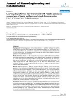
The patient presente d to accident and emergency the
same day where examination revealed bony tenderness
and obvious bruising and swelling to the injured a rea,
however x-rays failed to demonstrate a fracture
(Figure 1).
The patient’s hand was not placed in plaster and he
was referred to the orthopaedic outpatient clinic. Exami-
nation in clinic revealed bruising, swelling and bony ten-
derness to his 3
rd
and 4
th
MCP joints and due to the
high index of suspicion, further anter oposterior (AP),
lateral and carpal tunnel x-rays were requested. The AP
and carpal tu nnel radiographs showed no fracture, how-
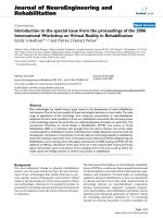
ever an oblique x- ray was mistakenly taken instead of
the requested lateral. This was an error on the part
of the radiographer’s. This oblique view revealed a
displaced hamate body fracture (Figure 2).
Under sedation in theatre, further examination
revealed 4
th
ray carpo-metacarpal subluxation on stres-
sing the joint indicating that this was a closed unstable
injury. Open reduction and internal fixation of this frac-
ture was successfully undertaken. Follow up at three
months revealed a well maintained reductio n of the
fracture which was healed (Figure 3). At one ye ar follow
up the patient was pain free with a stable joint and a
range of movement (ROM) of 0-90° which was consis-
tent with ROM in other unaffected MCP joints.
* Correspondence:
1
Vishal Borse, Room 346, Institute of Medical & Biological Engineering,
School of Mechanical Engineering, University of Leeds, Leeds, LS2 9JT, UK
Full list of author information is available at the end of the article
Borse et al. Journal of Orthopaedic Surgery and Research 2010, 5:64
/>© 2010 Borse et al; licensee BioMed Central Ltd. This is an Open Access article distribute d under the terms of the Creative Commons
Attribution License ( which permits unrestricted use, distribution, and reproduction in
any medium, provided the original work is properly cited.
Discussion
The hamate bon e is a roughly triangular-shaped bone,
which i s located in the distal carpal row farthest to the
ulnar side. It is bordered proximally by the pisiform and
the lunate in the proximal carpal row, radially by the
capitate, and distally by the bases of the fourth and fifth
metacarpals.
Hamate fractures are classified as either type I, invol-
ving the hook, or type II, involving the body, with type I
fractures being more common. Hamate fractures are
uncommon, particularly those involving the body of the
hamate, however they are the commonest fracture o f
the distal carpal row [1] and are increasing in incidence
possibly due to the increasing popularity of sports invol-
ving racquets, bats a nd clubs. They are associated with
instability and unless detected and managed appropri-
ately are associated with a poor outcome [2].
Traditionally, fractures and dislocation of the hamate are
identified on AP or lateral x-ray views [3]. Carpal tunnel
views and computed tomography (CT) [4,5] have also
been suggested to help. This case highlights some of the
problems encountered with traditional x-ray views, and
the need to consider oblique views as either standard pro-
cedure or as an adjunct whe re clinical suspicion remains
high even in the presence of normal x-rays. This point
remains valid even given the increasing use and availability
of other forms of radiological investigation [6-9].
We report the current case to highlight the following:
1. X-ray views
Andreson et al [1] concluded high resolution CT was
the imaging modality of choice for body and hamate
hook fractures. Their vitro experiments on 18 cadaver
hands showed that CT h ad 100% sensitivity and 94.4%
specificity and conventional X-ray showed 72.2% sensi-
tivity and 88. 8% specificity for detection of hamate frac-
tures. Howe ver, our case demonstrates with supporting
literature [1,2,10,11] the benefit of oblique views from
30 - 45° and the se should be considered standard with
anteroposterior, lateral and carpal tunnel views when
hamate fracture is suspected. If detected with these,
computerised tomography may be avoided.
2. Minimal palmar flexion injuries associated with carpal
bone fractures
It is commonly recognised that hyperextension injuries
to the hand are associated with c arpal bone fractures,
especially scaphoid. This case and others[2] establish a
link between minimal pa lmar flexed injuries and hamate
fractures.
Figure 1 Anteroposterior, lateral and carpal tunnel x-ray views (clockwise from left).
Figure 2 Pronated oblique 30° x-ray view. Blue arrow shows
fracture site.
Borse et al. Journal of Orthopaedic Surgery and Research 2010, 5:64
/>Page 2 of 4
We the authors believe that the ‘standard’ views for all
wrist injuries should include;
• PA (posteroanterior)
• PA with ulnar flexion
• Medial oblique
• Lateral.
We also believe that in injuries where the hamate is
thought to be involved OR where a high index of suspi-
cion for bony injury remains in the presence of normal
initial radiographs, that carpal tunnel views should be
carried out. Furthermore several other oblique projec-
tions may be needed until the plane of the fracture is
delineated clearly.
Conclusion
The authors believe that where a hamate fracture is sus-
pected an oblique x-ray view should be considered as
part of the initial diagnostic investigations. It can help
with diagnosis and give further important informatio n
to aid appropriate management. An oblique x-ray view
is of part icular use when clinical suspicion for hamate
fracture remains high in the light of otherwise normal
x-rays. Consideration and use of this view can negate
the need for costly, time-consuming CT scans. We
believe that the standard trauma series should be: PA;
PA with ulnar flexion; medial oblique and lateral X-rays.
With an additional carpal tunnel view where hamate
fracture is suspected.
Abbreviations
MCP: metacarpophalangeal; AP: anteroposterior; PA: posteroanterior; CT:
computed tomography; ROM: range of movement.
Consent
Written informed consent was obtained from the patient for publication of
this case report and accompanying images. A copy of the written consent is
available for review by the Editor-in-Chief of this journal.
Competing interests
The authors declare that they have no competing interests.
Authors’ contributions
VB collected the information and wrote the report. JH assisted with the
writing of the report and collected x-rays. AF had the initial idea for the
report and is guarantor. All authors read and approved the final manuscript.
Author details
1
Vishal Borse, Room 346, Institute of Medical & Biological Engineering,
School of Mechanical Engineering, University of Leeds, Leeds, LS2 9JT, UK.
2
James Hahnel, Department of Orthopaedics, Pinderfields General Hospital,
Aberford road, Wakefield, WF1 4DQ, UK.
3
Adnan Faraj, Department of
Orthopaedics, Airedale District General Hospital, Skipton road, Steeton,
Keighley, BD20 6TD, UK.
Received: 8 April 2010 Accepted: 27 August 2010
Published: 27 August 2010
References
1. Andreson R, Radmer S, Sparmann M, Bogusch G, Banzer D: Imaging of
Hamate Bone Fractures in Conventional X-Rays and High Resolution
Computerised Tomography: An In Vitro Study. Investigative Radiology
1999, 34(1):46-50.
2. Ebraheim NA, Skie MC, Savolaine ER, Jackson WT: Coronal Fracture of the
Body of the Hamate. Journal of Trauma Injury Infection and Critical Care
1995, 38(2):169-174.
3. Rockwood CA, Green DP: Fractures in Adults Lippincott-Raven: Philadelphia
1996, 1.
4. Resnick D: Bone and Joint Imaging W B Saunders Company: Philadelphia
1996.
5. Gella S, Borse VH, Rutten E: Coronal Fractures of the Hamate: are they
rare or rarely spotted? J Hand Surg Eur Vol 2007, 32(6):721-2.
6. Celi J, de Gautard G, Della Santa JD, Bianchi S: Sonographic Diagnosis of a
Radiographically Undiagnosed Hook of the Hamate Fracture. Journal of
Ultrasound in Medicine 2008, 27:1235-1239.
7. Royal College of Radiologists: Making the Best Use of a Department of
Clinical Radiology; Guidelines for Doctors London, UK: Royal College of
Radiologists, 5 2003.
Figure 3 Three month follow up anteroposterior (left) and oblique (right) x-ray views.
Borse et al. Journal of Orthopaedic Surgery and Research 2010, 5:64
/>Page 3 of 4
8. Murray IPC: Bone Scintigraphy in Trauma. In Nuclear Medicine in Clinical
Diagnosis and Treatment. Edited by: Ell PJ, Gambhir SS. New York: Churchill
Livingstone; , 3 2004:.
9. Mack MG, Keim S, Balzer JO, et al: Clinical Impact of MRI in Acute Wrist
Fractures. Eur Radiol 2003, 13:612-617.
10. Sherman GM, Seitz WH: Fractures and Dislocations of the wrist. Curr Opin
Orthop 1999, 10:237-251.
11. Jones BG, Hems TEJ: Simultaneous Fracture of the Body of the Hamate
and the Distal Pole of the Scaphoid. Journal of Trauma Injury Infection and
Critical Care 2001, 50:568-570.
doi:10.1186/1749-799X-5-64
Cite this article as: Borse et al.: Lessons to be learned from a missed
case of Hamate fracture: a case report. Journal of Orthopaedic Surgery
and Research 2010 5:64.
Submit your next manuscript to BioMed Central
and take full advantage of:
• Convenient online submission
• Thorough peer review
• No space constraints or color figure charges
• Immediate publication on acceptance
• Inclusion in PubMed, CAS, Scopus and Google Scholar
• Research which is freely available for redistribution
Submit your manuscript at
www.biomedcentral.com/submit
Borse et al. Journal of Orthopaedic Surgery and Research 2010, 5:64
/>Page 4 of 4