báo cáo hóa học:" Transarticular screw fixation for atlantoaxial instability - modified Magerl’s technique in 38 patients" docx
Bạn đang xem bản rút gọn của tài liệu. Xem và tải ngay bản đầy đủ của tài liệu tại đây (589.49 KB, 8 trang )
RESEARC H ARTIC LE Open Access
Transarticular screw fixation for atlantoaxial
instability - modified Magerl’s technique in
38 patients
Raj Bahadur
1,2†
, Tarun Goyal
3*†
, Saravdeep S Dhatt
4
, Sujit K Tripathy
4
Abstract
Background: Symptomatic atlantoaxial instability needs stabilization of the atlantoaxial joint. Among the various
techniques described in literature for the fixation of atlantoaxial joint, Magerl’s technique of transarticular screw
fixation remains the gold standard. Traditionally this technique combines placement of transarticular screws and
posterior wiring construct. The aim of this stud y is to evaluate clinical and radiological outcomes in subjects of
atlantoaxial instability who were operated using transarticular screws and iliac crest bone graft, without the use of
sublaminar wiring (a modification of Magerl’s technique).
Methods: We evaluated retrospectively 38 subjects with atlantoaxial instabilit y who were operated at our institute
using transarticular screw fixation. The subjects were followed up for pain, fusion rates, neurological status and
radiographic outcomes. Final outcome was graded both subjectively and objectively, using the scoring system
given by Grob et al.
Results: Instability in 34 subjects was secondary to trauma, in 3 due to rheumatoid arthritis and 1 had tuberculosis.
Neurological deficit was present in 17 subjects. Most common presenting symptom was neck pain, present in 35
of the 38 subjects.
Postoperatively residual neck and occipital pain was present in 8 subjects. Neurological deficit persisted in only 7
subjects. Vertebral artery injury was seen in 3 subjects. None of these subjects had any sign of neurological deficit
or vertebral insufficiency. Three cases had nonunion. At the latest follow up, subjectively, 24 subjects had good
result, 6 had fair and 8 had bad result. On objective grading, 24 had good result, 11 had fair and 3 had bad result.
The mean follow up duratio n was 41 months.
Conclusions: Transarticular screw fixation is an excellent technique for fusion of the atlantoaxial complex. It
provides highest fusion rates, and is particularly important in subjects at risk for nonunio n. Omitting the posterior
wiring construct that has been used along with the bone graft in the traditional Magerl’ s technique achieves
equally good fusion rates and is an important modification, thereby avo iding the complications of sublaminar wire
passage.
Background
Atlantoaxial articulation is the most unique part of the
spine. It is the most mobile segment of the spine, and
largely depends on the ligamentous supports an d the
integrity of the odontoid for its stability. Fusion of the
C1-C2 complex may be required in cases of atlantoaxial
instability. Its extreme mobility places heavy demand on
the atlantoaxial fixation construct for sufficient rigidity
required for its fusion. The causes of C1-C2 instability
are numerous and include trauma, congenital malforma-
tions, inflammatory arthritis, malignancies, skeletal dys-
plasias, rotator y subluxations and pharyngeal infections.
Symptoms of instability of the atlanto axial complex are
varied, such as neck pain, transient paresis, headaches,
ataxia and intermittent loss of consciousness.
Clinically or radiographically significant atlantoaxial
subluxation is best treated by reduction and fusion of
* Correspondence:
† Contributed equally
3
Dept of Orthopaedics, All India Institute of Medical Sciences, New Delhi,
India
Full list of author information is available at the end of the article
Bahadur et al. Journal of Orthopaedic Surgery and Research 2010, 5:87
/>© 2010 Bahadur et al; licensee BioMed Central Ltd. This is an Open Access article distributed under the terms of the Creative Commons
Attribution Lice nse ( which permits unrestricted use, distribution, and reproduction in
any medium, provided the original work is properly cited.
the C1-C2 joint. Posterior C1-C2 fusion using transarti-
cular screw (TAS), introduced by Magerl et al in 1979
[1] is the gold standard for atlantoaxial arthrodesis. It
has the advantage of a more rigid fixation with higher
rates of fusion, avoiding need for postoperativ e halo, no
placement of implant in the spinal canal, and possibility
of its use in anomalies of odontoid process or the pos-
terior arch [2-7]. Magerl et al used two transarticular
screws along with bone graft and interspinous wiring for
fusion. But the use of sublaminar wiring is fraught with
several complications, such as damage to the dura and
the cord during insertion of the wires and late comp res-
sionofthecordbywirebreakageorloosening[8-12].
Further, it has been found that there may be no impor-
tant contribution of the wires in holding the graft for
fusion, and comparable fusion rates hav e been achieved
in these studies [4,13,14].
Thus we designed our study to evaluate the outcome
of cases of atlantoaxial instability treated with transarti-
cular screw fixation. We did not include supplemental
wiring as described b y Magerl et al in our technique.
Postoperatively, the subjects were evaluated clinically
and radiographically for the improvement in clinical
scores, fusion rates of the arthrodesis and any associated
complications.
Methods
We studied 38 subjects of atlantoaxial instability who
underwent posterior fusion using transarticular screws.
All the cases were operated by the senior author (RB)
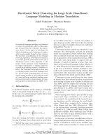
from 1995 to 2008. Instability was defined on flexion-
extension X-rays, using atlanto dens interval (ADI). ADI
of greater than 5 mm was taken as definition of atlan-
toaxial instability (figure 1).
All subjects were assessed with plain anteroposterior,
open mouth view and lateral flexion extension radio-
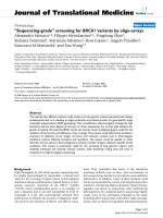
graphs. Later al radiographs help to verify that the
C1-C2 complex has been reduced adequately befor e the
surgery and to find the estimated length of the screws
to be used (figure 2).
A Computed T omography Scan with saggital, coro nal
and 3 D reconstruction was done in all the cases to look
at the transverse fo ramen of C2, understand the fracture
anatomy, C2 isthmus size, space available for the cord
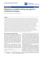
and integrity of the C1 lateral masses. Magnetic Reso-
nance Imaging (MRI) was done only in subjects with
neurological deficit, to study the lesion of the cord and
the degree of canal compromise, in order to plan poster-
ior decompression in these cases (figure 3).
Subjects who had pathology of the C1-C2 facets and
C1 lateral masses, such as comminuted fractures or the
tumors destroying the C1 lateral masses that preclude
screw placement were excluded from the study. Subjects
who were found to have anomalous course of the
vertebral artery o n Computed Tomography Scan were
also excluded from the study. This was studied using
axial and saggital cuts of the CT scan in the region of
transverse foramen of the C2 vertebra. High riding ver-
tebral artery was identified as having a too medial and/
Figure 1 Lateral radiograph of a subject with atlantoaxial
instability secondary to odontoid fracture showing marked
atlantoaxial displacement.
Figure 2 Post reduction film of the same subject using skeletal
traction in the ward. Further complete reduction was obtained
intraoperatively using skeletal traction with crutchfield tongs.
Bahadur et al. Journal of Orthopaedic Surgery and Research 2010, 5:87
/>Page 2 of 8
or a cranial course, recognized by the medial or cepha-
lad location of transverse foramen. This reduces the dis-
tance between the spinal canal and t he medial wa ll of
the transverse foramen, thereby placing the vertebral
artery in the path of the screw. The screw trajectory was
taken as neutral to about 15 degrees medial from the
starting point at the inferomedial angle of C2-C3 facet.
All traumatic cases were screened for other associated
spinal and extraspinal injuries, using clinical examina-
tion and necessary investigations. Other preoperative
variables that were assessed included risk factors for
nonunion, pathological abnormality responsible for
C1-C2 instability, subject’s clinical status including pain
and presenting radiological findings. The neurological
status was documented using Frankel’s Grades.
We used two transarticular screws for fixation of
C1-C2 complex combined with bone grafting. The pla-
cement of transarticular screws was similar in technical
details to the technique described by Magerl et al in
1979 [1]. We used iliac crest bone graft measuring
about 3 × 2 cm harvested from the posterior iliac crest.
The lamina of the C2 vertebra and C1 arch were decor-
ticated before application of the bone graft with a high
speed burr. C1 C2 facet joints were also curetted to
enhance fusion. Bone graft was placed between the pos-
terior arch of C1 and the spinou s process of the C2 ver-
tebra. The graft press f its in the space once nibbled to
appropriate shape. In subjects where posterior decom-
pression was carried out and laminectomy of the C1
was done (n = 10), this graft could not be placed in the
midline. We used morselised bone graft placed along
the bilateral facet joints in these cases.
Postoperatively, all subjects were kept in a Philadel-
phia collar for 6 weeks. The subjects were followed up
for pain, fusion rates, neurological status and radio-
graphic outcomes. Initial follow up was at 3 months,
then at 6 months and 1 year. Subsequent follow up was
done annually. Fu sion was defined radiologically as evi-
denceofcontinuityoftrabecularboneformation
between C1 and C2 acr oss the graft, without lucency or
resorption of the graft or hardware failure. Position of
the screws was assessed by transoral, anteroposterior
and lateral radiographs. A screw was considered well
positioned when both the lateral and anteroposterior
projections showed both screws lying entirely within the
bone and crossing the joint space in the anteroposterior
view. Stability was accepted if there was no change in
atlantodens interval during flexion and extension stu-
dies. Range of neck motion in rotation was also noted
in the follow up.
Final outcome was graded both subjectively and objec-
tively, using the scoring system given by Grob et al [6].
Subjectively, the results were graded as good (no serious
pain, no restriction of activity); fair (periods of pain,
working capacity reduced); or bad (permanent severe
pain and disability). The objective rating was good (no
pain, solid fusion); fair (moderate pain, solid fusion); or
bad (nonunion with severe pain) [15].
Results
A total of 38 subjects were studied. Of them 29 were
males (76%) and 9 were females. The mean age at the
time of surgery was 35 years (range 9 to 63 years).
Trauma was the most common cause of atlantoaxial
instability, seen in 34 (89.5%) subjects. Most common
mode of trauma was road traffic ac cident, in 29 of these
34 subjects. The distribution of subjects by etiology is
given in table 1. All subjects with traumatic atlantoaxial
instability had fracture of the odontoid process. Type II
D’ Olanzo fracture was seen in 30 of these subjects. In 4
subjects it was type I II fracture. Indications for arthrod-
esis in these subjects with odontoid fracture were estab-
lished nonunion or age more than 60 years. Ther e were
five cases of non union of odontoid fracture secondary
to failed anterior screw f ixation for the fracture of the
odontoid. They were operated after mean of 7 months
after injury. In 8 subjects the initial injury to the upper
cervical spine was missed at their initial referral center.
These subjects presented late with neck pain and stiff-
ness at 3-6 months from injury. In 21 subjects, fracture
Figure 3 MRI showing cord compression due to anterior
translation of the axis over atlas in a subject with atlantoaxial
instability.
Table 1 Etiology of atlantoaxial instability
Etiology Frequency Percentage
trauma 34 89.5%
rheumatoid arthritis 3 8%
tuberculosis 1 2.5%
Total 38 100%
Bahadur et al. Journal of Orthopaedic Surgery and Research 2010, 5:87
/>Page 3 of 8
odontoid was managed conservatively at their initial
referral centre with immobilization or traction. There
were three cases with rheumatoid arthritis. Mean ADI
in these cases was 10.5 mm. All these subjects had neu-
rological deficit.
Most common presenting symptom was neck pain,
present in 35 of the 38 subjects (92%) in our series.
Neurological deficit was present in 17 subjects (44 .7%).
Of these 15 subjects had quadriparesis and 2 s ubjects
had monoplegia. Out of the 34 traumatic cases 14 had
neurological deficit. All 3 subjects with rheumatoid
arthritis had neurological deficit. Worsening of neurolo-
gical deficit over time was seen in 3 subjects. Two of
these subjects had rheumatoid arthritis, and the third
had history of road side accident.
The most common risk factor for nonunion in our
subjects was smoking, seen in 8 subjects (21%). The
other factors included-rheumatoid arthritis, in 3 sub-
jects; steroid intake in 3 subjects and diabetes mellitus
in 3 subjects.
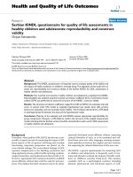
Postoperative radiographs showed adequate reduction
of C1 over C2 in 35 cases. Adequate screw placement
was seen in 31 cases (figure 4 & 5). In one patient only
one screw could be placed due to vertebral artery injury
on that side. Another subject had screw cutout. She was
a case of rheumatoid arthritis, and was taking steroids
for a lon g period. Radiographs were suggestive of mark-
edly reduced bone density . She did not progressed to
union, and neurological deficit persisted in her. In the
third patient the screw placem ent was a lit tle too lateral
and superior, and the screws penetrated out of the ante-
rior cortex of the anterior arch of C1. The future course
was uneventful in this p atient. The mean screw length
was 42 mm.
Posterior decompression was combined with the pro-
cedure in 10 subjects with neurological deficit and evi-
dence of cord compression on Magnetic Resonance
Imaging (MRI). All 3 cases with rheumatoid arthritis
had undergone posterior decompression. Vertebral
arteryinjurywasseenin3subjects.Noneofthesesub-
jects had anomalous transverse foramen or abnormally
narrow isthmus on preoperative Computed Tomography
(CT) scan. W hen vertebral artery injury was encoun-
tered intraoperatively, screw was placed in the drill hole
to provide a temponade effect. Placement o f screw on
the other side was not attempted for the fear of injuring
both the vertebral arteries. In one of these subjects only
one screw could be placed since the artery was hit on
the side being operated first. In none of these subjects
any sign of neurological deficit or vertebral insufficiency
was seen, probably because of sufficient collateral circu-
lation [16,17].
Fusion was seen in 35 cases. In three cases the graft
showed resorption, and there was no evidence of forma-
tion of bony bridge between C1 and C2. Earliest radiolo-
gical evidence of union could be seen in these patients
at a mean follow up of 3.6 months. There was no
instance of deep infection of the surgical site or the
graft site. Decubitus ulcers on the occiput were seen in
two subjects. Suboccipital paresth esia and numbness
was present in 3 patients.
The most common postoperative complaint was resi-
dual neck and occipital pain, seen in 8 subjects. At the
latest follow up, subjectively, 24 subjects had good
result, 6 had fair and 8 had bad result. On objective
grading, 24 had good result, 11 had fair and 3 had bad
result. The mean follow up duration was 41 months
(range 15-70 months).
Figure 4 Postop erative radiogr aph of the subject showing
placement of two transarticular screws across the reduced
atlantoaxial joints.
Figure 5 Anteroposterior open mouth view showing
placement of transarticular screws.
Bahadur et al. Journal of Orthopaedic Surgery and Research 2010, 5:87
/>Page 4 of 8
At admission 17 subjects had neurological deficit. Of
these 14 were Frankel’ s grade C and 3 were Frankel’s
grade D. At discharge 10 subjects had completely recov-
ered, with neurological deficit persisting in only 7 sub-
jects. All these 7 subjects belonged to Frankel’s grade C.
The mean range of neck motion was 40 degrees of lat-
eral rotation on left side and 35 degrees of rotation on
right side. Since atlantoaxial fusion virtually eliminates
the motion at C1-C2 joint, the residual rotation reflects
the subaxial component of the neck motion. This range
of motion was maintained on follow up. The mean
range of la teral rotation in cases of rheumatoid arthritis
was 25 degrees on each side. This is consistent with the
view that rheumatoid spine has restricted range of atlan-
toaxial and subaxial motion. The range of flexion and
extension was maintained after surgery. The mean flex-
ion and extension arc was 150 degrees. The mean range
of lateral bending was 45 degrees on each side.
Discussion
The aim of treatment of atlantoaxial instability is to
achieve a solid fusion between C1 and C2, virtually
eliminating a ny motion between them. This is expected
to relieve the neck pain and avoid the risk of further
neurological deficit. The posterior wiring techniques
popularized by Gallie et al [18] and Brooks and Jenkins
[19] had been the most common means of stabilization
in the past. In recent years, a variety of other techniques
have been used, such as, interlaminar cl amps, polyaxial
screw and rod fixation, transarticular screw fixation and
C1 lateral mass screws with C2 pars screw fixation. Pos-
terior C1-C2 transarticular screw fixation has become
thegoldstandardforatlantoaxial fusion. It has lead to
considerable improvement in the fusion rates upto more
than 95% [1-6] over C1-C2 wiring procedures, whose
failure rates range from 10% to 25% [20,21 ]. Taggard et
al [7] conducted a case control study to compare the
fusion rates using transarticular screws and posterior
wiring techniques. After a mean follow up of 31 months
they found that successful fusion was achieved in 1 3 of
14 subjects treated with the TAS technique as compared
to 5 out of 13 subjects who underwent a posterior wir-
ing technique. They observed that subjects with a radio-
graphically solid fusion were 21 more times likely to
have undergone TAS than posterior wi ring techni que (p
= 0.004). The position of the transarticular screws is clo-
ser to the centre of axis of rotation and l ateral bending,
which provides better control of movements than other
techniques which rely on peripheral fixation.
The biomechanics of surgical stabilization of the C1-C2
articulation can be divided into three different types.
One-point fixation stabilizes the motion segment only
posteriorly (e.g. Gallie wiring, Halifax clamps etc). Two-
point fixation construct includes transarticular screws
through the laterally placed facet joints. Thre e-point fixa-
tion consists of the combination of the two previous
principles, thus stabilizing the C1-C2 motion segment
both laterally and posteriorly. In biomechanical testing
three point fixation has been found to be superior to
both two-point and one-point fixations [22-26]. Thus the
tension band construct provides two advantages-first, it
enhances the stability of the TAS fixation; and s econd,
the structural bone graft is stabilized by the wire. But
sublaminar wire pa ssage carries the potential risk of neu-
rological complications [9-11], especially in cases where
the canal has already been compromised. Further this
wire-graft technique is technically demanding and time
consuming [2,27]. Some reports have shown that metal
wires or cables may bow anteriorly because of “spring
phenomenon” even without any breakage, leading to
encroachment upon the spinal cord [13,28].
It is controversial in literature whether posterior wir-
ing construct provide any additional contribution
towards fusion. Matsumo to et al reported 18 cases of
loosening of posterior wiring construct in 52 cases with
95% fusion rate [14]. In Ito’s series, all cases had loosen-
ing, but with 100% fusion rate. Thus, wire or cable loos-
ening did not lead to nonunion or ps eudarthrosis, but it
might endanger the spinal cord. From these observa-
tions, Ito et al came to the conclusion that adding wire
construct is not required [13]. Avoiding the placement
of posterior wires may be especially important in s itua-
tions where inflammatory disease with soft tissue swel-
ling and pannus has resulted in compromise of the
spinal canal, or in the case of C1-C2 subluxation which
is not completely reducible [8]. Significant degenerative
changes or osteoporosis of the posterior elements of C1
and C2 also preclude the use of posterior wiring techni-
ques. Wang et al [4] achieved solid fusion in all their 57
subjects, using only morselized autograft and transarti-
cular screw, without any posterior wiring construct. We
did not use the mors elized graft but a strut of iliac crest
graft well fitted in the space between the C1 lamina and
C2 spinous process. Thus, although from the biomecha-
nical viewpoint, bilateral TAS fixation may not be as
stable as the 3-point fixations, fusion rates have not
been altered. There is only slight micromotion left in
flexion-extension after fixation. We supposed that this
micromotion would not affect fusion. I n our series,
there is no loss of the reduction and the fusion rate is
92%. This is in unison with the fusion rates achieved by
other authors who used combination of Transarticular
screws and posterior wiring [1-7]. Randomized or a case
control study will be a better study design to study this
effect. But corre spondence of our results with those of
studies using Magerl’s fixation suggests that this techni-
que is a sound alternative thus simplifying the Magerl’s
technique.
Bahadur et al. Journal of Orthopaedic Surgery and Research 2010, 5:87
/>Page 5 of 8
Tho ugh single screw placement is expected to lead to
nonunion, there is no convincing data in this regard. In
our study single screw was placed in 1 subject. Solid
union was achieved in this subject at follow up. Song
et al [23] concluded that unilateral C1-C2 transarticular
screw fixation with interspinous bone graft wiring is an
excellent alternative in the treatment of atlantoaxial
instability when bilateral screw fixation is contraindi-
cated. They reported a solid fusion using this technique
in 18 of 19 subjects with atlantoaxial instability and uni-
lateral anomalies. Grob et al [6] found that nonunion
did not follow incorrect placement of one screw, so
bilateral fixation is not an indispensable condition for a
satisfactory outcome.
Posterior transarticul ar screw fixation has several
advantages over other fixation techniques. Contrary to
the traditional posterior fusion techniques, the integrity
of the ring of C1 is not necessary for t ransarticular screw
placement. Thus this technique can be used even in cases
of fracture or the absence of posterior arch of the atlas.
This technique also provides approach for laminectomy,
if needed for decompression of the cord. Further, there is
no implant inside the spinal canal as in the wiring techni-
ques and complications associated with wire loosening
are avoided. A very important advantage is that it avoids
the need for postoperative halo immobilization, when
compared to the posterior wiring techniques. This is an
important factor from the subjects’ point of view for the
selection of the procedure. Achieving preoperative reduc-
tion is imperative for safe atlantoaxial fusion. Displace-
ment of C1 on C2 decreases the space available for the
cord. This distorts the C1 C2 alignment, and the place-
ment of transarticular screws is not completely safe. This
also increases the risk with sublaminar wire passage,
because of increased chances of hitting the cord.
Although some authors have used transarticular screw
fixation for in situ fixation, the p recise limit beyond
which this technique is contraindicated is not defined.
Thus in large fixed displace ments of C1 on C2, occipito-
cervical fusion with C1 decompression, or anterior
decompression and fusion are indicated [8].
The disadvantages of this procedure include need for
an extensive skin incision and soft tissue dissection to
expose the entire dorsum of C2. This extensive posterior
exposure has been associated with a complication rate
as high as 10%, including superficial infections and occi-
pital nerve injury [8,29]. Screw placement requires an
acute angle for proper screw trajectory, which may be
impeded by kyphotic deformities or by moving the neck
anteriorly. Additionally, there is a steep learning curve
for this technique. Complications associated with this
technique include the potential for vertebral artery
injury,malpositionofscrews,pseudoarthrosis,implant
failure, dural tear, hypoglossal paresis, brain stem
infarc tion and death. Inconstant size and location of the
transverse foramen in the lateral mass of the axis places
the vertebral artery at risk during drilling and screw pla-
cement. Scans with sa ggital and coronal reconstructio ns
help to assess the relationship of transve rse foramen of
C2 and the C1-C2 facet joint to determine the correct
trajectory for the screw and avoid arterial injury [30,31].
Radiographic and anatomical studies of th e atlanto-axial
complex suggest that upto 20% of the subjects have
atlanto axial anatomy that precludes safe bilateral screw
placement [32-34]. We had 3 cases of vertebral artery
injury in our study (8%). Reported rates of vertebral
artery injury using this technique vary from 0-10% in
different series [17,8,29,32,35-39]. American Associat ion
of Neurological Surgeons/Congress of Neurological Sur-
geons (AANS/CNS) Section on disorders of Spinal
Nerves and Peripheral Nerves in their survey published
by Wright and Lauryssen [35], estimated the risk of ver-
tebral artery injury during C1-C2 transarticular screw
fixation to be 2.2% per screw inserted. The risk of neu-
rological deficit from vertebral artery injury was 0.2%
per subject, and the mortality rate was 0.1%. Thus injury
to vertebral artery is well tolerated in the majority of the
subjects. Despite numerous reports of vertebral artery
injuries, resultant neurological deficit is rare [8]. Coric
D et al [40] reported a case of vertebral artery to epi-
dural venous plexus fistula as a complication of poster-
ior atlantoaxial facet screw fixation. Madawi et al [33]
reported five cases of vertebral artery injury (8.2%) in
subjects who underwent this operation. He also pointed
out that incomplete reduction is a risk factor for inade-
quate screw placement. In cidence of dural tears has
been reported to be 0.3%. suboccipital numbness is rela-
tively common, seen in 16.8% patients in repo rt by
Wright and Lauryssen [35]. In most of them however it
resolved spontaneously with time.
Despite excluding all the patients with dangerous
anatomy of the vertebral artery, we still had 3 patients
in whom vertebral artery injury was observed. Two of
these patients were observed in the first half of the
study period when the experience of the surgeon with
this technique was relatively recent. This is a highly sur-
geon dependent technique and learning curve is high.
Surgeon has to be familiar with the anatomy of the
transverse foramen in the upper cervical spine. This
needs experience with studying a large number of CT
scans. Failure t o meticulously identify the danger in this
region may lead to catastrophy.
The studies of RA subjects showed relatively lower
rates of bony union than did the studies with smaller
percentages of RA subjects [29,41-43]. Literature sug-
gests that presence of rheumatoid arthritis entails the
risk of posterior graft nonuni on more than other disor-
ders [6,41-43]. We achieved union in only of the
Bahadur et al. Journal of Orthopaedic Surgery and Research 2010, 5:87
/>Page 6 of 8
3 patients with RA. Due to the small sample size with
only 3 subjects with rheumatoid arthritis, no statistically
significant conclusion regarding effect of rheumatoid
disease on fixation and union can be reached. Ito T et al
found that in 5 of the ir 7 subjects with rheumatoid
arthritis who had nonunion, C1-C2 complex was stable
due to fusion at the facet joints, as demonstrated by
functional radiographs and computed tomography scans
[13]. Thus atlantoaxial transarticular screws can bring
the facet fusion despite the posterior graft failure in
such cases.
Conclusions
Thus, transarticular screw fixation is an effective techni-
que for the fusion of t he atlantoaxial complex. It pro-
vides highest fusion rates, and is particularly important
in subjects at risk for nonunion. It has expanded the
indications for atlantoaxial fusion and is an important
salvage technique in subjects with pre vious failed proce-
dures. Although its l earning curve may be steep, it is
associated with few rates of complications in expert
hands.
Acknowledgements
Authors have not received any funding for the study or during preparation
of the manuscript.
Author details
1
Postgraduate Institute of Medical Education and Research, Chandigarh,
India.
2
Government Medical College and Hospital, Chandigarh, India.
3
Dept
of Orthopaedics, All India Institute of Medical Sciences, New Delhi, India.
4
Dept of Orthopaedics, Postgraduate Institute of Medical Education and
Research, Chandigarh, India.
Authors’ contributions
RB is the senior authors who carried out the surgical procedure, coordinated
the planning of preoperative and postoperative protocols, and helped to
draft the manuscript. TG had the instrumental role in the planning and
execution of perioperative and intraoperative design of the study and
preparation of the manuscript. SSD and ST helped in acquisition of data and
in drafting of the manuscript. All authors read and approved the final
manuscript.
Competing interests
The authors declare that they have no competing interests.
Received: 26 November 2009 Accepted: 22 November 2010
Published: 22 November 2010
References
1. Magerl F, Seemann PS: Stable posterior fusion of the atlas and axis by
transarticular screw fixation. In Cervical spine I. Volume 1. Edited by: Kehr P,
Weidner A. New York: Springer; 1987:322-327.
2. Stillerman CB, Wilson JA: Altanto-axial stabilization with posterior
transarticular screw fixation: technical description and report of 22
cases. Neurosurgery 1993, 32:948-955.
3. Blauth M, Richter M, Lange U: Transarticular screw fixation C1/2 in
traumatic atlantoaxial instabilities. Comparison between percutaneous
and open procedures. Orthopade 1999, 28:651-661.
4. Wang C, Yan M, Zhou H, Wang S, Dang G: Atlantoaxial transarticular
screw fixation with morselized autograft and without additional internal
fixation: technical description and report of 57 cases. Spine 2007,
32(6):643-646.
5. Jeanneret B, Magerl F: Primary Posterior Fusions C1-2 in Odontoid
Fractures: Indications, Technique, and Results of Transarticular Screw
Fixation. J Spinal Disord 1992, 5:464-475.
6. Grob D, Jeanneret B, Aebi M, Aebi M, Markwalder TM: Atlanto axial fusion
with transarticular screw fixation. J Bone Joint Surg Br 1991, 73(6):972-6.
7. Taggard DA, Kraut MA, Clark CR, Traynelis VC: Case-control study
comparing the efficacy of surgical techniques for C1-C2 arthrodesis. J
Spinal Disord Tech 2004, 17(3):189-94.
8. Smith MD, Phillips WA, Hensinger RN: Complications of fusion to the
upper cervical spine. Spine 1991, 16(7):702-5.
9. Coyne TJ, Fehlings MG, Wallace MC, Bernstein M, Tator CH: C1-C2 Posterior
Cervical Fusion: Long Term Evaluation of Results and Efficacy. Neurosurg
1995, 37:688-693.
10. Fraser AB, Sen C, Casden AM, Catalano PJ, Post KD: Cervical transdural
intramedullary migration of a sublaminar wire: a complication of cervical
fixation. Spine 1994, 19:456-9.
11. Cervellati S, Bettini N, Bianco T, Parisini P: Neurological complications in
segmental spinal instrumentation: analysis of 750 subjects. Eur Spine J
1996, 5:161-6.
12. Blacklock JB: Fracture of a sublaminar stainless steel cable in the upper
cervical spine with neurological injury. Case report. J Neurosurg 1994,
81:932-3.
13. Ito T, Hayashi M, Takei H: Loosening of supplemental cable in
transarticular screw fixation and bone grafting. J Orthop Surg 1998,
6:71-4.
14. Matsumoto M, Chiba K, Nakamura M, Ogawa Y, Toyama Y, Ogawa J: Impact
of interlaminar graft materials on the fusion status in atlantoaxial
transarticular screw fixation. J Neurosurg Spine 2005, 2(1):23-6.
15. McGuire RA Jr, Harkey HL: Modification of technique and results of
atlantoaxial transfacet stabilization. Orthopedics 1995, 18:1029-1032.
16. Taneichi H, Suda K, Kajino T, Kaneda K: Traumatically induced vertebral
artery occlusion associated with cervical spine injuries: prospective
study using magnetic resonance angiography. Spine 2005, 30:1955-62.
17. Neo M, Fujibayashi S, Miyata M, Takemoto M, Nakamura T: Vertebral artery
injury during cervical spine surgery: a survey of more than 5600
operations. Spine 2008, 33(7):779-85.
18. Gallie WE: Fractures and Dislocation of the Cervical Spine. Am J Surg
1939, 46:495-499.
19. Brooks AL, Jenkins EB: Atlanto-axial arthrodesis by wedge compression
method. J Bone Joint Surg Am 1978, 60:279-284.
20. Dickman CA, Sonntag VK: Posterior C1-C2 transarticular screw fixation for
atlantoaxial arthrodesis. Neurosurgery 1998, 43(2):275-80.
21. Farey ID, Nadkarni S, Smith N: Modified Gallie technique versus
transarticular screw fixation in C1-C2 fusion. Clin Orthop Relat Res 1999,
359:126-135.
22. Melcher RP, Puttlitz CM, Kleinstueck FS, Lotz JC, Harms J, Bradford DS:
Biomechanical testing of posterior atlantoaxial fixation techniques. Spine
2002, 27(22):2435-40.
23. Mitchell TC, Sadasivan KK, Ogden AL, Mayeux RH, Mukherjee DP,
Albright JA: Biomechanical study of atlantoaxial arthrodesis: transarticular
screw fixation versus modified Brooks posterior wiring. J Orthop Trauma
1999, 13(7):483-9.
24. Naderi S, Crawford NR, Song GS, Sonntag VK, Dickman CA: Biomechanical
comparison of C1-C2 posterior fixations. Cable, graft, and screw
combinations. Spine (Phila Pa 1976) 1998, 15(23):1946-55.
25. Montesano PX, Juach EC, Anderson PA, Benson DR, Hanson PB:
Biomechanics of cervical spine internal fixation. Spine (Phila Pa 1976)
1991, 16(3):S10-S16.
26. Grob D, Dvorak J, Panjabi MM, Hayek J: Dorsal atlantoaxial screw fixation.
A stability test in vitro and in vivo. Orthopade 1991, 20(2):154-162.
27. Guiot B, Fessler RG: Complex atlantoaxial fractures. J Neurosurg 1999,
91(Suppl 2):139-143.
28. Geremia GK, Kim KS, Cerullo L, Calenoff L: Complications of sublaminar
wiring. Surg Neurol 1985, 23(6):629-35.
29. Gluf WM, Schmidt MH, Apfelbaum RI: Atlantoaxial transarticular screw
fixation: a review of surgical indications, f usion rate, complications,
and lessons learned in 191 adult subjects. J Neu rosurg Spine 2005,
2(2):155-63.
30. Dull ST, Toselli RM:
Preoperative oblique axial computed tomographic
imaging for C1-C2 transarticular screw fixation: technical note. J
Neurosurg 1995, 37:150-1.
Bahadur et al. Journal of Orthopaedic Surgery and Research 2010, 5:87
/>Page 7 of 8
31. Nogueira-Barbosa MH, DeWno HLA: Multiplanar reconstructions of helical
computed tomography in planning of atlantoaxial transarticular fixation.
Eur Spine J 2005, 14:493-500.
32. Yoshida M, Neo M, Fujibayashi S, Nakamura T: Comparison of the
anatomical risk for vertebral artery injury associated with the C2-pedicle
screw and atlantoaxial transarticular screw. Spine 2006, 31:E513-7.
33. Abou Madawi A, Solanki G, Casey AT, Crockard HA: Variation of the groove
in the axis vertebra for the vertebral artery. Implications for
instrumentation. J Bone Joint Surg Br 1997, 79(5):820-823.
34. Miyata M, Neo M, Ito H, Yoshida M, Miyaki K, Fujibayashi S, Nakayama T,
Nakamura T: Is Rheumatoid Arthritis a Risk Factor for a High-Riding
Vertebral Artery? Spine 2008, 33(18):2007-2011.
35. Wright NM, Lauryssen C: Vertebral artery injury in C1-2 transarticular
screw fixation: results of a survey of the AANS/CNS section on disorders
of the spine and peripheral nerves. American Association of
Neurological Surgeons/Congress of Neurological Surgeons. J Neurosurg
1998, 88(4):634-640.
36. Neo M, Matsushita M, Iwashita Y, Yasuda T, Sakamoto T, Nakamura T:
Atlantoaxial transarticular screw fixation for a high-riding vertebral
artery. Spine 2003, 28(7):666-70.
37. Haid RW Jr, Subach BR, McLaughlin MR, Rodts GE Jr, Wahlig JB Jr: C1-C2
transarticular screw fixation for atlantoaxial instability: a 6-year
experience. Neurosurgery 2001, 49(1):65-8.
38. Liang ML, Huang MC, Cheng H, Huang WC, Yen YS, Shao KN, Huang CI,
Shih YH, Lee LS: Posterior transarticular screw fixation for chronic
atlanto-axial instability. J Clin Neurosci 2004, 11(4):368-72.
39. Campanelli M, Kattner KA, Stroink A, Gupta K, West S: Posterior C1-C2
transarticular screw fixation in the treatment of displaced type II
odontoid fractures in the geriatric population–review of seven cases.
Surg Neurol 1999, 51(6):596-600.
40. Coric D, Branch CL Jr, Wilson JA, Robinson JC: Arteriovenous fistula as a
complication of C1-2 transarticular screw fixation. Case report and
review of the literature. J Neurosurg 1996, 85(2):340-3.
41. Casey AT, Madawi AA, Veres R, Crockard HA: Is the technique of posterior
transarticular screw fixation suitable for rheumatoid atlanto-axial
subluxation? Br J Neurosurg 1997, 11(6):508-19.
42. Kandziora F, Mittlmeier T, Kerschbaumer F: Stage-related surgery for
cervical spine instability in rheumatoid arthritis. Eur Spine J 1999,
8(5):371-81.
43. Shen FH, Samartzis D, Jenis LG, An HS: Rheumatoid arthritis: evaluation
and surgical management of the cervical spine. Spine J 2004,
4(6):689-700.
doi:10.1186/1749-799X-5-87
Cite this article as: Bahadur et al.: Transarticular screw fixation for
atlantoaxial instability - modified Magerl’s technique in 38 patients.
Journal of Orthopaedic Surgery and Research 2010 5:87.
Submit your next manuscript to BioMed Central
and take full advantage of:
• Convenient online submission
• Thorough peer review
• No space constraints or color figure charges
• Immediate publication on acceptance
• Inclusion in PubMed, CAS, Scopus and Google Scholar
• Research which is freely available for redistribution
Submit your manuscript at
www.biomedcentral.com/submit
Bahadur et al. Journal of Orthopaedic Surgery and Research 2010, 5:87
/>Page 8 of 8