báo cáo hóa học:" The role of FGF-2 and BMP-2 in regulation of gene induction, cell proliferation and mineralization" pdf
Bạn đang xem bản rút gọn của tài liệu. Xem và tải ngay bản đầy đủ của tài liệu tại đây (896.97 KB, 8 trang )
RESEARCH ARTICLE Open Access
The role of FGF-2 and BMP-2 in regulation of
gene induction, cell proliferation and
mineralization
Millie Hughes-Fulford
1,2,3,4*
, Chai-Fei Li
4
Abstract
Introduction: The difficulty in re-growing and mineralizing new bone after severe fracture can result in loss of
ambulation or limb. Here we describe the sequential roles of FGF-2 in inducing gene expression, cell growth and
BMP-2 in gene expression and mineralization of bone.
Materials and me thods: The regulation of gene expression was determined using real-time RTPCR (qRTPCR) and
cell proliferation was measured by thymidine incorporation or fluorescent analysis of DNA content in MC3T3E1
osteoblast-like cells. Photomicroscopy was used to identify newly mineralized tissue and fluorescence was used to
quantify mineralization.
Results: Fibroblast growth factor-2 (FGF-2) had the greatest ability to induce proliferation after 24 hours of
treatment when compared to transforming growth factor beta (TGFb, insulin-like growth factor-1 (IGF-1), bone
morphogenic protein (BMP-2), platelet derived growth factor (PDGF) or prostaglandin E
2
(PGE
2
). We found that
FGF-2 caused the most significant induction of expression of early growth response-1 (egr-1), fgf-2, cyclo-oxygenase-
2 (cox-2), tgfb and matrix metalloproteinase-3 (mmp-3) associated with proliferation and expression of angiogenic
genes like vascular endothelial growth factor A (vegfA) and its receptor vegfr1. We found that FGF-2 significantly
reduced gene expression associated with mineralization, e.g. collagen type-1 (col1a1), fibronectin (fn), osteocalcin (oc),
IGF-1, noggin, bone morphogenic protein (bmp-2) and alkaline phosphatase (alp). In contrast, BMP-2 significantly
stimulated expression of the mineralization associated genes but had little or no effect on gene expression
associated with growth.
Conclusions: The ability of FGF-2 to re-program a mineralizing gene expression profile to one of proliferation
suggests that FGF-2 plays a critical role of osteoblast growth in early fracture repair while BMP-2 is instrumental in
stimulating mineralization.
Introduction
The mechanisms that regulate bone growth and minera-
lization remain poorly understood. The cellular events
involved in bone formation include chemotaxis of osteo-
blast p recursors, growth factor (GF) production, prolif-
eration of committed osteoblast precursors, and the
differentiation (mineralization) of osteoblasts. Bone for-
mation requires expr ession of structural proteins such
as collagen type I, osteocalcin, noggin and run x2 during
mineralization [1]. Numerous studies suggest that a
variety of growth factors such as FGF-2, TGFb,IGF-1,
PDGF and PGE
2
act as autocrine and paracrine hor-
mones to regulate bone cell proliferation [2]. FGF-2 is
an important modulator of bone formation in vitro and
in vivo [3,4]. FGF-2 is tightly bound to the bo ne matrix
and can be extracted as a biologically active GF [5] and
is thought to play a major role in wound healing [6,7].
To evaluate the physiological activity of FGF-2 and
other growth factors, we studied their relative ability to
influence proliferation of osteoblasts at a site of injury
in a mineralized culture. MC3T3-E1 is a cloned mouse
osteoblast-like cell line that retains synthetic functions
of bone. When treated with differentiation media, these
cultured osteoblasts have the ability to differentiate,
* Correspondence:
1
Department of Research, Veterans Affairs Medical Center, 4150 Clement
Street, San Francisco, CA 94121, USA
Full list of author information is available at the end of the article
Hughes-Fulford and Li Journal of Orthopaedic Surgery and Research 2011, 6:8
/>© 2011 Hughes-Fulfo rd and Li; licensee BioMed Central Ltd. This is an Open Access article distributed under the terms of t he Creative
Commons Attribution License (http ://creativeco mmons.org /licenses/by/2.0), which permits unrestricted use, distribution, and
reproduction in any medium, provided the original work is properly cited.
including synthesis of alkaline phosphatase [8], type I
collagen [9], osteocalcin [10,11] and mineralized matrix
containing hydroxyapatite crystals [12].
We have previously reported that FGF-2 is induced by
mechanical stress [13,14] and causes proliferation after
mechanical stress. FGF-2 is an immediate-early gene
that is regulated by both PKA and MAPK signal trans-
duction pathways [15]. Here we report that FGF-2
induces expression of growth-related genes and down-
regulates g enes responsible for differentiation and
mineralization. In addition, BMP-2 is considerably more
effective than FGF-2 in inducing new mineralization.
Materials and met hods
Materials
We obtained GFs from Amgen, Thousand Oaks, CA.
FGF-2 and IGF-1 from R & D Systems, Minneapolis,
MN. TGFb,PDGFanddmPGE
2
are from Cayman Che-
mical, Ann Arbor, Michigan. Cell culture supplies
(aMEM, fetal calf serum, trypsin and antibiotics) were
obtained through the tissue culture facility at the
University of California, San Francisco. Cell culture
dishes were purchased from Corning, Corning, New
York. Rhodamine-phalloidin is from Invitrogen, Carls-
bad, California. Tritiated thymidine and 35 S methionine
are from Amersham, Arlington Heights, IL. All other
materials came from standard laboratory suppliers.
MC3T3E1 osteoblast-like cells, a cloned cell line, estab-
lished by Kodama [8,12] were used in this study at early
passage number.
Methods
We maintained cloned MC3T3-E1 osteoblast-like cells in
normal media (NM) consisting of alpha MEM medium
with 10% fetal calf serum (FCS), 1% antibiotic solution
and 1% glutamine solution and subcultured the cells
every 3 to 4 days. The cells were subcultured by incubat-
ing with trypsin for five minutes and resuspending at a
concentration of 3 × 10⁵ cells/ml. For experiments, we
grew the cells in the NM above, using multi-well plates.
After three days, the cells reach confluence and minerali-
zation medium (MM) was added. MM is alpha MEM
medium with 5% fetal calf serum (FCS), 1% antibiotic
solution and 1% glutamine solution supplemented with
ascorbic acid (50 μg/ml) and b-glycerol phosphate
(10 mM) to support mineralization. The cultures were
then incubated for 1-2 more days for mineralization stu-
dies.Weusedatleasttriplicate independent biological
samples in multiple experiments for data collection.
Protein Assay
Protein concentration was determined by Bio-Rad DC
protein assay (Bio-Rad, CA) according to manufacturer’s
protocol.
Microscopy
At the conclusion of the 24 or 48 hour incubation, the
coverslip was removed. The specimen was rinsed five
times in room temperature phosphate buffered saline
(PBS) and fixed. We then visualized the mineralizing
cells with 2% Alizarin Red. After rinsing in distilled
water and air drying the samples, we mounted the cov-
erslips on microscope slides using Fluoromount and
examined and photographed the cells on a Zeiss Axios-
kop using 20×.
Tritiated thymidine incorporation into DNA
At the conclusion of the 24 hour incubation, the culture
medium was rem oved and the cells were incubated for
15 minutes at 37°C in 1 ml PBS containing trit iated thy-
midine (4 μCi/ml) as described previously [16]. Follow-
ing this incubation, the PBS was removed and the cells
were washed 3 times with ice cold trichloroacetic acid
(TCA) followed by ice cold ethanol and allowed to a ir
dry. Then 1 ml of sarkosyl lysing buffer was added to
each well; all the cells were solubilized after 30 minutes .
Finally, after mixing the resulting solution with a pip-
ette, radioactivity was c ounted in a scintillation counter
and protein content was measured. The data was calcu-
lated and expressed as disintegrations per mi nute
(DPM) per microgram protein.
Alizarin Red visualization of mineralization
Alizarin Red (2%) stained cells were incubated with 10%
acetic acid for 30 minutes to release bound Alizarin Red
into solution. The solution was neutralized with 10%
ammonium hydroxide and the absorbance of Alizarin
Red was measured at 450 nm using a microplate reader.
Data is expressed i n absolute amounts accor ding to a
standard curve.
RNA Isolation
RNA were isolated through the use of the RNeasy™-
Mini kit (QIAGEN, Valencia, CA) or TriReagent™
acco rding to the manuf acturer’s protocol. For RNeasy™
Mini kit RNA isolation, cells were seeded in 6-well
plates with aMEM media supple mented with 10% FCS,
then downregulated and activated as indicated in the
figure legends. Cells were lysed using 350 μlofbuffer
RLT (supplied in kit) containing 2-mercaptoethanol
(Biorad, Hercules, CA). The lysate was then placed into
QIAshredder homogenizer (QIAGEN, Valencia, CA)
and centrifuged at 20,000 rpm for 2 minutes. 350 μlof
70% ethano l was added to the flow through, mixed, and
centrifuged in the RNeasy™Mini column (supplied in
kit) for 15 s at 20 ,000 rpm. Flow through was discarded
and the column was washed with 700 μlofbufferRW1
(supplied in kit) for 15 s at 20,000 rpm. Two additional
washes were performed with 500 μl of buffer RPE
Hughes-Fulford and Li Journal of Orthopaedic Surgery and Research 2011, 6:8
/>Page 2 of 8
(supplied in kit) at 20,000 rpm for 15 s and 2 minutes,
respectively. The flow through was discarded and the
column placed in a sterile 1.5 ml collection tube.
Depending on the expected yield, 20-50 μlRNase-free
water is pipetted into the column and cent rifuged for
1 minute at 20,000 rpm. The samp les are then stored at
-80°C until further analysis.
Reverse Transcription (RT)
1.5 μg of RNA was added to 30 μl reverse transcriptase
(RT) reaction buff er containing 5 mM MgCl
2
,10mM
Tris-HCl (pH 8.3), 50 mM KCl, 1 mM dNTPs, 2.5 μM
oligo d(T) primer, 2.5 U/μlofMuLV,and1U/μlof
RNase in hibitor. The RT reaction was incubated at
room temperature for 10 min, 42°C for 30 min, inacti-
vated at 99°C for 5 min, and cooled at 5°C for 5 min.
Real-time Quantitative RT-PCR Reaction (qRTPCR)
2 μl of cDNA from the RT reaction w as added to 20 μl
real-time quantitative polymerase chain reaction (qPCR)
mixture containing 10 μl of 2× SYBR
®
Green PCR Mas-
ter Mix (Applied Biosystems, Foster City, CA) and
12 pmol oligonucleotide primers. PCR s were carried out
in a Bio-Rad MyiQ Single-Color Real-Time PCR Detec-
tion Sy stem (Bio-Rad, Hercules, CA). The thermal pro-
file was 50°C for 2 min, 95°C for 10 min to activate the
Taq polymerase, followed by 50 amplification c ycles,
consisting of denaturation at 95°C for 1 min 40 s,
annealing at 63°C for 1 min 10 s and elongation at 72°C
for 1 min 40 s. Fluorescence was measured and used for
quantitative purposes. A t the end of the amplification
period, melting curve analysis was performed to confirm
the specificity of the amplicon. RNA samples were nor-
malized to cyclophilin (CPHI) internal standard. Relative
quantification of gene expression was calculated by
using 2
-(CtgeneT-CtCPHIT)-(Ctgene0hr-CtCPHI0hr)
equation, where “C
t
gene T” represents the calculated
threshold cycle (C
t
) of a time point of each sample
other than 0 hr, or each treatment other than control.
Relative gene absolute abundance was calculated using
2 sup>(Ct gene T - Ct CPHI T) as p reviously described
[17] allows us to compare the abundance of the gene
between other genes and experiments. The resulting
numbers were then multiplied by 10,000 for better gra-
phical presentation. Primer sequence information was
previously published [18-22]. All data derived using
qRTPCR was fro m multiple experiment s with at least
triplicate independent biological samples.
Results
Growth factor effect on cell proliferation DNA synthesis
As seen in Table 1, in the absence of any added com-
pounds there were small and unremarkable changes in
DNA synthesis with IGF-1 and PDGF; in contrast,
FGF-2, TGFb and PGE2 significantly enhanced thymi-
dine incorporation within 24 hours of treatment. TGFb
stimulated thymidine incorporation more than 2 fold
while FGF-2 and PGE2 increased DNA synthesis more
than 4.5 and 3.3 fold respectively.
Regulation of FGF-2 induced gene expression
Using qRTPCR, we found that FGF-2 dramatically
induced egf-1 , fgf-2, cox -2, tgfb, mmp 3, vegfA and vegfr1
over a 24 hour period each displa ying a different sequen-
tial temporal pattern of gene induction (Figure 1). VegfA
and vegfr1 are associated with an giogenesi s while mmp3,
is associated with increased migration. Tgfb, fgf-2, egr-1
and cox-2 ar e key genes in regulation of osteoblast
proliferation.
Interestingly, we found that FGF-2 also significantly
decreased expression of othe r genes associated with
mineralization including col1a1, fn, bmp-2, oc, run-x,
and noggin. IGF-1, a known differentiation factor, was
significantly decreased by FGF-2 treatment. (Figure 2).
Relative abundance of genes regulated
by FGF-2 and BMP-2
Since FGF-2 increased growth associated genes, we used
BMP-2, a known promoter of mineralization, to study
relative abundance of gene expression in mineralizing
cells after 24 hours of treatment. As seen in Table 2, we
found that BMP-2 treatment caused significant increases
in genes associated with mineralization including cola1,
fn, noggin and oc. Moreover, BMP-2 treatment caused
little or no changes in expression of genes associated
with angiogenesis and migration e.g. VEGF and MMP3.
When compared with relative gene abundance of FGF-2
treated cells (Figure 3) we found that in general, BM P-2
maintained the mineralizing RNA profile of igf-1, alp,
and bmp-2 and significantly increased expression of
other genes associated with mineralization like col1a1,
fn, ilgf-1, noggin and oc. Fgf-2, on the other ha nd, signif-
icantly suppressed expression of mineralizing genes.
Table 1 Effect of growth factors on protein synthesis in
wounded mineralized osteoblasts
Treatment Thymidine incorporation DPM × 10
3
/ug protein
con 37.6 ±2.9
IGF-1 42.3 ± 4.2
FGF-2 114.3 ± 11
TGFb 65.2 ± 12
PDGF 39.8 ± 7.2
BMP-2 41.5 ± 5.6
PGE2 84.1 ± 23.1
Representative experiment showing the effects of IGF-1 (20 ng/ml), FGF-2
(2.0 ng/ml), TGFb (2 ng/ml), PDGF (3 ng/ml), BMP-2 (100 ng), PGE
2
(2 μg/ml)
on proliferation/mg protein of MC3T3-E1 osteoblasts after 24 hours of
treatment (n = 4).
Hughes-Fulford and Li Journal of Orthopaedic Surgery and Research 2011, 6:8
/>Page 3 of 8
Relative mineralization of FGF-2 and BMP-2 treated cells
As seen in Figure 4 and Table 3, BMP-2 treatment
enhances mineralization of th e cells as shown by uptake
and presence of Alizarin Red after cultures were grown
to confluence and then treated with BMP-2 or FGF-2
for 24 to 48 hours. Cells were then washed and stained
with 2% Alizarin Red and results determined using
photography or fluorescence analysis at 48 hours of
treatment.
Discussion
Bone formation during injury repair is a multi-step series
of events modulated by an integrated cascade of gene
expression that initially supp orts the proli feration stage.
The later mineralization stage is associated with the
sequential expression of genes that support biosynthesis,
organization and mineralization of the bone extracellular
matrix. Mineralization requires expression of structural
proteins such as collagen type I, osteocalcin, as well as
noggin and runx2 which aid in mineralization [1].
Transcriptional control de fines the regulatory events
necessary for both stages of bone formation [23]. There
is a general consensus that during injury GFs are released
from the wounded bone matrix and promote healing
[24]. In this study, we have documented the relative effi-
ciency of bone growth factors FGF-2, TGFb, and PGE2
markedly enhanced the synthesis of the total protein con-
tent of the dishes (Table 1)
Rate of proliferation was dependent on the specific
GF. FGF-2, TGFb and PGE
2
significantly promote
growth, with FGF-2 having the highest efficacy and the
lowest dose. FGF-2 produced a distinct patte rn of gene
expression. FGF-2 down regulates genes associated with
mineralization while it induces genes associated with
proliferation and angiogenesis, a finding supported by
observations of others [25]. Since cox-2 had a 27-fold
induction by FGF-2, we examined the effect of the
COX-2 p roduct, PGE
2
on proliferation. We found that
PGE
2
increased DNA synthesis by 3.3 fold significantly
higher than TGFb, IGF-1, PDGF, suggesting that its
induction by FGF-2 helps complete the FGF-2 full
induction of osteoblast growth. These data also s uggest
that FGF-2 may be an important regulator of migration,
angiogenesis and proliferation during the first stage of
healing a critical defect since it induces mm p3, vegfa
and vegfr1 expression. In data not shown, FGF-2 had
no effect on expression of mmp-1. Moreover, FGF-2
Figure 1 qRTPCR analysis of gene induction of proliferation
and angiogenesis; qRTPCR analysis of gene reduction of genes
over 24 hours of treatment with FGF-2 shows a significant increase
in genes associated with proliferation and angiogenesis. Cultures
were cultured and harvested for RNA as described in Materials and
Methods. Each bar represents mean ± SD triplicate independent
biological samples each time point corrected to cyclophilin. (*p <
0.05; **p < 0.01 with two-tail student t-test compared to 0 hour of
each gene).
Figure 2 qRTPCR analysis of FGF-2 regulated genes associated
with mineralization; qRTPCR analysis of gene reduction of genes
over a 24 hours of treatment with FGF-2 shows a marked reduction
in genes associated with mineralization. Cultures were cultured and
harvested for RNA as described in Materials and Methods. Each bar
represents mean ± SD triplicate independent biological samples at
each time point corrected to cyclophilin. (*p < 0.05; **p < 0.01 with
two-tail student t-test compare to 0 hour of each gene.).
Hughes-Fulford and Li Journal of Orthopaedic Surgery and Research 2011, 6:8
/>Page 4 of 8
induced its own message as w ell as TGF b, but signifi-
cantly reduced expression of BMP-2, osteocalcin, nog-
gin, runx2, collagen type I and IGF-1, genes which are
associated with mineralization.
As described by others, bone formation is divided into
two phases, proliferation and minera lization [2,26-29].
These two stages are the result of a specific sequential reg-
ulation of gene expression from the early phase of osteo-
blast proli feration to the final steps of mineralization.
Once the cells start mineralizing, cell division and DNA
synthesis dramatically slow down and eventually cease.
When an injury occurs in mineralized tissue, GFs l ike
FGF -2 are released and st art a new proliferation stage to
heal the defect. The increase in cell replication in a miner-
alizing cell likely represents a de-differentiation from th e
mineralizing phase to the growing phase, and increases
expression of GFs most likely induce proliferation. Treat-
ment of the mineralized defect model with FGF-2 resulted
in gene expression that corresponds to de-differentiation
(e.g. significant i ncreases in growth related genes egf -1, fgf-2,
cox-2, TGFb, vegfA, vegfr and mmp3 and down-regulation
of mineralizing related genes). Vegf and vegfr1 are primary
regulators of angiogenesis, w hile MMP3 is thought to
play a major role on cell behaviors such as proliferation
and migration [30] which may explain the ability of the
FGF-2 to enable the cultured cells to fill the defect void
efficiently. The f act that FGF-2 induce s its own expres-
sion suggests that after injury, the FGF-2 released from
the wound matrix could promote it’s own expression,
making it a feed-forward loop.
Although Figures 1 and 2 demonstrate the relative
FGF-2 regulation and sequential expression of growth,
angiogenic and chemotactic genes and depresses expres-
sion of mineralization-related genes, these figures do not
tell us the relative abundance of the genes. In Table 2,
we determined the relative abundance of genes in three
groups after 24 hours; with or without treatment with
FGF-2 or BMP-2. FGF-2 caused a significant increase in
abundance of genes associated with proliferation, che-
motaxis and angiogenesis. Moreover, the addition of
FGF-2 to the mineralized wounded cultures caused a
marked decrease in abundance of col1a1 as well as fn,
igf-1, noggin, oc, bmp-2 and alp message. In the early
stages of mineralization, the major protein (greater than
20%) synthesized by the osteoblast is collagen, however
Table 2 Relative abundance of gene expression in FGF-2 and BMP-2 treated cells
Non-treated FGF-2 treated BMP-2 treated FGF-2 vs BMP-2
Gene Average SD Average SD Average SD p-value
Collagen Type I 85,081.73 2,5316.39 **678.21 358.27 *170,243.43 24,493.77 0.0003
Fibronectin 55,827.93 1,2119.18 *28,432.19 1195.92 **239,750.67 23,464.19 0.0001
IGF1 3,249.41 689.70 **50.65 13.30 4,193.34 739.19 0.0006
RUNX2 349.09 40.63 **674.95 63.04 1,043.65 783.29 n.s.
VEGFA 109.49 38.86 **5,132.66 755.22 537.13 379.66 0.0007
TGFb 93.08 10.55 **245.40 41.93 *185.20 38.34 n.s.
ALP 58.30 34.81 13.39 11.68 91.77 23.15 0.0064
OC 16.20 3.19 **1.38 0.65 *34.04 6.11 0.0008
Noggin 7.11 2.77 *1.61 0.49 2.41 1.76 n.s.
BMP-2 0.40 0.12 **0.06 0.01 0.38 0.05 0.0004
MMP3 0.03 0.03 **4.04 0.97 0.12 0.14 0.0023
This table shows the relative abundance of gene expression in mineralizing MC3T3-E1 cells after 24 hours of treatment with FGF-2 (5 n g) or BMP-2 (100 ng).
Total RNA was harvested 24 hours after the addition using Qiagen RNeasy kit. A two-step RT-qPCR was preformed. Each data point represents the mean ± SD of
three biological independent samples. *p < 0.05; **p < 0.01; ***p < 0.0001 against 0 hour control samples with 2 tail student t test.
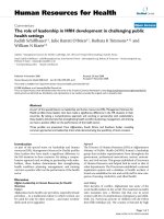
Figure 3 FGF-2 and BMP-2, the yin and yang of mineralization:
Contrast of effect of 24 hours of treatment with FGF-2 or BMP-
2 on fold increase in abundance of mineralization-related gene
expression. Mineralizing MC3T3-E1 cells were prepared as
described in Materials and Methods. They were then treated with
either FGF-2 or BMP-2 for 24 hours at which time RNA was
collected and analyzed for relative abundance using qRTPCR. Each
bar represents mean ± SD triplicate independent biological samples
each time point corrected to cyclophilin. (*p < 0.05; **p < 0.01 with
two-tail student t-test compare to 0 hour of each gene.) *<0.05;
**<0.01; ***<0.0001.
Hughes-Fulford and Li Journal of Orthopaedic Surgery and Research 2011, 6:8
/>Page 5 of 8
collagen is not a major component of the proliferating
cell, suggesting that FGF-2 stimulates proliferation partly
through its ability to drastically reduce the relative
abundance of a majority of the mineralizing-associated
genes.ThedramaticreductionofIGF-1byFGF-2sug-
gests a role for IGF-1 in minera lization, this is i n agree-
ment with findings of others that demonstrated IGF-1
to be a major factor in bone mineralization [31-33]
using the IGF-1 null mouse. In contrast, in cells treated
with BMP-2, the relative abundance of col1a1, fn, oc,
and tgfb were dramatically induced while BMP-2 had no
significant effect on genes related to growth, angiogen-
esis or chemot axis. These data suggest that BMP-2 may
bethebestGFtouseforthemineralizationstagebut
not the proliferation stage of bone formation. This find-
ing may help explain studies by others [34] who discov-
ered that a delayed ad ministration of BMP- 2 to a
fracture resulted in better repair of critical size defects.
It is likely that the delay of BMP-2 treatment allowed a
longer period of proliferation prior to BMP-2 promotion
of mineralization. Our findings in Table 2, 3 and Figure 3
support the hypothesis that FGF-2 and BMP-2 are
required at different stages of bone repair.
Conclusions
These data demonstrate the de-differentiation (reduction
of mineralization genes) effect of FGF-2 likely plays a
key role in osteoblast proliferation, the first stage of
bone formation. Some h ave expressed concern that
ex-vivo proliferation of human stem cells by a growth fac-
tor like FGF-2 might change the osteogenic characteristics
of a pre-osteoblast; however others have shown that
expansion of the population do es not affect later osteo-
genic potential [35] of stem cells. Therefore, an expansion
of osteoblast cells by FGF-2 might be an excellent strategy
for first stage re-population of a critical defect since FGF-2
Table 3 Mineralization of cells with BMP-2
Treatment Relative abundance
NM 5.6 ± 1.7
NM + 5 ng/ml FGF-2 5.3 ± .1
NM + 50 ng/ml BMP-2 16.2 ± 4.2
MM 9.1 ± 2.0
MM + 5 ng/ml FGF-2 4.9 ± 1.1
MM + 50 ng/ml BMP-2 55.2 ± 12.7
The Alizarin Red (2%) stained cells were incubated with 10% acetic acid for
30 minutes to release bound Alizarin Red into solution. The solution was
neutralized with 10% ammonium hydroxide and the absorbance of Alizarin
Red was measured at 450 nm using a microplate reader (n = 6). Data is
expressed at in absolute amounts according to a standard curve.
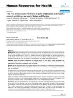
5%NormalMedia 5%NormalMedia+5ng/mlFGFͲ25%NormalMedia+50ng/mlBMPͲ2
5%MineralizingMedia 5%MineralizingMedia+5ng/mlFGFͲ25%MineralizingMedia+50ng/mlBMPͲ2
Figure 4 Alizarin Staining of Mineralizing Osteoblast cells. MC3T3-E1 osteoblasts were seeded at 3000 cells/well in 96 well CELLBIND
®
plates
in normal medium. Once cells were confluent, media was changed to 5% NM or 5% mineralizing media with or without 5 ng/ml FGF-2 or 50
ng/ml BMP-2. Two days after treatment, media was removed and cells were fixed in 10% formalin and stored at 4°C until subsequent analysis.
Cells were stained for calcium with 2% Alizarin Red for 10 minutes and visualized under 20× objectives for photography. Many areas of
mineralization, as seen by bright red staining, were present in the cells treated with 5% MM plus 50 ng/ml BMP-2 (FIG. 11). Little to no
mineralization was seen with other 5 treatments.
Hughes-Fulford and Li Journal of Orthopaedic Surgery and Research 2011, 6:8
/>Page 6 of 8
has the needed efficacy for promoting proliferation. These
data also suggest that the final stage of bone repair is best
accomplished with BMP-2 due to its promotion of differ-
entiation and mineralization.
Acknowledgements
This work is supported by MHF’s US Army Medical Research and Materiel
Command US Army grant W81WH-07-1-0427, NASA grant NAG-2-1086 and
in part by NASA grants NAG-2-1286, NCC2-1361 and the Department of
Veterans Affairs Medical Research Service in support of MHF and this project.
We thank Sandra Spurlock for the data plate reading and data analysis of
Table 3. We would like to thank Tammy Chang for her thoughtful
comments and suggestions during this work and Joe Meissler, Tara
Candelario, Esmeralda Aguayo and Jesus Aguado for their thoughtful
comments on the manuscript.
Author details
1
Department of Research, Veterans Affairs Medical Center, 4150 Clement
Street, San Francisco, CA 94121, USA.
2
Department of Medicine, University of
California, 4150 Clement Street, San Francisco, CA 94121, USA.
3
Department
of Urology, University of California, 4150 Clement Street, San Francisco, CA
94121, USA.
4
Hughes-Fulford Laboratory, Northern California Institute for
Research and Education, 4150 Clement Street, San Francisco, CA 94121, USA.
Authors’ contributions
MHF conceived the study, designed the study, directed the research and
wrote the manuscript. C-FL made substantive intellectual contribution in the
acquisition of data, analysis and has contributed to the manuscript. Both
authors have read and approved the final manuscript.
Competing interests
The Department of Veterans Affairs has filed and owns a patent using some
of the data found in this manuscript.
Received: 29 July 2010 Accepted: 9 February 2011
Published: 9 February 2011
References
1. Stein GS, Lian JB, Stein JL, Van Wijnen AJ, Montecino M: Transcriptional
control of osteoblast growth and differentiation. Physiol Rev 1996,
76:593-629.
2. Mundy GR, Chen D, Zhao M, Dallas S, Xu C, Harris S: Growth regulatory
factors and bone. Rev Endocr Metab Disord 2001, 2:105-115.
3. Canalis E, McCarthy TL, Centrella M: Growth factors and cytokines in bone
cell metabolism. Annu Rev Med 1991, 42:17-24.
4. Montero A, Okada Y, Tomita M, Ito M, Tsurukami H, Nakamura T,
Doetschman T, Coffin JD, Hurley MM: Disruption of the fibroblast growth
factor-2 gene results in decreased bone mass and bone formation. J Clin
Invest 2000, 105:1085-1093.
5. Burgess WH, Maciag T: The heparin-binding (fibroblast) growth factor
family of proteins. Annu Rev Biochem 1989, 58:575-606.
6. Santiago FS, Lowe HC, Day FL, Chesterman CN, Khachigian LM: Early
growth response factor-1 induction by injury is triggered by release and
paracrine activation by fibroblast growth factor-2. Am J Pathol 1999,
154:937-944.
7. Grazul-Bilska AT, Johnson ML, Bilski JJ, Redmer DA, Reynolds LP, Abdullah A,
Abdullah KM: Wound healing: the role of growth factors. Drugs Today
(Barc) 2003, 787-800.
8. Kodama HA, Amagai Y, Koyama H, Kasai S: A new preadipose cell line
derived from newborn mouse calvaria can promote the proliferation of
pluripotent hemopoietic stem cells in vitro. J Cell Physiol 1982, 112:89-95.
9. Ibbotson KJ, Orcutt CM, Anglin AM, D’Souza SM: Effects of transforming
growth factors beta 1 and beta 2 on a mouse clonal, osteoblastlike cell
line MC3T3-E1. J Bone Miner Res 1989, 4:37-45.
10. Katsuyama H, Otsuki T, Tomita M, Fukunaga M, Fukunaga T, Suzuki N,
Saijoh K, Fushimi S, Sunami S: Menaquinone-7 regulates the expressions
of osteocalcin, OPG, RANKL and RANK in osteoblastic MC3T3E1 cells. Int
J Mol Med 2005, 15:231-236.
11. Sierra OL, Cheng SL, Loewy AP, Charlton-Kachigian N, Towler DA: MINT, the
Msx2 interacting nuclear matrix target, enhances Runx2-dependent
activation of the osteocalcin fibroblast growth factor response element.
J Biol Chem 2004, 279:32913-32923.
12. Sudo H, Kodama HA, Amagai Y, Yamamoto S, Kasai S: In vitro
differentiation and calcification in a new clonal osteogenic cell line
derived from newborn mouse calvaria. J Cell Biol 1983, 96:191-198.
13. Hatton JP, Pooran M, Li CF, Luzzio C, Hughes-Fulford M: A short pulse of
mechanical force induces gene expression and growth in MC3T3-E1
osteoblasts via an ERK 1/2 pathway. J Bone Miner Res 2003, 18:58-66.
14. Hughes-Fulford M, Rodenacker K, Jutting U: Reduction of anabolic signals
and alteration of osteoblast nuclear morphology in microgravity.
J Cell
Biochem 2006, 99:435-449.
15.
Li CF, Hughes-Fulford M: Fibroblast growth factor-2 is an immediate-early
gene induced by mechanical stress in osteogenic cells. J Bone Miner Res
2006, 21:946-955.
16. Wiley MH, Feingold KR, Grunfeld C, Quesney-Huneeus V, Wu JM: Evidence
for cAMP-independent inhibition of S-phase DNA synthesis by
prostaglandins. J Biol Chem 1983, 258:491-496.
17. Johnson RF, Mitchell CM, Giles WB, Bisits A, Zakar T: Mechanisms
regulating prostaglandin H2 synthase-2 mRNA level in the amnion and
chorion during pregnancy. J Endocrinol 2006, 188:603-610.
18. Hughes-Fulford M, Gilbertson V: Osteoblast fibronectin mRNA, protein
synthesis, and matrix are unchanged after exposure to microgravity.
Faseb J 1999, 13:S121-127.
19. Hughes-Fulford M, Tjandrawinata R, Fitzgerald J, Gasuad K, Gilbertson V:
Effects of microgravity on osteoblast growth. Gravit Space Biol Bull 1998,
11:51-60.
20. Tjandrawinata RR, Hughes-Fulford M: Up-regulation of cyclooxygenase-2
by product-prostaglandin E2. Adv Exp Med Biol 1997, 407:163-170.
21. Hughes-Fulford M: Prostaglandin regulation of gene expression and
growth in normal and malignant tissues. Adv Exp Med Biol 1997, 269-278.
22. Hughes-Fulford M, Lewis ML: Effects of microgravity on osteoblast growth
activation. Exp Cell Res 1996, 224:103-109.
23. Stein GS, Lian JB, Stein JL, Van Wijnen AJ, Montecino M: Transcriptional
control of osteoblast growth and differentiation. Physiol Rev 1996,
76:593-629.
24. Hauschka PV, Chen TL, Mavrakos AE: Polypeptide growth factors in bone
matrix. Ciba Found Symp 1988, 136:207-225.
25. Tsuboi T, Mizutani S, Nakano M, Hirukawa K, Togari A: Fgf-2 regulates
enamel and dentine formation in mouse tooth germ. Calcif Tissue Int
2003, 73:496-501.
26. Gerstenfeld LC, Chipman SD, Kelly CM, Hodgens KJ, Lee DD, Landis WJ:
Collagen expression, ultrastructural assembly, and mineralization in
cultures of chicken embryo osteoblasts. J Cell Biol 1988, 106:979-989.
27. Vary CP, Li V, Raouf A, Kitching R, Kola I, Franceschi C, Venanzoni M, Seth A:
Involvement of Ets transcription factors and targets in osteoblast
differentiation and matrix mineralization. Exp Cell Res 2000, 257 :213-222.
28. Chen D, Zhao M, Mundy GR: Bone morphogenetic proteins. Growth
Factors 2004, 22:233-241.
29. Mundy GR:
Regulation of bone formation by bone morphogenetic
proteins
and other growth factors. Clin Orthop Relat Res 1996, 24-28.
30. Laurent M, Martinerie C, Thibout H, Hoffman MP, Verrecchia F, Le Bouc Y,
Mauviel A, Kleinman HK: NOVH increases MMP3 expression and cell
migration in glioblastoma cells via a PDGFR-alpha-dependent
mechanism. Faseb J 2003, 17:1919-1921.
31. Koch H, Jadlowiec JA, Campbell PG: Insulin-like growth factor-I induces
early osteoblast gene expression in human mesenchymal stem cells.
Stem Cells Dev 2005, 14:621-631.
32. Wang Y, Nishida S, Sakata T, Elalieh HZ, Chang W, Halloran BP, Doty SB,
Bikle DD: Insulin-like growth factor-I is essential for embryonic bone
development. Endocrinology 2006, 147:4753-4761.
33. Burghardt AJ, Wang Y, Elalieh H, Thibault X, Bikle D, Peyrin F, Majumdar S:
Evaluation of fetal bone structure and mineralization in IGF-I deficient
mice using synchrotron radiation microtomography and Fourier
transform infrared spectroscopy. Bone 2007, 40:160-168.
34. Betz OB, Betz VM, Nazarian A, Egermann M, Gerstenfeld LC, Einhorn TA,
Vrahas MS, Bouxsein ML, Evans CH: Delayed administration of adenoviral
BMP-2 vector improves the formation of bone in osseous defects. Gene
Ther 2007, 14:1039-1044.
Hughes-Fulford and Li Journal of Orthopaedic Surgery and Research 2011, 6:8
/>Page 7 of 8
35. Kulterer B, Friedl G, Jandrositz A, Sanchez-Cabo F, Prokesch A, Paar C,
Scheideler M, Windhager R, Preisegger KH, Trajanoski Z: Gene expression
profiling of human mesenchymal stem cells derived from bone marrow
during expansion and osteoblast differentiation. BMC Genomics 2007,
8:70.
doi:10.1186/1749-799X-6-8
Cite this article as: Hughes-Fulford and Li: The role of FGF-2 and BMP-2
in regulation of gene induction, cell proliferation and mineralization.
Journal of Orthopaedic Surgery and Research 2011 6:8.
Submit your next manuscript to BioMed Central
and take full advantage of:
• Convenient online submission
• Thorough peer review
• No space constraints or color figure charges
• Immediate publication on acceptance
• Inclusion in PubMed, CAS, Scopus and Google Scholar
• Research which is freely available for redistribution
Submit your manuscript at
www.biomedcentral.com/submit
Hughes-Fulford and Li Journal of Orthopaedic Surgery and Research 2011, 6:8
/>Page 8 of 8