báo cáo hóa học:" The calcar screw in angular stable plate fixation of proximal humeral fractures - a case study" potx
Bạn đang xem bản rút gọn của tài liệu. Xem và tải ngay bản đầy đủ của tài liệu tại đây (1.39 MB, 6 trang )
RESEARCH ARTICLE Open Access
The calcar screw in angular stable plate fixation
of proximal humeral fractures - a case study
Georg Osterhoff, Christian Ossendorf, Guido A Wanner, Hans-Peter Simmen and Clément M Werner
*
Abstract
Background: With new minimally-invasive approaches for angular stable plate fixation of proximal humeral
fractures, the need for the placement of oblique inferomedial screws (’calcar screw’) has increasingly been
discussed. The purpose of this study was to investigate the influence of calcar screws on secondary loss of
reduction and on the occurrence of complications.
Methods: Patients with a proximal humera l fracture who underwent angular stable plate fixation between 01/2007
and 07/2009 were included. On AP views of the shoulder, the difference in height between humeral head and the
proximal end of the plate were determined postoperatively and at follow-up. Addition ally, the occurrence of
complications was documented. Patients with calcar screws were assigned to group C+, patients without to group
C
Results: Follow-up was possible in 60 patients (C+ 6.7 ± 5.6 M/C- 5.0 ± 2.8 M). Humeral head necrosis occurred in
6 (C+, 15.4%) and 3 (C-, 14.3%) cases. Cut-out of the proximal screws was observed in 3 (C+, 7.7%) and 1 (C-, 4.8%)
cases. In each group, 1 patient showed delayed union. Implant failure or lesions of the axillary nerve were not
observed. In 44 patients, true AP and Neer views were available to measure the head-plate distance. There was a
significant loss of reduction in group C- (2.56 ± 2.65 mm) compared to C+ (0.77 ± 1.44 mm; p = 0.01).
Conclusions: The placement of calcar screw s in the angular stable plate fixation of proximal humeral fractures is
associated with less secondary loss of reduction by providing inferomedial support. An increased risk for
complications could not be shown.
Keywords: Proximal humerus, fracture, locked screw, locking plate
Background
Patients with minimally displaced or stable fractures of
the proximal humerus are treated conservatively in the
majority of cases [1]. In contras t, patients with fractures
fulfilling the criteria of instability, referred to as dis-
placed or unstable fractures, benefit from surgical inter-
vention wh ich mostly renders reliable results, both,
clinically and radi ographically [2,3]. However, surgery of
displaced proximal humeral fractures is technically
demanding. A wide array of surgical options has been
described and controversially discussed [4-10].
The introduction of locking plate systems represents a
milestoneinfracturetreatment with the advantage of
improved osseous anchorage and higher resistance to
failure by combining axial and angular stability [11,12].
These plates are suitable for pathologic and osteoporotic
fractures. Additionally, locking plates do not depend on
friction or compression between plate and bone to stabi-
lize the fracture and theref ore do not compro mise peri-
osteal blood supply [13,14].
In proximal humeral fractures, the particular proxi-
mity of tendinous and neurovascular structures of the
joint and the characteristic bone strength distribution of
the humeral head [15] require a fixation system with
predetermined screw settings. The Philos plate system
(Synthes, Oberdorf, Switzerland) was developed to meet
these requirements by using a tridimensionally-fash-
ione d locking system for the proximal screws. However,
several studies with short- to mid-term experiences after
Philos plate fixation suggest that-in spite of good overall
clinical results-the implant’s stiffness might lead to a
* Correspondence:
Division of Trauma Surgery, University Hospital Zurich, Rämistrasse 100, 8091
Zurich, Switzerland
Osterhoff et al . Journal of Orthopaedic Surgery and Research 2011, 6:50
/>© 2011 Osterhoff et al; licensee BioMed Central Ltd. This is an Open Acces s ar ticle distributed under the terms of the Creative
Commons Attribution License ( which permits unrestricted use, distribution, and
reproduction in any medium, provided the original work is properly cite d.
higher rate of screw cut-out or c ut-through [16-27]. A
lack of medial support was suggested to be one possible
reason [28]. In addition, the presence or absence of
medial support was described as a significant predictor
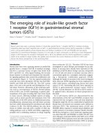
of loss of fracture reduction [29]. One simple way o f
gaining medial support is the insertion of one or two
screws running tangentially to the medial curvature of
the humeral surgical neck (calcar screws, Figure 1). Y et,
with new minimal ly-invasive approaches for the angular
stable plate osteosynthesi s the need for these calcar
screws has increa sing ly been discussed. It has been sug-
gested that the proximity to the anterior [30] and pos-
terior [31] circumflex arteries might compromise
perfusion of the humeral head and by this lead to
delayed-union or non-union or to osteonecrosis. As they
are supposed to additionally stiffen the osteosynthetic
construct, calcar screws mayalsoincreasetheriskof
cut-out [32].
Therefore, orthopaedic surgeons cannot be sure if they
either increase the risks of complications or potentially
miss a b etter long term fracture reduction implicating a
better treatment outcome.
Thus, the purpose of this study was to investigate if
the presence of calcar screws can reduce secondary loss
of reduction and if it has influence on the occurrence of
possible complications-especially cut-out and axillary
nerve lesions.
Methods
All patients with a proximal humeral fracture who
underwent angular stable plate fixation (PHILOS,
Synthes, Oberdorf, Switzerland) in our hospital between
01/2007 and 07/2009 were included in the present
study. All data w as collected according to the terms of
reference specified by the local ethics committee.
Criteria for exclusion were: age younger than 18 years,
previous ipsilateral fractures of the humerus and bony
metastases. The indication for surgery was set when
posttraumatic radiographs showed evidence of displace-
ment of > 1 cm or an angulation > 45° according to
Neer’s criteria for displaced fractures [33]. Fracture mor-
phology was classified in two-, three- and four-part frac-
tures on posttraumatic true AP and Neer radio graphs.
Surgery was performed either via a deltopectoral
approach or in minimally-invasive technique via short
delta-split approach combined with skin incisions for
the distal screws, depending on the surgeons’ choice.
All patients underwent a standardized postoperative
treatment schedule characterized by early passive
motion under physiotherapeutic surveillance.
Differences in height between humeral head and the
proximal end of the plate were determined on true AP
radiographs of the shoulder, postoperatively and at fol-
low-up, as described previously [29] (Figure 2). The dis-
tance between two lines orthogonal to the plate axis was
measured, one line running through the proximal end of
the plate and one through the tip of the humeral head.
All measurements were performed by the first author
using a digital caliper tool of the standard viewer soft-
ware of our institution (Agfa Study Viewer 5.0.1, Agfa
HealthCare, Mortsel, Belgium). An average value of 3
measurements of each radiographic dista nce was com-
puted. A decrease was interpreted as a loss of reduction.
Subsequently, the presen ce of screw s running tangen-
tially to the medial curvature of the humeral surgical
neck (calcar screws) was determined (Figure 1).
Figure 1 Angular stable plate fixation with (A) and without (B)
calcar screws. Arrows pointing at calcar screws.
Figure 2 Method of measuring the distance between humeral
head and the proximal end of the plate (A) postoperatively and
(B) after follow-up (as previously described by Gardner et al.)
Osterhoff et al . Journal of Orthopaedic Surgery and Research 2011, 6:50
/>Page 2 of 6
Patients with one or two calcar screws were assigned
to group C+, patients without a calcar screw to group
C The surgical reports of all patients were checked for
the a pproach that was used. Complications were evalu-
ated based on follow- up radiographs (AP and Neer) and
a retrospective chart review of the patients’ medical
records. The incidence of humeral head necrosis,
delayed union, implant failure or neurolo gical deficits
was documented. Cut-out was defined as penetration of
the proximal screws (humeral head s crews) into the
joint cavity in the absence of humeral head necrosis.
Humeral head necrosis was determined by a c ollapse of
the humeral head with an unrounding of the articular
surface.
Statistical Analysis
Statistical analysis o f nominal data was done using 2-
sided Fisher’ s Exact Tests, and metric data was pro-
cessed using the Mann-Whitney Test with SPSS for
windows 17.0 (SPSS, Chicago, Illinois, USA). Differences
were considered significant for values of p <0.05.A
post-hoc power analysis for comparing loss of reduction
was calculated using PS Power and Sample Size Calcula-
tions 3.0 (alpha error: 0.05) [34].
Results
A total of 68 patients with proximal humeral fracture
underwent angular stable plate fixation within the obser-
vation period. One patient died shortly after surgery
because of non-related diagnoses. Six patients were lost
to f ollow-up as they did not appea r at their outpatient-
clinic appointments for unknown reasons. One patient
(gr oup C+) presented with an early wound infect which
made it necessary to remove the plate just 13 days post-
operatively. Thus, follow-up was possible only in 60
patients (mean age: 57.9 ± 17.5 years). Twenty-one
patients formed group C- (mean ag e 54 ± 20). Thirty
nine patients formed the Group C+ (mean age 60 ± 16).
A short delta-split (minimally-invasive) approach was
used in twelve patients (57.1%) of group C- but in only
one patient (2.6%) of group C+. Mean follow-up was 6.1
± 4.8 months (range C+ 6.7 ± 5.6 months/C- 5.0 ± 2.8
months). Out of these, humeral head necrosis occurred
in 6 (15.4%) cases in patients with calcar screws and 3
(14.3%) without calcar screws (p = 1). It could be
noticed that fracture morp hology differed between both
groups and group C+ included considerably more com-
plex fractures (Table 1). Head necrosis, in fact, was seen
only in three- or four-part fractures. Cut-out of the
proximal screws (Figure 3) was observed in 3 (C+, 7.7%)
and in 1 (C-, 4.8%) cases (p = 1). In each group one
patient showed delayed fracture union (p = 1). Impla nt
failure or loosening of the screw heads in the plate was
not observed. Revision surgery due to the complications
namedabovewasrequiredin6(C+,15.4%)and4(C-,
19.0%) patients (Table 2). No neurological deficits were
observed in group C-, while in group C+ one patient
had persistent dysaesthesia in his palm, most likely
because of intraoperative stretch of the brachial plexus.
Another patient in group C+ complained about par-
esthesia in all fingers of the operated arm although an
electroneuromyography revealed no traceable nerval
lesion and his underlying schizophrenic disease might
have influenced the patient’s perception. There was no
clinical indication o f a lesion to the axillary nerve in any
of the 60 patients (Table 3). The measurement of the
head-plate distance was only possible in 44 patients (C-:
n = 16, C+: n = 28) due to i ncorrect projection of the
radiographs in 16 patients. Measurements of head-plate
distance (Figure 4) yielded a significant loss of reduction
in group C- (2.56 ± 2.65 mm) compared to C+ ( 0.77 ±
1.44 mm; p = 0.01). Post-hoc analysis revealed a power
of 0.97 for measurements of a loss of reduction (n = 44).
Table 1 Fracture morphology
2 part 3 part 4 part Total
Calcar +
N 7141839
% 17.9 35.9 46.2 100
Calcar -
(n = 21)
N 88521
% 38.1 38.1 23.8 100
Total
(n = 60)
N 15 22 23 60
% 25.0 36.7 38.3 100
Figure 3 Example of a failed plate fixation without calcar
screws at 6 weeks (A) and 9 months (B) after surgery. Notice
non union at the medial cortex (white arrow).
Osterhoff et al . Journal of Orthopaedic Surgery and Research 2011, 6:50
/>Page 3 of 6
Inthosepatientsthatwerestabilizedusingashort
delta split approach, loss of reduction was significantly
higher (2.33 ± 1.99 mm) when compared with those sta-
bilized using a deltopectoral approach (1.08 ± 1.93 mm;
p = 0.23). Due to the small number of patients with a
minimally-invasive delta split approach (n = 12), how-
ever, post-hoc analysis revealed a power of only 0.44 for
this statement.
Discussion
With new minimally-invasive approaches for the angular
stable plate o steosynthesis, the need for calcar screws
has been d iscussed increasingly. In order not to harm
the axillary nerve some surgeons tend to avoid
placement of calcar screws, especially when done percu-
taneously in minimal-invasive plating. In the present
studyitwasshownthatalossofreductionovertime
could be prevented by the placement o f one or two
screws running tangentially to the medial curvature of
the humeral surgical neck, commonly referred to calcar
screws. It has been sugg ested that the placement of cal-
car screws in minimally- invasive approaches increases
the risk for lesions to the axillary nerve [35]. In our
study, the insertion of calcar screws did not increase the
risk of adverse events like damage to the axillary nerve,
cut-out, delayed union. H umeral head necrosis occurred
similarly in both groups-as far as this conclusion can be
drawn with a follow-up of 6 months. Since the rate of
humeral head nec rosis after locking plates is increasing
over time [36], a follow-up of 6 months is too short to
draw definitive conclusions about humeral head necro-
sis. For the evaluation of varus malalignment and conse-
cutive cut-out, however, this time period seems
sufficient as the bone-plate-interface plate osteosynthesis
of proximal humeral fractures usually fails during the
first three, four weeks postoperatively [37].
Loss of fracture reduction was linked to the presence
or absence of medial support in locking-plate fixation of
proximal humeral fractures by Gardner et al. [29,38].
Yet, this study did not distinguish between anatomic
cortical reduction, head-shaft-impaction or an inferome-
dial screw (analogous to the calcar screw in the present
study). In the clinical setup or during surgery, however,
it might b e difficult to properly evaluate the first two
named entities. Moreover, in some cases cortex-to-cor-
tex reduction can result in varus fixation with the clini-
cal problems associated with varus malunions [28].
Even though our findings concerning the measure-
ments of loss of reduction were statistically significant,
one has to consider statistical effects as sociated with the
relatively small number of pati ents. Radiogra phic loss of
reduction indicates humeral varus mal-union, thus
Table 2 List of patients that required revision surgery
Patient 1
st
-2
nd
Group Approach Complication Intervention
SO, 35 y 8 w C- delt pect. head necrosis implant
removal
CG, 57 y 12 w C- mipo head necrosis screw
replacement
CP, 77 y 36 w C- mipo head necrosis implant
removal
BB, 81 y 8 w C- mipo l o r w/cut-
out
screw
replacement
NU, 49 y 51 w C+ delt pect. head necrosis arthroplasty
AH, 73 y 20 w C+ delt pect. l o r w/cut-
out
implant
removal
WL, 70 y 16 w C+ delt pect. head necrosis arthroplasty
ED, 58 y 8 w C+ delt pect. l o r w/cut-
out
screw
replacement
WB, 52 y 7 w C+ delt pect. head necrosis head
resection
JJ, 68 y 16 w C+ delt pect. l o r w/cut-
out
screw
replacement
1
st
-2
nd
: time between fracture fixation and secondary intervention. y: years.
w: weeks.
delt pect.: deltopectoral approach. mipo: minimally-invasive short delta split
approach.
l o r w/cut-out: loss of reduction with cut-out of the proximal screws.
Table 3 Complications and Reoperations due to complications
Head necrosis Delayed union Cut-Out/-Through Neurological deficits Second surgery
Calcar +
(n = 39)
N 61 3 2 6
% 15.4 2.6 7.7 5.1 15.4
Calcar -
(n = 21)
N 31 1 0 4
% 14.3 4.8 4.8 0.0 19.0
Total
(n = 60)
N 92 4 2 10
% 15.0 3.3 6.7 3.3 16.7
Osterhoff et al . Journal of Orthopaedic Surgery and Research 2011, 6:50
/>Page 4 of 6
resulting in a shorter lever arm of the rotator cuff
[39,40] and subacromial impingement due to a reduced
acromio-humeral distance [40,41].
The method of measuring the head-plate distance has
bee n described previously [29], but highly depends on a
similar humeral rotati on on the true AP radiographs. In
our institution the latter one is usually defined by rotat-
ing the patient 40° towards the affected side, hands lying
on the abdomen [42,43]. Due to pain, in some patients
it was not possible to rotate the arm accordingly. This
implies a considerable variance of humeral rotation and
is the main reason urging us to exclude 14 patients
from the evaluation of loss of reduction.
We did not take into account bone quality or differ-
ences of fracture morphology between the two groups.
The complexity of fractures influences the incidence of
sustaining nonimplant-related complications [17], and
humeral head necrosis is associated with more complex
fractures[44]asthisissuggestedbyourdataaswell
(no 2 part fractures were followed by o steonecrosis). In
our study, the occurrence of complications (cut-out,
axillary nerve lesion, delayed union) and the rate of
humeral head necrosis did not differ significantly among
the two groups, however.
On the other hand, age and complexity of fractures
was higher in group C+, suggesting lower complication
rates in the presence of calcar screws.
It is know n that the surgical approach to the gleno-
humeral joint influences the functional but not the radi-
ological outcome [45]. The effect of the surgical
approach in the present study is not clear. Seemingly,
patients with a short delta-split had higher radi ographic
loss of reduct ion. A possibl e explanation would that the
minimally-invasive procedure hardened reduction.
However, power of these results is insufficient due to
the small number of patients with a delta-split approach.
Undoubtedly, no axillary nerve lesions were observed in
our study population. Yet, in almost all patients with a
delta-split (11/12) the s urgeon refrained from placing a
calcar screw. Thus, a final statement concerning the
influence of the approach on loss of reduction and other
complications can not be made.
Conclusions
The placement of calcar screws in the angular stable
plate fixation of proximal humeral fractures is associated
with less secondary loss of reduction by providing infer-
omedial support. An increased risk for cut-out, delayed
union or axillary nerve lesion could not be shown.
Future studies should consider the importance of medial
calcar support.
Ethics committee approval
All data was collected according to the terms of reference
specified by the local ethics committee .
zh.ch/internet/gesundheitsdirektion/kek/de/home.html.
Authors’ contributions
GO participated in designing the study, carried out the radiographical
measurements, analysed and drafted the manuscript. CO participated in
drafting the manuscript. GW and HPS were involved in the surgical
procedures, the classification of the fractures and revised the manuscript.
CW participated in designing the study, was involved in the surgical
procedures, the classification of the fractures, and the analysis of the data
and revised the manuscript. All authors read and approved the final
manuscript.
Competing interests
The authors declare that they have no competing interests.
Received: 1 September 2010 Accepted: 24 September 2011
Published: 24 September 2011
References
1. Hanson B, Neidenbach P, de Boer P, Stengel D: Functional outcomes after
nonoperative management of fractures of the proximal humerus. Journal
of Shoulder and Elbow Surgery 2009, 18:612-621.
2. Platzer PMD, Thalhammer GMD, Oberleitner GMD, Kutscha-Lissberg FMD,
Wieland TMD, Vecsei VMD, Gaebler CMD: Displaced Fractures of the
Greater Tuberosity: A Comparison of Operative and Nonoperative
Treatment. Journal of Trauma-Injury Infection & Critical Care 2008,
65:843-848.
3. Misra A, Kapur R, Maffulli N: Complex proximal humeral fractures in
adults–a systematic review of management. Injury 2001, 32:363-372.
4. Kristiansen B, Christensen SW: Plate fixation of proximal humeral fractures.
Acta Orthop Scand 1986, 57:320-323.
5. Mittlmeier TW, Stedtfeld HW, Ewert A, Beck M, Frosch B, Gradl G:
Stabilization of proximal humeral fractures with an angular and sliding
stable antegrade locking nail (Targon PH). J Bone Joint Surg Am 2003, 85-
A:136-146.
6. Esser R: Open reduction and internal fixation of three- and four-part
fractures of the proximal humerus. Clin Orthop Relat Res 1994,
299:244-251.
7. Wanner GA, Wanner-Schmid E, Romero J, Hersche O, von Smekal A,
Trentz O, Ertel W: Internal fixation of displaced proximal humeral
fractures with two one-third tubular plates. J Trauma 2003, 54:536-544.
Figure 4 Example of a patient in group C- (no calcar screw)
with a loss of reduction of 1.2 mm when comparing
postoperative (A) and follow-up radiographs at 3 months (B).
Osterhoff et al . Journal of Orthopaedic Surgery and Research 2011, 6:50
/>Page 5 of 6
8. Duda G, Epari D, Babst R, Lambert S, Matthys R, NP S: Mechanical
evaluation of a new minimally invasive device for stabilization of
proximal humeral fractures in elderly patients: a cadaver study. Acta
Orthop 2007, 78:430-435.
9. Robinson CM, Page RS, Hill RM, Sanders DL, Court-Brown CM, Wakefield AE:
Primary hemiarthroplasty for treatment of proximal humeral fractures. J
Bone Joint Surg Am 2003, 85-A:1215-1223.
10. Kocialkowski A, Wallace WA: Closed percutaneous K-wire stabilization for
displaced fractures of the surgical neck of the humerus. Injury 1990,
21:209-212.
11. Walsh S, Reindl R, Harvey E, Berry G, Beckman L, Steffen T: Biomechanical
comparison of a unique locking plate versus a standard plate for
internal fixation of proximal humerus fractures in a cadaveric model. Clin
Biomech (Bristol, Avon) 2006, 21:1027-1031.
12. Seebeck J, Goldhahn J, Städele H, Messmer P, Morlock M, Schneider E:
Effect of cortical thickness and cancellous bone density on the holding
strength of internal fixator screws. J Orthop Res 2004, 22:1237-1242.
13. Schumer RA, Muckley KL, Markert RJ, Prayson MJ, Heflin J, Konstantakos EK,
Goswami T: Biomechanical comparison of a proximal humeral locking
plate using two methods of head fixation. J Shoulder Elbow Surg 2010,
19:495-501.
14. Egol KAMD, Kubiak ENMD, Fulkerson EMD, Kummer FJP, Koval KJMD:
Biomechanics of Locked Plates and Screws. Journal of Orthopaedic
Trauma 2004, 18:488-493.
15. Hepp P, Lill H, Bail H, Korner J, Niederhagen M, Haas N, Josten C, Duda G:
Where should implants be anchored in the humeral head? Clin Orthop
Relat Res 2003, 415:139-147.
16. Rose PS, Adams CR, Torchia ME, Jacofsky DJ, Haidukewych GG,
Steinmann SP: Locking plate fixation for proximal humeral fractures:
Initial results with a new implant. Journal of Shoulder and Elbow Surgery
2007, 16:202-207.
17. Brunner F, Sommer C, Bahrs C, Heuwinkel R, Hafner C, Rillmann P, Kohut G,
Ekelund A, Muller M, Audige L, Babst R: Open reduction and internal
fixation of proximal humerus fractures using a proximal humeral locked
plate: a prospective multicenter analysis. J Orthop Trauma 2009,
23:163-172.
18. Fazal MA, Haddad FS: Philos plate fixation for displaced proximal
humeral fractures. J Orthop Surg (Hong Kong) 2009, 17:15-18.
19. Papadopoulos P, Karataglis D, Stavridis SI, Petsatodis G, Christodoulou A:
Mid-term results of internal fixation of proximal humeral fractures with
the Philos plate. Injury 2009, 40:1292-1296.
20. Martinez AA, Cuenca J, Herrera A: Philos plate fixation for proximal
humeral fractures. J Orthop Surg (Hong Kong) 2009, 17:10-14.
21. Shahid R, Mushtaq A, Northover J, Maqsood M: Outcome of proximal
humerus fractures treated by PHILOS plate internal fixation. Experience
of a district general hospital. Acta Orthop Belg 2008, 74
:602-608.
22. Charalambous C, Siddique I, Valluripalli K, Kovacevic M, Panose P,
Srinivasan M, Marynissen H: Proximal humeral internal locking system
(PHILOS) for the treatment of proximal humeral fractures. Archives of
Orthopaedic and Trauma Surgery 2007, 127:205-210.
23. Koukakis A, Apostolou CD, Taneja T, Korres DS, Amini A: Fixation of
proximal humerus fractures using the PHILOS plate: early experience.
Clin Orthop Relat Res 2006, 442:115-120.
24. Kettler M, Biberthaler P, Braunstein V, Zeiler C, Kroetz M, Mutschler W: Die
winkelstabile Osteosynthese am proximalen Humerus mit der PHILOS-
Platte. Der Unfallchirurg 2006, 109:1032-1040.
25. Hente R, Kampshoff J, Kinner B, Füchtmeier B, Nerlich M: Die Versorgung
dislozierter 3- und 4-Fragmentfrakturen des proximalen Humerus mit
einem winkelstabilen Plattenfixateur. Der Unfallchirurg 2004, 107:769-782.
26. Bjorkenheim JM, Pajarinen J, Savolainen V: Internal fixation of proximal
humeral fractures with a locking compression plate: a retrospective
evaluation of 72 patients followed for a minimum of 1 year. Acta Orthop
Scand 2004, 75:741-745.
27. Thanasas C, Kontakis G, Angoules A, Limb D, Giannoudis P: Treatment of
proximal humerus fractures with locking plates: a systematic review. J
Shoulder Elbow Surg 2009, 18:837-844.
28. Lescheid J, Zdero R, Shah S, Kuzyk PR, Schemitsch EH: The Biomechanics
of Locked Plating for Repairing Proximal Humerus Fractures With or
Without Medial Cortical Support. J Trauma 2010.
29. Gardner MJ, Weil Y, Barker JU, Kelly BT, Helfet DL, Lorich DG: The
Importance of Medial Support in Locked Plating of Proximal Humerus
Fractures. Journal of Orthopaedic Trauma 2007, 21:185-191.
30. Gerber C, Schneeberger A, Vinh T: The arterial vascularization of the
humeral head. An anatomical study. J Bone Joint Surg Am 1990,
72:1486-1494.
31. Hertel R, Hempfing A, Stiehler M, Leunig M: Predictors of humeral head
ischemia after intracapsular fracture of the proximal humerus. J Shoulder
Elbow Surg 2004, 13:427-433.
32. Lill H, Hepp P, Korner J, Kassi JP, Verheyden AP, Josten C, Duda GN:
Proximal humeral fractures: how stiff should an implant be? A
comparative mechanical study with new implants in human specimens.
Arch Orthop Trauma Surg 2003, 123:74-81.
33. Neer Cn: Four-segment classification of proximal humeral fractures:
purpose and reliable use. J Shoulder Elbow Surg 2002, 11:389-400.
34. Dupont WD, Plummer WD: Power and Sample Size Calculations for
studies Involving Linear Regression. Controlled Clinical Trials 1998,
19:589-601.
35. Stecco C, Gagliano G, Lancerotto L, Tiengo C, Macchi V, Porzionato A, De
Caro R, Aldegheri R: Surgical anatomy of the axillary nerve and its
implication in the transdeltoid approaches to the shoulder. J Shoulder
Elbow Surg 2010,
19:1166-1174.
36. Greiner S, Kääb MJ, Haas NP, Bail HJ: Humeral head necrosis rate at mid-
term follow-up after open reduction and angular stable plate fixation
for proximal humeral fractures. Injury 2009, 40:186-191.
37. Micic ID, Kim KC, Shin DJ, Shin SJ, Kim PT, Park IH, Jeon IH: Analysis of
early failure of the locking compression plate in osteoporotic proximal
humerus fractures. J Orthop Sci 2009, 14:596-601.
38. Gardner M, Lorich D, Werner C, Helfet D: Second-generation concepts for
locked plating of proximal humerus fractures. Am J Orthop 2007,
36:460-465.
39. Josten C, Hepp P, Lill H: Korrektureingriffe bei fehlverheilten Frakturen,
Pseudarthrosen und Infektionen. In Die proximale Humerusfraktur. Edited
by: H L. Stuttgart: Thieme; 2006:181-199.
40. Benegas E, Zoppi Filho A, Ferreira Filho AA, Ferreira Neto AA, Negri JH,
Prada FS, Zumiotti AV: Surgical treatment of varus malunion of the
proximal humerus with valgus osteotomy. Journal of Shoulder and Elbow
Surgery 2007, 16:55-59.
41. Siwach R, Singh R, Rohilla R, Kadian V, Sangwan S, Dhanda M: Internal
fixation of proximal humeral fractures with locking proximal humeral
plate (LPHP) in elderly patients with osteoporosis. J Orthop Trauma 2008,
9:149-153.
42. Greenspan A: Orthopaedic Radiology: A practical approach Philadelphia:
Lippincott Williams & Wilkins; 2004.
43. Lutz K: Einstelltechniken in der Traumatologie Stuttgart New York: Thieme;
1992.
44. Frangen TM, Dudda M, Martin D, Arens S, Greif S, Muhr G, Kälicke T:
Proximal humeral fractures with angle-stable plate osteosynthesis–is
everything better now? Zentralbl Chir 2007, 132:60-69.
45. Hepp P, Theopold J, Voigt C, Engel T, Josten C, Lill H: The surgical
approach for locking plate osteosynthesis of displaced proximal humeral
fractures influences the functional outcome. Journal of Shoulder and
Elbow Surgery 2008, 17:21-28.
doi:10.1186/1749-799X-6-50
Cite this article as: Osterhoff et al.: The calcar screw in angular stable
plate fixation of proximal humeral fractures - a case study. Journal of
Orthopaedic Surgery and Research 2011 6:50.
Osterhoff et al . Journal of Orthopaedic Surgery and Research 2011, 6:50
/>Page 6 of 6