báo cáo hóa học:" Osseointegration of porous titanium implants with and without electrochemically deposited DCPD coating in an ovine model" potx
Bạn đang xem bản rút gọn của tài liệu. Xem và tải ngay bản đầy đủ của tài liệu tại đây (4.91 MB, 8 trang )
RESEARCH ARTIC LE Open Access
Osseointegration of porous titanium implants
with and without electrochemically deposited
DCPD coating in an ovine model
Dong Chen
1
, Nicky Bertollo
1
, Abe Lau
1
, Naoya Taki
2
, Tomofumi Nishino
3
, Hajime Mishima
3
, Haruo Kawamura
4
and
William R Walsh
1*
Abstract
Background: Uncemented fixation of components in joint arthroplasty is achieved primarily through de novo bone
formation at the bone-implant interface and establishment of a biological and mechanical interlock. In order to
enhance bone-implant integration osteoconductive coatings and the methods of application thereof are
continuously being developed and applied to highly porous and roughened implant substrates. In this study the
effects of an electrochemically-deposited dicalcium phosphate dihydrate (DCPD) coating of a porous substrate on
implant osseointegration was assessed using a standard uncemented implant fixation model in sheep.
Methods: Plasma sprayed titanium implants with and without a DCPD coating were inserted into defects drilled
into the cancellous and cortical sites of the femur and tibia. Cancellous implants were inserted in a press-fit
scenario whilst cortical implants were inserted in a line-to-line fit. Specimens were retrieved at 1, 2, 4, 8 and 12
weeks postoperatively. Interfacial shear-strength of the cortical sites was assessed using a push-out test, whilst
bone ingrowth, ongrowth and remodelling were investigated using histologic and histomorphometric endpoints.
Results: DCPD coating significantly improved cancellous bon e ingrowth at 4 weeks but had no significant effect
on mechanical stability in cortical bon e up to 12 weeks postoperatively. Whilst a significant reduction in cancellous
bone ongrowth was observed from 4 to 12 weeks for the DCPD coating, no other statistically significant
differences in ongrowth or ingrowth in either the cancellous or cortical sites were observ ed between TiPS and
DCPD groups.
Conclusion: The application of a DCPD coating to porous titanium substrates may improve the extent of
cancellous bone ingrowth in the early postoperative phase following uncemented arthroplasty.
Keywords: Bone ingrowth, Interfacial shear strength, Calcium phosphate, Osteoconduction, Bone remodeling
Background
Uncemented fixation has been a major method
employed in arthroplasty for decades [1,2]. To this end
various rough and porous surfaces have been developed
and applied in clinical use [3]. Aseptic loosening, how-
ever, is still a main cause of prosthesis failure [4]. In
order to further improve bone-implant integration,
highly porous or rough structures and surface coatings
are continuously being investigated to enhance osteo-
genesis at the implant surface.
The recruitment and migration of osteogenic cells to
the surface of implants to differentiate to osteoblasts
forming new bone directly on the implant is referred to
as contact osteogenesis [5,6]. Porous or rough surfaces
can greatly increase surface area so as to attach large
amount of surface adsorbing fibrins, which in turn cause
increased numbers of osteo-differentiating cells to
migrate to the bone-implant interface [5,6]. Plasma
spraying is one of the most popular techniques used in
the fabrication of porous surfaces for uncemented
implantation [7,8]. It has been recognised that plasma
* Correspondence:
1
Surgical & Orthopaedic Research Laboratories, Prince of Wales Hospital,
University of New South Wales, Sydney, Australia
Full list of author information is available at the end of the article
Chen et al. Journal of Orthopaedic Surgery and Research 2011, 6:56
/>© 2011 Chen et al; licensee BioMed Central Ltd. T his is an Open Access article distributed under the terms of the Creati ve Commons
Attribution License ( which permits unrestricted use, distribution, and reprodu ction in
any medium, provided the original work is properly cited.
spraying produces highly porous surfaces with open and
interconnected pores, which can vastly improve bone
ingro wth characteristics [7,9]. In addition, depending on
porosity and the thickness of the porous coating, the
compressive modulus of the porous substrate can be tai-
lored to match that of cancellous bone, thus reducing
the problems associated with stress shielding [7].
The osteoconductive nature of calcium phosphates
can facilitate improved de novo bone formation at the
bone-implant interface [10]. As such, they are often
applied to implant substrates to improve bone-implant
fixation [11,12]. Conventional hydroxyapatite (HA) coat-
ings are also typically applied by a plasma spraying tech-
nique [13]. A limitation of this particular method is that
HA may interfer e with the structure, openness and
interconnectivity of pores. An a lternative method, elec-
trochemical cathodic depositi on, is performed in a solu-
tion containing dissolved calcium and phosphorus ions
resulting in the deposition of a thin and uniform layer
of calcium phosphate compound on the 3D porous sub-
strate, with grain size ranging from the sub-micron scale
to several microm eters [14]. Dicalcium phosphate dihy-
drate (DCPD) is one such osteoconductive coating
which can be applied to a porous substrate by this
method, without compromising pore openness and
interconnectivity [15]. Ho wever, DCPD exists in living
bone in a metastable phase [16], meaning that the
length of time present in vivo is limited.
We conducted this study to determine whether an
electrochemically-deposited DCPD coating could
improve the extent of ingrowth and ongrowth for a
highly porous titanium surface and whether the coating
could enhance bone-implant interfacial shear stren gth.
Our null hypothesis was that the DCPD coating would
have no effect on interfacial cortical shear strength and
osseointegration in either cortical or cancellous sites.
Materials and methods
Implants
One hundred and fifty plasma sprayed titanium implants
(6 mm diameter, 22 mm long) without (TiPS group; n =
75) and with a DCPD coating (DCPD group; n = 75)
were assessed in this study. The TiPS group s erved as
the control, representing a medium used commonly in
uncemented fixation. Pore size of the TiPS coating ran-
ged from 50 to 200 μm with a microporosity of 35%
and a thickness o f 350 μm. The DCPD layer, applied
using a process of electrochemical cathodic deposition
exhibited an average thickness and dihydrate crystal size
of 20 μmand1-3μm, respectively. Whilst not directly
measured as part of this study it stands to reason that
following the app licatio n of t he DCPD coating effective
pore size was in the order of 10 - 160 μm. Implants
were manufactured by Aesculap AG, Germany.
Experimental animal model
Twenty-one skeletally mature sheep (cross-bred Merino
Wethers, 18 month-old, 54 ± 2 kg) were used in this
study with ethical consent from our institutional Animal
Care and Ethics Committee. Implants were inserted into
cylindrical defects drilled bilaterally in the cancellous
bone (n = 4 per animal) of the distal femur and proxi-
mal tibia and cortical bone ( n = 2 per animal) of the
tibial diaphysis. Sheep were sacrificed and specimens
retrieved at five postoperative tim epoints: 1 (n = 3), 2 (n
=3),4(n=6),8(n=3)and12(n=6)weeks.Three
sheep per time point provided a total of 6 cortical and
12 cancellous specimens per group at each timepoint.
Three additional animals were allocated to the 4 and 12
week groups to ensure a sufficient s ample size and sta-
tistical power to detect a significant difference in interfa-
cial shear strength. In these animals an additional 4
cortical implants were inserted as described below.
These timepoints were chosen based on our previous
publications with this animal model [10,17,18].
The bilateral surgical implantation model used in this
study has previously been described in detail [10,17,18].
For cancellous implantation, a 4 cm longitudinal inci-
sion was made from the medial epicondyle across the
knee joint line to a point approximately 2 cm below
medial tibial plateau. The medial femoral condyle and
the medial tibial plateau were exposed. The implantation
centre in the femur was positioned approximately 1 cm
anterior and 1 cm inferior to the medial epicondyle,
with the axis of the drilled defect being perpendicular to
the surface of medial femoral condyle. The implantation
point in tibial plateau was midway along the anteropos-
terior dimension of the tibial plateau and 8 mm distal to
the proximal tibial joint surface. A 5 mm diameter hole
was first drilled in cancellous bone which was then
over-drilled to a 5.5 mm diameter. The 6.0 mm dia-
meter implant was inserted in a press fit manner using
a custom-made impactor.
For cortical implantation a second inc ision was made
to expose the tibial diaphysis. Three bicortical holes
werecreatedusing5mmand6mmdiameterdrillsin
sequence. Holes in the tibial shaft were spaced approxi-
mately 2 cm apart in an effort to avoid stress concentra-
tions and decrease the likelihood of fracture. Cortical
implants were inserted in a line-to-line fashion.
Sheep were free to mobilize in their pen and fully
weight-bear. Implants were retrieved at harvest and pro-
cessed for mechanical, histologic and histomorphometric
endpoints.
Mechanical testing
Mechani cal testing was con ducted to eva luate interfacial
shear strength of cortical bone samples as previously
described [10,17,18]. Implants were displaced at a
Chen et al. Journal of Orthopaedic Surgery and Research 2011, 6:56
/>Page 2 of 8
constant rate (5 mm.min
-1
)usingan858BionixServo-
hydraulic Materials Testing Machine (MTS Systems
Inc.,MN,USA).Peakpushoutforce(N),stiffness(N/
mm) and energy-to-failure (J) were determined from
load- displacement output using Matlab (Matlab R2009a,
MathWorks Inc. MA, USA). Interfacial shear-strength
(MPa) values were derived from the combination of
peak pushout force and mean cortical thickness (mm)
determined from the PMMA embedded sections (as
described below).
Histology
Retrieved cancellous and mechanically-tested cortical
bone specimens were fixed in 10% phosphate buffered
formalin, subseque ntly dehydrated in increasing concen-
trations of alcohol (70 - 100%) and embedded in poly-
methyl methacrylate (PMMA) for histological and
histomorphometric assessment. Two sections were cut
from each embedded cancellous specimen and one from
each cortical specimen using a Buehler Isomet Saw
(Buehler, IL, USA). For the cancellous samples, multiple
sections were taken perpendicular to the long axis of
the implant, whilst for cortical samples the single sec-
tion was coincident with the implant long axis. Sections
were ground, polished and s putter-coated in gold (25
nm thickness) using an Emitech K550× Gold Sputter
Coater (Quorum Technologies Ltd, Ashford, UK), fol-
lowed by imaging with back scattered electron micro-
scopy (BSEM) imaging on a HITACHI S-3400 SEM
(Hitachi High-Technologies Corporation, Tokyo, Japan).
Low power overviews of the cortical specimens were
used to obtain values for cortical thickness in the deriva-
tion of interfacial shear strength.
Following analysis by SEM a 30 μm thick section was
cut from each embedded specimen using a Leica
SP1600 saw microtome (Leica Microsystems, Nussloch,
Germany) and stained with methylene blue and basic
fuchsin and observed under a light microscope.
Histomorphometry
Percentage bone ingrowth was calculated based on SEM
images using Bioquant Nova Prime image analysis soft-
ware (BIOQUANT Image Analysis Corporation, TN,
USA). Both cancellous and cortical specimens were ana-
lysed using similar techniques. The porous coating region
of the specimen, new bone and void, was selected using a
rectangular region of interest (ROI). Bone ingrowth frac-
tion was calculated as bone volume divided by available
void (i.e. total pixel area minus the pixels occupied by
titanium). In this was bone ingrowth was normalised to
the amount of available void. Bone ongrowth rate was
calculated on SEM images using Matlab. Percentage
bone ongrowth was also determined, defined as bone
contact area divided by implant perimeter in each ROI.
Statistical analysis
Mechanical and histomorphometric data were analysed
with SPSS 17.0 software (SPSS Inc., IL, USA). Data were
analysed using an ANOVA with Tukey’s post hoc testing .
Statistical significance was considered where P < 0.05.
Results
Bone-implant interface mechanical properties
Interfacial shear-strength data is summarised in Table 1.
No significant difference in interfacial shear-strength,
stiffness and energy-to-failure between the DCPD and
TiPS groups at each timepoint was found (P >0.05).The
DCPD coating had no effect on implant f ixation in the
cortical sites up to 12 weeks postoperatively. Interfacial
shear-strength increased significa ntly with time for both
implant types (P <0.05).FortheDCPDgroup,shear-
strength increased after 2 weeks and the differences were
significant between 4 and 8 weeks, 4 and 12 weeks, as
well as 2 and 8 weeks (P-values of 0.036, 0.001 and 0.005,
respectively). For the TiPS group, interfacial shear
strength also increased with time, with the increase being
significant between 4 and 12 weeks as well as 2 and 8
weeks (P-values of 0.001 and 0.024, respectively).
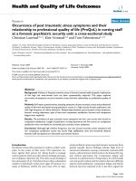
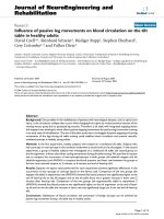
The mode of failure for the plasma sprayed titanium
implants is illustrated in Figure 1, where the fracture
plane was typically coincident with the host bone/de
novo bone interface. An exception to this rule were the
1 and 2 week timepoints, where the fracture plane was
coincident with the de novo bone implant interface, and
which may be indicative of insufficient appositional
bone growth. For all mechanical testing samples, regard-
less of timepoint, no damag e to or delamination of the
porous titanium domain was observed, despite mean
ultimate interfacial shear strength values 12 weeks post-
operatively of 28.3 ± 5.43 MPa and 29.06 ± 8.22 MPa
for the DCPD and TiPS groups, respectively.
Bone ongrowth
No significant differences in ongrowth were found
between DCPD and TiPS groups in either the cancellous
Table 1 Interfacial shear strength results for the DCPD
and TiPS implant groups as a function of postoperative
timepoint.
Time (weeks) Shear Strength (MPa)
DCPD TiPS P value
1 2.38 (1.81) 0.11 (0.02) 0.999
2 2.15 (2.64) 2.29 (2.02) 0.999
4 10.61 (4.35) 16.99 (11.34) 0.608
8 24.88 (4.35) 22.29 (6.09) 0.999
12 28.32 (5.43) 29.06 (8.22) 0.999
(Mean ± SD)
Chen et al. Journal of Orthopaedic Surgery and Research 2011, 6:56
/>Page 3 of 8
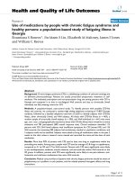
or cortical implantation sites (P > 0.05) (Figure 2). Mean
ongrowth in the cancellous site decreased from 4 to 12
weeks in both groups, where this reduction was signifi-
cant for the DCPD coating (P < 0.001) only. On the
contrary, mean cortical bone ongrowth increased from 4
to 12 weeks for both groups, where this increase was
significant for the TiPS coating (P = 0.002). Mean per-
centage bone ongrowth for the cortical implantation
sites appeared lower than cancellous site at 4 weeks in
both DCPD and TiPS groups, although the difference
was not significant. However, cortical bone ongrowth
rate surpassed cancellous ongrowth rate in both groups
at 12 weeks, which was significant for the DCPD coating
(P = 0.001).
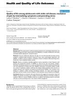
Bone ingrowth
Mean percentage bone ingrowth for the DCPD and
TiPS groups in the cancellous sites ranged from 29% to
69% and 18% to 60%, respective ly (Figure 3). DCPD
implants showed higher mean percentage bone ingrowth
at all time points, with the difference being significant at
4 weeks (P = 0. 003) only. In the cortical sites no signifi-
cant difference in bone ingrowth rate was observed
between DCPD and TiPS at either timepoint (P > 0.05).
Mean bone ingrowt h was gen erally higher in cancel-
lous bone than cortical bone for both TiPS and DCPD
groups at 4 weeks, although the differences were not
significant (P > 0.05). In contrast, cortical sites generally
exhibited higher bone ingrowth rate than cancellous site
at 12 weeks, which was significant for the TiPS coating
(P < 0.001).
Histological findings
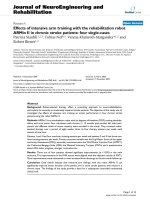
At 1 week following surgery, bone debris still could be
seen around both TiPS and DCPD implants, indicating
it had yet to be fully resorbed. Only traces of DCPD
coating were visible from the BSEM images (Figure 4),
suggesting substantial resorption of DCPD coating had
taken place following 1 week in situ.
Analysis of TiPS and DCPD implants at 2 weeks illu-
strated the initial de novo woven bone formation a nd
resorption of bone debris. The new bone appeared as a
deep red colour in the histology images, indicating
newlyformedbonegrowingdirectlyontheimplant
Figure 1 SEM image of an im plant from the DCPD group
depicting the failure location (black arrow) after push-out
testing.
Figure 2 Mean percentage bone ongrowth for TiPS and D CPD
groups as a function of implantation site and time. Note that
implant group and timepoint are combined in the x-axis categorical
variable (Mean ± SE).
Figure 3 Mean percentage ingrowth for both implant groups
in cancellous bone as a function of time. * denotes P = 0.003.
(Mean ± SE).
Chen et al. Journal of Orthopaedic Surgery and Research 2011, 6:56
/>Page 4 of 8
surface (Figure 5). Osteoblast lines could be seen on
newly formed bone directly on the porous implant sur-
face. The osteoblasts appeared enlarged, roundish and in
layers, indicating contact osteogenesis had been active at
2 weeks. They could also be seen on nearby new bone,
suggesting distance osteogen esis. New bone could also
be observed growing deep into the pores, extending to
the cylindrical implant substrate (Figure 6). Both osteo-
genic mechanisms were evident in the TiPS and DCPD
specimens. There was no evidence of residual DCPD
coating at 2 weeks post-implantation.
Images of both the TiPS and DCPD mediums at 4
weeks illustrated that the newly deposited bone
resembled normal trabeculae, growing from outside to
inside pores and exhibiting continuous curves, despite
the intervening presence of the titanium pore walls (Fig-
ure 7). Have rsian canals were occasionally seen in the
images at 4 and 8 weeks, indicating remodelling. At 12
weeks, mature Haversian canals could be seen in both
TiPS and DCPD implants. Osteocytes were more evenly
distributed and lamellar bone could be clearly identified
(Figure 8).
Discussion
Electrochemical cathodic deposition is a method
employed to apply a thin and uniform layer of calcium
phosphate coating on a porous impl ant surface. Metallic
implants are submerged in an electrolyte bath
Figure 4 Trac es of DCPD were visible from DCPD sections at 1
week.
Figure 5 Osteoblasts were enlarged, ro undish and in layers on
newly formed bones directly on porous implant surface and
on opposite surrounding bone. Image taken 2 weeks
postoperatively.
Figure 6 SEM image depicting de novo bone formation on and
extending to within the porous surface at 2 weeks
postoperatively.
Figure 7 SEM image depicting a continuation of the trabecular
structure of the cancellous bone to within the porous implant
domain, despite the barrier provided by the coating itself.In
this image bone can be seen growing onto the cylindrical implant
substrate.
Chen et al. Journal of Orthopaedic Surgery and Research 2011, 6:56
/>Page 5 of 8
containing dissolved calcium and phosphorus ions and
connected to an external power s upply [14]. A thin
DCPD layer with grain size ranging from 1-3 μmis
deposited on and within the porous implant surface,
without compromising pore openness and interconnec-
tivity [15]. DCPD dissolution is mainly affected by
volume diffusion [19]. In this study the DCPD laye r was
found t o be mostly dissolved at 1 week, with only trace
amounts present at 2 weeks, which is consistent with
other reports in the literature [20,21].
DCPD is believed to act as a heterogeneous centre for
HA growth in early bone formation [22]. For this reason
it has been postulated that the thin calcium phosphate
coating will improve bone ongrowth and ingrowth of
porous implant surfaces to achieve rapid and early
bone-implant interface integration and stability. Our
results suggest that a DCPD coating has the potential to
improve the extent of cancellous bone ingrowth in the
early postoperative period (Figure 3). This finding is
consistent with an in vit ro study showing higher cell
attachment ability on calcium phosphate compound
samples in the early stages [23]. Simank et al [15]
detected no significant difference in the mechanical fixa-
tion or bone formation throughout porous titanium
implants coated with either an osteoinductive growth
and diff erentiation factor-5 (GDF-5) or osteocond uctive
DCPD coating [15]. The mean bone ingrowth rate in
the DCPD group was approximately 66% in cortical
bone at 4 weeks, which compares well with values of
60% and 48% previously reported for a porous beaded
coating with and without a 50 μmHAcoatingat4
weeks in an ovine model [10].
In this study the cancellous implantation sites pre-
sented with higher mean bone ingrowth and ongrowth
values than in the cortical bone sites at 4 weeks post-
operatively for both DCPD and TiPS groups. Whilst this
mean increase was not statistically distinguishable this
finding is consistent with the kn owledge that cancellous
bone heals at a faster rate than cortical bone [24]. On
the other hand, bone ingrowth and ongrowth in cortical
bone sites showed generally higher percentage values
than in cancellous bone at 12 weeks for both groups.
Ostensibly, this result at 12 weeks is indicative of the
compact nature of cortical bone. In joint arthroplasty
the primary mode of fixation for uncemented tibial
trays, femoral components and acetabular cups is indeed
via cancellous bone ongrowth and ingrowth. Possible
effects which the differential in ongrowth and ingrowth
patterns observed in this study may have on uncemen-
ted fixation of joint components remains unknown.
Another striking feature in the current study was the
seeming preservation of trabecular bone structure to
within the porous coating domain (Figure 7), despite the
presence of intervening titanium. Because trabecular
bone tends to adapt to direction of mechanical stress
[25,26] this phenomenon may indicate that mechanical
loads were indeed transmitted through the thin titanium
pore walls. This observatio n supports the potential of
selective manufacturing to limit the effects of stress-
shielding by tailoring the elastic modulus of mediums
for hard tissue infiltration. Ryan and colleagues [7] have
demonstra ted that the c ompress ive modulus of porous
metals is better matched to cancellous bone as com-
pared to solid metals. This phenomenon of the conti-
nuation of trabecular bone architecture to within the
porous coating has not previously been observed for
thick-walled porous surfaces, such as beaded constructs
[18].
An implant exhibiting an osteoconductive coating can
sti mulate new bone growth directly on the implant sur-
face [8,9] and improve uncemented prosthesis fixation
in the early postoperative period [27,28]. In this study,
the plasma sprayed titanium porous surface both with
and without the electrochemically-deposited DCPD
coating exhibited de novo bone formation on the
implant surface as early as two weeks after implantation
(Figure 5 and Figure 6). At this timepoint osteoblasts
were seen lining new bone on both the implant surface
and adjacent host bone (Figure 5). Contact osteogenesis
in both DCPD and TiPS groups was in agreement with
a report that porous concave coatings can stimulate
osteogenic cells differentiating to osteoblasts [29].
The percentage ingrowth for both test materials in the
current study averaged approximately 37% at 2 weeks,
as compared to the 13% ingrowth obtained for a porous
tantalum implant [30]. Tantalum has been recognized as
having excellent bone and fibrous ingrowth properties,
allowing for r apid and substantial bone and soft tissue
attachment [31]. Direct comparison of these results is
fraught with difficulty, though, due to differences
Figure 8 Haversian canals and lamellar bone, indicative of
mature bone were clearly seen at 12 weeks.
Chen et al. Journal of Orthopaedic Surgery and Research 2011, 6:56
/>Page 6 of 8
between studies in terms of implant parameters (poros-
ity and coating thickness), implantation site and species.
Regardless, the results of the current study support the
osteoconductive potential of a highly porous titanium
surface with a DCPD coating.
Evidence of remodeling in the cancellous sites was
observed in both DCPD and TiPS groups as early as 4
and 8 weeks, with Haversian can als identified at 12
weeks (Figure 5). In addition, considerable amounts of
lamellar bone and evenly distributed osteocytes were
clearly seen in surrounding bone on both DCPD and
TiPS sections at 12 weeks. The rate of remodeling in
the current study is in contrast to other previous unce-
mented implant fixation studies in sheep [32,33] where
woven bone and lamellar matrix persisted three months
postoperatively. This remodeling rate may be attributed
to the highly porous surface and the press-fit insertion
manner adopted in current study.
Mechanical testing revealed no difference between
DCPD and TiPS at either timepoi nt. When selecting a
soluble material for coatings, the match of resorption
rate and bone regeneration rate must be taken into
account. If resorption rate is faster than regene ration,
there may be a void left by the absorbed material, which
can potentially compromise bone and implant contact
[13]. The shear strength of DCPD group was not lower
than the control group in the current study, although
the DCPD coating appeared mostly absorbed at 1 week
and almost completely at 2 weeks. The mechanical simi-
larity between DCPD and TiPS group in the two early
time points indicated the thin (20 μm) and highly solu-
ble DCPD coating will not co mpromise bone-implant
interface mechanical stability in early stage.
The failure mode for both implan t types from 4 to 12
weeks postoperatively was primarily at the interface
between de novo formed and host bone. The failure
mode illustrated that shear strength depends on the
amount and strength of surrounding new bone, which
can also be correlated to a study showing that mechani-
cal stability of rough titanium implants depends on the
amount of bony tissue surrounding the implant [15].
Thismaybethereasonwhyhigheringrowthdidnot
result in higher shear strength in DCPD implants. The
increase of mechanical strength with increasing time
may be due to the increasing amounts of mature sur-
rounding bone.
Conclusion
The study of plasma sprayed porous titanium surface
coated with and without DCPD demonstrated electro-
chemically deposited thin layer of DCPD with fine grain
size can improve bone ingrowth in vivo. Mechanical
results indicate that the thin and soluble DCPD had
neither a positive nor negative effect on interfacial shear
strength and implant stability in cortical bone. More-
over, analysis of the failure mode su ggests that the bone
bonding strength of the porous surface depends on the
amount and maturity of surrounding new bone for both
groups. As expected, an improvement in interfacial
shear strength for both implant types with time was
observed, continuous with the mechanical advantage of
bony remodeling.
Cancellous bone implantation was associate d with
higher bone in growth and ongrowth at the early stage,
whilst cortical bone implantation had more bone
ingrowth and ongrowth than cancellous bone at 12
weeks. The continuity of trabecular bone to within t he
porous coatings (Figure 7) a lso indicates the adaptation
of the highly porous surface structure to cancellous
bone. The implantation of the porous surface implants
by press-fit insertion demonstrated excellent early new
bone formation and remodelling.
Finally, electrochemical deposition has the potential to
produce calcium phosphate compounds with sub-
micron sized grains which may lead to high er cell adhe-
sion and osteoblast activity [34]. The effect of such coat-
ings may be examined in the future.
Author details
1
Surgical & Orthopaedic Research Laboratories, Prince of Wales Hospital,
University of New South Wales, Sydney, Australia.
2
Yokohama City University
Medical Center, Yokohama, Japan.
3
University of Tsukuba, Tsukuba, Japan.
4
Ryugasaki Saiseikai Hospital, Ryugasaki, Japan.
Authors’ contributions
WRW is credited with both conception and design of the study. DC
performed the animal surgery and, along with WRW, AL and NB was also
involved with and responsible for the processing of data, statistical analysis
and interpretation of results. All authors contributed equally to drafting and
critical review of the manuscript.
Competing interests
Funds for this study were received by our Institution from BBraun Aesculap
Japan. Co. No author of this paper was a direct beneficiary of such funding.
Received: 23 March 2011 Accepted: 3 November 2011
Published: 3 November 2011
References
1. Morscher EW: European experience with cementless total hip
replacements. Hip 1983, 190-203.
2. Yamada H, Yoshihara Y, Henmi O, Morita M, Shiromoto Y, Kawano T,
Kanaji A, Ando K, Nakagawa M, Kosaki N, Fukaya E: Cementless total hip
replacement: past, present, and future. J Orthop Sci 2009, 14(2):228-41.
3. Fyhrie DP, Carter DR, Schurman DJ: Effects of ingrowth, geometry, and
material on stress transfer under porous-coated hip surface
replacements. J Orthop Res 1988, 6(3):425-33.
4. Ito S, Matsumoto T, Enomoto H, Shindo H: Histological analysis and
biological effects of granulation tissue around loosened hip prostheses
in the development of osteolysis. J Orthop Sci 2004, 9(5):478-87.
5. Davies JE: Understanding peri-implant endosseous healing. J Dent Educ
2003, 67(8):932-949.
6. Davies JE: Bone bonding at natural and biomaterial surfaces. Biomaterials
2007, 28(34):5058-5067.
7. Ryan G, Pandit A, Apatsidis DP: Fabrication methods of porous metals for
use in orthopaedic applications. Biomaterials 2006, 27(13):2651-2670.
Chen et al. Journal of Orthopaedic Surgery and Research 2011, 6:56
/>Page 7 of 8
8. Takemoto M, Fujibayashi S, Neo M, Suzuki J, Kokubo T, Nakamura T:
Mechanical properties and osteoconductivity of porous bioactive
titanium. Biomaterials 2005, 26(30):6014-23.
9. Otsuki B, Takemoto M, Fujibayashi S, Neo M, Kokubo T, Nakamura T: Pore
throat size and connectivity determine bone and tissue ingrowth into
porous implants: Three-dimensional micro-CT based structural analyses
of porous bioactive titanium implants. Biomaterials 2006,
27(35):5892-5900.
10. Svehla M, Morberg P, Zicat B, Bruce W, Sonnabend D, Walsh WR:
Morphometric and mechanical evaluation of titanium implant
integration: comparison of five surface structures. J Biomed Mater Res
2000, 51(1):15-22.
11. Chambers B, St Clair SF, Froimson MI: Hydroxyapatite-coated tapered
cementless femoral components in total hip arthroplasty. J Arthroplasty
2007, 22(4 Suppl 1):71-4.
12. Daugaard H, Elmengaard B, Bechtold JE, Jensen T, Soballe K: The effect on
bone growth enhancement of implant coatings with hydroxyapatite and
collagen deposited electrochemically and by plasma spray. J Biomed
Mater Res A 2009.
13. Hench LL, Best S: Ceramics, glasses and glass-ceramics. In Biomaterials
Science. Edited by: Ratner BD, et al. Elsevier Inc; 2004:153-170.
14. Kim KH, Ramaswamy N: Electrochemical surface modification of titanium
in dentistry. Dent Mater J 2009, 28(1):20-36.
15. Simank HG, Stuber M, Frahm R, Helbig L, van Lenthe H, Muller R: The
influence of surface coatings of dicalcium phosphate (DCPD) and
growth and differentiation factor-5 (GDF-5) on the stability of titanium
implants in vivo. Biomaterials 2006, 27(21):3988-94.
16. Vereecke G, LemaÓtre J: Calculation of the solubility diagrams in the
system Ca(OH)2-H3PO4-KOH-HNO3-CO2-H2O. Journal of Crystal Growth
1990, 104(4):820-832.
17. Bertollo N, Matsubara M, Shinoda T, Chen D, Kumar M, Walsh WR: Effect of
Surgical Fit on Integration of Cancellous Bone and Implant Cortical Bone
Shear Strength for a Porous Titanium. Journal of Arthroplasty 2011.
18. Svehla M, Morberg P, Bruce W, Zicat B, Walsh WR: The effect of substrate
roughness and hydroxyapatite coating thickness on implant shear
strength. The Journal of Arthroplasty 2002, 17(3):304-311.
19. Zhang J, Nancollas GH: Interpretation of dissolution kinetics of dicalcium
phosphate dihydrate. Journal of Crystal Growth 1992, 125(1-2):251-269.
20. Bohner M, Theiss F, Apelt D, Hirsiger W, Houriet R, Rizzoli G, Gnos E, Frei C,
Auer JA, von Rechenberg B: Compositional changes of a dicalcium
phosphate dihydrate cement after implantation in sheep. Biomaterials
2003, 24(20):3463-3474.
21. Wang Y, Wei M, Gao J: Improve corrosion resistance of magnesium in
simulated body fluid by dicalcium phosphate dihydrate coating.
Materials Science and Engineering: C 2009, 29(4):1311-1316.
22. Kanzaki N, Onuma K, Treboux G, Ito A: Dissolution kinetics of dicalcium
phosphate dihydrate under pseudophysiological conditions. Journal of
Crystal Growth 2002, 235(1-4):465-470.
23. Duheyne P, Beight J, Cuckler J, Evans B, Radin S: Effect of calcium
phosphate coating characteristics on early post-operative bone tissue
ingrowth. Biomaterials 1990, 11(8):531-540.
24. Waris P, Karaharju E, Slatis P, Paavolainen P: Radiographic healing and
remodelling of cortical and cancellous bone grafts after rigid plate
fixation. Experiments in the rabbit. Acta Radiol Diagn (Stockh) 1980,
21(1):107-13.
25. Allori AC, Sailon AM, Pan JH, Warren SM: Biological basis of bone
formation, remodeling, and repair-part III: biomechanical forces. Tissue
Eng Part B Rev 2008, 14(3):285-93.
26. Turner CH, Pavalko FM: Mechanotransduction and functional response of
the skeleton to physical stress: the mechanisms and mechanics of bone
adaptation. J Orthop Sci 1998, 3(6):346-55.
27. Marco F, Milena F, Gianluca G, Vittoria O: Peri-implant osteogenesis in
health and osteoporosis. Micron 2005, 36(7-8):630-44.
28. Nikolidakis D, Meijer GJ, Oortgiesen DA, Walboomers XF, Jansen JA: The
effect of a low dose of transforming growth factor beta1 (TGF-beta1) on
the early bone-healing around oral implants inserted in trabecular bone.
Biomaterials 2009, 30(1):94-9.
29. Nimb L, Gotfredsen K, Steen Jensen J: Mechanical failure of
hydroxyapatite-coated titanium and cobalt-chromium-molybdenum
alloy implants. An animal study. Acta Orthop Belg 1993, 59(4):333-8.
30. Bobyn JD, Stackpool GJ, Hacking SA, Tanzer M, Krygier JJ: Characteristics of
bone ingrowth and interface mechanics of a new porous tantalum
biomaterial. J Bone Joint Surg Br 1999, 81(5):907-14.
31. Levine BR, Sporer S, Poggie RA, Della Valle CJ, Jacobs JJ: Experimental and
clinical performance of porous tantalum in orthopedic surgery.
Biomaterials 2006, 27(27):4671-81.
32. Chappard D, Aguado E, Hure G, Grizon F, Basle MF: The early remodeling
phases around titanium implants: a histomorphometric assessment of
bone quality in a 3- and 6-month study in sheep. Int J Oral Maxillofac
Implants 1999, 14(2):189-96.
33. Mavrogenis AF, Dimitriou R, Parvizi J, Babis GC: Biology of implant
osseointegration. J Musculoskelet Neuronal Interact 2009, 9(2):61-71.
34. Narayanan R, Kim SY, Kwon TY, Kim KH: Nanocrystalline hydroxyapatite
coatings from ultrasonated electrolyte: preparation, characterization, and
osteoblast responses. J Biomed Mater Res A 2008, 87(4):1053-60.
doi:10.1186/1749-799X-6-56
Cite this article as: Chen et al.: Osseointegration of porous titanium
implants with and without electrochemically deposited DCPD coating
in an ovine model. Journal of Orthopaedic Surgery and Research 2011 6:56.
Submit your next manuscript to BioMed Central
and take full advantage of:
• Convenient online submission
• Thorough peer review
• No space constraints or color figure charges
• Immediate publication on acceptance
• Inclusion in PubMed, CAS, Scopus and Google Scholar
• Research which is freely available for redistribution
Submit your manuscript at
www.biomedcentral.com/submit
Chen et al. Journal of Orthopaedic Surgery and Research 2011, 6:56
/>Page 8 of 8