báo cáo hóa học:" A Rat Model of Early Stage Osteonecrosis Induced by Glucocorticoids" potx
Bạn đang xem bản rút gọn của tài liệu. Xem và tải ngay bản đầy đủ của tài liệu tại đây (894.61 KB, 23 trang )
This Provisional PDF corresponds to the article as it appeared upon acceptance. Fully formatted
PDF and full text (HTML) versions will be made available soon.
A Rat Model of Early Stage Osteonecrosis Induced by Glucocorticoids
Journal of Orthopaedic Surgery and Research 2011, 6:62 doi:10.1186/1749-799X-6-62
Mohammad Amin Kerachian ()
Edward J. Harvey ()
Denis Cournoyer ()
Terry Y. Chow ()
Ayoub Nahal ()
Chantal Seguin ()
ISSN 1749-799X
Article type Research article
Submission date 11 March 2011
Acceptance date 21 December 2011
Publication date 21 December 2011
Article URL />This peer-reviewed article was published immediately upon acceptance. It can be downloaded,
printed and distributed freely for any purposes (see copyright notice below).
Articles in Journal of Orthopaedic Surgery and Research are listed in PubMed and archived at
PubMed Central.
For information about publishing your research in Journal of Orthopaedic Surgery and Research or
any BioMed Central journal, go to
/>For information about other BioMed Central publications go to
/>Journal of Orthopaedic Surgery
and Research
© 2011 Kerachian et al. ; licensee BioMed Central Ltd.
This is an open access article distributed under the terms of the Creative Commons Attribution License ( />which permits unrestricted use, distribution, and reproduction in any medium, provided the original work is properly cited.
A Rat Model of Early Stage Osteonecrosis Induced by
Glucocorticoids
Mohammad Amin Kerachian,
1,2
Edward J. Harvey,
3
Denis Cournoyer,
1,4,5
Terry Y.
Chow,
5
Ayoub Nahal,
6
and Chantal Séguin
4,5
*
1
Department of Human Genetics, McGill University Health Center (MUHC), 1650 Cedar
Avenue, Montreal, QC H3G 1A4, Canada
2
Department of Medical Genetics, Mashhad University of Medical Sciences (MUMS),
Azadi Square, Mashhad, 917794-8564, Iran
3
Division of Orthopaedic Surgery, McGill University Health Center (MUHC), 1650
Cedar Avenue, Montreal, QC H3G 1A4, Canada
4
Department of Medicine, Division of Haematology, McGill University Health Center
(MUHC), 1650 Cedar Avenue, Montreal, QC H3G 1A4, Canada
5
Department of Oncology, McGill University Health Center (MUHC), 1650 Cedar
Avenue, Montreal, QC H3G 1A4, Canada
6
Department of Pathology, McGill University Health Center (MUHC), 1650 Cedar
Avenue, Montreal, QC H3G 1A4, Canada
*Corresponding author
Email addresses:
MAK:
EJH :
DC:
TYC:
AN:
CS:
Abstract
Background: Glucocorticoid (GC)-induced osteonecrosis (ON) is an important
complication of medical therapy. The exact pathomechanisms of ON has not been clearly
elucidated. There is a need for a reproducible animal model that better approximates the
clinical scenario.
Methods: To determine the genetic susceptibility of rats to develop GC-induced femoral
head ON, we evaluated 5 different inbred strains of rats (Spontaneous Hypertensive Rat,
Wistar Kyoto, Wistar Furth, SASCO Fisher and Lewis). Prednisone pellets (dosage of
1.82-2.56 mg/kg/day) were implanted subcutaneously for 90. After 90 days, the femurs
were resected and examined histologically and radiographically. Pathological and
histological examination was performed. Hematoxylin and eosin (H & E) staining was
used to delineate the femoral head osteonecrosis lesions as well as abnormalities of
articular cartilage and growth plate.
Results: The greatest differences in H & E staining were seen in the Wistar Kyoto and
Wistar Furth groups. In these groups 4 out of 5 and 3 out of 5, respectively, steroid-
induced rats revealed growth plate disruption with acellular areas. The TUNEL apoptosis
staining assay for apoptosis revealed that 4 out of 5 of Wistar Kyoto rats, 5 out of 5 of
Wistar Furth, 2 out of 4 of surviving Lewis and 2 out of 2 of the surviving spontaneous
hypertensive rats had apoptotic osteocytes in trabeculae, whereas none of the Fisher rats
showed apoptotic osteocytes.
Conclusions: We postulate that Wistar Kyoto, Wistar Furth and spontaneous hypertensive
rats may be strains of rats more susceptible to develop ON of the femoral head while
Fisher rats were the most resistant.
Background
Glucocorticoids (GCs) are widely prescribed in cases of rheumatoid arthritis, asthma,
systemic lupus erythematosus, cancer, organ transplantation and many other medical
conditions. The therapeutic use of GCs has been accompanied by marked side effects,
especially with the long-term usage of this drug. The adverse effect of GCs on bone has
been recognized for more than 60 years [1-3]. The bone effect is characterized by
decreased bone formation and in situ death of isolated segments of
bone which may be
associated with osteonecrosis (ON) particularly important clinically for the femoral head.
ON in the femoral head gradually progresses to fracture of the subchondral bone, collapse
of the surface and hip arthritis. Although ON has been linked to a variety of conditions,
GC usage remains the predisposing factor most commonly associated with the
development of non-traumatic ON. There is considerable interest in identifying which
patients are at highest risk for ON, with the long-term goal of modifying regimens to
decrease the risk of adverse effects of therapy. Despite the strong association of GC
administration with ON, the role of potential underlying risk factors such as
hyperlipidemia, thrombophilia, and hyperfibrinolysis in the circulatory system remain
unclear [2,4]. It has been clearly established that among patients receiving a specific dose
of GC, only an unpredictable subset will develop ON. This underscores the existence of
individual variability in the action of GCs and the potential presence of additional
mechanisms and/or risk factors such as a genetic predisposition. On the other hand,
studying the clinical pathology of ON in the early disease stage (before radiographic
findings) is extremely difficult in human subjects. Thus, animal experiments are needed
to elucidate the pathophysiology of the disorder. Having a suitable animal model would
allow for the systemic evaluation of host-related (ie. genetic variations) as well as
acquired (ie. treatment-related) risk factors. GC-induced ON has been induced in rabbit
models [5-8], bipedal animals (e.g., chickens, emus) [9,10] and recently, in BALB/cJ
mice [11]. GC-induced ON has been described in mature Japonese white rabbits (Kbs-
JW) [5,8] but the genome of rabbit has only been incompletely sequenced, thus limiting
the usefulness of that model for the identification of genes affecting the risk of
developing ON. The biped models are difficult to interpret in the context of bone healing
as we do not have a full grasp of avian bone healing. Although a mouse model of GC-
induced ON is interesting, the very small diameter of the femoral head of mice limits the
application of numerous experiments and monitoring techniques. It is currently
impossible to read an MRI or radiograph from a mouse with the goal of differentiating a
normal hip from a hip with ON changes. A rat model would allow easier radiographic
interpretation, allow facile surgical interventions, allow existing small animal facilities to
be used as well as be in an animal where the genetics of healing is much better
understood. To date, there has been no rat model of GC-induced ON unless it has been
combined with a surgical procedure [12] or in combination with immune responses
stimuli [13]. These blood interruption studies do not faithfully model the more prevalent
non-traumatic ON. In this study, our goal was to establish a rat model of GC-induced
ON by screening different strains of rats in order to uncover those whose constitutive
phenotype might predispose to the development of ON.
Methods
Maintenance and experimental animals
In this pilot study, female retired breeder (aged 6-8 months) Fisher, Lewis,
Spontaneous Hypertensive, Wistar Kyoto, and Wistar Furth rats (6 of each strain) were
obtained from Charles River Laboratories (Pointe-Claire, QC, Canada). The rats were
tagged and housed in plastic cages (2 animals per cage) under standard laboratory
conditions with a 12-hour dark/12-hour light cycle, a constant temperature of 20 °C, and
humidity of 48%. Food and water were provided ad libitum with a standard rodent diet.
The weight of the rats were followed before and after the implant of a prednisone pellet
for the first 4 consecutive days, then every week until the end of the experiment. All
experiments were conducted under an animal protocol (Protocol No. 4935) approved by
the McGill Animal Care Department, Montreal, Canada.
Glucocorticoid administration
Slow-release prednisone pellets (Innovative Research of America, Sarasota,
Florid, USA) were implanted subcutaneously in 5 inbred rats composing each group
(Fisher, Lewis, Spontaneous Hypertensive, Wistar Kyoto and Wistar Furth). Each pellet
was implanted underneath the skin on the lateral side of the neck by surgically making an
incision and developing a pocket about 2 cm beyond the incision site. The pellet was
placed in the pocket and the incision was sutured. Based on the manufacturer’s
instructions the pellet releases a constant dose of the drug subcutaneously. The average
dose release from the pellet was equivalent to 1.82-2.56 mg/kg/day (mean: 2.26, SD:
0.19) for a period of 90 days. This dosage is an equivalent dosage to humans that
commonly causes ON changes. Thus, each group had 5 GC-induced rats along with 1
control rat in each group not treated with prednisone (the control rat did not receive a
placebo pellet).
Histological Examination
The rats were sacrificed with an overdose of ketamine/xylazine following 90 days
of the experiment. Tissue samples were obtained from the femoral head. Bone samples
were fixed in 10% neutral buffered formalin overnight, then decalcified in 4%
ethylenediamine tetraacetic acid (pH 7.2) (Sigma-Aldrich, St. Louis, MO, USA). The
specimens were processed routinely and embedded in paraffin. Tissue sections were cut
parasagitally with a rotary microtome to obtain 4 to 5 microns thickness, stained with
hematoxylin and eosin (H & E) and evaluated by light microscopy.
Tissue samples were analyzed in a blinded fashion by an experienced bone pathologist
(AN). GC-induced ON was diagnosed based on bone and growth plate changes. The
histological findings of an established ON were defined as dead trabeculae exhibiting
empty lacunae with or without appositional bone formation [14]. Occasional empty
lacunae possibly created by sectioning through the edge of a lacunae was not considered
as a sign for ON. The growth plate changes were considered as thinning, discontinuity
pattern and disruption of articular cartilage alignment or growth plate alignment.
Tissue sections were also examined according to the criteria of Arlet et al. namely
presence of degeneration, necrosis, and disappearance of marrow cells as well as the
nuclear disappearance and hypochromasia of trabecular osteocytes as early signs of ON
[15]. Early signs of ON was also considered when apoptosis occurred in the osteocytes
and osteoblasts. Positivity for apoptosis was defined by the authors as 2 to 3 apoptotic
osteocytes and/or osteoblasts considered as one plus, between 3 to 6 as two plus and more
than 6 as three plus recognized in a high magnification field (×200). The experiments
were performed in triplicate.
Measurement of apoptosis in undecalcified bone section
We used terminal deoxynucleotidyl transferase dUTP nick end labeling (TUNEL
assay) to detect DNA fragmentation by labeling the terminal end of nucleic acids. In Situ
Cell Death Detection Kit was obtained from Roche (Germany). TUNEL assay on
paraffin-embedded tissue sections was performed as recommended by the manufacturer.
Briefly, after deparaffinization and permeabilization of the tissue sections with proteinase
K, the slides were incubated with the TUNEL reaction mixture containing TUNEL-
Enzyme solution and TUNEL-Label solution for 1 hour at 37 °C inside a humidified
chamber. After washing steps, samples were analyzed under a fluorescence microscope
(in a drop of 1X PBS). The excitation wavelength ranged between 450-500 nm whereas
the detection wavelength ranged between 515-565 nm (green). DNase I-treated tissue
section was used as a positive control. Negative controls for the study constituted of
sample slides processed using the same procedure but only treated with TUNEL-Label
solution.
Faxitron X-ray
Based on the histological results Faxitron x-ray analysis was performed initially
on a group of Wistar Kyoto rats (5 rats, 10 femoral head samples) (Model MX-20).
Previous work has shown that radiographic changes were a late finding in steroid induced
ON in the rat model used. We performed the Faxitron radiographs on this group to ensure
there were no significant changes despite changes on histology.
Statistical Analysis
Comparison between groups was made with Fisher’s Exact test. Significant
differences were considered at P values less than 0.05.
Results
We observed a high mortality rate in some strains of rats after prednisone
implantation. Interestingly, the Lewis and spontaneous hypertensive strains of rats
seemed to be at highest risk (mortality rate was 1 out of 5 and 3 out of 5, respectively and
no mortality for other strains). There was an overall mortality rate of 16% among the
steroid-treated rats in our pilot study related to the development of GC-induced
hyperglycemia in these “older” rats (a two to three times fold increase compare to control
rats).
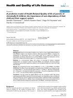
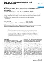
Growth plate changes were observed in Wistar Kyoto and Wistar Furth rats (Fig. 1). In
these groups 4 out of 5 and 3 out of 5 of steroid-induced rats revealed growth plate
disruption with acellular areas, respectively. Osteocyte necrosis and empty lacunae were
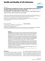
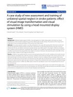
not detected in any samples. TUNEL assay for apoptosis revealed that 4/5 of Wistar
Kyoto, 5/5 of Wistar Furth, 2/4 of Lewis and 2/2 Spontaneous Hypertensive rats had
apoptotic osteocytes in trabeculae, whereas none of the Fisher rats showed apoptotic
osteocytes (Table 1, Fig. 2). In the Lewis group, apoptosis of osteocytes and osteoblasts
without any degeneration of the growth plate was observed. Overall, the most apoptosis
rate was in spontaneous hypertensive rats (+++) and then Wistar Furth (++) and Wistar
Kyoto (++). The apoptosis level in Lewis and Fisher rats was (+) and zero, respectively.
Bone marrow and chondrocyte apoptotic cells were seen in all strains of rats, even the
control rats as expected. There were no signs of inflammation and necrosis, such as
hyperemia, round cell infiltration, or lipid cyst formation. Plain x-rays obtained from
Faxitron analysis did not reveal any significant anomaly in the initial group of Wystar
Kyoto rats. Often diminished bone density was noticed in rats exposed to glucocorticoid.
The radiographs were not performed in the other groups because of the lack of changes.
If there had been changes in the initial group the other strains would have been tested.
This finding also confirmed that plain x-rays are not a suitable method to diagnose early
stages of ON in rats.
Discussion
A strain dependant genetic predisposition may be responsible for the high
mortality rate observed in some strains of rats after prednisone implantation. Recognition
of this complication of hyperglycemia has been reported in young rats [16] and seems to
be important when choosing a model for ON. It would make these two strains less
desirable for this usage.
The rat growth plate is present throughout the life-cycle. This may confound the findings
of ON for late stage ON in that reparative changes will overcome the initiating stimulus-
particularly in traumatic vascular interruption studies. For this GC study, the high
dosages of steroid given will result in early ongoing changes in the rat femoral head
despite reparative processes from the growth plate. Because of this a rodent model is
possible for early ON.
Growth plate disruption was observed particularly in Wistar Kyoto and Wister Furth
strains. This was observed in early stages of the disease- before radiographic change was
evident. Other studies using blood supply interruption (ischemic model) have also shown
growth plate changes. Trueta and Amato used animal models and showed that the blood
supply to the cartilage of the growth plate of the femoral head originates from the
epiphyseal vessels [17], while the metaphysis is supplied by metaphyseal vessels and
nutrient arteries coming from the medullary cavity. Mechanical damage to the
metaphyseal arteries leads to destruction of the growth plate and, eventually, a physeal
bridge [18]. It is possible that thrombosis in the metaphyseal arteries reported in ON of
the femoral head could cause injury and disruption of the growth plate with areas lacking
normal cells. Sato et al. have also shown that apoptosis tended to occur in early stages of
ON. In their rat study of ischemic ON, apoptosis occurred 12 hours after the mechanical
insult, whereas no evidence of apoptosis remained after 96 hours, at which time only
empty lacunae were detected [19]. They postulated that the mechanism of cell death
involved in ischemic ON was apoptosis as indicated by DNA fragmentation and the
presence of apoptosis bodies in osteocytes [11,20]. In the present study, apoptosis of
chrondrocytes were not only detected in GC-induced rats but also in control rats
indicating that apoptosis of chondrocytes is not characteristic of ON but probably more
indicative of normal bone turnover if observed in small amounts. Other studies have
shown that apoptosis of osteocytes and osteoblasts is an important process in developing
ON, especially in the early stages of ON [21]. Kabata and his colleagues demonstrated
extensive osteocyte apoptosis in a rabbit model of GC-induced ON [22]. Shibahara et al.
also reported the presence of a large number of apoptotic osteocytes around necrotic
areas [23]. In the present study, we observed that apoptosis occurred at the level of
osteocytes, osteoblasts, and bone marrow cells in the early stages of GC-induced ON
lesions in three strains of inbred rats: Wistar Kyoto, Wistar Furth and Spontaneous
Hypertensive. Apoptosis has been shown to play an important role in maintaining
haematopoietic stem cells (HSC) homeostasis in in vivo. Thus, apoptosis of the HSC
could occur as part of the normal physiology in bone marrow cells [24]. Previously, it has
been shown that apoptosis could happen in bone marrow of control rats [25]. Apoptosis
could result from a direct effect of GCs on the bone cells or could be secondary to the
dysfunction/activation of other cells such as the femoral head endothelial cells [26].
Fisher rats were resistant to the development of osteocyte apoptosis in response to GC
induction. Given the observed inter-strain variability of susceptibility to the development
of GC-induced ON lesions, it is probable that genetic factors are involved in ON
developing in response to GCs. To date different genetic variations and mutations
accounting for ON have been reported. A 4G/4G mutation of the plasminogen activator
inhibitor-1 gene [27], a G >A transition in exon 50 (p.G1170S) of collagen type II
(COL2A1) [28] and a promoter polymorphisms of the vascular endothelial growth factor
(VEGF) gene [29] were reported to be correlated with the occurrence of ON of the
femoral head. Identifying the genetic factors may prove relevant to the human disorder
and facilitate the identification of individuals at increased risk of developing ON.
Conclusions
Based on these findings, Wistar Kyoto, Wistar Furth and Spontaneously
Hypertensive rats were the most susceptible strains to develop GC-induced ON of the
femoral head. The Spontaneously Hypertensive rats had a high mortality rate that is
unacceptable for a study model. Fisher rats were resistant to the development of ON at
the GC dosage used, based on the absence of osteocyte apoptosis in early stages of the
disease process. Although several other investigations have reported ON in rats following
the administration of GCs, our rat model has shown early stage disease regardless of
additional adjuvants such as surgery [12] or immune response stimuli [13,30] as
previously reported. It is possible that extended exposure to GCs could establish the
histological criteria of the later stages of ON.
Competing interests
The author(s) declare that they have no competing interests.
Authors' contributions
All authors participated in the study. MAK made a major contribution to the writing of
the manuscript's first draft, and conducted the experiments involved in the study. CS
made a major contribution to the design of the study, data interpretation and scientific
revision of the manuscript. DC, EJH and TYC made equal contributions to data
interpretation and scientific revision of the manuscript. EJH made a major contribution to
the editing and grammar of the manuscript. AN made major contributions to the
histological experiments involved in the study. All authors participated in the manuscript
preparation and revision. All authors read and approved the final manuscript.
Acknowledgments
This work has been supported by the Montreal General Hospital Foundation (C.S.), by
the generous research award from Mr John D. Miller (C.S.) and support from FRSQ
Chercheur-Boursier Clinicien Senior (E.J.H.).
References
1. Cushing H: The basophil adenomas of the pituitary body and their clinical
manifestations (pituitary basophilism). 1932. Obes Res 1994, 2:486-508.
2. Kerachian MA, Séguin C, Harvey EJ: Glucocorticoids In Osteonecrosis of the
Femoral Head: A New Understanding of the Mechanisms of Action. J Steroid
Biochem Mol Biol 2009, 114:121-8.
3. Yao W, Cheng Z, Busse C, Pham A, Nakamura MC, Lane NE: Glucocorticoid excess
in mice results in early activation of osteoclastogenesis and adipogenesis and
prolonged suppression of osteogenesis: a longitudinal study of gene expression in
bone tissue from glucocorticoid-treated mice. Arthritis Rheum 2008, 58:1674-86.
4. Jones LC, Mont MA, Le TB, Petri M, Hungerford DS, Wang P, Glueck CJ:
Procoagulants and osteonecrosis. J Rheumatol 2003, 30:783-91.
5. Zhang G, Sheng H, He YX, et al: Continuous occurrence of both insufficient
neovascularization and elevated vascular permeability in rabbit proximal femur
during inadequate repair of steroid-associated osteonecrotic lesions. Arthritis Rheum
2009, 60:2966-77.
6. Zhang G, Wang XL, Sheng H, et al: Constitutional flavonoids derived from
Epimedium dose-dependently reduce incidence of steroid-associated osteonecrosis
not via direct action by themselves on potential cellular targets. PLoS One 2009, 4:
e6419
7. Zhang G, Qin L, Sheng H, Yeung KW, et al: Epimedium-derived phytoestrogen
exert beneficial effect on preventing steroid-associated osteonecrosis in rabbits with
inhibition of both thrombosis and lipid-deposition. Bone 2007, 40:685-92.
8. Sheng HH, Zhang GG, Cheung WH, et al: Elevated adipogenesis of marrow
mesenchymal stem cells during early steroid-associated osteonecrosis development.
J Ortho Surg Res 2007, 2:15-22.
9. Wang GJ, Cui Q, Balian G: The Nicolas Andry award. The pathogenesis and
prevention of steroid-induced osteonecrosis. Clin Orthop Relat Res 2000, 370:295-
310.
10. Conzemius MG, Brown TD, Zhang Y, Robinson RA: A new animal model of
femoral head osteonecrosis: one that progresses to human-like mechanical failure. J
Orthop Res 2002, 20:303-309.
11. Yang L, Boyd K, Kaste SC, Kamdem KL, Rahija RJ, Relling MV: A mouse model
for glucocorticoid-induced osteonecrosis: effect of a steroid holiday. J Orthop Res
2009, 27:169-175.
12. Chen Y, Huang K, Lang F, Huang Y, Huang H, Huang H et al: Experimental study
on cheng zai wan for treatment of necrosis of the femoral head. J Tradit Chin Med
2003, 23:292-298.
13. Okazaki S, Nishitani Y, Nagoya S et al: Femoral head osteonecrosis can be caused
by disruption of the systemic immune response via the toll-like receptor 4 signalling
pathway. Rheumatology (Oxford) 2009, 48:227-32
14. Wada M, Kumagai K, Murata M, Yamashita Y, Shindo H: Warfarin reduces the
incidence of osteonecrosis of the femoral head in spontaneously hypertensive rats. J
Orthop Sci 2004, 9:585-590.
15. Arlet J: A traumatic necrosis of the femoral head: general report. In Bone
circulation and vascularization in normal and pathological conditions. Edited by
Schoutens A, Arlet J, Gardeniers JWM, Hughes SPF:New York: Plenum; 1993, 235-240.
16. Rafacho A, Cestari TM, Taboga SR, Boschero AC, Bosqueiro JR: High doses of
dexamethasone induce increased beta-cell proliferation in pancreatic rat islets. Am J
Physiol Endocrinol Metab 2009, 296:E681-9.
17. Trueta J, Amato VP: The vascular contribution to osteogenesis. III. Changes in
the growth cartilage caused by experimentally induced ischaemia. J Bone Joint Surg
Br 1960, 42-B:571-587.
18. Nyska M, Shabat S, Long PH, Howard C, Ezov N, Levin-Harrus T et al:
Disseminated thrombosis-induced growth plate necrosis in rat: a unique model for
growth plate arrest. J Pediatr Orthop 2005, 25:346-350.
19. Sato M, Sugano N, Ohzono K, Nomura S, Kitamura Y, Tsukamoto Y et al:
Apoptosis and expression of stress protein (ORP150, HO1) during development of
ischaemic osteonecrosis in the rat. J Bone Joint Surg Br 2001, 83:751-759.
20. Weinstein RS, Manolagas SC: Apoptosis in glucocorticoid-induced bone disease.
Curr Opin Endocrinol Diabetes 2008, 12:219-223.
21. Jones LC, Hungerford DS: The pathogenesis of osteonecrosis. Instr Course Lect
2007, 56:179-196.
22. Kabata T, Kubo T, Matsumoto T, Nishino M, Tomita K, Katsuda S et al: Apoptotic
cell death in steroid induced osteonecrosis: an experimental study in rabbits. J
Rheumatol 2000, 27:2166-2171.
23. Shibahara M, Nishida K, Asahara H, Yoshikawa T, Mitani S, Kondo Y, Inoue H:
Increased osteocyte apoptosis during the development of femoral head osteonecrosis
in spontaneously hypertensive rats. Acta Med Okayama 2000, 54:67-74.
24. Domen J: The role of apoptosis in regulating hematopoietic stem cell numbers.
Apoptosis 2001, 6:239-52.
25. Matsouka P, Mylonas P, Papandoniou E, Dimitropoulou I, Floratou K, Alexandridis
T, Kardamakis D: Abdominal radiation initiates apoptotic mechanism in rat femur
bone marrow cells in vivo that is reversed by IGF-1 administration. J Radiat Res
(Tokyo) 2008, 49:41-7.
26. Kerachian MA, Harvey EJ, Cournoyer D, Chow TY, Séguin C: Avascular Necrosis
of the Femoral Head: Vascular Hypotheses. Endothelium 2006, 13:237-244.
27. Glueck CJ, Fontaine RN, Gruppo R, Stroop D, Sieve-Smith L, Tracy T, Wang P:
The plasminogen activator inhibitor-1 gene, hypofibrinolysis, and osteonecrosis.
Clin Orthop Relat Res. 1999, 336:133-46.
28. Liu YF, Chen WM, Lin YF, Yang RC, Lin MW, et al: Type II collagen gene
variants and inherited osteonecrosis of the femoral head. N Engl J Med 2005,
352:2294-2301.
29. Kim TH, Hong JM, Lee JY, Oh B, Park EK, Lee CK, Bae SC, Kim SY.
Promoterpolymorphisms of the vascular endothelial growth factor gene is
associated withan osteonecrosis of the femoral head in the Korean population.
Osteoarthritis Cartilage 2008, 16:287-291.
30. Okazaki S, Nishitani Y, Nagoya S, Kaya M, Yamashita T, Matsumoto H: Femoral
head osteonecrosis can be caused by disruption of the systemic immune response
via the toll-like receptor 4 signalling pathway. Rheumatology (Oxford) 2009, 48:227-
32
Figures
Figure 1. Photomicrographs showing histological findings in steroid-induced rat
models. H & E staining in a control rat (A) versus glucocorticoid-induced rats of all
groups (B: Lewis, C: Fisher, D: Wistar Kyoto, E: Wistar Furth and F: Spontaneous
Hypertensive rats ). Normal growth plate and its discontinuity pattern are shown by
arrows and arrowheads, respectively. Magnification x200.
Figure 2. Photomicrographs showing apoptosis of osteocytes as a marker of early
osteonecrosis of the femoral head. TUNEL staining apoptosis assay analyzed by (A):
florescence microscope, (B): superimposed florescence and optical view on femoral head
in II: Lewis, III: Fisher, IV: Wistar Kyoto, V: Wistar Furth and VI: Spontaneous
Hypertensive rats induced with steroids for 3 months versus a control sample (I). The
nucleus of apoptotic cells are shown in green. Magnification x200.
Tables
Table 1. Apoptosis of the femoral head of GC-induced inbred rats from 5 different strains
(WKY: Wistar Kyoto, WF: Wistar Furth, SHR: Spontaneous Hypertensive rat,).
Strain Bone Marrow Osteocyte Chondrocyte
Lewis 4/4 2/4* 4/4
Fischer 5/5 0/5* 5/5
WKY 5/5 4/5* 5/5
WF 5/5 5/5* 5/5
SHR 2/2 2/2* 2/2
*Fisher’s Exact Test (P value =0.0039).
Figure 1
Figure 2