báo cáo hóa học:" Luteinizing hormone-induced Akt phosphorylation and androgen production are modulated by MAP Kinase in bovine theca cells" doc
Bạn đang xem bản rút gọn của tài liệu. Xem và tải ngay bản đầy đủ của tài liệu tại đây (533.79 KB, 8 trang )
BioMed Central
Page 1 of 8
(page number not for citation purposes)
Journal of Ovarian Research
Open Access
Research
Luteinizing hormone-induced Akt phosphorylation and androgen
production are modulated by MAP Kinase in bovine theca cells
Shin Fukuda
1
, Makoto Orisaka*
1,2
, Kimihisa Tajima
1
, Katsushige Hattori
1
and Fumikazu Kotsuji
1
Address:
1
Department of Obstetrics & Gynecology, University of Fukui, Matsuoka, Fukui, 910-1193, Japan and
2
University of Fukui, 23-3
Shimoaiduki, Matsuoka, Eiheiji-cho, Yoshida-gun, Fukui, 910-1193, Japan
Email: Shin Fukuda - ; Makoto Orisaka* - ; Kimihisa Tajima - ;
Katsushige Hattori - ; Fumikazu Kotsuji -
* Corresponding author
Abstract
Background: Theca cells play an important role in controlling ovarian steroidogenesis by
providing aromatizable androgens for granulosa cell estrogen biosynthesis. Although it is well
established that the steroidogenic activity of theca cells is mainly regulated by LH, the intracellular
signal transduction mechanisms that regulate thecal proliferation and/or steroidogenesis remain
obscure. In this study, we examined whether and how LH controls the PI3K/Akt signaling pathway
and androgen production in bovine theca cells. We also explored whether this LH-induced PI3K/
Akt activation is modulated with other signaling pathways (i.e. PKA and MAPK).
Methods: Ovarian theca cells were isolated from bovine small antral follicles and were incubated
with LH for various durations. Phospho-Akt and total-Akt content in the cultured theca cells were
examined using Western blotting. Androstenedione levels in the spent media were determined
using EIA. Semi-quantitative RT-PCR analyses were conducted to analyze the mRNA levels of
CYP17A1 and StAR in the theca cells. To examine whether Akt activity is involved in theca cell
androgen production, the PI3K inhibitors wortmannin and LY294002 were also added to the cells.
Results: Akt is constitutively expressed, but is gradually phosphorylated in cultured bovine theca
cells through exposure to LH. LH significantly increased androstenedione production in bovine
theca cells, whereas addition of the wortmannin and LY294002 significantly decreased LH-induced
androstenedione production. LH significantly increased CYP17A1 mRNA level in theca cells,
whereas addition of LY294002 significantly decreased LH-induced CYP17A1 expression. Neither
LH nor PI3K inhibitors alter the mRNA levels of StAR in theca cells. Although H89 (a selective
inhibitor of PKA) does not affect LH-mediated changes in Akt, U0126 (a potent MEK inhibitor)
suppressed LH-induced Akt phosphorylation, CYP17A1 expression, and androgen production in
theca cells.
Conclusion: These results indicate that LH stimulates CYP17 mRNA expression and androgen
production in theca cells via activation of the PI3K/Akt pathway. The LH-induced Akt
phosphorylation and androgen production are modulated by the MAPK signaling in bovine theca
cells.
Published: 16 November 2009
Journal of Ovarian Research 2009, 2:17 doi:10.1186/1757-2215-2-17
Received: 13 July 2009
Accepted: 16 November 2009
This article is available from: />© 2009 Fukuda et al; licensee BioMed Central Ltd.
This is an Open Access article distributed under the terms of the Creative Commons Attribution License ( />),
which permits unrestricted use, distribution, and reproduction in any medium, provided the original work is properly cited.
Journal of Ovarian Research 2009, 2:17 />Page 2 of 8
(page number not for citation purposes)
Background
The principal function of ovarian theca cells is steroid hor-
mone production. Theca cells play an important role in
controlling ovarian steroidogenesis by providing aroma-
tizable androgens for granulosa cell estrogen biosynthesis
[1]. Androgens also function as local regulators of ovarian
folliculogenesis upon binding androgen receptors local-
ized to granulosa cells, stromal cells, and oocytes [2].
Androgen receptor null mice culminate in reduced fertility
and premature ovarian failure [3], indicating that andro-
gens are necessary for reproductive function and fertility.
Normal ovarian function requires accurate regulation of
steroidogenic activity of theca cells through extraovarian
and intraovarian mechanisms. Thecal steroidogenic
hyperactivity can cause ovarian dysfunction, such as poly-
cystic ovary syndrome (PCOS) [4].
It is well established that theca cell steroidogenesis is
under the primary control of luteinizing hormone (LH)
through the second-messenger cAMP-protein kinase A
(PKA) pathway [5,6]. Moreover, LH stimulates theca cells
to produce androgens and to maintain progesterone pro-
duction by the induction of genes involved in steroido-
genesis: cytochrome P450 side-chain cleavage enzyme
(CYP11A1), 3β-hydroxysteroid dehydrogenase, 17α-
hydroxylase/C17-20 lyase cytochrome P450 (CYP17A1),
and steroidogenic acute regulatory protein (StAR) [7-10].
Intracellular signaling mechanisms that regulate ovarian
follicular development and/or steroidogenesis remain
obscure [11]. Nevertheless, LH reportedly activates the
extracellular-signal-regulated kinases (ERK)/mitogen acti-
vated protein kinase (MAPK) pathway in ovarian granu-
losa and theca cells [12]. Although FSH and several
growth factors are known to activate the phosphatidyli-
nositol 3' kinase (PI3K)/Akt pathway in granulosa cells
[13-15], whether LH stimulates the PI3K/Akt cascade in
theca cells is not clear. Although LH augments androgen
production in theca cells, it remains unknown whether
this response is mediated via activation of the PI3K/Akt
pathway.
In this study, we examined whether and by what means
LH controls PI3K/Akt signaling and androgen production
using cultured bovine theca cells. We demonstrated that
LH stimulates CYP17A1 mRNA expression and androgen
production in theca cells via activation of the PI3K path-
way. Both the PI3K and the MAPK pathways coordinately
regulate androgen production in bovine theca cells.
Methods
Exprimental design
Experiment 1
To examine whether LH stimulates PI3K/Akt signaling in
theca cells, bovine theca cells from small antral follicles
were incubated with LH for various durations (0, 5 min,
20 min, 1 h, 2 h, 4 h, 6 h, 8 h, 12 h, 24 h, and 48 h), and
phospho-Akt and total-Akt content were examined using
Western blotting.
Experiment 2
To examine whether Akt activity is involved in theca cell
androgen production, theca cells were pretreated for 30
min with the PI3K inhibitors, wortmannin (0.1 μM) and
LY294002 (25 μM). The cells were subsequently stimu-
lated with LH (100 ng/ml) for 24 h. Androstenedione lev-
els in the spent media were determined using EIA.
Experiment 3
Along with examining androstenedione production,
semi-quantitative RT-PCR analyses were conducted to
analyze the mRNA levels of CYP17A1 and StAR in the cul-
tured theca cells at 12 h of incubation.
Experiment 4
Whether PKA or MAPK pathway influence LH-induced
Akt phosphorylation in theca cells was explored. Theca
cells were pretreated with H89 (i.e. a selective inhibitor of
PKA [16]), and U0126 (i.e. a potent MEK inhibitor) for 30
min. The cells were subsequently stimulated with LH (100
ng/ml) for 24 h. Phospho-Akt and total-Akt content in the
cultured theca cells were examined using Western blot at
24 h of the culture. CYP17A1 mRNA levels in the theca
cells and androstenedione levels in the spent media were
also determined.
Antibodies
Rabbit polyclonal anti-phospho-Akt (i.e. active Akt) anti-
bodies and anti-total-Akt antibodies were purchased from
Cell Signaling Technologies (Beverly, MA). Goat anti-rab-
bit IgG coupled to horseradish peroxidase was purchased
from Santa Cruz Biotechnology, Inc. (Santa Cruz, CA).
Reagents
Human LH was provided by the National Institutes of
Health and Dr. A. F. Parlow (National Hormone and Pep-
tide Program, Torrance, CA). LY294002 (a PI3K inhibitor)
was from Sigma Chemical Co. (St. Louis, MO), and wort-
mannin (a PI3K inhibitor), H89 (a selective inhibitor of
PKA), and U0126 (a potent MEK inhibitor) were pur-
chased from Calbiochem Novabiochem Corp. (San
Diego, CA).
Theca cell culture
Bovine ovaries were collected less than 15 min after
slaughter at a local abattoir. The ovaries were placed in an
ice-cold buffered salt solution and transferred to the labo-
ratory less than 90 min after collection. The estrous cycle
stage was determined morphologically, as described pre-
viously by Ireland et al [17]; only those ovaries with a
regressing corpus luteum were used for this study. Theca
cells were isolated from the ovaries under sterile condi-
Journal of Ovarian Research 2009, 2:17 />Page 3 of 8
(page number not for citation purposes)
tions, as described previously [18]. Briefly, small antral
follicles (2-4 mm diameter) with clear surfaces were cut
into halves and theca interna removed in situ using fine
forceps. Granulosa cells, together with part of the theca
cell layer, were removed by scraping with a scalpel under
a stereomicroscope. The resultant thin thecal layer was
minced and subsequently treated with a Hanks'-HEPES
buffer containing collagenase (2150 U/ml, type 1; Sigma)
and DNase (100 U/ml; Sigma), 0.4% (vol/vol) BSA, and
0.2% (wt/vol) glucose (pH 7.4). Cell dissociation was
allowed to continue for 30-60 min at 37°C with continu-
ous stirring at 80 rpm and 0.25% (wt/vol) pancreatin
(Sigma) in a Hanks'-HEPES buffer for 7 min. Dispersed
cells were washed three times. Cell viability, as deter-
mined using the trypan blue-dye exclusion test, was 90-
93%. Purity of the theca cell preparation used in this study
was substantiated by the secretion of estradiol; prepared
theca cells did not produce estradiol in the presence or
absence of forskolin, whereas granulosa cells obtained
from the same follicle secret significant (data not shown).
Isolated theca cells were plated onto serum-coated dishes
with serum-free medium for 36 h. Then they were stimu-
lated with LH (100 ng/ml) for various durations (0, 5
min, 20 min, 1 h, 2 h, 4 h, 6 h, 8 h, 12 h, 24 h, and 48 h).
Preliminary data indicated that 100 ng/ml of LH is the
minimal effective concentration for inducing a significant
increase in androgen production and CYP17A1 expres-
sion in our culture system.
Western blot analysis
Western blot analysis was conducted as described previ-
ously [12]. Briefly, primary cultures at the end of incuba-
tion with the appropriate stimulant or no stimulation as
indicated in each experiment were rinsed with ice-cold
PBS and once with buffer A [50 mM β-glycerophosphate
(pH 7.3), 1.5 mM EGTA, 1 mM EDTA, 1 mM dithiothrei-
tol, and 0.1 mM sodium vanadate] and were subsequently
harvested in buffer A plus proteinase inhibitors. Cell
lysates were centrifuged at 20,000 × g for 20 min. The
supernatant was assayed for protein content and subjected
to Western blot analysis to detect anti-phospho-Akt and
anti-total-Akt. Samples containing equal amounts of pro-
tein (40 μg) were separated by 10% acrylamide SDS-
PAGE. The relevant proteins were detected on blots using
their specific antibodies.
Determination of androstenedione levels
Androstenedione levels were determined using EIA at the
end of the stimulation. Protein was quantified using the
Bradford method.
RNA extraction and RT-PCR
Total RNA was isolated using TRIzol (Invitrogen Corp.,
Carlsbad, CA) according to the manufacturer's instruc-
tions. The RNA pellets were ethanol precipitated, washed,
and resuspended in sterile ribonuclease-free water. Qual-
ity of the RNA was assessed by fractionating it on 1% aga-
rose gel and observing the presence of the typical 28S and
18S rRNA under UV light. RT-PCR analyses for bovine
CYP17A1, StAR, and 36B4 (an acidic ribosomal phospho-
protein as an internal control) were performed on total
RNAs from cultured theca cells using specific primers.
Primers used for bovine CYP17A1 were 5'-TCAGA-
GAAGTGCTCCGAATCC-3' and 5'-TGCCACTCCTTCT-
CACTGTGA-3'; those for bovine StAR were 5'-
TCGCGGCTCTCTCCTAGGT-3' and 5'-CTGCCG-
GCTCTCCTTCTTC-3', and those for bovine 36B4 were 5'-
GGCGACCTGGAAGTCCAACT-3' and 5'-GGATCTGCT-
GCATCTGCTTG-3', respectively. In each case, RNAs were
reverse transcribed in a final volume of 40 μl solution con-
taining 1× first-strand buffer [3 mM MgCl
2
, 75 mM KCl,
50 mM Tris-HCl (pH 8.3)], 500 μM each deoxynucleotide
triphosphate, 10 mM dithiothreitol, 200 U SuperScript III
RNase H-free reverse transcriptase (Invitrogen Corp.), 200
ng random hexamers, and 2 μg total RNA. The target
cDNAs were amplified for 30 cycles (CYP17A1 and StAR)
and 25 cycles (36B4, internal control), respectively, in a
thermal cycler (94 C for 20 s, 60 C for 30 s, and 72 C for
60 s) using deoxynucleotide triphosphate (0.2 mM) and
1.5 U of TaKaRa Ex Taq (Takara Shuzo Co. Ltd., Kyoto,
Japan). Aliquots of PCR products were electrophoresed on
1.5% agarose gels and stained with ethidium bromide.
The relative integrated density of each band was scanned
and digitized using FluorChem (Alpha Innotech Corpora-
tion, San Leandro, CA); the ratios of densitometric read-
ings of the amplified target cDNA and internal control,
36B4, DNA were analyzed.
Statistical analysis
All experiments were repeated at least three times using
theca cells obtained from separate groups of bovines. Data
were subjected to ANOVA. Group means were contrasted
using Tukey's post hoc multiple comparison test. P < 0.05
was considered significant. All values are expressed as
mean ± SEM.
Results
Experiment 1
LH increases phospho-Akt content in bovine theca cells
Total-Akt was present in theca cells at 0 h and remained
constant during culture with LH. During the 5 min to 8 h
of culture, Akt was not phosphorylated by LH. However,
the amount of phospho-Akt began to increase at 12 h and
reached its highest level (five-fold higher than baseline) at
24 h after addition of LH (Fig. 1).
Experiment 2
Effects of the PI3K inhibitors on LH-induced androgen production in
theca cells
Results show that LH significantly increased androstene-
dione production in bovine theca cells. Addition of the
PI3K inhibitors wortmannin and LY294002 significantly
Journal of Ovarian Research 2009, 2:17 />Page 4 of 8
(page number not for citation purposes)
decreased LH-induced androstenedione production in
theca cells (Fig. 2).
Experiment 3
Effects of the PI3K inhibitors on CYP17 and StAR mRNA expressions
in theca cells
Results show that LH significantly increased CYP17A1
mRNA level in the theca cells. Addition of LY294002, but
not wortmannin, significantly decreased LH-induced
CYP17A1 mRNA expression (Fig. 3). Neither LH nor the
PI3K inhibitors alter the mRNA levels of StAR in the theca
cells.
Experiment 4
Effect of PKA inhibitor and MEK inhibitor on LH-induced Akt
phosphorylation
In fact, H89 (i.e. a selective inhibitor of PKA) did not
affect LH-mediated changes in Akt. On the other hand,
U0126 (i.e. a potent MEK inhibitor) inhibited LH-
induced Akt phosphorylation in the theca cells (Fig. 4).
Although LH stimulated CYP17A1 mRNA expression and
androstenedione production in the theca cells, the MAPK
cascade inhibitor (U0126) completely blocked these
responses (Fig. 5).
Discussion
In this study, we demonstrated that: 1) Akt is constitu-
tively expressed, but is gradually phosphorylated in cul-
tured bovine theca cells through exposure to LH; 2) LH
stimulated androstenedione production in theca cells,
although addition of the PI3K inhibitors (i.e. wortmannin
and LY294002) attenuated LH-induced androstenedione
production; 3) LH increased CYP17A1 mRNA level in
theca cells, whereas addition of LY294002 suppressed LH-
induced CYP17A1 expression in theca cells; 4) although
H89 (i.e. a selective inhibitor of PKA) did not affect LH-
mediated changes in Akt, U0126 (i.e. a potent MEK inhib-
itor) inhibited the LH-induced Akt phosphorylation,
CYP17A1 expression, and androgen production in theca
cells. These results suggest that LH stimulates CYP17A1
mRNA expression and androgen production in theca cells
via activation of the PI3K/Akt pathway, and that the
MAPK, not PKA, is involved in LH stimulation of the
PI3K/Akt cascade in bovine theca cells.
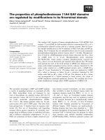
Time-course effect of LH on Akt phosphorylation in bovine theca cellsFigure 1
Time-course effect of LH on Akt phosphorylation in
bovine theca cells. Theca cells were plated onto serum-
coated dishes with serum-free medium for 36 h and then
stimulated with LH (100 ng/ml) for the stated times.
Cytosolic extracts (20 μg) were subjected to immunoblotting
with anti-phosphorylated-Akt antibody and anti-total-Akt
antibody. Representative images (Top) and densitometric
data of phospho-Akt contents (Bottom), expressed as ratio of
phospho-Akt to total-Akt, are shown. * denotes means that
are significantly different from 0 h (P < 0.01). ** denotes
means that are significantly different from 0 h (P < 0.001).
Phospho-Akt
Total-Akt
hours 046 8122448
0
2
4
6
Ratio of phospho-Akt/total-Akt
0
*
*
**
1/12 1/3 1 2 4 6 8 12 24 48
Time after LH (h)
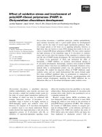
Effects of PI3K inhibitors on androstenedione production in bovine theca cellsFigure 2
Effects of PI3K inhibitors on androstenedione pro-
duction in bovine theca cells. Bovine theca cells were
stimulated with LH (100 ng/ml), wortmannin (W, 0.1 μM),
LY294002 (LY, 25 μM), or their combination for 24 h in
serum-coated dishes with serum-free medium. Control cells
(CTL) were cultured in the absence of added treatments.
Culture media were assayed for androstenedione by EIA.
Values are means ± SEM for four experiments. Different let-
ters denote a significant difference of means (P < 0.05).
0
100
200
300
a
a
b
c
a,c
a
Andr ostenedione (pg/ml)
LH
–––
+++
CTL W LY
Journal of Ovarian Research 2009, 2:17 />Page 5 of 8
(page number not for citation purposes)
PI3K converts phosphatidylinositol-4,5-biphosphate to
phosphatidylinositol-3,4,5-triphosphate, leading to acti-
vation of downstream kinases including Akt, which in
turn phosphorylates Bad, forkhead in rhabdomyosar-
coma (FKHR), Fas-associated death domain-like IL-1β-
converting enzyme-like inhibitory protein (FLIP), and X-
linked inhibitor of apoptosis protein (XIAP) [19]. The
PI3K/Akt activation drives cell through many biological
functions, including gene expression, cell cycle, survival,
glucidic metabolism, endocytosis and vesicular traffick-
ing, cell transformation, and oncogenesis [20]. In ovary,
FSH and several growth factors are known to activate the
PI3K/Akt pathway and prevent apoptosis in granulosa
cells and cultured follicles [13-15]. Although LH has been
reported to activate the cAMP/PKA pathway [4] and the
ERK/MAPK pathway [12] in theca cells, whether LH stim-
ulates the PI3K/Akt cascade in theca cells remains unclear.
Results of this study show for the first time that 1) LH
stimulates Akt phosphorylation in cultured bovine theca
cells, and that 2) activation of PI3K/Akt is involved in
CYP17A1 mRNA expression and androgen production in
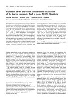
Effects of PI3K inhibitors on mRNA expression of CYP17A1 and StAR in bovine theca cellsFigure 3
Effects of PI3K inhibitors on mRNA expression of
CYP17A1 and StAR in bovine theca cells. Theca cells
were incubated with LH in the presence or absence of wort-
mannin (0.1 μM) or LY294002 (25 μM) in serum-coated
dishes with serum free medium for 12 h. Control cells (CTL)
were cultured in the absence of added treatments. Then RT-
PCR was conducted using CYP17A1, StAR, and 36B4 (inter-
nal control) primers using total RNA isolated from the cells.
The products were fractionated on 1% agarose gel and
stained with ethidium bromide. The mRNA levels of
CYP17A1 and StAR were expressed as ratio to 36B4 values.
Data are the mean ± SEM (n = 5). Different letters represent
statistically significant differences of means (P < 0.05).
0
5
10
CYP17A1 mRNA (fold increase)
LH
–––
+++
CTL W LY
a
b
a
a
a
a,b
0
1
2
LH
–––
+++
CTL W LY
a
a
a
a
a
a
StAR mRNA (fold increase)
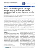
Effects of PKA inhibitor, MEK inhibitor and PI3K inhibitors on Akt phosphorylation in bovine theca cellsFigure 4
Effects of PKA inhibitor, MEK inhibitor and PI3K
inhibitors on Akt phosphorylation in bovine theca
cells. Subconfluent cultures were pretreated with PKA inhib-
itor (H89, 3 μM), MEK inhibitor (U0126, 10 μM), or PI3K
inhibitors (wortmannin, 0.1 μM; LY294002, 25 μM) for 30
min. Then they were stimulated with LH (100 ng/ml) for 24
h. Control cells (CTL) were cultured in the absence of added
treatments. Cell lysates (20 μg) were subjected to SDS-
PAGE and Western blot using anti-phosphorylated-Akt anti-
body (Phospho-Akt) or anti-total-Akt antibody (Total-Akt).
Representative images (Top) and densitometric data of phos-
pho-Akt contents (Bottom), expressed as a ratio of phospho-
Akt to total-Akt, are shown. Values show the mean ± SEM
for three experiments. Each experiment was reproduced at
least three times. Different letters denote significant differ-
ences of means (P < 0.05).
Phospho-Akt
Total-Akt
LH
–
+
CTL W LY
–
+
–
+
–
+
–
+
H89 U0126
0
1
2
3
a
a
a
a
a
a
b
b
a,c
a,c
LH
CTL W LY
–
+
H89 U0126
–
+
–
+
–
+
–
+
Ratio of phospho-Akt/total-Akt
Journal of Ovarian Research 2009, 2:17 />Page 6 of 8
(page number not for citation purposes)
theca cells. Reportedly, LH induced Akt phosphorylation
in whole rat ovary [21], and the PI3K inhibitor,
LY294002, suppressed androstenedione production by
theca cells in rat [22] and cattle [11]. It is possible that LH-
stimulated Akt phosphorylation in theca cells is responsi-
ble for these observations reported earlier.
Both wortmannin and LY294002 are inhibitors of the
lipid-modifying enzymes known as PI3K, and many
researchers perform a parallel study by using both inhibi-
tors to probe the roles of PI3K in biological processes.
However, depending on the concentration examined,
these inhibitors could be non-specific and cytotoxic and
could complicate the interpretation of their findings. In
our system, the 0.1 μM of wortmannin and 25 μM of
LY294002 are the minimal effective concentrations for
blocking the LH-induced androstenedione production in
theca cells. Nevertheless, only LY294002 suppressed LH-
induced CYP17A1 mRNA expression, whereas wortman-
nin did not affect this response. While the reason for this
apparent discrepancy is not clear, it is worth noting that
wortmannin has been reported to be unstable in aqueous
solution and less specific than LY294002 [23,24]. Higher
concentration (> 0.1 μM) of wortmannin induced theca
cell detachment and apoptosis in our serum-free culture
system.
Numerous reports have described that an activation of the
intracellular signaling (i.e. cAMP/PKA, ERK/MAPK, and
PI3K/Akt) is a rapid reaction in most cells. However, in
this study, it took 12 h for LH-induced increase in phos-
pho-Akt content in theca cells. It is of interest whether
PKA pathway, which is considered to be a major mediator
of the LH-generated signaling, and/or the MAPK pathway
influence LH-induced Akt phosphorylation or not. Exper-
iment 4 was performed to verify this point.
As described earlier, H89, a potent and selective inhibitor
of PKA, did not affect LH-mediated changes in phospho-
Akt, indicating that a pathway distinct from that of PKA is
involved in LH-induced Akt phosphorylation in theca
cells. Until recently, the effects of cAMP were generally
thought to be mediated by activation of cAMP-dependent
PKA, a major cAMP target, followed by phosphorylation
of many intracellular targets, such as cAMP responsive ele-
ment binding protein (CREB) [25], resulting in changes in
ovarian gene expression such as CYP17A1. Nevertheless,
some effects of cAMP appear to be inexplicable by activa-
tion of PKA. For instance, TSH and cAMP regulate prolif-
eration of thyroid cells by mechanisms independent of
PKA [26-29]. Actually, cAMP binds specific guanine nucle-
otide exchange factors: cAMP-GEFs (also called exchange
protein activated by cAMP, Epac) [30,31]. Gonzalez-
Robayna et al. reported that cAMP-GEFs are expressed in
rat granulosa cells and that the cAMP-GEFs play a role in
FSH-induced activation of the PI3K/Akt pathway in gran-
ulosa cells by PKA-independent manner [32]. Whether
theca cells also express these regulatory components and
whether the (PKA-independent) cAMP-GEFs mechanism
Effects of MEK inhibitor on CYP17A1 mRNA expression and androstenedione production in bovine theca cellsFigure 5
Effects of MEK inhibitor on CYP17A1 mRNA expres-
sion and androstenedione production in bovine theca
cells. Subconfluent cultures were pretreated with MEK
inhibitor (U0126, 10 μM) for 30 min. Then they were stimu-
lated with LH (100 ng/ml) for 12-24 h. Control cells (CTL)
were cultured in the absence of added treatments. RT-PCR
was conducted using CYP17A1 and 36B4 (internal control)
primers using total RNA isolated from the cells. The mRNA
level of CYP17A1 were expressed as ratio to 36B4 values
(Top). Culture media were also assayed for androstenedione
by EIA (Bottom). Data are the mean ± SEM (n = 4). Each
experiment was reproduced at least three times. Different let-
ters represent statistically significant differences of means (P <
0.05).
0
2
4
6
CYP17A1 mRNA (fold increase)
LH
––
++
CTL U0126
a
b
a
a
0
20
40
200
300
LH
––
++
CTL U0126
a
b
a
a
Andr ostenedione
(pg/ml)
Journal of Ovarian Research 2009, 2:17 />Page 7 of 8
(page number not for citation purposes)
is involved in LH-induced Akt phosphorylation in theca
cells remains to be elucidated.
In contrast to PKA inhibitor, the MEK inhibitor (U0126)
blocked LH-mediated Akt phosphorylation and androgen
production in theca cells. Reportedly, the MAPK inhibitor
also inhibits FSH-mediated Akt phosphorylation in rat
granulosa cells [32]. While the precise mechanism for the
activation of PI3K pathway by LH in theca cells is not
known, it is possible that the LH-induced phospho-Akt
up-regulation may involve MAPK-mediated down-regula-
tion of phosphatase and tensin homologue (PTEN; a
tumor suppressor which negatively regulates Akt phos-
phorylation). In this context, it has been shown that PI3K
is required for estradiol-stimulated hepatic cell growth
and that the MAPK pathway reduces the level of PTEN,
allowing estradiol-induced phosphorylation of Akt [20].
Whether this indeed is the case in the theca cells awaits
further investigation.
As a mechanism explaining why phospho-Akt content in
theca cells was increased only after 12 h of incubation
with LH, we are also interested in autocrine effects of insu-
lin-like growth factor-II (IGF-II) and nerve growth factor
(NGF) on theca cells. Reportedly, theca cells express IGF-
II and NGF in cattle, and each of IGF-II and NGF stimulate
androgen production [33,34]. Whether LH induces gene/
protein expression of these growth factors, and whether it
modulates the LH-mediated Akt phosphorylation in theca
cells, are subjects that are currently under investigation in
our laboratory.
Conclusion
Taking this evidence together, we conclude that LH stim-
ulates CYP17A1 mRNA expression and androgen produc-
tion in theca cells via activation of the PI3K/Akt pathway.
LH acts in theca cells by PKA-independent mechanisms as
well as PKA-dependent mechanisms, each of which con-
trols androgen production. Both the PI3K and the MAPK
pathways coordinately regulate androgen production in
bovine theca cells. Clarification of the LH-mediated intra-
cellular signaling events is essential for better understand-
ing of not only ovarian physiology, but also of the
pathophysiology of PCOS.
Abbreviations
LH: luteinizing hormone; cAMP: cyclic adenosine mono-
phosphate; PKA: protein kinase A; CYP17A1: 17α-hydrox-
ylase/C17-20 lyase cytochrome P450; StAR: steroidogenic
acute regulatory protein; ERK: extracellular-signal regu-
lated kinase; MAPK: mitogen activated protein kinase;
PI3K: phosphatidyl inositol 3-kinase; EIA: enzyme immu-
noassay; RT-PCR: reverse transcription polymerase chain
reaction; MEK: MAPK/ERK kinase; 36B4: acidic ribosomal
phosphoprotein; GEFs: guanine nucleotide exchange fac-
tors; PTEN: phosphatase and tensin homologue; PCOS:
polycystic ovary syndrome.
Competing interests
The authors declare that they have no competing interests.
Authors' contributions
SF, MO, KT, KH, and FK conceived of the study, partici-
pated in its design and coordination and drafted the man-
uscript. All authors read and approved the final version of
the manuscript.
Acknowledgements
This research was supported by a Grant-in-Aid for Scientific Research from
the Ministry of Education, Culture, Sports, Science, and Technology, Japan
(MEXT; Grant 19591892 and 21592093 to M.O.).
References
1. Erickson GF, Magoffin DA, Dyer CA, Hofeditz C: The ovarian
androgen producing cells: a review of structure/function
relationships. Endocr Rev 1985, 6:371-99.
2. Weil SJ, Vendola K, Zhou J, Adesanya OO, Wang J, Okafor J, Bondy
CA: Androgen receptor gene expression in the primate
ovary: cellular localization, regulation, and functional corre-
lations. J Clin Endocrinol Metab 1998, 83:2479-85.
3. Hu YC, Wang PH, Yeh S, Wang RS, Xie C, Xu Q, Zhou X, Chao HT,
Tsai MY, Chang C: Subfertility and defective folliculogenesis in
female mice lacking androgen receptor. Proc Natl Acad Sci USA
2004, 101:11209-14.
4. Magoffin DA: Ovarian theca cell. Int J Biochem Cell Biol 2005,
37:1344-9.
5. Erickson GF, Ryan KJ: Stimulation of testosterone production
in isolated rabbit thecal tissue by LH/FSH, dibutyryl cyclic
AMP, PGE2alpha, and PGE2. Endocrinology 1976, 99:452-8.
6. Richards JS, Hedin L, Caston L: Differentiation of rat ovarian the-
cal cells: evidence for functional luteinization. Endocrinology
1986, 118:1660-8.
7. Bogovich K, Richards JS: Androgen biosynthesis in developing
ovarian follicles: evidence that luteinizing hormone regu-
lates thecal 17 alpha-hydroxylase and C17-20-lyase activities.
Endocrinology 1982, 111:1201-8.
8. Magoffin DA, Kurtz KM, Erickson GF: Insulin-like growth factor-I
selectively stimulates cholesterol side-chain cleavage
expression in ovarian theca-interstitial cells. Mol Endocrinol
1990, 4:489-96.
9. Magoffin DA, Weitsman SR: Differentiation of ovarian theca-
interstitial cells in vitro: regulation of 17 alpha-hydroxylase
messenger ribonucleic acid expression by luteinizing hor-
mone and insulin-like growth factor-I. Endocrinology 1993,
132:1945-51.
10. Magoffin DA, Weitsman SR: Insulin-like growth factor-I stimu-
lates the expression of 3 beta-hydroxysteroid dehydroge-
nase messenger ribonucleic acid in ovarian theca-interstitial
cells.
Biol Reprod 1993, 48:1166-73.
11. Ryan KE, Glister C, Lonergan P, Martin F, Knight PG, Evans AC:
Functional significance of the signal transduction pathways
Akt and Erk in ovarian follicles: in vitro and in vivo studies in
cattle and sheep. J Ovarian Res 2008, 1:2.
12. Tajima K, Yoshii K, Fukuda S, Orisaka M, Miyamoto K, Amsterdam A,
Kotsuji F: Luteinizing hormone-induced extracellular-signal
regulated kinase activation differently modulates progester-
one and androstenedione production in bovine theca cells.
Endocrinology 2005, 146:2903-10.
13. Tilly JL, Pru JK, Rueda BR: Apoptosis in ovarian development,
function, and failure. The ovary 2nd edition. 2004:321-52.
14. Hu CL, Cowan RG, Harman RM, Quirk SM: Cell cycle progression
and activation of Akt kinase are required for insulin-like
growth factor I-mediated suppression of apoptosis in granu-
losa cells. Mol Endocrinol 2004, 18:326-38.
Publish with BioMed Central and every
scientist can read your work free of charge
"BioMed Central will be the most significant development for
disseminating the results of biomedical research in our lifetime."
Sir Paul Nurse, Cancer Research UK
Your research papers will be:
available free of charge to the entire biomedical community
peer reviewed and published immediately upon acceptance
cited in PubMed and archived on PubMed Central
yours — you keep the copyright
Submit your manuscript here:
/>BioMedcentral
Journal of Ovarian Research 2009, 2:17 />Page 8 of 8
(page number not for citation purposes)
15. Orisaka M, Orisaka S, Jiang JY, Craig J, Wang Y, Kotsuji F, Tsang BK:
Growth differentiation factor 9 is antiapoptotic during follic-
ular development from preantral to early antral stage. Mol
Endocrinol 2006, 20:2456-68.
16. Hidaka H, Watanabe M, Kobayashi R: Properties and use of H-
series compounds as protein kinase inhibitors. Methods Enzy-
mol 1991, 201:328-39.
17. Ireland JJ, Murphee RL, Coulson PB: Accuracy of predicting
stages of bovine estrous cycle by gross appearance of the
corpus luteum. J Dairy Sci 1980, 63:155-60.
18. Tajima K, Orisaka M, Hosokawa K, Amsterdam A, Kotsuji F: Effects
of ovarian theca cells on apoptosis and proliferation of gran-
ulosa cells: changes during bovine follicular maturation. Biol
Reprod 2002, 66:1635-9.
19. Franke TF, Kaplan DR, Cantley LC: PI3K: downstream AKTion
blocks apoptosis. Cell 1997, 88:435-7.
20. Marino M, Acconcia F, Trentalance A: Biphasic estradiol-induced
AKT phosphorylation is modulated by PTEN via MAP kinase
in HepG2 cells. Mol Biol Cell 2003, 14:2583-91.
21. Carvalho CR, Carvalheira JB, Lima MH, Zimmerman SF, Caperuto LC,
Amanso A, Gasparetti AL, Meneghetti V, Zimmerman LF, Velloso LA,
Saad MJ: Novel signal transduction pathway for luteinizing
hormone and its interaction with insulin: activation of Janus
kinase/signal transducer and activator of transcription and
phosphoinositol 3-kinase/Akt pathways. Endocrinology 2003,
144:638-47.
22. Zerbinatti CV, Mayer LP, Audet RG, Dyer CA: Apolipoprotein E is
a putative autocrine regulator of the rat ovarian theca cell
compartment. Biol Reprod 2001, 64:1080-9.
23. Vlahos CJ, Matter WF, Hui KY, Brown RF: A specific inhibitor of
phosphatidylinositol 3-kinase, 2-(4-morpholinyl)-8-phenyl-
4H-1-benzopyran-4-one (LY294002). J Biol Chem 1994,
269:5241-8.
24. Stein RC, Waterfield MD: PI3-kinase inhibition: a target for
drug development? Mol Med Today
2000, 6:347-57.
25. Gonzalez GA, Montminy MR: Cyclic AMP stimulates somatosta-
tin gene transcription by phosphorylation of CREB at serine
133. Cell 1989, 59:675-80.
26. Cass LA, Summers SA, Prendergast GV, Backer JM, Birnbaum MJ,
Meinkoth JL: Protein kinase A-dependent and -independent
signaling pathways contribute to cyclic AMP-stimulated pro-
liferation. Mol Cell Biol 1999, 19:5882-91.
27. Dremier S, Pohl V, Poteet-Smith C, Roger PP, Corbin J, Doskeland
SO, et al.: Activation of cyclic AMP-dependent kinase is
required but may not be sufficient to mimic cyclic AMP-
dependent DNA synthesis and thyroglobulin expression in
dog thyroid cells. Mol Cell Biol 1997, 17:6717-26.
28. Kupperman E, Wen W, Meinkoth JL: Inhibition of thyrotropin-
stimulated DNA synthesis by microinjection of inhibitors of
cellular Ras and cyclic AMP-dependent protein kinase. Mol
Cell Biol 1993, 13:4477-84.
29. Cass LA, Meinkoth JL: Differential effects of cyclic adenosine
3',5'-monophosphate on p70 ribosomal S6 kinase. Endocrinol-
ogy 1998, 139:1991-8.
30. de Rooij J, Zwartkruis FJ, Verheijen MH, Cool RH, Nijman SM, Wit-
tinghofer A, Bos JL: Epac is a Rap1 guanine-nucleotide-
exchange factor directly activated by cyclic AMP. Nature
1998, 396:474-7.
31. Kawasaki H, Springett GM, Mochizuki N, Toki S, Nakaya M, Matsuda
M, Housman DE, Graybiel AM: A family of cAMP-binding pro-
teins that directly activate Rap1. Science 1998, 282:2275-9.
32. Gonzalez-Robayna IJ, Falender AE, Ochsner S, Firestone GL, Richards
JS: Follicle-Stimulating hormone (FSH) stimulates phosphor-
ylation and activation of protein kinase B (PKB/Akt) and
serum and glucocorticoid-lnduced kinase (Sgk): evidence for
A kinase-independent signaling by FSH in granulosa cells.
Mol Endocrinol 2000, 14:1283-300.
33. Spicer LJ, Voge JL, Allen DT: Insulin-like growth factor-II stimu-
lates steroidogenesis in cultured bovine thecal cells. Mol Cell
Endocrinol 2004, 227:
1-7.
34. Dissen GA, Parrott JA, Skinner MK, Hill DF, Costa ME, Ojeda SR:
Direct effects of nerve growth factor on thecal cells from
antral ovarian follicles. Endocrinology 2000, 141:4736-50.