báo cáo hóa học:" Development of a syngeneic mouse model of epithelial ovarian cancer" ppt
Bạn đang xem bản rút gọn của tài liệu. Xem và tải ngay bản đầy đủ của tài liệu tại đây (5.09 MB, 17 trang )
RESEARC H Open Access
Development of a syngeneic mouse model of
epithelial ovarian cancer
Bridget A Quinn
1,5
, Fang Xiao
1
, Laura Bickel
1
, Lainie Martin
1
, Xiang Hua
2
, Andres Klein-Szanto
3,4
,
Denise C Connolly
1*
Abstract
Background: Most cases of ovarian cancer are epithelial in origin and diagnosed at advanced stage when the
cancer is widely disseminated in the peritoneal cavity. The objective of this study was to establish an
immunocompetent syngeneic mouse model of disseminated epithelial ovarian cancer (EOC) to facilitate laboratory-
based studies of ovarian tumor biology and preclinical therapeutic strategies.
Methods: Individual lines of TgMISIIR-TAg transgenic mice were phenotypically characterized and backcrossed to
inbred C57BL/6 mice. In addition to a previously described line of EOC-prone mice, two lines (TgMISIIR-TAg-Low)
were isolated that express the oncogenic transgene, but have little or no susceptibility to tumor development.
Independent murine ovarian carcinoma (MOVCAR) cell lines were established from the ascites of tumor-bearing
C57BL/6 TgMISIIR-TAg transgenic mice, characterized and tested for engraftment in the following recipient mice:
1) severe immunocompromised immunodeficient (SCID), 2) wild type C57BL/6, 3) oophorectomized tumor-prone
C57BL/6 TgMISIIR-TAg transgenic and 4) non-tumor prone C57BL/6 TgMISIIR-TAg-Low transgenic. Lastly, MOVCAR
cells transduced with a luciferase reporter were implanted in TgMISIIR-TAg-Low mice and in vivo tumor gro wth
monitored by non-invasive optical imaging.
Results: Engraftment of MOVCAR cells by i.p. injection resulted in the development of disseminated peritoneal
carcinomatosis in SCID, but not wild type C57BL/6 mice. Oophorectomized tumor-prone TgMISIIR-TAg mice
developed peritoneal carcinomas with high frequency, rendering them unsuitable as allograft recipients. Orthotopic
or pseudo-orthotopic implantation of MOVCAR cells in TgMISIIR-TAg-Low mice resulted in the development of
disseminated peritoneal tumors, frequently accompanied by the production of malignant ascites. Tumors arising in
the engrafted mice bore histopathological resemblance to human high-grade serous EOC and exhibited a similar
pattern of peritoneal disease spread.
Conclusions: A syngeneic mouse model of human EOC was created by pseudo-orthotopic and orthotopic
implantation of MOVCAR cells in a susceptible inbred transgenic host. This immunocompetent syngeneic mouse
model presents a flexible system that can be used to study the consequences of altered gene expression (e.g., by
ectopic expression or RNA interference strategies) in an established MOVCAR tumor cell line within the ovarian
tumor microenvironment and for the development and analysis of preclinical therapeutic agents including EOC
vaccines and immunotherapeutic agents.
Background
Ovarian cancer is the most common cause of death
from gynecologic malignancies and the fifth most com-
mon cause of cancer death in women in the United
States [1]. Ovarian adenocarcinomas account for 85-90%
of all cancers of the ovary. The initiating cell population
for EOC remains to be exactly defined, with different
evidence suggesting tumors originate from the ovarian
surface epithelium (OSE), inclusion cysts lined by OSE
[2-5] or alternatively, the fallopian tube epithelium [6]
or components of the secondary Müllerian system,
including the epithelial cells of the rete ovarii, paraovar-
ian/paratubal cysts, endosalpingiosis, endometriosis or
endomucinosis [7]. The lack of clarity regarding tumor
* Correspondence:
1
Women’s Cancer Program, Fox Chase Cancer Center, 333 Cottman Avenue,
Philadelphia, PA 19111-2497, USA
Full list of author information is available at the end of the article
Quinn et al. Journal of Ovarian Research 2010, 3:24
/>© 2010 Quinn et al; licensee BioMed Central Ltd. This is an Open Access article distributed under the terms of the Creative C ommons
Attribution License ( g/li censes/by/2.0), which p ermits unrestricted use, distribution, and reproduction in
any medium, provided the orig inal work is properly cited.
origin stems from the fact that unlike epithelial cancers
arising in other organs, a well-defined disease spectrum
consisting of benign, invasive and metastatic lesions has
not been identified for EOC. This is due at least in part
to that fact that the majority of cases are identified at
advanced stage when disease has spread beyond the
ovary. Another reason is the morphologic complexity of
common EOCs which consist of sever al distinct histolo-
gic s ubtypes; these include serous, endometrioid, muci-
nous and clear cell cancers.
Progress in ovarian cancer research has been slowed
by the lack of suitable animal models that exhibit fea-
tures of human disease. Genetically manipulable mam-
malian models of spontaneous ovarian cancer are rare,
particularly those representing ovarian adenocarcinomas.
Human and rodent models of spontaneous ex vivo
transformation of OSE have been described [8-10]. One
of these models, a syngeneic mouse model of EOC [10],
has been extensively used for precl inical studies of ther-
apeutic agents and studies of the tumor microenviron-
ment [11-18]. Early attempts to produce murine EOC
models using transgenic or other genetic engineering
approaches resulted in the development of granulosa
cell tumors [19-24]. More recently, a number of labora-
tories have developed genetically engineered mouse
(GEM) models of EOC by using ex vivo transformation
[25,26], transgenic [27,28] and conditional gene expres-
sion strategies [29-31]. To date, due to the lack of a sui-
table GEM model expressing Cre-r ecombinase, the
strategy most frequently employed for conditional gene
expression in the ovarian epithelium involves survival
surgery for intrabursal injection of recombinant Adeno-
virus-Cre [29-34].
Recently, our group developed a spontaneous trans-
genic mouse model of EOC by expressing the oncogenic
early region of SV40 under the transcriptional control of
the Müllerian inhibiting substance type II receptor gene
promoter [27,28]. Although SV40 TAg expression is not
directly associated with the d evelopment of human can-
cer, its expression results in functional inactivation of
the critical tum or suppressors p53 and Rb. Mutation of
TP53 is, by far, the most common genetic alteration
observed in EOC, particularly the serous subtype
[35,36]. Direct mutati on or loss of Rb or its downstream
signaling mediators are also common in EOCs [37-41].
Via binding and inhibition of PP2A, SV40 tag also
results in activation of PI3K/AKT and mitogen activated
protein kinase (MAPK) signaling [42], pathways fre-
quently activated in human EOC [43]. A stable trans-
genic line of TgMISIIR-TAg mice was established in
which female mice develop bilateral ovarian carcinoma
with 100% penetrance [28]. To date, this is the only
GEM model that develops spontaneous EOC with
pathological features of serous EOC that does not
require extensive surgical manipulation to induce the
phenotype. Like human EOC, female TgMISIIR-TAg
mice with significant tumor burden exhibit no apparent
symptoms of illness and disease dissemination is typi-
cally restricted to the peritoneum [27,28]. Murine ovar-
ian carcinoma (MOVCAR) cell lines isolated from the
ascites and primary tumors of these mice share many
molecular features with human tumors [27,28,44-48]
and are well suited to experimental analysis in vitro.
With these reagents, the expression levels of specific
genes can be experimentally manipulated and properties
of MOVCAR cell lines can be assessed in vitro.How-
ever, the lack of a syngeneic recipient for manipulated
MOVCAR cells has limited the analysis of the in viv o
effects of genetic alterations in the mo del to studies in
immunodeficient mice. The present study describes the
identification of non-tumor prone lines of TgMISIIR-
TAg transgenic mice that can be used as syngeneic reci-
pients for MOVCAR cell allografts. The availability of
this syngeneic model affords the opportunity to study
the in vivo effects of genetic alterations on tumor prop-
erties and on interactions between tumor cells and their
microenvironment in an immunocompetent host. More-
over, this immunocompetent mouse model of EOC is
suitable for studies of immune-based therapeutic strate-
gies and vaccine development.
Methods
Transgenic mice and backcrosses
All procedures involving mice were approved by the Fox
Chase Cancer Center (FCCC) Institutional Animal Care
and Use Committee (IACUC) and all mice were main-
tained under specific pathogen free conditions. Indivi-
dual transgenic TgMISI IR-TAg founder mice were
generated in the FCCC Transgenic Facility in a first
generation hybrid genetic background of C57BL/6 and
C3H (B6C3F1) and genotyped by P CR amplification as
previously described [27]. Transgenic founders were
crossed with wild type C57BL/6 mice (obtained from
the FCCC Laboratory Animal Facility) to establish
breeding lines. Relevant lines of EOC-prone and non-
tumor-prone TgMISIIR-TAg mice were maintained as
hemizygotes and backcrossed for a minimum of ten
generations to wild type C57BL/6 mice to generate
genetically pure lines of C57BL/6 TgMISIIR-TAg mice.
Cell lines and culture conditions
Pure C57BL/6 MOVCAR cell lines, including MOVCAR
12, 5009 [49], 5025, 5183, 5438, 5447 and 5612, were
established from bulk ascites isolated from individual
ovarian tumor-bearing C57BL/6 TgMISIIR-TAg mice as
previously described [27]. Tumorigenic spontaneously
transformed murine ovarian surface epithelial cell
(MOSEC) lines ID-8, IF-5 and IG-10 were a gift from
Quinn et al. Journal of Ovarian Research 2010, 3:24
/>Page 2 of 17
Dr. Katherine Roby, University of Kansas Medical Cen-
ter, and ID-8 cells stably ov erexpressing murine
VEGF164 were a gift from Dr. George Coukos, Univer-
sity of Pennsylvania. All MOVCAR and MOSEC cells
were maintained in DMEM suppl emented with 4% FBS,
1× Insulin/Transferrin/Selenium-A (ITS, supplied as
100× stock from Gibco/Invitrogen), penicillin/strepto-
mycin (100 units/mL and 100 μg/mL, respectively) and
2 mM l-glutamine and incubated at 37°C in 5% CO
2
.
Culture medium was changed once weekly and cells
were trypsinized and passaged at 4-5 day intervals when
they reached confluence. MOVCAR cells were prepared
for in vivo injection as described [49]. For in vivo ima-
ging, cells were transduced with a retroviral construct
encoding the firefly luciferase gene (pWZL-Luc, gener-
ously provided by Dr. Maureen Murphy, FCCC) us ing
standard methods.
Immunoblot and immunoprecipitation
To prepare lysates for immunoblot an alysis, cells were
washed with cold PBS, lysed with M-PER
mammalian
protein extraction reagent (Thermo Scientific, Rockford,
IL) supplemented with a cocktail of protease inhibitors
(Complete Mini, Roche, Indianapolis, IN) and protein
concentration was determined by BCA method (Thermo
Scientific, Rockford, IL). Equal amounts of protein sam-
ples were resolved by SDS-PAGE gel electrophoresis on
12% acrylamide gels a nd transferred to p olyvinylidene
difluoride membra ne (Immobilo n, Millipore Corp., Bed-
ford, MA). Membranes were blocked in 5% milk and
0.1% Tween-20 in 1× PBS for 1 h prior to i ncubation
with primary antibodies recognizing SV40 TAg (Pab
101) and mouse p53 (Pab 240) obtained from Santa
Cruz Biotechnology, Inc. at 1:1000 dilution. Horseradish
peroxidase-conjugated secondary antibodies were used
according to manufacturer’s protocols. Immunoreactivity
was visualized using the ECL system and was exposed to
BioMax MR film (Eastman Kodak Co.).
For immunoprecipitation, cells were grown in 100-mm
plates and lysed in 1 ml M-PER mammalian protein
extraction reagent. The whole cell lysates were incu-
bated with SV40 TAg antibody (Pab101) at a dilution of
1:100 at 4°C overnight with constant mixing. Protein A
beads (40 μl)wereaddedandmixedfor3hat4°C.
Immunoprecipitates were then washed 5 times with M-
PER mammalian protein extraction reagent and pellets
resuspended in Laemli buffer for protein electrophoresis
and immunoblot blot analysis performed as described
above with antibodies against TAg and p53.
Cell cycle analysis
Cells were prepared for cell cycle anal ysis using the
fluorescent nuclear stain propidium iodide and fluore s-
cent sorting was carried out using the Guava P ersonal
Cell Analysis machine exactly as described by the manu-
facturer (Guava Technologies).
RNA preparation, quantitative reverse transcription PCR
Total RNA was isolated from MOVCAR cells using the
RNA Easy Mini Kit (Qiagen). With the assistance of the
FCCC Genomics Facility, levels of Mdm2 mRNA
expression were evaluated by rea l-time quantitative
reverse transcription PCR (qRT-PCR) using Taqman
technology with probe sets for Mdm2 and Hprt1
obtained from Applied Biosystems, Carlsbad, CA.
Quantitation of secreted VEGF by ELISA
Cells (5 × 10
5
) were plated in triplicate in 6-well dishes
and grown in complete medium for 72 hours. The con-
ditioned culture medium was remo ved and the level of
secreted VEGF present in the medium was determined
by ELISA using the Mouse VEGF Quantikine Elisa Kit
(R&D systems, Minneapolis, MN). After removal of the
conditioned culture supernatant, cells were immediately
rinsed with PBS, trypsinized and the number of cells
present in each well was counted. Secreted VEGF levels
were normalized to the total number of cells present in
the sample to determine the amoun t of VEGF/10
4
cells.
Three independent assays were performed and the
amount of secreted VEGF/10
4
cellsexpressedasthe
mean value for each cell line tested.
Oophorectomy and MOVCAR cell allografts
Four to six week-old ovarian tumor-prone Tg MISIIR-
TAg mice were anesthetized by i.p. injection of 95 μl per
10 gram body weight of 10 mg/mL Ketamine hydro-
chloride and 1 mg/mL Xylazine hydrochloride in sterile
saline and subjected to oophorectomy using a standard
asceptic surgical procedure commonly used for trans-
genic embryo injection to expose the ovarian fat pad
and ovary (described in detail in [49]). Once exposed, a
small incision was made in the ovarian bursa that
enabled removal of the resident ovary an d/or fallopian
tube. The ovarian bursa was sealed with surgical glue
and the re productive tract returned through th e incision
in the body wall. The surgical incision was closed with
wound clips. The same surgical procedure was used for
orthoto pic (i.b.) injectio n of MOVCAR cells into recipi-
ent mice. Methods for i.b. and i.p. (pseudo-orthotopic)
injections of MOVCAR cells were previously described
in detail [49].
Preparation and analysis of tissues, histology and
immunohistochemistry
All mice were euthanized by CO
2
asphyxiation, necrop-
sied and examined for gross abnormalities. Pathologi-
cally altered organs, entire reproductive tracts and
represent ative specim ens of multiple organs and tissues,
Quinn et al. Journal of Ovarian Research 2010, 3:24
/>Page 3 of 17
including the brain, lung, liver, kidney, spleen, pancreas
and intestine were removed at necropsy, fixed in 10%
(v/v) neutral buffered formalin (NBF) overnight, trans-
ferred to 70% ethanol and paraffin-embedded. In mice
with evident tumor, specimens of the tumor tissue were
also excise d, snap frozen in liquid N
2
and stored at -80°
C. For histological analysis, 5 μm formalin fixed paraffin
embedded tissue sections were cut for either H&E stain-
ing or immunohistochemistry (IHC). Histo pathological
analysis was performed by a pathologist with expertise
in human and murine malignancies (AKS).
Sections of tumor tissue for IHC staining were cut on
SuperFrost Plus charged slides (Fisher). Unstained sec-
tions were deparaffinized, subjected to antigen retrieval
and stained with antibody against SV40 TAg (Pab 101,
1:100) as described [27].
Bioluminescent imaging (BLI)
For detection of in vivo growth of pWZL-Luc trans-
duced MOVCAR tumor cells, mice were anesthetized
with 2% isofluorane and given i.p. injections of 100 mg/
kg luciferin substrate (Caliper Life Sciences) ten minutes
prior to imaging using the IVIS Spectrum in vivo ima-
ging system (Caliper Life Sciences) as described [49].
Image analysis was performed and total flux emission
(photons/second) in the region of interest (ROI) was
determined using the Living Image Software for the
IVIS Spectrum.
Results
Allografted MOVCAR cells grow in immunodeficient mice,
but not in wild type C57BL/6 mice
Previous work showed that MOVCAR cell lines could
be readily established from the malignant ascites of indi-
vidual female TgMISIIR-TAg founder mice with ovarian
tumors and t hat these cells were tumorigenic in i mmu-
nocompromised SCID mice [27]. Subsequently, MOV-
CAR cell lines have been isolated from the EOC-bearing
female offspring o f a fully penetrant stable transgenic
line of EOC-prone mice, TgMISIIR-TAg-DR26, derived
from a male founder [28]. These cells exhibited the
capacity for pseudo-orthotopic tumor growth giving rise
to disseminated peritoneal tumors in SCID mice similar
to advanced EOC observed in humans (data not shown).
While the ability to g row tumor cells in vivo in immu-
nodeficient anim als is high ly valuab le for tumor biology
studies, it is somewhat limited in that important contri-
butions of immune cell signaling in the tumor microen-
vironment are lacking. Therefore, the ability to grow
tumor cells in a syngeneic host is highly desirable. In
establishing such a model, important considerations
include the genetic background of both the host from
which the tumor cells were isolated and the recipient
animal into which they will be all ografted. An additional
consideration is the potential immunogenicity of
the transgene protein product if it is not expressed
endogenously in wild type mice, as is the case for SV40
TAg. All TgMISI IR-TAg transgenic mice were initially
established in a B6C3F1 first generation hybrid genetic
background and maintained by crossing to wild type
C57BL/6 mice, thus resulting in a mixed genetic back-
ground of the offspring and any cell lines derived from
these mice. To address this issue, male TgMISIIR-TAg-
DR6 mice were maintained as hemizygotes with respect
to the TAg transgene and backcrossed to wild type
female C57BL/6 mice for a minimum of ten generations
to ensure >99% purity of the C57BL/6 genetic back-
ground. No changes in either tumor latency or TAg
expression patterns in ovarian tumors and reproductive
tracts of female mice were observed during the process
of backcrossing. Several new MOVCAR cell lines
(MOVCAR 12, 5009, 5025, 543 8, 5447 and 5612) were
established from t he ascites of ovarian tumor bearing
pure C57BL/6 TgMISIIR-TAg-DR 6 mice and tested for
tumorigenic potential following i.p. injection of 5 × 10
6
-1×10
7
cells in SCID mice. Tumors developed within
one to five months in SCID mice injected with all six
cell lines tested (Figure 1, Table 1 and data not shown).
In additio n to the presence of peritoneal tumor nodules
on the pancreas, omentum, mesentery, body wall and
diaphragm, several of the SCID mice exhibited grossly
enlarged ovaries at necropsy and histopathological
review of H&E and TAg stained sections confirmed the
presence of TAg positive tumor around and within the
ovarian cortex. Tumors exhibited histology similar to
high-grade serous ovarian carcinomas in women. Next,
we similarly tested the tumorigenicity of MOVCAR cells
in wild type C57BL/6 mice (n= 5 - 10 mice/group).
Although each cell line tested was tumorigenic in SCID
mice, none of the cell lines engrafted in immunocompe-
tent wild type C57BL/6 mice (Table 1 and data not
shown). The lack of tumor development in the immuno-
competent C57BL/6 mice suggests, as previous studies
have shown [50], that the expression of TAg proteins in
the MOVCAR cells was immunogenic in wild type
C57BL/6 recipients.
Analysis of SV40 TAg expression and function in MOVCAR
cell lines
One of the principle mechanisms of oncogenicity of
SV40 virus is the capacity of the large TAg protein to
bind to and functionally inactivate the p53 and Rb
tumor suppressor prot eins [51]. Expression of t he large
TAg protein was verified by Western blot in all o f the
MOVCAR cell lines, but absent in murine NIH3T3 cells
(data not shown) or MOSEC cell lines IF-5, ID-8, and
IG-10 (Figure 2A). In cells expressing wild type p53, p53
protein is k ept at low, typically undetectable levels by
Quinn et al. Journal of Ovarian Research 2010, 3:24
/>Page 4 of 17
ubiquitin mediated proteasomal degradation [52]. How-
ever, in cells expressing SV40 Large TAg, p 53 protein
remains bound to the TAg, resulting in p53 protein sta-
bilization [52]. Consistent with these previous observa-
tions and our own published results showing p53
protein stabilization in TgMISIIR-TAg ovarian tumors
[27], we obse rved consistently high l evels of p53 protein
in MOVCAR cell lines, but not in MOSEC cell lines IF-
5, ID-8, and IG-10 or NIH3T3 cells (Figure 2A and data
not shown). Physical interaction of the TAg and p53
proteins in MOVCAR cells was confirmed by coimmu-
noprecipitation assay. Whole cell lysates immunopreci-
pitated with a TAg-specific antibody (Pab 101) and
probed for p53 showed that p53 protein co-precipitated
with TAg in all of the MOVCAR cells tested (Figure 2A,
lower panels). To confirm that TAg binding results in
the functional abrogation of p53, MOVCAR cells were
treated with 200 nM etoposide for 0, 8 and 24 hours.
The capacity for a p53-mediated response to etoposide
treatment was assessed by evaluation of p53 protein
expression and st abilization, induction of the p53
responsive gene Mdm2 and induction of cell cycle
arrest. Treatment of the TAg negative ID-8 cells with
etoposide resulted in induction and stabilization o f p53
protein (Figure 2B), suggesting that p53 is functional in
these cells. However, in TAg expressing MOVCAR cells,
p53 protein was already stabilized and no further induc-
tion or stabilization of p53 was observed in the etopo-
side treated c ompared to untreated cells (F igure 2B). In
etoposide treated ID-8 cells, qRT-PCR analysis showed
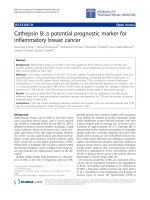
Figure 1 Cell lines derived from C57BL/6 mice are tumorigenic in SCID mice. Individual MOVCAR cell lines isolated from C57BL/6 mice
(MOVCAR 12, 5612, 5447 and 5438) were tested for tumorigenicity in SCID mice by i.p. injection of 0.5 - 1.0 × 10
7
cells. H&E stained sections
show the presence of tumor cells in the ovary (a-d) and peritoneum (i-l). The tumors derived from all cell lines were poorly differentiated
carcinomas. The neoplastic cells were usually arranged in solid sheets and occasionally formed glandular structures and/or irregular slit-like
spaces. On the peritoneal surface, these cells also formed papillary structures. Immunohistochemical detection of TAg (e-h and m-p) shows
positively staining tumor cells with no staining of surrounding normal tissue. All micrographs were taken at the same magnification and the
calibration bar shown in panel p corresponds to 100 μm.
Quinn et al. Journal of Ovarian Research 2010, 3:24
/>Page 5 of 17
greater than four-fold induction of Mdm2 expression
(Figure 2C) and cell cyle analysis showed growth arrest
indicat ed by accumulation of cells in G2/M (Figure 2D).
None of the similarly treated TAg positive MOVCAR
cell lines exhibited robust induction Mdm2 expression
or G2/M growth arrest. Taken together, these results
confirm the functional activity of TAg in MOVCAR cell
lines.
VEGF secretion in MOVCAR cell lines
In culture, MOVCAR cells exhibit differences in growth
rates and expression of signaling proteins associated
with EOC, including VEGF among others (Additional
file 1, Table S1 and data not shown). Differences in
tumor growth rates and ascites production among dif-
ferent MOVCAR cell lines were also apparent in vivo.
Peritoneal implantation of MOVCAR 5009 or 5025 cells
in SCID mice resulted in rapid tumor growth and the
production of voluminous ascites that necessitated
euthanasia within 4-6 weeks. In SCID mice injected
with MOVCAR 5183, 5438, 5447 and 5612 cells, the
time to development of tumors necessitating euthanasia
was between 12 and 20 weeks and mice generally exhib-
ited lower volumes of ascites at the time of necropsy
(Table 1 and data not shown). The cell l ines expressing
the highest levels of secreted VEGF in vitro (e.g., MOV-
CAR 5009 and 5025) resulted in more r apid tumor
growth and ascites production in vivo than cell lines
with lower VEGF levels. This observation is consistent
with a previous study showing that enforced expression
of VEGF in the spontaneously transformed MOSEC line
ID-8 led to more aggressive in vivo tumor growth and
Table 1 Growth of MOVCAR cells in C57BL/6 and SCID mice
Host MOVCAR cell line # cells injected i.p. Survival
(days post tumor cell injection)
Tumor location Ascites
(>1.0 mL)
C57BL/6 12 1 × 10
7
243 None
C57BL/6 12 1 × 10
7
256 None
C57BL/6 12 1 × 10
7
256 None
C57BL/6 12 1 × 10
7
256 None
C57BL/6 12 2 × 10
7
326 None
C57BL/6 12 2 × 10
7
326 None
C57BL/6 12 3 × 10
7
208 None
C57BL/6 12 3 × 10
7
208 None
C57BL/6 12 3 × 10
7
303 None
C57BL/6 12 3 × 10
7
303 None
SCID 12 1 × 10
7
93 Peritoneal cavity, invasion of ovarian cortex +
SCID 12 1 × 10
7
100 Peritoneal cavity, invasion of ovarian cortex +
SCID 12 1 × 10
7
103 Peritoneal cavity, invasion of ovarian cortex +
SCID 12 1 × 10
7
103 Peritoneal cavity, invasion of ovarian cortex +
SCID 12 1 × 10
7
105 Peritoneal cavity, invasion of ovarian cortex +
SCID 5009 1 × 10
7
25 Peritoneal cavity +
SCID 5009 1 × 10
7
25 Peritoneal cavity +
SCID 5009 1 × 10
7
34 Peritoneal cavity +
SCID 5009 1 × 10
7
34 Peritoneal cavity +
SCID 5009 1 × 10
7
34 Peritoneal cavity +
SCID 5009 1 × 10
7
34 Peritoneal cavity +
SCID 5183 1 × 10
7
109 Peritoneal cavity, invasion of ovarian cortex +
SCID 5183 1 × 10
7
116 Peritoneal cavity, invasion of ovarian cortex
SCID 5183 1 × 10
7
116 Peritoneal cavity, invasion of ovarian cortex
SCID 5348 1 × 10
7
141 Peritoneal cavity +
SCID 5348 1 × 10
7
141 Peritoneal cavity, invasion of ovarian cortex +
SCID 5348 5 × 10
6
141 Peritoneal cavity +
SCID 5447 1 × 10
7
95 Peritoneal cavity, invasion of ovarian cortex +
SCID 5447 1 × 10
7
95 Peritoneal cavity, invasion of ovarian cortex +
SCID 5447 5 × 10
6
95 Peritoneal cavity
SCID 5447 5 × 10
6
97 Peritoneal cavity, invasion of ovarian cortex +
SCID 5612 1 × 10
7
74 Peritoneal cavity, invasion of ovarian cortex
SCID 5612 5 × 10
6
97 Peritoneal cavity +
Quinn et al. Journal of Ovarian Research 2010, 3:24
/>Page 6 of 17
more ascites production than the parental cell line [18].
Like individual MOSEC lines [10], the results also sug-
gest that although MOVCAR cell lines are der ived from
ascites from an inbred strain of transgenic mice, indivi-
dual cell lines exhibit intrinsic differences.
Oophorectomized C57BL/6 TgMISIIR-TAg-DR6 mice
develop intrabursal and disseminated peritoneal
carcinomas
In order to identify a suitable syngeneic recipient strain
for in vivo growth, one potential strategy to overcome
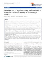
Figure 2 Analysis of SV40 TAg expression and function in MOVCAR cells. A) Whole cell lysates of spontaneously transformed MOSEC lines
(IF-5, ID-8 and IG-10) and MOVCAR cell lines (12-3, 5025, 5183, 5438, 5447, 5612 and 5009) were evaluated by immunoblot analysis to determine
relative levels of SV40 TAg, p53 and Actin (loading control) protein expression. Lysates were also immunoprecipitated with anti-TAg antibody
Pab 101 followed by immunoblot analysis of TAg and p53 protein present in the immunoprecipitates. B) Induction of p53 protein was evaluated
by immunoblot following treatment of ID-8 and MOVCAR 5025, 5447 and 5612 cells with 200 nM etoposide for 0, 8 and 24 hr. C) Levels of
Mdm2 gene expression in ID-8 and MOVCAR 5025, 5447 and 5612 cells following treatment with 200 nM etoposide for 0, 8 and 24 hr were
evaluated by qRT-PCR. D) Cell cycle analysis was performed on ID-8 and MOVCAR 5025, 5447 and 5612 cells following treatment with 200 nM
etoposide.
Quinn et al. Journal of Ovarian Research 2010, 3:24
/>Page 7 of 17
immunogenicity of the TAg transgene proteins is to
grow MOVCAR cells in t umor-prone C57BL/6 TgMI-
SIIR-TAg-DR6 transgenic mice. We hypothesized that
removaloftheovariesofyoungTgMISIIR-TAg-DR 26
transgenic mice might abrogate tumor development and
render these mice suitable for engraftment of MOVCAR
cells. In addition to TAg expression detected in tumor
cells, TAg staining was also commonly observed in the
uterine and fallopian tube epithelia of 28 day-old mice
(Figure 3 and [28]), although neither uterine nor fallo-
pian tube carcinomas we re observed at the time of
euthanasia. However, it is possible that o varian carci-
noma development was sufficiently rapid that it out-
paced carcinoma development in the endometrium or
oviduct. To determine whet her removal of the ovar ies
from TgMISIIR-TAg-DR26 transgenic mice was suffi-
cient to inhibit tumor formation, a series of oophorect-
omy experiments were performed (summarized in Table
2). Mice were oophorectomized between four and six
weeks of age, which is prior to the age of onset of
cyclivity at 48 days in C57BL/6 mice [53] and prior to
any obvious enlargement of the ovaries (Figure 3, [28]
and data not shown). Female C57BL/6 TgMISIIR-TAg-
DR26 transgenic mice were subjected to the following
surgical manipulations: 1) bilateral oophorectomy (n =
9), 2) bil ateral oophorectomy and salpingectomy (n = 5)
and 3) bilateral oophorectomy and salpingectomy with
remo val of the ovarian bursa (n = 8) and the results are
Figure 3 Early tumor formation in TgMISIIR-TAg-DR26 mice. T he presence and extent of tumor formation in a 28-day old fem ale TgMISIIR-
TAg-DR26 mouse was confirmed by histopathological evaluation. A) Low power magnification (40x) of an H&E stained section of the
reproductive tract showing the ovary and segments of the fallopian tube and uterus reveals an early-stage ovarian tumor indicated by the
arrow. B) Immunostaining of an adjacent section shows TAg positive tumor cells in the ovary (arrowheads). TAg positive staining cells were also
apparent in the epithelium of the fallopian tube and endometrium. The segment contained within the box is shown in (C) at higher
magnification (100X). D) High power magnification (400X) of the boxed area in (C) showing the TAg positive epithelial cells of the endometrial
glands. Calibration bar: A and B, 1 mm; C, 250 μm; D, 125 μm.
Quinn et al. Journal of Ovarian Research 2010, 3:24
/>Page 8 of 17
summarized in Table 2. Tumor formation was detected
in most mice and h istopathological evaluation revealed
the presence of carcinomas that were similar to those
that occurred spontaneously in TgMISIIR-TAg-DR26
transgenic mice. Tumors arising in the TgMISIIR-TAg-
DR26 mice in which the ovarian bursa was removed at
the time of bilateral oophorectomy and salpingectomy
were widely disseminated in the peritoneal cavity and
resembled primary peritoneal carcinomatosis. The origin
of the tumors remains uncertain as 28 day-old mice
already exhibited the presence of TAg positive tumor
cells in the ovary and TAg positive cells in the fallopian
tube and uterus (Figure 3 and [28]). Tumors arising in
ovariectomized mice may originate from residual tumor
cells shed from the ovaries prior to the time of surgery,
or alternatively, from TAg positive c ells present in the
fallopian tubes or the uterus. Although we cannot defi-
nitively distinguish between these possibilities, the his-
tology of tumors arising in oophorectomized mice
resembled the high-grade serous ovarian adenocarcino-
mas and disseminated peritoneal carcinomatosis that
occurs spontaneously in TgMISIIR-TAg-DR26 mice sug-
gesting that ovarian tumors arise from the ovaries and/
or fallopian tube and t hat tumor initiation occurs early
in these mice. There was no evidence of endometrial
carcinomas in any of the groups, suggesting that
although the SV40 TAg transgene protein is expressed
in the endometrium, this expression is not sufficient for
full oncogenic transformation of this tissue. Importantly,
as surgical removal of the ovaries and oviduct are not
suffi cient to prevent tumor development, these mice are
unsuitable as allograft hosts for implantation of MOV-
CAR cells.
Phenotypes of TgMISIIR-TAg transgenic mice
As an al ternative means to c ircumvent the problem of
TAg immunogenicity in recipient mice, we used a strat-
egy previously described by Mintz and Silvers [54] in
which inbred transgenic mice with low expression of the
tumor promoting transgene, and hence little or no sus-
ceptibility to tumor formation, were utilized as allograft
rec ipients. To identify such transgenic lines, we isolated
and phenotypically analyzed a total of 96 TAg positive
TgMISIIR-TAg transgenic founders. Among these, 36
were female, and as previously reported [27], 18/36
(50%) developed early onset, bilateral, moderately to
poorly differentiated ovarian carcinomas with wide-
spread peritoneal dissemination. Tumors exhibiting dif-
ferentiated morphology resembled high-grade serous
EOCs. Among the remaining female founders, 3/36 (8%)
developed non-ovarian tumors and 15/36 (42%) lacked
detectable TAg transgene protein expression, failed to
transmit the transgene or failed to breed. Therefore,
TAgpositivemalefounderswerebredandoffspring
were analyzed to identify stable transgenic lines of mice
that transmitted the TAg transgene. Similar to female
TgMISIIR-TAg founder mice, male founders were fre-
quently infertile, sub-fertile or did not transmit trans-
gene expression. Among the fertile transgenic lines
establishedfromTgMISIIR-TAg male founders, several
exhibited TAg transgene expression in the fallopian
tubes of female o ffspring without obvious pathology.
Further phenotypic characterization of female offspring
of two of these transgenic lines, TgMISIIR-TAg-DQ62
and TgMISIIR-TAg-EE73, showed that although t he
mice expressed the TAg transgene, they exhibited nor-
mal fertility and lifespan and failed to develop tumors.
The expression of TAg protein in these mice was
detected in a limited number of epithelial cells lining
the fallopian tube (Figure 4). These transgenic lines are
referred to as “TgMISIIR-TAg-Low” mice due to the
relatively limited expression of TAg protein. Previous
work [54] suggested that because t hese mice normally
expressed the TAg protein and exhibited little or no
susceptibility tumor formation, they could serve as suita-
ble hosts for implan tation of TAg expressing MOVCAR
cells.
MOVCAR cells grow as i.p. and orthotopic allografts in
C57BL/6 TgMISIIR-TAg-Low recipients
Prior to testing whether the TgMISIIR-TAg-Low trans-
genic lines, DQ62 and EE73, could se rve as recipients
for allografted MOVCAR cells, each was backcrossed to
wild type C57BL/6 mice for a minimum of ten genera-
tions to ensure genetic purity. No changes in TAg
expression patterns in the reproductive tracts of female
mice were observed during the backcrossing process. To
test whether MOVCAR cells could be grown as allo-
grafts in female C57BL/6 TgMISIIR -TAg-Low transgenic
mice, three TgMISIIR-TAg-DQ62 mice and three TgMI-
SIIR-TAg-EE73 mice were each injected i.p. with 2 × 10
7
MOVCAR 12 cells. Simi lar to SCID mice, C57BL/6
TgMISIIR-TAg-DQ62 and TgMISIIR-TAg-EE73 mice
injected i.p. with MOVCAR 12 cells developed tumors
that necessitated euthanasia within three months (Figure
5 and Table 3). At necropsy, disseminated peritoneal
tumors were detected and several mice exhibit ed
enlarged ovaries. In addition to the presence of
Table 2 Oophorectomized TgMISIIR-TAg transgenic mice
develop epithelial tumors
Surgical procedure Number of mice with
tumors
1) Remove TgMISIIR-TAg ovaries 9/9
2) Remove TgMISIIR-TAg ovaries and fallopian
tubes
4/5
3) Remove TgMISIIR-TAg ovaries, fallopian
tubes and bursa
6/8
Quinn et al. Journal of Ovarian Research 2010, 3:24
/>Page 9 of 17
disseminated perito neal adenocarcinoma infiltrating the
pancreas,omentum,mesentery,diaphragmandabdom-
inal wall, histopathological review of H&E and TAg
stained sections revealed the presence of tumor cells
growing within the intrabursal space surrounding the
ovaries and within the ovarian cortex of both the
C57BL/6 TgMISIIR-TAg-DQ62 and TgMISIIR-TAg-EE73
mice (Figure 5 and Table 3). Detect ion of the TAg posi-
tive tumor cells in the ovaries of both SCID (Figure 1)
and syngeneic C57BL/6 TgMISIIR-TAg-Low mice (Fig-
ure 5) suggests that MOVCAR cells exhibit a strong
propensity for organot ropic homing to ovary. To en sure
that the observed results were not cell line-specific, five
additional MOVCAR cell lines (MOVCAR 5009, 5025,
5183, 5447 and 5612) were tested for tumorigenic
potential following i.p. and i.b. injection. All five cell
linestestedgrewasallograftsinC57BL/6TgMISIIR-
TAg-Low mice (Figure 6, Table 3 and data not shown)
producing disseminated peritoneal adenocarcinoma fre-
quently accompanied by intrabursal and intra-ovarian
tumor growth. Like the allograft experiments performed
in SCID mice, individual cell lines exhibited differences
in t umor latency and dissemination pattern in C57BL/6
TgMISIIR-TAg-Low mice. However, the tumor latency
and dissemination pattern for any individual cell line are
similar in SCID and C57BL/6 TgMISIIR-TAg-Low allo-
graft recipients (compare data summarized in Tables 1
and 3). Taken together, these results show that both
lines of C57BL/6 TgMISIIR-TAg-Low mice can serve as
immunocompetent syngeneic recipients for the growth
of MOVCAR tumor cells isolated from individual tumor
bearing C57BL/6 TgMISIIR-TAg-DR6 mice.
Tumor growth in TgMISIIR-TAg-Low mice can be
monitored in vivo by bioluminescent imaging
Although orthotopic or pseudo-orthotopic implantation
of EOC c ells represents a more highly relevant tumor
microenvironment for tumor growth, there are inheren t
difficulties in detection and quantitation of tumor
growth and progression in deeply embedded tumors
growing within the intrabursal space or as dissemin ated
peritoneal disease. To facilitate detection and quantita-
tion of tumor growth in vivo, MOVCAR 5009 and 5447
cell s were transduced with a retroviral cons truct encod-
ing firefly luciferase. Stably transduced cells were
implanted into C57BL/6 TgMISIIR-TAg-Low mice by i.p.
or i.b. injection and tumor growth was then monitored
non-invasively by bioluminescent imaging (BLI).
Figure 4 TgMISIIR-TAg-EE73 and TgMISIIR-TAg-DQ62 mice exhibit restricted TAg expression. Histopathological evaluation of H&E (a-d) and
TAg (e-h) stained sections of female TgMISIIR-TAg-EE73 (a, b, e and f) and TgMISIIR-TAg-DQ62 (c,d, g and h) mice show TAg positive cells present
in the oviduct (e and g), but not in the OSE and bursal epithelium of the same mice (f and h). All micrographs were taken at the same
magnification and the calibration bar shown in panel h corresponds to 100 μm.
Quinn et al. Journal of Ovarian Research 2010, 3:24
/>Page 10 of 17
Age matched female C57BL/6 TgMISIIR-TAg-Low
(DQ62) mice injected i.p. with 5.0 × 10
6
MOVCAR-
5009-Luc cells rapidly developed disseminated peritonea l
carcinomatosis readily detectable by BLI (data not
shown). We predicted that detection of bioluminescent
signal emanating from orthotopically implanted tumors
would be technically more challenging in C57BL/6
TgMISIIR-TAg-Low mice due to the relatively low num-
ber of cells that can be implanted by this method (e.g.,
8.0 × 10
5
cells) and by the presence of black pigment in
the C57BL/6 mice compared to white mice. Therefore,
SCID mice injected i.b. with the same number o f either
MOVCAR-5009-Luc or MOVCAR-5447-Luc cells were
used as positive controls for BLI of orthotopically
implanted tumor cells in age-matched female C57BL/6
TgMISIIR-TAg-Low mice injected i.b. with 8.0 × 10
5
MOVCAR-5009-Luc (mouse numbers EE73 7245 and
EE73 7263) or MOVCAR-5 447-Luc (mouse numbers
EE73 7244 and EE73 7261) cells (Figure 7). Tumor
growth was monitored by BLI for up to 11 weeks and
showed that orthotopic implantation of cells resulted in
proscribed luminescent signals that were confined to the
site of intrabursal injection in both SCID and C57BL/6
TgMISIIR-TAg-Low mice (Figure 7 and Table 4).
Although the same numbers of cells were injected in all
mice, signal intensities appear stronger in SCID mice due
to the lack of pigment and therefore, are not directly
comparable to BLI signals in the C57BL/6 TgMISIIR-
TAg-Low mice. In vitro, MOVCAR-5009-Luc cells grow
much more rapidly than MOVCAR-5447-Luc cells. This
pattern was also observed in vivo, with rapid accel eration
of tumor growth detected three weeks post injection of
MOVCAR-5009-Luc cells, while signal intensiti es
detected in mice injected with MOVCAR-544 7-Luc cells
did not increase significantly until ten weeks post injec-
tion (Figure 7). Taken together, these data show that
growth of MOVCAR cells engineered to express firefly
luciferase can be monitored non-invasively by BLI and
that differences in in vivo growth rate s of individual
MOVCAR cell lines can be detected using this method.
Discussion
Utilization of animal models with an intact i mmune sys-
tem is critical for the evaluation of immune-based thera-
peutic strategies and vaccine development. An SV40 TAg
transgenic model of prostate cancer [55] has been used to
study the effects of combining blockade of cytotoxic T
lymphocyte antigen 4 (CTLA-4) and vaccination with
granulocyte macrophage colony stimulating factor (GM-
CSF;Gvax) and subsequent derivatives of this vaccine
strategy [56-60]. The C57BL/6 syngeneic mouse ovarian
cancer model developed by Roby et al, [10] has been used
for studies of the contribution of cells in the tumor micro-
environment, including epithelial-stromal cell interactions,
VEGF induced-effects on tumor vasculature and tumor
cell-secreted factors that stimulate cytokine production,
macrophage infiltration and vasculariza tion that favor
tumor growth and progression [14,15,18]. Simil ar studi es
would be difficult to impossible to conduct in immunode-
ficient mice. The availability of an additional syngeneic
mouse model of EOC will allow cross-comparison of
mouse models and validation of key findings.
The functional utility of animal models of human can-
cer depends largely on the extent to which the animal
model recapitulates the histology and biological behavior
of the disease in humans. Many transgenic tumor mod-
els have been developed using the immediate early
region of the SV40 virus co ntaining the potently onco-
genic large and small T antigen (TAg and tag) genes
Figure 5 MOVCAR 12 cells are tumorigenic in TgMISIIR-TAg-
DQ62 and TgMISIIR-TAg-EE73 mice. Micrographs of tumors arising
in TgMISIIR-TAg-DQ62 (a, c, e and h) and TgMISIIR-TAg-EE7 (b, d, f
and h) mice injected i.p. with 2 × 10
7
MOVCAR 12 cells. H&E (a, b, e
and f) and TAg (c, d, g and h) stained sections of tumors located
within the ovarian cortex (a-d) and peritoneal tumors invading the
omentum and pancreas (e-h). The tumors were similar to those
described in Figure 1. All micrographs were taken at the same
magnification and the calibration bar shown in panel h corresponds
to 100 μm.
Quinn et al. Journal of Ovarian Research 2010, 3:24
/>Page 11 of 17
[55,61-66]. The continued utility of SV40 TAg models in
studying cancer is underscored by seminal contributi ons
to our understanding of the “angiogenic switch” [67-71]
and tumor progression and invasion [72]. Importantly, a
recent study [73] identified an i ntegrated gene expres-
sion signature from three distinct TAg mouse models (i.
e., mammary, prostate and lung cancer models) that is
comparable to a signature associated with the aggressiv e
biological behavior and prognosis for several human
epithelial tumors, including breast cancers. Results from
this study showed that tumors arising in TAg-based
mouse models share common features of gene expres-
sion with human cancer and are relevant preclinical
models [73].
Female transgenic C57BL/6 Tg MISIIR-TAg-DR26 mice
develop spontaneous bilateral ovarian carcinoma with
100% penetrance [28]. Tumor p rogression in these mice
is characterized by widespread peritoneal dissemination
and the development of malignant ascites and tumor
morphology and histology of the tumors closely resem-
bles high-grade serous adenocarcinomas, the most com-
mon histologic subtype of EOC detected in women.
Tumor s and cell lines derived from primary tumors and
ascites of tumor bearing mice exhibit several character-
istics in common with human EOC cell lines and
tumors including AKT/mTOR activation, COX1 overex-
pression and VEGF overexpression and secretion
([28,44- 47] and the present study). In addition, a verapi-
mil-sensitive Hoescht dye-excluding ovarian carcinoma
side population (SP), a potential population of ovarian
cancer initiating cells, was identified in MOVCAR cell
lines [48]. Ovarian tumors arising in C57BL/6 TgMI-
SIIR-TAg-DR26 mice are sensitive to standard combina-
tion platinum and taxane chemotherapy and to mTOR
inhibition with Everolimus (RAD001) [28,45]. These
observations underscore the potential utility of these
transgenic mice for preclinical evaluation of therapeutic
agents. However, reflecting its relation to the biology of
human EOC, tumor format ion in this transgenic model
is also stochastic, resulting in variation in the latency of
tumor formation and time to metastasis. This necessi-
tates relatively large cohorts of mice and non-invasive
longitudinal in vivo imaging such as MRI to optimize
results of therapeutics studies.
To overcome the limitations encountered with sponta-
neous tumor development, we isolated individual trans-
genic lines of non-tumor prone C57BL/6 TgMIS IIR-TAg
transgenic mice that can serve as syngeneic immuno-
competent hosts for allografted TAg expressing MOV-
CAR cells isolated from tumor bearing C57BL/6
TgMISIIR-TAg-DR26 mice. Syngeneic mouse models of
EOC in whic h spontaneously transformed ID-8 MOSEC
grown as allografts in C57BL/6 recipients [10] or HM-1
tumor cells grown as allografts in B6C3F1 recipients
[74] have been previously described. These syngeneic
models have been used successfully for preclinical eva-
luation of therapeutic agents and studies of the role of
the tumor microenvironment on ovarian tumor growth
and progression [11-18,75]; however, these models each
rely on single mouse ovarian carcinoma cell lines in
which the underlying molecular mechanisms of malig-
nant transformation remain undefined.
Table 3 Growth of MOVCAR cells in TgMISIIR-TAg-Low mice
Host MOVCAR cell line # cells injected i.p. Survival
(days post tumor cell injection)
Tumor location Ascites
(>1.0 mL)
DQ62 12 2 × 10
7
96 Peritoneal cavity, invasion of ovarian cortex +
DQ62 12 2 × 10
7
90 Peritoneal cavity, invasion of ovarian cortex +
DQ62 12 2 × 10
7
96 Peritoneal cavity, invasion of ovarian cortex +
EE73 12 2 × 10
7
90 Peritoneal cavity, invasion of ovarian cortex
EE73 12 2 × 10
7
96 Peritoneal cavity, invasion of ovarian cortex
EE73 12 2 × 10
7
90 Peritoneal cavity, invasion of ovarian cortex
DQ62 5009 5 × 10
6
28 Peritoneal cavity +
DQ62 5009 5 × 10
6
28 Peritoneal cavity +
DQ62 5009 5 × 10
6
28 Peritoneal cavity +
DQ62 5009 5 × 10
6
28 Peritoneal cavity +
DQ62 5025 5 × 10
6
30 Peritoneal cavity, invasion of ovarian cortex +
DQ62 5025 1 × 10
7
30 Peritoneal cavity, invasion of ovarian cortex +
DQ62 5025 1 × 10
7
37 Peritoneal cavity, invasion of ovarian cortex +
DQ62 5025 1 × 10
7
37 Peritoneal cavity, invasion of ovarian cortex +
DQ62 5183 2 × 10
7
108 Peritoneal cavity
DQ62 5183 2 × 10
7
49 Peritoneal cavity +
DQ62 5612 1.5 × 10
7
71 Peritoneal cavity +
DQ62 5612 1.5 × 10
7
71 Peritoneal cavity +
Quinn et al. Journal of Ovarian Research 2010, 3:24
/>Page 12 of 17
The ease of establishment of TAg-transformed
MOVCARcelllinesinculturehasenabledtheisola-
tion of a large number of distinct cell lines, several of
which are described in the present study. Although
derived from an inbred strain of mice, the stochastic
manner in which tumors arise in C57BL/6 TgMISIIR-
TAg-DR26 mice results in intrinsic differences in
MOVCAR cell lines derived from individual tumor-
bearing mice. MOVCAR cell lines grown in culture
exhibit different growth rates and expression of pro-
teins associated with EOC, such as levels of secreted
VEGF. These cell lines also exhibit differences when
grown in vivo. For example, some cells lead to very
rapid growth and production of voluminous malignant
ascites, whereas other cells are slower growing a nd
produce less ascites. Interestingly, the cell lines that
result in the highest levels of ascites production in vivo
are the cell lines that exhibit the highest levels of
VEGF secretion in vitro. These observations suggest
that although the primary oncogenic stimulus driving
tumorigenesis in C57BL/6 TgMISIIR-TAg-DR26 trans-
genicmiceisthesameinallanimals,therearelikely
additional genetic, epigenetic and/or gene expression
alterations that contribute to ovarian tumor progres-
sion, and identification of these alterations may contri-
bute to our understanding of human EOC. Moreover,
once identified, the role of specific alterations in gene
function in ovarian tumorigenesis can be studied in
these cell lines as they are readily amenable to direct
manipulation using established strategies for ectopic
gene expression or RNA interference.
With regard to preclinical evaluation of nove l therapeutic
agents, our syngeneic mouse model of EOC provides sev-
eral advantages. First, tumors are grown in fully immuno-
competent mice enabling the evaluation of vaccine and
immune-based therapeutic strategies. Second, TgMISIIR-
Figure 6 Pseudo-orthotopic and orthotopic implantation of MOVCAR cell in TgMISIIR-TAg-Low mice.H&E(a,c,e,g,I,k,mando)and
TAg (b, d, f, h, j, l, n and p) stained sections of tumors arising in TgMISIIR-TAg-Low mice injected i.p (a-h) or i.b. (i-p) with MOVCAR 5009 (a, b, e,
f, i, j, m and n) and 5447 (c, d, g, h, k, l, o and p) cells. The tumors are similar to those shown in the previous figures, i.e., poorly differentiated
carcinomas with few areas of tubular differentiation and occasional papillary structures, e.g., panels c and g. All micrographs were taken at the
same magnification and the calibration bar shown in panel p corresponds to 100 μm.
Quinn et al. Journal of Ovarian Research 2010, 3:24
/>Page 13 of 17
TAg-Low transgenic mice have been fully backcrossed to a
pure C57BL/6 genetic background, exhibit normal fertility
and lifespan and do not develop tumors. Thus, large
cohorts of mice can be established for synchronous allo-
graft initiation without interference of tumor growth
initiated from the host. Third, the availability of multiple
distinct MOVCAR cells lin es for ev aluation avoids issues of
cell line-specific effects, and because MOVCAR cells are
easily manipula ted in culture, on-target effe cts of therapeu-
tics can be confirmed in parallel using RNAi based strate-
gies for direct target knockdown. Finally, the ability to
easily express reporter genes in MOVCAR cells facilitates
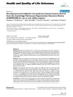
Figure 7 Orthotop ic tumor growth in TgMISIIR-TAg-Low mice monitored and quantifi ed in vivo by BLI.SCIDandTgMISIIR-TAg-EE7 mice
were given unilateral or bilateral intrabursal injections with 2 × 10
5
MOVCAR 5009 or 5447 cells and subjected to weekly bioluminescent
imaging to monitor tumor growth. A) Quantitative analysis of total photon counts from dorsal images of TgMISIIR-TAg-EE7 mice injected i.b. with
MOVCAR 5009 cells (mice 7245 and 7263) and MOVCAR 5447 cells (mice 7244 and 7261). B) Dorsal images of control SCID and TgMISIIR-TAg-EE7
mice injected i.b. with MOVCAR 5009 cells (mice 7245 and 7263) and MOVCAR 5447 cells (mice 7244 and 7261) showing proscribed luminescent
signals at the site of unilateral (mice 7245 and 7244) or bilateral (mice 7263 and 7261) i.b. injection.
Quinn et al. Journal of Ovarian Research 2010, 3:24
/>Page 14 of 17
strategies for non-invasive in vivo optical imaging such as
bioluminescent, fluorescent and near infrared fluorescent
imaging.
Conclusions
In conclusion, we have developed an immunocompetent
syngeneic mouse model of EOC consisting of C57BL/6
TgMISIIR-TAg-Low transgenic mice that can serve as
immunocompetent syngeneic allograft recipients for
MOVCAR cell line s. Based on distinct characte ristics of
these cell lines and their amenability to in vitro manipu-
lation of gene expression, this model represents a flex-
ible system to study ovarian tumor biology and to
evaluate the efficacy of novel therapeutic strategies.
Additional material
Additional file 1: Levels of secreted VEGF protein in MOVCAR cells.
The amount of secreted VEGF protein present in conditioned medium of
seven independent MOVCAR cell lines was determined by ELISA assay.
List of abbreviations
EOC: epithelial ovarian cancer; TAg: T antigen; SCID: severe combined
immunodeficient; MOVCAR: murine ovarian carcinoma; OSE: ovarian surface
epithelium; GEM: genetically engineered mouse; MISIIR: Müllerian inhibiting
substance type II receptor; IHC: immunohistochemistry; BLI: bioluminescent
imaging.
Acknowledgements
The authors gratefully acknowledge Drs. Thomas Hamilton, Maureen Murphy
and Julia Pimkina for suggestions and helpful discussions. The authors are
also grateful for the critical review of this manuscript by Drs. Alana O’Reilly,
Andrew Godwin and Erica Golemis. DCC is supported by NIH RO1
CA136596, the Ovarian Cancer Research Fund, the Fox Chase Cancer Center
Ovarian Cancer Spore (P50 CA083638), the Fox Chase Cancer Core Grant
(P30 CA006927) and the Keystone Program in Personalized Risk and
Prevention.
Author details
1
Women’s Cancer Program, Fox Chase Cancer Center, 333 Cottman Avenue,
Philadelphia, PA 19111-2497, USA.
2
Transgenic Facility Fox Chase Cancer
Center, 333 Cottman Avenue, Philadelphia, PA 19111-2497, USA.
3
Department of Pathology, Fox Chase Cancer Center, 333 Cottman Avenue,
Philadelphia, PA 19111-2497, USA.
4
Cancer Biology Program, Fox Chase
Cancer Center, 333 Cottman Avenue, Philadelphia, PA 19111-2497, USA.
5
Department of Human and Molecular Genetics Virginia Commonwealth
University School of Medicine 1220 E. Broad Street Room 7003 Richmond,
VA 23298, USA.
Authors’ contributions
BAQ, FX, LB and LM conducted the studies and participated in the data
analysis. XH performed oophorectomies, ovarian transplants and orthotopic
implantation of tumor cells and AKS conducted the histopathological
evaluation of tumor tissues. DCC conceived and designed experiments,
analyzed the data and wrote the manuscript. All authors have read and
approved the final manuscript.
Competing interests
The authors declare that they have no competing interests.
Received: 13 July 2010 Accepted: 19 October 2010
Published: 19 October 2010
References
1. Jemal A, Siegel R, Ward E, Hao Y, Xu J, Thun MJ: Cancer statistics, 2009. CA
Cancer J Clin 2009, 59:225-249.
2. Auersperg N, Maines-Bandiera SL, Dyck HG: Ovarian carcinogenesis and
the biology of ovarian surface epithelium. J Cell Physiol 1997, 173:261-265.
3. Pothuri B, Leitao MM, Levine DA, Viale A, Olshen AB, Arroyo C,
Bogomolniy F, Olvera N, Lin O, Soslow RA, et al: Genetic analysis of the
early natural history of epithelial ovarian carcinoma. PLoS One 5:e10358.
4. Salazar H, Godwin AK, Daly MB, Laub PB, Hogan WM, Rosenblum N,
Boente MP, Lynch HT, Hamilton TC: Microscopic benign and invasive
malignant neoplasms and a cancer-prone phenotype in prophylactic
oophorectomies. J Natl Cancer Inst 1996, 88:1810-1820.
5. Scully RE: Pathology of ovarian cancer precursors. J Cell Biochem Suppl
1995, 23:208-218.
6. Crum CP, Drapkin R, Miron A, Ince TA, Muto M, Kindelberger DW, Lee Y:
The distal fallopian tube: a new model for pelvic serous carcinogenesis.
Curr Opin Obstet Gynecol 2007, 19:3-9.
7. Dubeau L: The cell of origin of ovarian epithelial tumors and the surface
epithelium dogma: does the emperor have no clothes? Gynecol Oncol
1999, 72:437-442.
8. Auersperg N, Maines-Bandiera S, Booth JH, Lynch HT, Godwin AK,
Hamilton TC: Expression of two mucin antigens in cultured human
ovarian surface epithelium: influence of a family history of ovarian
cancer. Am J Obstet Gynecol 1995, 173:558-565.
9. Godwin AK, Testa JR, Handel LM, Liu Z, Vanderveer LA, Tracey PA,
Hamilton TC: Spontaneous transformation of rat ovarian surface
epithelial cells: association with cytogenetic changes and implications of
repeated ovulation in the etiology of ovarian cancer. J Natl Cancer Inst
1992, 84:592-601.
10. Roby KF, Taylor CC, Sweetwood JP, Cheng Y, Pace JL, Tawfik O, Persons DL,
Smith PG, Terranova PF: Development of a syngeneic mouse model for
events related to ovarian cancer. Carcinogenesis 2000, 21:585-591.
11. Chen T, Pengetnze Y, Taylor CC: Src inhibition enhances paclitaxel
cytotoxicity in ovarian cancer cells by caspase-9-independent activation
of caspase-3. Mol Cancer Ther 2005, 4:217-224.
12. George JA, Chen T, Taylor CC: SRC tyrosine kinase and multidrug
resistance protein-1 inhibitions act independently but cooperatively to
restore paclitaxel sensitivity to paclitaxel-resistant ovarian cancer cells.
Cancer Res 2005, 65:10381-10388.
13. Greenaway J, Henkin J, Lawler J, Moorehead R, Petrik J: ABT-510 induces
tumor cell apoptosis and inhibits ovarian tumor growth in an
orthotopic, syngeneic model of epithelial ovarian cancer. Mol Cancer Ther
2009, 8:64-74.
Table 4 Intrabursal growth of MOVCAR-Luciferase cells in TgMISIIR-TAg-Low mice
Host MOVCAR cell
line
# cells injected
i.b.
site of
injection
Survival
(days post tumor cell
injection)
Right ovary tumor
volume
(mm
3
)
Left ovary tumor
volume
(mm
3
)
Ascites
(>1.0
mL)
EE73 5009 8 × 10
5
left 50 n/a 167 +
EE73 5009 8 × 10
5
bilateral 50 151 176 +
EE73 5447 8 × 10
5
left 81 n/a 57
EE73 5447 8 × 10
5
bilateral 81 32 76
n/a: not applicable
Quinn et al. Journal of Ovarian Research 2010, 3:24
/>Page 15 of 17
14. Greenaway J, Moorehead R, Shaw P, Petrik J: Epithelial-stromal interaction
increases cell proliferation, survival and tumorigenicity in a mouse
model of human epithelial ovarian cancer. Gynecol Oncol 2008,
108:385-394.
15. Hagemann T, Robinson SC, Thompson RG, Charles K, Kulbe H, Balkwill FR:
Ovarian cancer cell-derived migration inhibitory factor enhances tumor
growth, progression, and angiogenesis. Mol Cancer Ther 2007,
6:1993-2002.
16. Pengetnze Y, Steed M, Roby KF, Terranova PF, Taylor CC: Src tyrosine
kinase promotes survival and resistance to chemotherapeutics in a
mouse ovarian cancer cell line. Biochem Biophys Res Commun 2003,
309:377-383.
17. Roby KF, Niu F, Rajewski RA, Decedue C, Subramaniam B, Terranova PF:
Syngeneic mouse model of epithelial ovarian cancer: effects of
nanoparticulate paclitaxel, Nanotax. Adv Exp Med Biol 2008, 622:169-181.
18. Zhang L, Yang N, Garcia JR, Mohamed A, Benencia F, Rubin SC, Allman D,
Coukos G: Generation of a syngeneic mouse model to study the effects
of vascular endothelial growth factor in ovarian carcinoma. Am J Pathol
2002, 161:2295-2309.
19. Risma KA, Clay CM, Nett TM, Wagner T, Yun J, Nilson JH: Targeted
overexpression of luteinizing hormone in transgenic mice leads to
infertility, polycystic ovaries, and ovarian tumors. Proc Natl Acad Sci USA
1995, 92:1322-1326.
20. Keri RA, Lozada KL, Abdul-Karim FW, Nadeau JH, Nilson JH: Luteinizing
hormone induction of ovarian tumors: oligogenic differences between
mouse strains dictates tumor disposition. Proc Natl Acad Sci USA 2000,
97:383-387.
21. Rahman NA, Huhtaniemi IT: Ovarian tumorigenesis in mice transgenic for
murine inhibin alpha subunit promoter-driven Simian Virus 40 T-
antigen: ontogeny, functional characteristics, and endocrine effects. Biol
Reprod 2001, 64:1122-1130.
22. Kumar TR, Palapattu G, Wang P, Woodruff TK, Boime I, Byrne MC,
Matzuk MM: Transgenic models to study gonadotropin function: the role
of follicle-stimulating hormone in gonadal growth and tumorigenesis.
Mol Endocrinol 1999, 13:851-865.
23. Kananen K, Markkula M, Rainio E, Su JG, Hsueh AJ, Huhtaniemi IT: Gonadal
tumorigenesis in transgenic mice bearing the mouse inhibin alpha-
subunit promoter/simian virus T-antigen fusion gene: characterization of
ovarian tumors and establishment of gonadotropin-responsive
granulosa cell lines. Mol Endocrinol 1995, 9:616-627.
24. Dutertre M, Gouedard L, Xavier F, Long WQ, di Clemente N, Picard JY,
Rey R: Ovarian granulosa cell tumors express a functional membrane
receptor for anti-Mullerian hormone in transgenic mice. Endocrinology
2001, 142:4040-4046.
25. Orsulic S, Li Y, Soslow RA, Vitale-Cross LA, Gutkind JS, Varmus HE: Induction
of ovarian cancer by defined multiple genetic changes in a mouse
model system. Cancer Cell 2002, 1:53-62.
26. Xing D, Orsulic S: A mouse model for the molecular characterization of
brca1-associated ovarian carcinoma. Cancer Res 2006, 66:8949-8953.
27. Connolly DC, Bao R, Nikitin AY, Stephens KC, Poole TW, Hua X, Harris SS,
Vanderhyden BC, Hamilton TC: Female mice chimeric for expression of
the SV40 TAg under control of the MISIIR promoter develop epithelial
ovarian cancer. Cancer Research
2003, 63:1389-1397.
28. Hensley H, Quinn BA, Wolf RL, Litwin SL, Mabuchi S, Williams SJ, Williams C,
Hamilton TC, Connolly DC: Magnetic Resonance Imaging for Detection
and Determination of Tumor Volume in a Genetically Engineered Mouse
Model of Ovarian Cancer. Cancer Biol Ther 2007, 6.
29. Dinulescu DM, Ince TA, Quade BJ, Shafer SA, Crowley D, Jacks T: Role of K-
ras and Pten in the development of mouse models of endometriosis
and endometrioid ovarian cancer. Nat Med 2005, 11:63-70.
30. Flesken-Nikitin A, Choi KC, Eng JP, Shmidt EN, Nikitin AY: Induction of
carcinogenesis by concurrent inactivation of p53 and Rb1 in the mouse
ovarian surface epithelium. Cancer Res 2003, 63:3459-3463.
31. Wu R, Hendrix-Lucas N, Kuick R, Zhai Y, Schwartz DR, Akyol A, Hanash S,
Misek DE, Katabuchi H, Williams BO, et al: Mouse model of human ovarian
endometrioid adenocarcinoma based on somatic defects in the Wnt/
beta-catenin and PI3K/Pten signaling pathways. Cancer Cell 2007,
11:321-333.
32. Clark-Knowles KV, Garson K, Jonkers J, Vanderhyden BC: Conditional
inactivation of Brca1 in the mouse ovarian surface epithelium results in
an increase in preneoplastic changes. Exp Cell Res 2007, 313:133-145.
33. Clark-Knowles KV, Senterman MK, Collins O, Vanderhyden BC: Conditional
inactivation of Brca1, p53 and Rb in mouse ovaries results in the
development of leiomyosarcomas. PLoS One 2009, 4:e8534.
34. Quinn BA, Brake T, Hua X, Baxter-Jones K, Litwin S, Ellenson LH,
Connolly DC: Induction of ovarian leiomyosarcomas in mice by
conditional inactivation of Brca1 and p53. PLoS One 2009, 4:e8404.
35. Aunoble B, Sanches R, Didier E, Bignon YJ: Major oncogenes and tumor
suppressor genes involved in epithelial ovarian cancer (review). Int J
Oncol 2000, 16:567-576.
36. Feeley KM, Wells M: Precursor lesions of ovarian epithelial malignancy.
Histopathology 2001, 38:87-95.
37. Dodson MK, Cliby WA, Xu HJ, DeLacey KA, Hu SX, Keeney GL, Li J,
Podratz KC, Jenkins RB, Benedict WF: Evidence of functional RB protein in
epithelial ovarian carcinomas despite loss of heterozygosity at the RB
locus. Cancer Res 1994, 54:610-613.
38. Farley J, Smith LM, Darcy KM, Sobel E, O’Connor D, Henderson B,
Morrison LE, Birrer MJ: Cyclin E expression is a significant predictor of
survival in advanced, suboptimally debulked ovarian epithelial cancers: a
Gynecologic Oncology Group study. Cancer Res 2003, 63:1235-1241.
39. Hashiguchi Y, Tsuda H, Yamamoto K, Inoue T, Ishiko O, Ogita S: Combined
analysis of p53 and RB pathways in epithelial ovarian cancer. Hum
Pathol 2001, 32:988-996.
40. Havrilesky LJ, Berchuck A: Molecular Alterations in Sporadic Ovarian
Cancer.
In Ovarian Cancer. Edited by: Rubin SC, Sutton GP. Philadelphia:
Lippincott Williams , Second 2001:23-42.
41. Tsuda H, Bandera CA, Birrer MJ, Hashiguchi Y, Berkowitz RS, Mok SC: Cyclin
E amplification and overexpression in clear cell adenocarcinoma of the
ovary. Oncology 2004, 67:291-299.
42. Arroyo JD, Hahn WC: Involvement of PP2A in viral and cellular
transformation. Oncogene 2005, 24:7746-7755.
43. Altomare DA, Wang HQ, Skele KL, De Rienzo A, Klein-Szanto AJ, Godwin AK,
Testa JR: AKT and mTOR phosphorylation is frequently detected in
ovarian cancer and can be targeted to disrupt ovarian tumor cell
growth. Oncogene 2004, 23:5853-5857.
44. Daikoku T, Tranguch S, Trofimova IN, Dinulescu DM, Jacks T, Nikitin AY,
Connolly DC, Dey SK: Cyclooxygenase-1 is overexpressed in multiple
genetically engineered mouse models of epithelial ovarian cancer.
Cancer Res 2006, 66:2527-2531.
45. Mabuchi S, Altomare DA, Connolly DC, Klein-Szanto A, Litwin S, Hoelzle MK,
Hensley HH, Hamilton TC, Testa JR: RAD001 (Everolimus) delays tumor
onset and progression in a transgenic mouse model of ovarian cancer.
Cancer Res 2007, 67:2408-2413.
46. Pieretti-Vanmarcke R, Donahoe PK, Pearsall LA, Dinulescu DM, Connolly DC,
Halpern EF, Seiden MV, MacLaughlin DT: Mullerian Inhibiting Substance
enhances subclinical doses of chemotherapeutic agents to inhibit
human and mouse ovarian cancer. Proc Natl Acad Sci USA 2006,
103:17426-17431.
47. Pieretti-Vanmarcke R, Donahoe PK, Szotek P, Manganaro T, Lorenzen MK,
Lorenzen J, Connolly DC, Halpern EF, MacLaughlin DT: Recombinant
human Mullerian inhibiting substance inhibits long-term growth of MIS
type II receptor-directed transgenic mouse ovarian cancers in vivo. Clin
Cancer Res 2006, 12:1593-1598.
48. Szotek PP, Pieretti-Vanmarcke R, Masiakos PT, Dinulescu DM, Connolly D,
Foster R, Dombkowski D, Preffer F, Maclaughlin DT, Donahoe PK: Ovarian
cancer side population defines cells with stem cell-like characteristics
and Mullerian Inhibiting Substance responsiveness. Proc Natl Acad Sci
USA 2006, 103:11154-11159.
49. Connolly DC, Hensley HH: Xenograft and Transgenic Mouse Models of
Epithelial Ovarian Cancer and Non-Invasive Imaging Modalities to
Monitor Ovarian Tumor Growth In Situ: Applications in Evaluating Novel
Therapeutic Agents. Current Protoc Pharmacol 2009, 45:14.12.11-14.12.36.
50. Wettstein PJ, Jewett L, Faas S, Brinster RL, Knowles BB: SV40 T-antigen is a
histocompatibility antigen of SV40-transgenic mice. Immunogenetics 1988,
27:436-441.
51. Ali SH, DeCaprio JA: Cellular transformation by SV40 large T antigen:
interaction with host proteins. Semin Cancer Biol 2001, 11:15-23.
52. Pipas JM, Levine AJ: Role of T antigen interactions with p53 in
tumorigenesis. Semin Cancer Biol 2001, 11:23-30.
53. Pinter O, Beda Z, Csaba Z, Gerendai I: Differences in the onset of puberty
in selected inbred mouse strains. Endocrine Abstracts 2007,
14:P617.
Quinn et al. Journal of Ovarian Research 2010, 3:24
/>Page 16 of 17
54. Mintz B, Silvers WK: Transgenic mouse model of malignant skin
melanoma. Proc Natl Acad Sci USA 1993, 90:8817-8821.
55. Greenberg NM, DeMayo F, Finegold MJ, Medina D, Tilley WD, Aspinall JO,
Cunha GR, Donjacour AA, Matusik RJ, Rosen JM: Prostate cancer in a
transgenic mouse. Proc Natl Acad Sci USA 1995, 92:3439-3443.
56. Curran MA, Allison JP: Tumor vaccines expressing flt3 ligand synergize
with ctla-4 blockade to reject preimplanted tumors. Cancer Res 2009,
69:7747-7755.
57. Fasso M, Waitz R, Hou Y, Rim T, Greenberg NM, Shastri N, Fong L, Allison JP:
SPAS-1 (stimulator of prostatic adenocarcinoma-specific T cells)/
SH3GLB2: A prostate tumor antigen identified by CTLA-4 blockade. Proc
Natl Acad Sci USA 2008, 105:3509-3514.
58. Hurwitz AA, Foster BA, Kwon ED, Truong T, Choi EM, Greenberg NM,
Burg MB, Allison JP: Combination immunotherapy of primary prostate
cancer in a transgenic mouse model using CTLA-4 blockade. Cancer Res
2000, 60:2444-2448.
59. Kwon ED, Foster BA, Hurwitz AA, Madias C, Allison JP, Greenberg NM,
Burg MB: Elimination of residual metastatic prostate cancer after surgery
and adjunctive cytotoxic T lymphocyte-associated antigen 4 (CTLA-4)
blockade immunotherapy. Proc Natl Acad Sci USA 1999, 96:15074-15079.
60. Kwon ED, Hurwitz AA, Foster BA, Madias C, Feldhaus AL, Greenberg NM,
Burg MB, Allison JP: Manipulation of T cell costimulatory and inhibitory
signals for immunotherapy of prostate cancer. Proc Natl Acad Sci USA
1997, 94:8099-8103.
61. Hanahan D: Heritable formation of pancreatic beta-cell tumours in
transgenic mice expressing recombinant insulin/simian virus 40
oncogenes. Nature 1985, 315:115-122.
62. Chen J, Tobin GJ, Pipas JM, Van Dyke T: T-antigen mutant activities in
vivo: roles of p53 and pRB binding in tumorigenesis of the choroid
plexus. Oncogene 1992, 7:1167-1175.
63. Maroulakou IG, Anver M, Garrett L, Green JE: Prostate and mammary
adenocarcinoma in transgenic mice carrying a rat C3(1) simian virus 40
large tumor antigen fusion gene. Proc Natl Acad Sci USA 1994,
91:11236-11240.
64. Kasper S, Sheppard PC, Yan Y, Pettigrew N, Borowsky AD, Prins GS,
Dodd JG, Duckworth ML, Matusik RJ: Development, progression, and
androgen-dependence of prostate tumors in probasin-large T antigen
transgenic mice: a model for prostate cancer. Lab Invest 1998, 78:i-xv.
65. Grippo PJ, Sandgren EP: Highly invasive transitional cell carcinoma of the
bladder in a simian virus 40 T-antigen transgenic mouse model. Am J
Pathol 2000, 157:805-813.
66. Chailley-Heu B, Rambaud C, Barlier-Mur AM, Galateau-Salle F, Perret C,
Capron F, Lacaze-Masmonteil T: A model of pulmonary adenocarcinoma
in transgenic mice expressing the simian virus 40 T antigen driven by
the rat Calbindin-D9K (CaBP9K) promoter. J Pathol 2001, 195:482-489.
67. Bergers G, Brekken R, McMahon G, Vu TH, Itoh T, Tamaki K, Tanzawa K,
Thorpe P, Itohara S, Werb Z, Hanahan D: Matrix metalloproteinase-9
triggers the angiogenic switch during carcinogenesis. Nat Cell Biol 2000,
2:737-744.
68. Bergers G, Javaherian K, Lo KM, Folkman J, Hanahan D: Effects of
angiogenesis inhibitors on multistage carcinogenesis in mice. Science
1999, 284:808-812.
69. Casanovas O, Hicklin DJ, Bergers G, Hanahan D: Drug resistance by
evasion of antiangiogenic targeting of VEGF signaling in late-stage
pancreatic islet tumors. Cancer Cell 2005, 8:299-309.
70. Inoue M, Hager JH, Ferrara N, Gerber HP, Hanahan D: VEGF-A has a critical,
nonredundant role in angiogenic switching and pancreatic beta cell
carcinogenesis. Cancer Cell 2002, 1:193-202.
71. Nozawa H, Chiu C, Hanahan D: Infiltrating neutrophils mediate the initial
angiogenic switch in a mouse model of multistage carcinogenesis. Proc
Natl Acad Sci USA 2006, 103:12493-12498.
72. Du YC, Lewis BC, Hanahan D, Varmus H: Assessing tumor progression
factors by somatic gene transfer into a mouse model: Bcl-xL promotes
islet tumor cell invasion. PLoS Biol 2007, 5:2255-2269.
73. Deeb KK, Michalowska AM, Yoon CY, Krummey SM, Hoenerhoff MJ,
Kavanaugh C, Li MC, Demayo FJ, Linnoila I, Deng CX, et al: Identification of
an integrated SV40 T/t-antigen cancer signature in aggressive human
breast, prostate, and lung carcinomas with poor prognosis. Cancer Res
2007, 67:8065-8080.
74. Toyoshima M, Tanaka Y, Matumoto M, Yamazaki M, Nagase S, Sugamura K,
Yaegashi N: Generation of a syngeneic mouse model to study the
intraperitoneal dissemination of ovarian cancer with in vivo luciferase
imaging. Luminescence 2009, 24:324-331.
75. Said N, Socha MJ, Olearczyk JJ, Elmarakby AA, Imig JD, Motamed K:
Normalization of the ovarian cancer microenvironment by SPARC. Mol
Cancer Res 2007, 5:1015-1030.
doi:10.1186/1757-2215-3-24
Cite this article as: Quinn et al .: Development of a syngeneic mouse
model of epithelial ovarian cancer. Journal of Ovarian Research 2010 3:24.
Submit your next manuscript to BioMed Central
and take full advantage of:
• Convenient online submission
• Thorough peer review
• No space constraints or color figure charges
• Immediate publication on acceptance
• Inclusion in PubMed, CAS, Scopus and Google Scholar
• Research which is freely available for redistribution
Submit your manuscript at
www.biomedcentral.com/submit
Quinn et al. Journal of Ovarian Research 2010, 3:24
/>Page 17 of 17