Báo cáo toán học: " Visual diagnosis: Rectal foreign body: A primer for emergency physicians" potx
Bạn đang xem bản rút gọn của tài liệu. Xem và tải ngay bản đầy đủ của tài liệu tại đây (1.54 MB, 3 trang )
CAS E REP O R T Open Access
Visual diagnosis: Rectal foreign body: A primer for
emergency physicians
Bobby Desai
Abstract
We present a case that is occasionally seen within emergency departments, namely a rectal foreign body. After
presentation of the case, a discussion concerning this entity is given, with practical information on necessity of an
accurate and thorough history and removal of the object for clinicians.
Case
A 39-year-old male presented to the Emergency Depart-
ment with vague complaints of abdominal pain and con-
stipation. He stated that the abdominal pain was dull
and crampy in nature and generalized in distribution.
Furthermore, he stated that he had not had a bowel
movement in 2 days, though he felt as if he had to have
one. He denied constitutional complaints of fevers,
chills, nausea, and vomiting, and denied urinary com-
plaints as well.
The patient’ s vital signs were: temperature 37.2°C,
pulse 87 beats per minute, respiratory rate of 20 per
minute, and blood pressure 130/84 mmHg. The patient
was awake, alert, and oriented to time, person, and
place. His head, neck, cardiovascular, respiratory, a nd
neurologic exams were all documented as within normal
limits. His abdominal exam revealed a flat abdomen, dif-
fusely tender with bowel sounds in all four quadrants.
The physician noted a palpable mass in the left lower
quadrant. Upon further examination, the mass felt “very
hard” and had an “oblong” shape according to the physi-
cian notes. The patient was subsequently re-questioned
about a family history of cancer, which the patient
denied. The physician subsequently ordered basic
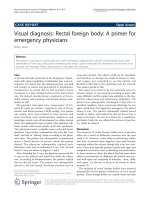
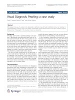
laboratory tests and an abdominal X-ray. The AP and
lateral X-rays are shown in Figures 1 and 2.
After obtaining the X-rays, the physician presented the
X-rays to the patient and asked him what the object
was. According to documentation, the patient r eplied
that he did not know. The patient was subsequently
placed in the left lat eral decu bitus position and an
anoscope inserted. The object could not be visualized,
and therefore no attempt was made to remove it. Gen-
eral surgery was consulted to see the patient and
decided to take him to the operating room for removal.
The patient agreed to this.
The object was noted to be the extension arm of a
vacuum cleaner. It was removed according to notes with
some difficulty and the patient was admitted to the hos-
pital for observation and intravenous antibiotics. The
patient was subsequently discharge d 2 days later in
excellent condition. Upon social work discharge, he was
again asked how that apparatus managed t o be placed
where it was. The pa tient vehemently denied sexual
assault or abuse, and insisted he did not know how it
came to be there. He met no criteria for a mandatory
psychiatric hold, but was offered the services of psychia-
try, which he refused.
Discussion
The majority of rectal foreign bodies seen in practice
today are a re sult of deliberate insertion into the anal
canal [1,2]. However, some sharp rectal foreign bodies
that have traversed entire digestive tract may become
impacted within the rectum, though this is far less com-
mon. These may typically present acutely with signs and
symptoms of trauma, such as bleeding and perforation.
In those instances where the object has had some delay
either in presentation or di agnosis, the patient may pre-
sent with signs and symptoms of infection - fever, chills,
and sepsis. An abscess is likely to be found in these
patients [3].
The majority of rectal foreign bodies have inserted
purposefully by the p atient themselves or by a sexual
part ner. These foreign bodies are usually blunt and take
Correspondence:
Department of Emergency Medicine, University of Florida, PO Box 100186,
Gainesville 32610, FL, USA
Desai International Journal of Emergency Medicine 2011, 4:73
/>© 2011 Desai; licensee Springer. This is an Open Access article distributed under the terms of the Creative Commons Attribution
License (http://creativecom mons.org/licenses/by/2.0), which permits unrestricted use, distribu tion, and reprod ucti on in any medium,
provided the original work is properly cited.
the shape of male genitalia [4,5]. Patients that repeatedly
place these types of objects within the anal canal over
time find that due to the increasing laxity of their rectal
tone, they can insert objects of a higher caliber. These
may be difficult for the patient to remove. Victims of
sexual assault may present with objec ts of varying cali-
ber,andthesemaynotnecessarilybeofablunttype.
These patients require careful examination to ensure
that perforation has not occurred. Drug mules have
been known to either s wallow latex balloons or directly
place them within the anus.
Due to the sensitive nature of the complaint, it is
occasionally difficult to elicit a history of the present ill-
ness. Furthermore, patients may be too embarrassed to
present early to an Emergency Department. Common
presenting complaints included abdominal pain, rectal
pain, rectal bleeding, and constipation. For those
patients who may have a bowel perforation, signs and
symptoms of this may be present, including severe
guarding, rebound tenderness, and fever, and these
patients may present septic [6].
The physician should make every effort to ensure the
patient feels comfortable during the history because of
the necessity of gaining accurate inf ormation about the
foreign body. Information should be sought as to the
objects approximate size, shape, material, length of time
since insertion, and any attempts at removal.
For examination, the patient should be placed in
either the lateral decubitus position or lithotomy posi-
tion. However, if the clinician suspects sharp foreign
objects, a plain abdominal X-ray should be obtained
first prior to examination to lessen the likelihood of
inadvertent injury to either the patient or clinician. If
sharp objects are noted, the exam should be deferred
and surgery consulted. Furthermore, if there are signs
and symptoms of bowel perforation, attempts at removal
should cease and surgery should be consulted emer-
gently as well. Plain abdominal X-rays are indicated in
almost all cases; CT s cans should be reserved for those
with potential sepsis or equivocal peritoneal signs [3].
Hollow objects may have a gas pattern in their general
shape. Radiolucent objects may require the use of rectal
contrast ; however, in these cases computed tomogra phy
may be the better modality to definitively diagnose t he
foreign body.
If this is not the case, the examination may proceed
with a general survey of the anal area, noting fissures,
excoriations, lacerations, and hemorrhoids. A digital rec-
tal exam followe d by anoscopy may reveal the object or
signs of trauma proximal to the anal verge.
Treatment entirely depends on the location of the for-
eign body. Low-lying foreign bodies by definition are
within the rectal ampulla, can often be palpated, and
potentially can be removed in the emergency
Figure 1 AP view.
Figure 2 Lateral view.
Desai International Journal of Emergency Medicine 2011, 4:73
/>Page 2 of 3
department [7]. High-lying objects usually require con-
sultation as these are located proximal to the recto-sig-
moid junction and require endoscopy for removal [7].
Due to the curvature of the sigm oid, the se objects typi-
cally are unable to pass beyond this area [8].
Prior to attempting removal, the physician should con-
sider medication with agents that relax not only the
patient, but the anal sphincter as well. If the patient can
tolerate the procedure without procedural sedation, they
may be able to assist the physician by performing the
Valsalva maneuver [9]. Regional anesthesia may be con-
sidered using a perianal block, though most emergency
physicians will have limited experience with this [10].
Removal may be accomplished by having the patient
perform the Valsalva maneuver while the physician
applies pressure to the suprapubic area while simulta-
neously trying to grasp the foreign body through the
anus. Either a finger or forceps may be used; forceps
would be ideal if the object has a graspable edge. To
improve visualization , an anoscope or other type of
retractor may be used. If the object cannot be removed
in this fashion, a Foley catheter may be used. A standard
Foley usually cannot be used because of its inherent
flexibility, and it often times may be difficult to pass the
Foley past the object because of the object’s diameter or
length. Therefore, it is recommended that a three-way
Foley catheter with a large balloon be used. A well-
lubricated catheter is advanced past the object and the
balloon inflated. If a three-way Foley is unavailable, a
small-diameter endotracheal tube can be used. In either
case, the catheter with the balloon inflated or the endo-
tracheal tube is then slowly withdrawn. However, care
must be taken not to force either tube past the object
because o f the risk of iatrogenic perforation. Two Foley
catheters can be utilized if the object tapers nea r its dis-
tal end.
Complications of removal include hemorrhage, per-
foration, and mucosal tears [3]. Most experts agree that
routine sigmoidoscopy shoul d be undertaken for all
patients subsequent to foreign body removal [6,7]. The
emergency physician should observe the patient for
signs of perforation after removal. The length of obser-
vation entirely depends on patient presentation and sub-
sequent clinical status post-extraction.
Consent
Written informed consent was obtained from the patient
for publication of this case report and any accompany-
ing images. A copy of the written consent is available
for review from the Editor-in-Chief of this journal.
Authors’ contributions
BD wrote, edited, and revised the entire report.
Competing interests
The author declares that they have no competing interests.
Received: 29 July 2011 Accepted: 7 December 2011
Published: 7 December 2011
References
1. Lyons MF, Tsuchida AM: Foreign bodies of the gastrointestinal tract. Med
Clin North Am 1993, 77(5):1101-1114.
2. Moreira CA, Wongpakdee S, Gennaro AR: A foreign body (chicken bone)
in the rectum causing extensive perirectal and scrota1 abscess: report of
a case. Dis Colon Rectum 1975, 18(5):407-409.
3. Anderson KL, Dean AF: Foreign bodies in the gastrointestinal tract and
anorectal emergencies. Emerg Med Clin N Am 2011, 29:369-400.
4. Fry RD: Anorectal trauma and foreign bodies. Surg Clin North Am 1994,
74(6):1491-1505.
5. Clarke DL, Buccimazza I, Anderson FA, et al: Colorectal foreign bodies.
Colorectal Dis 2005, 7(1):98-103.
6. Goldberg JE, Steele SR: Rectal foreign bodies. Surg Clin North Am 2010,
91(1):173-184.
7. Eftaiha M, Hambrick E, Abcarian H: Principles of management of colorectal
foreign bodies. Arch Surg 1977, 112(6):691-695.
8. Barone JE, Sohn N, Nealton TF: Perforations and foreign bodies of the
rectum:report of 28 cases. Ann Surg 1976, 184(5) :601-604.
9. Johnson SO, Hartranft TH: Nonsurgical removal of a rectal foreign body
using a vacuum extractor. Report of a case. Dis Colon Rectum 1996,
39(8):935-937.
10. Wigle RL: Emergency department management of retained rectal foreign
bodies. Am J Emerg Med 1988, 6(4):385-389.
doi:10.1186/1865-1380-4-73
Cite this article as: Desai: Visual diagnosis: Rectal foreign body: A
primer for emergency physicians. International Journal of Emergency
Medicine 2011 4:73.
Submit your manuscript to a
journal and benefi t from:
7 Convenient online submission
7 Rigorous peer review
7 Immediate publication on acceptance
7 Open access: articles freely available online
7 High visibility within the fi eld
7 Retaining the copyright to your article
Submit your next manuscript at 7 springeropen.com
Desai International Journal of Emergency Medicine 2011, 4:73
/>Page 3 of 3