Báo cáo hóa học: " The application of carbon nanotubes in target drug delivery systems for cancer therapies" docx
Bạn đang xem bản rút gọn của tài liệu. Xem và tải ngay bản đầy đủ của tài liệu tại đây (2.63 MB, 22 trang )
NANO REVIEW Open Access
The application of carbon nanotubes in target
drug delivery systems for cancer therapies
Wuxu Zhang
1
, Zhenzhong Zhang
2*
and Yingge Zhang
1*
Abstract
Among all cancer treatment options, chemotherapy continues to play a major role in killing free cancer cells and
removing undetectable tumor micro-focuses. Although chemotherapies are successful in some cases, systemic
toxicity may develop at the same time due to lack of selectivity of the drugs for cancer tissues and cells, which
often leads to the failure of chemotherapies. Obviously, the therapeutic effects will be revolutionarily improved if
human can deliver the anticancer drugs with high selectivity to cancer cells or cancer tissues. This selective delivery
of the drugs has been called target treatment. To realize target treatment, the first step of the strategies is to build
up effective target drug delivery systems. Generally speaking, such a system is often made up of the carriers and
drugs, of which the carriers play the roles of target delivery. An ideal carrier for target drug delivery systems should
have three pre-requisites for their functions: (1) they themselves have target effects; (2) they have sufficiently strong
adsorptive effects for anticancer drugs to ensure they can transport the drugs to the effect-relevant sites; and (3)
they can release the drugs from them in the effect-relevant sites, and only in this way can the treatment effects
develop. The transporting capabilities of carbon nanotubes combined with appropriate surface modifications and
their unique physicochemical properties show great promise to meet the three pre-requisites. Here, we review the
progress in the study on the application of carbon nanotubes as target carriers in drug delivery systems for cancer
therapies.
Keywords: carbon nanotubes, cancer therap ies, drug delivery systems, target chemotherapy
Introduction
Cancers are a kind of the diseases that are hardest to
cure, and most cancer patients definitely die even when
treated with highly developed modern medicinal techni-
que s. Surger y can remove cancer focus es but cannot do
the same for the micro-focuses and neither can extin-
guish the free cancer cells that are often the origin of
relapse. Chemotherapy with anticancer drugs is the
main auxiliary treatment but often fails because of their
toxic and side effects that are not endurable for the
patients. Over the past few decades, the field of cancer
biology has progressed at a phenomenal rate. However,
despite astounding advances in fundamental cancer biol-
ogy, these results have not been translated into
comparable advances in clinics. Inadequacies in the abil-
ity to adminis ter therapeutic agents with high selecti vity
and minimum side effects largely account for the discre-
pancies encompassing cancer therapies. Hence, consid-
erable efforts are being directed to such a drug delivery
system that selectively target the cancerous tissue with
minimal damage to normal tissue outside of the cancer
focuses. However, most of this research is still in the
preclinical stage and the successful clinical implementa-
tion is still in a remote dream. The development of such
a system is not dependent only on the identification of
special biomarkers f or neoplastic diseases but also on
the constructing of a system for the biomarker-targeted
delivery of therapeutic agents that avoid going into nor-
mal tissues, which remains a major challenge [1]. With
the development of nanotechnology, few nanomaterial-
based products have shown promise in the treatment of
cancers and many have been approved for clinical
research, such as nanoparticles, liposomes, and polymer-
drug conjugates. The requirements for new drug
* Correspondence: ;
1
Institute of Pharmacology and Toxicology and Key Laboratory of
Nanopharmacology and Nanotoxicology, Beijing Academy of Medical
Science, Zhengzhou, Henan, People’s Republic of China
2
Nanotechnology Research Center for Drugs, Zhengzhou University,
Zhengzhou, Henan, People’s Republic of China
Full list of author information is available at the end of the article
Zhang et al. Nanoscale Research Letters 2011, 6:555
/>© 2011 Zhang et al; licensee Springer. This is an Open Access article distributed under the terms of the Creative Commons Attribution
License ( which permits unrestricted use, distribution, and reproduction in any medium,
provided the original work is properly cited.
delivery systems to improve the pharmacological profiles
while decreasing the toxicological effects of the delivered
drugs have also envisaged carbon nanotubes (CNTs) as
one of the potential cargos for the cancer therapy.
CNTs belong to the fullerene family of carbon allotropes
with cylindrical shape. The unique physicochemical
properties [2,3] of CNTs with easy surface modification
have led to a surge in the number of publications in this
interesting field. Apart from their uses in the cellular
imaging with diagnostic effects in nanomedicine [4,5],
CNTs are promising drug carriers in the target drug
delivery systems for cancer therapies. Unlike other nao-
carriers, such as liposomes/micelles that emerged in the
1960s and nanoparticles/dendrimers that emerged in
1980s, it has emerged no more than 20 years for carbon
nanotubes to be envisaged as target drug carriers. In
this chapter, the works that have been carried out with
CNTs in the field of cancer therapy are briefly
introduced.
Physicochemical properties of CNTs
Carbon nanotubes are a huge cylindrical large molecules
consisting of a hexagonal arrangement of sp
2
hybridized
carbon atoms (C-C distance is about 1.4 Ǻ). The wall of
CNTs is single or mult iple layers o f graphene sheets, of
which those formed by rolling up of single sheet are
called single-walled c arbon nanotubes (SWCNTs) and
those formed by rolling up of more than one sheets are
called multi-walled CNTs (MWCNTs). Both SWCNTs
andMWCNTsarecappedatbothendsofthetubesin
a hemispherical arrangement of carbon networks called
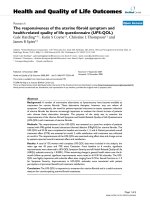
fuller enes warped up by the graphene sheet (Figure 1A).
The interlayer separation of the graphene layers of
MWCNTs is approximately 0.34 nm in average, each
one forming an individual tube, w ith all the tubes hav-
ing a larger outer diameter (2.5 to 100 nm) than
SWCNTs (0.6 to 2.4 nm). SWCNTs have a better
defined wall, whereas MWCNTs are more likely to have
structural defects, resulting in a less stable nanostruc-
ture, yet they continue to be featured in many publica-
tions due to ease of processing. As for their use as drug
carriers, there remain no conclusive advantages of
SWCNTs relative to MWCNTs; the defined sm aller dia-
meter may be suitable for their quality control while the
defects and less stable structure make their modification
easier. CNTs vary significantly in length and diameter
depending on the chosen synthetic procedure. SWCNTs
and MWCNTs have strong tendency to bundle together
in ropes as a consequence of attractive van der Waals
forces. Bundles contain many nanotubes and can be
considerably longer and wider than the original ones
from which they are formed. This phenomenon could
be of important toxicological significance [6,7]. CNTs
exist in different forms depending upon the orientation
of hexagons in the graphene sheet and possess a very
high aspect ratio and large surface areas. The available
surface area is dependent upon the length, diameter,
and degree of bundling. Theoretically, discrete SWCNTs
have special surface areas of approximately 1300 m
2
/g,
whereas MWCNTs generally have special surface areas
of a few hundred square meters per gram. The bundling
of SWCNTs dramatically decreases the special surface
area of most samples of SWCNT to approximately 300
m
2
/g or less, although this is still a very high value [8,9].
The markedly CNTs have various lengths from several
hundreds of nanometers to several micrometers and can
be shortened chemically or physically for their suitability
for drug carriers (Figure 1B) [10] by making their two
ends open with useful wall defects for intratube drug
loading and chemical functionalization (Figure 1B).
Functionalization of CNTs
As drug carriers, the solubility of CNTs in aqueous sol-
vent is a prerequisite for gastrointestinal absorption,
blood transportat ion, secreti on, and biocompatibility and
so on; hence, CNT com posites invo lved in therape utic
delivery system must meet this basic requirement. Simi-
larly, it is important that such CNT dispersions should
be uniform and stable in a sufficient degree, so as to
obtain accurate concentration data. In this regard, t he
solubilization of pristine CNTs in aqueous solvents is
one of the key obstacles in the way for them to be devel-
oped as practical drug carriers owing to the rather hydro-
phobic character of the graphene side walls, coupled with
the strong π-π interactio ns between the individual tubes.
These properties cause aggregation of CNTs into bun-
dles. For the successful dispers ion of CNTs, the medium
should be capable of both wetting the hydrophobic tube
surfaces and modifying the tube surfaces to decrease
tube’s bundle formation. To obtain desirable dispersion,
Foldvari et al. have proposed four basic approaches [11]:
(1) surfactant-assisted disper sion, (2) solvent dispers ion,
(3) functionalization of side walls, and (4) biomolecular
dispersion. Among the above described approaches, func-
tionalization has been the m ost effective approach. In
addition, functionalization has been shown capable of
decreasing cytotoxicity, improving biocompatibility, and
giving opportunity to appendage molecules of drugs, pro-
teins, or genes for the construction of delivery systems
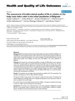
[12]. Up to now, there have been a lot of literatures on
the functionalization of CNTs with various molecules
(Figure 2A). The functionalization can be divided into
two main subcategories: non-covalent functionalization
and covalent functionalization (Figure 2B).
Non-covalent functionalization
Many small, as well as large, polymeric anticancer
agents can be adsorbed non-covalently onto the surface
Zhang et al. Nanoscale Research Letters 2011, 6:555
/>Page 2 of 22
Figure 1 The formation of SWCNT and its physical and chemical treatment for use as drug carriers.(A) The schematic illustration of the
structure formation of SWCNTs with the two ends closed. (B) The schematic illustration of the strategy for the preparation of the CNT-based
drug delivery systems.
Figure 2 The modification of CNTs. Schematic illustration of modification of CNTs with various molecules. 1, Dhar et al. [70]; 2, Jia et al. [13]; 3,
Georgakilas et al. 2002 [16]; 4, Peng et al. 1998; 5, Liu et al. [91]; 6, Gu et al. 2008; 7, Son et al. 2008; 8, Klingeler et al. 2009.
Zhang et al. Nanoscale Research Letters 2011, 6:555
/>Page 3 of 22
of pristine CNTs. Forces that govern such adsorption
are the hydrophobic and π-π stack ing interactions
between the chains of the adsorbed molecules and the
surface of CNTs. Since many anticancer drugs are
hydrophobicinnatureorhavehydrophobicmoieties,
the hydrophobic forces are the main driving forces for
the loading of such drugs into or onto CNTs. The pre-
sence of charge on the nanotube surface due to chemi-
cal treatment can enable the adsorption of the charged
molecules through ionic interactions [13,14]. Aromatic
molecules or the molecules with aromatic groups can be
embarked on the debunching and solubilization of
CNTs using nucleic acids and amphiphilic peptides
based on the π-π stacking interactions between the
CNT surface and aromatic bases/amino acids in the
structural backbone of these functional biomolecules.
Noncovalent functionalization of CNT is particularly
attractive because it offers the possibility of attaching
chemical handles without affecting the e lectronic net-
work of the tubes.
Oxide surfaces modified with pyrene through π-π
stacking interactions have been employed for the pat-
terned assembly of single-walled carbon nanomate rials
[15]. The carbo n graphitic structure can be recognized
by pyrene functional groups with distinct molecular
properties. The interactions between bifunctional mole-
cules (with amino and silane groups) and the hydroxyl
groups on an oxide substrate can generate an amine-
covered surface. This was followed by a coupling step
where molecules with pyrene groups were allowed to
react with amines. The patterned assembly of a single
layer of SWCNT could be achieved through π-π stack-
ing with the area covered with pyrenyl groups. Alkyl-
modified iron oxide nanoparticles have been attached
onto CNT by using pyrenecarboxylic acid derivative as
chemical cross-linker [16]. The resulting material had
an increased solubility in organic media due to the che-
mical functions of the inorganic nanoparticles.
Surfactant s were initially involved as di spersing agents
[17] in the purification protocols of raw carbon material.
Then, surfactants were used to stabilize dispersions of
CNT for spectroscopic characterization [18], optical lim-
iting property studies, and compatibility enhancement of
composite materials.
Functionalized nanotube surface can be achieved sim-
ply by exposing CNTs to vapors containing functionali-
zation sp ecies t hat non-covalently bonds to the
nanotube surface while providing chemically functional
groups at the nanotube surface [19]. A stable functiona-
lized nanotube surface can be obtained by exposing it to
vapor stabilization species that reacts with the functio-
nalization layer to form a stabilization layer against des-
orption from the nanotube surface while depositing
chemically functional groups at the nanotube surface.
Thestabilizednanotubesurface can be exposed further
to at least another material layer precursor species that
can deposit as a new layer of materials.
A patent [20] is pertinent to dispersions of CNTs in a
host polymer or copolymer with delocalized electron
orbitals, so that a dispersion interaction occurs between
the host polymer or copol ymer and the CNTs dispersed
in that matrix. Such a dispersion interact ion has advan-
tageous results if the monomers of the host polymer/
copolymer include an aromatic moiety, e.g., phenyl
rings or their derivatives. It is claimed that dispersion
force can be further enh anced if the aromatic moiety is
naphthalenyl and anthracenyl. A new non-wrapping
approach to functionalizing CNTs has been introduced
by Chen et al. [21]. By this appro ach, the fun cti onaliza-
tion can be realized in organic and inorganic solvents.
With a functionally conjugated polymer that includes
functional groups, CNT surfaces can be functionalized
in a non-wrapping or non-packaging fashion. Through
further functionalization, various other desirable func-
tional groups can be added to this conjugated polymer.
This approach provided the possibility of further tailor-
ing, even after functionalization. A process registered by
Stoddart et al. [22] involves CNTs treated with poly{(5-
alkoxy-m-phenylenev inylene)-co-[(2,5-dioctyloxy-p-phe-
nylene) vinyl-ene]} (PAmPV) polymers and their deriva-
tives for noncovalent functionalizat ion of the nanotubes
which increases solubility and enhances other properties
of interest. Pseudorotaxanes are grafted along the walls
of the nanotubes in a periodic fashion by wrapping of
SWCNTs with these functionalized PAmPV polymers.
Many biomolecules can interact with CNTs without
producing of covalent conjugates. Proteins are an
important class of substrates that possess high affinity
with the graphitic network. Nanotube walls can adsorb
proteins strongly on their external sides, and the pro-
ducts can be visualized clearlybymicroscopytechni-
ques. Metallothionein proteins were adsorbed onto the
surface of multi-walled CNT, as evidenced by high-reso-
lution transmission electron microscopy (TEM) [23].
DNA strands have been reported by several groups to
interact strongly with CNT to form stable hybrids effec-
tively dispersed in aqueous solutions [24,25]. Kim et al.
[26] reported the solubilization of nanotubes with amy-
lose by using dimethyl sulfoxide/water mixtures. The
polysaccharide adopts an interrupted loose helix struc-
ture in these media. The studies of the same gro up on
the dispersion capability of pullulan and carboxymethyl
amylase demonstrated that these substances could also
solubilize CNTs but to a lesser extent than amylose.
There are also some literatures that reported several
other examples of helical wrapping of linear or
branched polysaccharides around the surface of CNT
[27].
Zhang et al. Nanoscale Research Letters 2011, 6:555
/>Page 4 of 22
Covalent functionalization
Covalent functionalization gives the more secure con-
junction of functional molecules. CNTs can be oxidased,
giving CNTs hydrophilic groups as OH, COOH, and so
on. Strong acid solution treatment can create defects in
the side walls of CNTs, and the carboxylic acid groups
are generated at the defect point, predominantly on the
open ends. Excessive surface defects possibly change the
electronic properties and cut longer CNTs in to short
ones. as drug carriers may need CNTs with different
electronic properties and different lengths. In the pre-
paration of some drug delivery systems, CNTs are delib-
erately cut into short pieces. The functional groups on
the oxidized CNTs can further react with SOCl and car-
bodiimide to yield functional materials with great pro-
pensity for reacting with other compounds [28,29].
Covalent functionali zation of SWCNTs using addition
chemistryisbelievedtobeverypromisingforCNT
modification and derivatization. However, it is difficult
to achieve complete control over the chemo- and region
selectivity of such additions and require very special spe-
cies such as arynes, carbenes, or halogens, and the reac-
tions often occur only in extreme conditions for the
formation of covalent bonds. Furthermore, the charac-
terizatiuon of functionalized SWCNT s and the determi-
nation of t he precise location and mode of addition are
also very difficult [30]. The covalent chemistry of CNTs
is not particula rly rich with respect to variet y chemical
reactions to date. As regard to functionalization beha-
vior of SWCNTs and MWCNTs, it has been reported
that functionalization percentage of MWCNTs is lower
than that of SWCNTs with the similar process [31],
which is assumingly attributed to the larger outer dia-
meter and sheathed nature of MWCNTs that render
many of their sidewalls inaccessible; nonetheless, a com-
parative study on functionalizing single-walled and
multi-walled carbon nanotubes is scarce hitherto in
open literature.
In comparison with non-covalent functionalization,
there are ne w substances to develop and t herefore most
patents regarding functionalization of CNTs registered
to date are based on covalent chemistry. Though cova-
lent procedures are not highly diverse yet, the end pro-
ducts vary exceedingly in terms of characteristics
depending upon the incorporated species.
Methotrexate functionalization can be realized
through 1,3-cycloaddition reaction [32]. Azomethine
ylides consisting of a carbanion adjacent to an immo-
nium ion are organic 1,3-dipoles, which give pyrrolidine
intermediates upon cycloaddition to dipolarophiles.
Through decarboxylation of immonium salts obtained
from the condensation of a-amino acids with aldehydes
or ketones, azomethine ylides can be easily produced.
These compounds can make CNTs fused with
pyrrolidine rings with varied s ubstituent s depending on
the structure of used a-amino acids and aldehydes.
Using acyl peroxides can generate carbon-centered
free radicals for functionalization of CNTs [33]. The
promising method allows the chemical attachment of a
variety of functional groups to the wall or end-cap of
CNTs through covalent carbon bonds without destroy-
ing the wall or end-cap structure of CNTs [34], unlike
in the case of treating with strong acid. Carbon-centered
radicals generated from acyl peroxides can have terminal
groups that render the modificated sites capable of
further reaction with other compounds. For example,
organic groups with terminal carboxylic acid fu nction al-
ity can further react with acyl chloride and an amine to
form an amide or with a diamine to form an amide with
terminal amine. The reactiv e functional groups attached
to CNTs not only render solvent dispersibility improved
but also offer reaction sites for monomers to incorpo-
rate in polymeric structures. Free radicals for functiona-
lization can also be produced by organic sulfoxides. The
key feature of this free radical method is its simplicity
coupled with a reasonable choice of radical generating
compounds [35].
A method for producing polymer/CNTs composites
invented by Ford et al. [36] allows covalent attachments
of polymers to CNTs. The resultant composites disperse
in liquid media to form stable colloidal dispersions with-
out separating for prolonged periods ranging from hours
to months. The polymer functionalized CNTs are also
capable of being dispersed into the parent polymer. The
method has been effectively and conveniently used in
the functionalization, solubilization, and purification of
CNTs, although the stabilization of these dispersions is
greatly dependent upon given colloidal systems.
A three-step method has been proposed by Barrera et
al. [37], in which functionalized CNTs are used to pre-
pare polymer composite in first place and then these
CNTs are defuntionalized therein returning them to ori-
ginal chemistry. The first step i s dispersing functiona-
lized CNTs in a solvent to form a dispersion; the second
is incorporating the dispersion of functionalized CNTs
into a polymer host matrix to form a functionalized
CNTs-polymer composite; and the third is modifying
the functionalized CNTs-polymer composite with radia-
tion, wherein the modifying comprises defunctionaliza-
tion of the functionalized CNTs via radiation selected
from the group consisting of protons, neutrons, alpha
particles, heavy ions, cosmic radiation, etc. The feature
of this method is that the functionalization is carried
out only as assist dispersion, and CNTs are returned to
its original characteristics after incorporating in polymer
matrix.
A method to crea te new polymer/composite materials
has been devised by Tour et al. [38] by blending
Zhang et al. Nanoscale Research Letters 2011, 6:555
/>Page 5 of 22
derivatized carbon nanotubes into polymer matrices.
Modificat ion with suitable chemical groups using diazo-
nium chemistry made CNTs chemically compatible with
a polymer matrix, which allows the p roperties of CNTs
to transfer to that of the product composite material as
a whole. This method is simple and convenient. The
reaction can be achieved by physical blending of deriva-
tized CNTs w ith the polymeric material, no matter at
ambient or e levated temperature. T his method can be
used in the fixation of functional groups to CNTs
further covalently bonding to the matrix of h ost poly-
mers or directly between two tubes themselves. Further-
more, CNTs can be derivatized with a functional group
that is an active part of a polymerization process, result -
ing in a composite material in which CNTs are chemi-
cally involved as generator of polymer growth. This
procedure ensures an excellent interaction between the
matrix and CNTs since CNTs aid polymerization and
growth of polymer c hains that render them more com-
patible with the host polymer, although it does not
address the question of CNT dispersion. Stanislaus et al.
[39] functionalized the sidewalls of a plurality of CNTs
with oxygen moieties. This procedure exposed CNT dis-
persion to an ozo ne/oxygen mixture to form a plurality
of ozonized CNTs. The plurality of ozonized CNTs
reacted with a cleaving agent to form a plurality of side-
wall-functionalized CNTs.
As mentioned above, functionalization of CNTs can
be achieved in acidic media [40]. Bundled CNTs can be
separated as individual CNTs by dispersing them in an
acidic medium, which exposes the sidewalls of CNTs,
facilitating the functionalization. Once CNTs are dis-
persed in unbundled state, the functionalizing reaction
occurs. This method is of great promising because it is
easily scalable, providing for sidewall-functionalized
CNTs in large, industrial quantities. In acidic medium,
CNTs can be shortened, which causes loss of some
properties of CNTs, but this shortening are sometimes
needed for special purposes such as in the case of CNTs
are used as oral drug carriers [10].
For studies on the use of CNTs in neurology at the
nanometer scale, Mark et al. constructed an implant sys-
tem [41], composed of CNTs and neurons growing from
there. CNTs are functionalized with neuronal growth
promoting agents selected from a group chemicals con-
sisting of 4-hydroxynonenal, acetylcholine, dopamine,
GABA (g-aminobutyric acid), glutamate, serotonin,
somatostatin, nitrins, semaphorins, roundabout, calcium,
etc. Functionalized CNTs in this system are employed
for promoting the growth of neurons, which are clini-
cally significant because it is possible to be used for
effectively promoting nerve regeneration, bringing
opportunity for stroke patients to recover from their
paralyzed states.
CNTs have been demonstrated to be rather inert due
to the seamless arrangement of hexagon rings without
any dangling bonds in the sidewalls. The fullerene-like
tips in the ends o f the tubes are more reactive than the
sidewalls. Various chemical reagents can react with the
tips to attach chemical groups on them. However, it
remains a challenge to realize the asymmetric functiona-
lization of CNTs with each of their two endtips attached
by different chemical reagents. The method of asym-
metric end-functionalization has been tried by Dai and
Lee [42] who employ physicochemical process to pro-
duce asymmetric end-functionalization of CNTs.
A method for functionalizing CNTs with organosilane
species has be en devised by Enrique et al. [43]. Hydro-
xyl-functionalized CNTs are prepared by reacting fluori-
nated CNTs with moieties comprising terminal hydroxyl
groups and then to obtain organosilane-functionalized
polymer-interacting CNTs by reacting the hydroxyl-
functionalized CNTs with organofunctionalized silanol
(hydrolyzed organoalkoxysilanes) bearing “ polym er-
interacting” functional moieties. Such CNTs can interact
chemically with a polymer host material. This method
hastwobenefits.Thefirstisthatthefunctionalized
CNTs can provide strong a ttachment to both fiber
(other CNTs) and matrix (polymer) via chemical bonds.
With polymer compatible organo functional silane, func-
tionalized CNTs can be directly included into polymer
matrices. The second is a high level of CNT unroping
and the formation of relatively soluble materials in com-
mon organic solvents, offering opportunity for homoge-
neous dispersion in polymer matrices . Valery et al . [44],
also invented a method regarding the functionalization
of SWCNT sidewall through C-N bond substitution
reactions with fluorinated SWCNTs (fluoronanotubes).
Ford et al. patented a very convenient and simple
method of solubilizing CNTs that involves mixing and
heating of CNTs and urea to initiate a polymerization
reaction of the isocyanic aci d and/or cyanate ion to
yield modified CNTs [45].
As a summary, there have been a lot of literatures and
patents regarding the functionalization of CNTs. Of
these techniques, most have not been used, but they are
identical with those used in drug delivery systems.
These functionalization methods provided candidate
techniques, and there are great possibilities for t hem to
be used in the construction of drug delivery systems in
not too long a time. The functionalization of CNTs
used in the construction of drug delivery systems will be
discussed in later sections.
In vivo behavior of functionalized CNTs
For all pharmaceuticals, precise determination of phar-
macological parameters, such as the absorption, trans-
portation, target deliver y effects, blood circulation time,
Zhang et al. Nanoscale Research Letters 2011, 6:555
/>Page 6 of 22
clearance half-life, organ biodistri buti on, and accumula-
tion, are essential prerequisites for them to be developed
into practically usable drugs [46]. For their drug carrier
use, CNTs must be absorbed from the administration
site into the body. There are quite a few ways for the
administration of drugs, such as oral, vein injection,
muscle injection, subcutaneous injection, and local
injection and so on. The absorbed CNTs must be trans-
ported from the administration sites to the effects-
related sites, such as cancer focuses, infection focuses,
ischemia focuses, and so on. For the excretion, CNTs
must be transported from everywhere in the body to the
excretion organs such as kidney, liver, and so on. All of
these questions must be made clear for the biosafety of
CNTs used as drug carriers. Unfortunately , the data
about these questions are still insufficient, although
remarkable progress has been achieved.
Administration, absorption, and transportation
As drug carriers, the administration, absorption, and
transportation of CNTs must be considered for obtain-
ing desired treatment effects. The studied ways of CNT
administration include oral and injections such as sub-
cutaneous injection, abdominal injec tion, and intrave-
nous injection. There are different ways for the
absorption and transportation when CNTs are adminis-
tered in different ways. The absorbed CNTs are trans-
ported from the administration sites to the effects-
relevant sites by blood or lymphatic circulation.
After administration, absorption is the first key step
for drug carriers to complete their drug-delivering mis-
sion. However, there have been very few literatures on
the absorption of CNTs from their administrati on sites.
Yukako et al. studied the absorption of erythropoietin
(EPO) loaded in CNTs from rat small intestine and the
effect of fiber length on it. Erythropoietin-loaded carbon
nanotubes (CNTs) with surfactant as an absorption
enhancer were prepared for the oral delivery of EPO
using two types of CNTs, long and short fiber length
CNTs. The results of ELISA measurements revealed
that serum EPO level reached to C
max
, 69.0 ± 3.9 mIU/
ml, at 3.5 ± 0.1 h, and the area under the curve (AUC)
was 175.7 ± 13.8 mIU h/ml in free EPO group, which
was approximately half of that obtained with that loaded
into short fiber length CNTs, of which C
max
was 143.1
± 15.2 mIU/ml and AUC was 256.3 ± 9.7 mIU h/ml
[47]. When amphoteric surfactant, lipomin LA, sodium
b-alkylaminopropionic acid, was used to accelerate the
disagg regation of long fiber length CNTs, C
max
was 36.0
± 4.9 and AUC was 96.9 ± 11.9, showing less bioavail-
ability of EPO. These results suggest that CNTs them-
selves are capable of being absorbed and that the short
fiber length CNTs deliver more both EPO and absorp-
tion enhancer to the absorptive cells of the rat small
intes tine and the aggregation of CNTs is not the critical
factor for the oral delivery of EPO. Our recent works
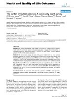
further demonstrated that t he physically shortened
CNTs orally administered can be absorbed through the
columnar cells of intestinal mucous membrane, which
was confirmed by transmission electron microscope
(Figure 3) [10]. In the experiment, high-speed shearing-
shortened SWCNTs were used. The absorb ability of
intes tinal tract for CNTs is of great significance because
this makes it possible to develop oral drug delivery sys-
tems based on CNTs.
When subcutaneously and abdomenally administered,
a part of CNTs exist persistently in the local tissues
while some of them may be absorbed through lymphatic
canal. Because the fenestra in the endothelial cells of
blood capillaries are 30 nm to approximately 50 nm
while that in the endothelial cells of lymphatic capil-
laries are larger than 100 nm in diameter, the lymph
absorption of bundled CNTs seemed to be easier than
blood absorption. The lymphatically absorbed CNTs
migrate along the lymph canal and are accumulated in
the lymph node, which is in fact a lymphatic target
effects. This is cl inically important because lymphatic
metastasis occurs extensively in cancers, resulting in fre-
quent tumor recurrence, even after extended lymph
node dissection. If anticancer drugs are loaded into
CNTs, they will be delivered into lymph system, where
the drugs will be r eleased to kill metastatic cancer cells.
Ji et al. successfully delivered gemcitabine to lymph
nodes with high efficiency by using lymphatic targeted
drug delivery system based on magnetic MWCNTs
under the magnetic field guidance [48,49]. The result
suggests that the anticancer drug delivery system based
on CNTs is advantageous over the current ways to deli-
ver chemotherapeutic agents to lymph nodes. In another
approach [50], water-soluble MWCNTs were subcuta-
neously injected into the left rear foot pad of rat; the
biopsy found that the accumulation of MWCNTs in left
popliteal lymph nodes was more obvious than in other
regions, and micropathology revealed large MWCNT
collections in the popliteal lymph nodes. At the same
time, the biopsy experiments found no presence of
MWCNTs in the major internal organs such as liver,
kidney, and lung, which suggests the properties of
MWCNT lymphatic targets.
When administered through veins, CNTs can directly
get into blood circulation and distribute in many inter-
nal organs, such as liver, spleen, heart and kidney
(unpublished date). Some studies demonstrated that the
blood clearance of intravenously injected CNTs largely
depends upon the surface modification. Singh et al.
found that, following intravenous administration,
111
In-
labeled water- soluble SWCNTs functionalized with
diethylenetriaminepenta acetic acid (average diameter, 1
Zhang et al. Nanoscale Research Letters 2011, 6:555
/>Page 7 of 22
nm; average length, approximately 300 to 1,000 nm) can
be eliminated rapidly from blood in the form of intact
CNT molecules, displaying a half-life of 3 h, with no
specific organ accumulation [51]. In a recent literature,
it was found that the clearance of 1,4,7,10-tetraazacyclo-
dod ecane-1,4,7,10 -tetra-acetic acid functionalized CNTs
complexed with yttrium-86 or
111
In and anti-CD20 anti-
body rituximab for targeting to malignant B cells was
B
Ve
Vi
200 nm
C
a
c
b
A
10 Pm
Figure 3 The absorption of SWCNTs through intestinal columnar epithelial cells [10]. (A) SWCNTs (arrows) found in the intestinal muc ous
membrane. (B) Magnification of the cell indicated by the left arrow in (A). Ve, transportion vehicles; Vi, villus of the columnar cells. (C)
Magnification of the Ve, which has membrane with double lipid layers (arrow).
Zhang et al. Nanoscale Research Letters 2011, 6:555
/>Page 8 of 22
rapid, although the blood half-lives have not been
reported [52].
Polyethylene glycol(PEG)ylation is believed to be one
of the most important strategies to prolong the circula-
tion time of CNTs in blood because the surface cover-
age with PEG lowers the immunogenicity of the carriers
and prevents their nonspecific phagocytosis by the reti-
culoendothelial system (RES); thus, their half-life in
blood circulation is prolonged. In fact, it has been found
that PEGylated CNTs can persistently exist within liver
and spleen macrophages for 4 months with excellent
compatibility [53]. In a recent investigation, it was
observed that fluorescein isothiocyanate (FITC)-labeled
PEGylated SWCNTs can penetrate the nuclear mem-
brane and get into nucleus in an energy-independent
way[54].ThepresenceofFITC-PEG-SWCNTsin
nucleus did not produce any significant ultrastructural
change in the nuclear organization and had no signifi-
cant effects on the growth kinetics and cell cycl e distri -
bution for up to 5 days. Surprisingly, upon removal of
the FITC-PEG-SWCNTs from the culture medium, the
internalized FITC-PEG-SWCNTs rapidly moved out of
the nucleus and were released from the cells, suggesting
that the internalizat ion of CNTs into and e xcretion of
CNTs from the cells are a bidirectional reversible pro-
cess. These results illustrated well the successful exploi-
tation of SWCNTs as ideal nanovectors for biomedical
and pharmaceutica l appli cations and, they will drive the
concern about the excretion problems out of people ’s
heart.
Distribution
Distribution indicates the sites or places the absorbed
CNTs can arrive and exist there, which are of great
importance in clinical pharmacology and toxicology of
CNTs as drug carriers.
There have been experiments to investigate in vivo
and ex vivo biodistributions, as well as tumor targeting
ability of radiolabeled SWCNTs (diameter, approxi-
mately 1 to 5 nm; length, approximately 100 to 300 nm)
noncovalently functionalized with phospholipids(PL)-
PEG in mice using positron emission tom ography and
Raman spectroscopy, re spectively. It was interesting to
note that the PEG chain lengths determine the biodistri-
bution and circulation of CNTs. PEG-5400-modified
SWCNTs have a circulation time (t
1/2
=2h)much
longer than th at of PEG-2000-modified counterpart (t
1/2
= 0.5 h), which may be attributed to the lower uptake of
the former by the RES as compared with that of the
later. By further functionalization of these PEGylated
SWCNTs with arginine-glycine-aspartic acid (RGD)
peptide, the accumulation in integrin-positive U87MG
tumors was significantly improved from approximately
3% to 4% to approximately 10% to 15% of the total
injected dose (ID)/g, owing to the specific RGD-integrin
a
v
b
3
recognition. Raman signatures of SWCNTs were
furtherusedtodirectlyprobethepresenceofCNTsin
mice tissues and confirmed the radiolabel-based results
[55]. In another experiment to evaluate the influences of
PEG cha in lengths on cellular uptake of PEGylated
SWCNTs, it has been found that adsorbing shorter
chain PEG (PL-PEG-2000) to SWCNTs was incapable of
protecting CNTs from macrophagocytosis both in vitro
and in vivo, while adsorbing longer chain PEG (PL-
PEG-5000) effectively reduced their nonspecific uptake
of CNTs in vivo [56]. Functionalization of SWCNTs
with PEG grafted branched polymers, namely poly(mal-
eicanhydride-alt-1-octadecene)-PEG methyl ethers
(PMHC18-mPEG) and poly (g-glutamic acid)-pyrine
(30%)-PEG methylethers (70%) (gPGA-Py-mPEG), the
blood circulation time was remarkably prolonged (half-
life of 22.1 h for gPGA-Py-mPEG and 18.9 h for
MHC18-mPEG) after intravenous injection into mice
[57]. Further research revea ls that the tumo r accumula-
tion of PEG-SWCNTs was 8% ID/g and 9% ID/g of the
intravenously administered doses in EMF6 model (breast
cancer in BABL/c mice) and the Lewis model (lung can-
cer in C57BL mice), respectively. SWCNTs covalently
modified with PEG showed longer half-life in blood cir-
culation in comparison with those noncovalently modi-
fied with PEG of similar chain lengths. SWCNTs
covalently conjugated with branched chains of 7-kDa
PEG effectively increased the half-life of SWCNTs up to
1 day, which is the longest among all of the tested
PEGs. And this length chain PEG-modified SWCNTs
had near-complete clearance from the main organs in
approximately 2 months. There seemed to be a length
limits in the relations between PEG chain lengths and
their effects to increase the blood circulation time.
Further increase in molecular weight from 7 to 12 kDa
had no influence on the blood circulation time and RES
uptake [58].
There are few literatures on the in vivo biodistribution
properties of radionuclide-filled CNTs, although they
have been extensively used as drug delivery systems or
radiotracers. A very recent study revealed that surface
functionalization of
125
I-filled SWCNTs offers versatility
towards modulation of biodistribution of these radio-
emitting crystals, in a manner determined by the system
that delivers them, which gave great promises for the
develo pment of organ-based therapeutics [59]. Nanoen-
capsulation of iodide within SWCNTs facilitated its bio-
distribution in tissues, and SWCNTs was completely
redirected from tissue with intrinsic affinity (thyroid) to
lungs. In this experiment, Na
125
I-filled glyco-SWCNTs
were intravenously administered into mice and tracked
in vivo b y single photon emission comp uted tomogra-
phy. Tissue-specific accumulation (lung in this case),
Zhang et al. Nanoscale Research Letters 2011, 6:555
/>Page 9 of 22
coupled with high in vivo stability, prevented excretion
or leakage of radionuclide to other high-affinity organs
(thyroid/stomach), allowing ultrasensitive imaging and
delivery of unprecedented radiodose density [60].
Metabolism and excretion
The nonbiodegradability in the body and non-eliminat-
ability from the body interrogate on the possib ility of
their successful use in clinical practice, which has been
always concerned about.
Functionalized SWCNTs seem to b e metabolizable in
animal body. For example, SWCNTs with carboxylated
surfaces have demonstrated their unique ability to
undergo 90-day degradation in a phagolysosomal simu-
lant, resulting in shortenin g of length and accumulation
of ultrafine solid carbonaceous debris. Unmodifi ed, ozo-
nolyzed, aryl-sulfonated SWCNTs exhibit no degrada-
tion under similar conditions. The observed metabolism
phenomenon may be accredited to the unique chemistry
of acid carboxylation, which, in addition to introducing
the reactive, modifiable COOH groups on CNT surfaces,
also induces a collateral damage to the tubular graphe-
nic backbone in the form of neighb oring active sites
that provide points of attack for further oxidative degra-
dation [59]. Some experiment s demonstrated that CNTs
persisted inside cells for up to 5 months after adminis-
tration; short (< 300 nm) and well-dispersed SWCNTs
effectively managed to escape the RES and finally were
excreted through the kidneys and bile ducts [61].
A very recent investigation reveals that the biodegra-
dation of SWCNTs can be catalyzed by hypochlorite
and reactive radical interme diates of the human neutro-
phil enzyme myeloperoxidase in neutrophils. The phe-
nomenon of CNT metabolism can also be seen in
macrophages to a lesser degree. Molecular modeling
further reveals that the interacti on between basic amino
acid residues on the enzyme backbone and carboxyl
acid groups of CNTs is favorable to orient the nano-
tubes close to the catalytic site. Notably, when aspirated
into the lungs of mice, the biodegradation of the nano-
tubes does not engender an inflammatory response.
These findings imply that the biodegradation of CNTs
may be a key determinant of the degree and severity of
theinflammatoryresponsesin individuals exposed to
them. However, further studies are still required in
order to draw an appropriate conclusion [62].
CNT-based drug delivery
While attachment of drugs to suitable carriers signifi-
cantly improves their bioavailability, owing to their
increased residence time in blood circulation and
enhanced solubility, the therapeutic efficacy of the drug
can be improved by the site-selective accumulation in
the pathological zone of interest that sometimes were
called therapeutic-effects-related sites. The unique cap-
ability of CNTs to penetrate cell membranes paves the
road for using them as carriers to deliver therapeutic
agents into the cytoplasm and, in many cases, into the
nucleus. The intrinsic spectroscopic properties of CNTs,
such as Raman and photoluminescence, afford addi-
tional advantages for tracking and real-time monitoring
of drug delivery efficacy in vivo.
Intracellular drug delivery
To study the cellular drug delivery, in vitro experiments
have unique advantages, which are convenient to carry
out; experiment conditions are easy to control and can
give reliable results, although they cannot completely
represent in vivo case.
Small molecules
Most of the anticancer agents are small molecules and
canbeloadedintoorontoCNTseitherbyphysical
adsorption through p-p stacking interactions between
pseudoaromatic double bonds of the graphene sheet and
the drug molecules, or covalent immobilization of the
interest drug molecules onto the reactive functional
groups present on the sidewalls of CNTs.
Recently, Borowiak-Palen et al. reported that cisplatin,
a small molecule, can be loaded into SWCNTs with a
diameter of 1.3 to 1.6 nm [63]. The cisplatin incorpo-
rated into the tubes was proved with Raman spectro-
scopy, infrared spectroscopy, and high-resolution
transmission electron microscopy (TEM). Drug-release
study using dialysis membrane method revealed that cis-
platin was continually released for almost a week, with
maximum release during 72 h and up to 1 week. The
encapsulation was 21 μg of drug per 100 μg of SWCNTs
as revealed by thermogravimetric analysis. Cytotoxicity
studies carried out on DU145 and PC3 human prostate
cancer cell lines using 3-(4,5)-dimeth ylthiahiazo (-z-y1)-
3,5-di- phenytetrazoliumromide (MTT) cell proliferation
assay showed that the cell viability decreased with an
increase in the concentration of the CNT-based nano-
vector, whereas blank CNTs showed no significant
effects. Computational methods revealed the feasibility
of interactions between CNTs and drug molecules [64].
For cisplatin acceptance or incorporation, CNTs must
havearadiusofatleast4.785Å(0.4785nm).Infact,
most of the experimentally u sed CNTs have diameters
greater than 4.785 Å. So, it is inferred that cisplatin is
likely to be encapsulated inside the nanotubes [65].
Doxorubicin can be loaded on CNT to form supra mo-
lecular complexes based on p-p stacking interactions by
simply mixing t he drug with an aqueous dispersion of
CNTs stabilized by Pluronic F127 (nonionic surfactant).
The doxorubicin loading on MWCNTs was observed by
measuring the emission spectrum of doxorubicin via
fluorescence spectrophotometry. With the increase in
Zhang et al. Nanoscale Research Letters 2011, 6:555
/>Page 10 of 22
the concentration of MWC NTs from 5 to 20 μg/ml, the
fluorescence intensity of doxorubicin dramatically
decreased with th e final concentration in the suspension
remaining constant (10 μg/ml), a part of which is attrib-
uted to the quenching of fluorescence. It was found that
a mass ratio of 1:2 is optimum for maximum interac-
tion/quenching ratio. TEM structural characterization
revealed that CNTs present as well-individualized, dis-
persed nanotubes, confirming the polymer molecules’
ability to disperse the CNTs effectively. On MCF-7
human breast cancer cell line, it was revealed that the
doxorubicin-MWCNT complex shows enhanced cyto-
toxity in compari son with both doxorubicin alone and
doxorubicin-Pluronic complexes. The enhanced cyto-
toxicity obtained with the doxorubicin-MWCNT com-
plex indicates that MWCNTs can effectively enhance
the delivery of doxorubicin and hence improve the cel-
lular uptake of the drug [66], although in vivo studies
are essential in order to further validate the efficiency of
the reported system. Some other groups have also devel-
oped doxorubicin-loaded nanotubes but with more com-
plex system structures. The system was composed of
oxidized SWCNTs trifunctionalized with doxorubicin, a
monoclonal antibody (mAb), and a fluorescent marker
and therefore can be used for targeting, imaging, and
the rape utic effects simultaneously. Confocal microscopy
observation revealed that the complex was efficiently
taken up by cancer cel ls and the do xorubicin was
released subsequently and then translocated to the
nucleus, while SWCNTs remain in the cytoplasm [67].
Of course, such a complex system requires rigorously
investigating in order to check the integrity of the
SWCNT hybrids during the course through biological
milieu for its in vivo use. Zhang et al. have successfully
prepared biocompatible and water-dispersible multifunc-
tional drug delivery system with doxorubicin-loaded
polysaccharide functionalized SWCNTs, which presents
stimuli-responsive drug-release characteristics in addi-
tion to simultaneous targeting. The chitosan/alginate
polymer chains have been wrapped around the CNTs
simply by sonication and stirring of a chitosan/alginate
solution containing CNTs. At acidic pH, hydrophilicity
of doxorubicin is enhanced, which facilitates its detach-
ment from the CNT surface. Compared with normal tis-
sues, physiological pH condition of tumor environment
and intracellular lysosomes is more acidic, and therefore,
this system seemed to be able to intelligently release
drugs in tumor tissues. Through tethering the free
amino groups of chitosan with folic acid (FA), the tar-
geting effects may be further improved. Such nanocar-
rier-based drug delivery systems for doxorubicin could
be more selective and effective than the free drug and
have the promise to result in reduced toxicity and side
effects in patients, along with a smaller d rug dose
needed for c hemotherapy [68]. Thus, such CNT-based
supramolecular systems with structural uniqueness of
the doxorubicin are capable of self-targeting due to the
aforementioned mechanisms. Furthermore, this can be
bolstered through ligand-based targeting by incorpora-
tion of special ligands, similar to RGD peptide, which
targets integrin receptors, making it a multitargeted
modality complementing each other for selective action
at cancerous tissue [69].
Antioxidants have been considered to play a signifi-
cant role in cancer therapy owing to their ability to
combat oxidative stress. However, their poor solubility
mitigates the reaping of the benefits from these com-
pounds. This drives us to bring them under the canopy
of nanocarriers in order to use them as practical phar-
maceuticals. Covalently PEGylated ultrashort SWCNTs
can be linked to the antioxidant, amino butylated
hydroxy toluene, through ionic interactions by simple
stirring of the mixture. Residual c arboxylic acid groups
on the oxidized CNT allow the ionic interactions with
the amine group of the butylated hydroxy toluene. The
formulation was evaluated using an oxygen radical
absorbance capacity assay, in which a fluorescent probe’s
loss of fluorescent intensity is monitored in the presence
of oxygen radicals. Oxygen-radical scave ngers can keep
the fluorescence of the probes. The fluorescence inten-
sity remains unaffected until the radical scavenger is
consumed when oxygen- radical scavengers are added to
the system. The assay readout can be compared to the
radical scavenging ability of a known radical scavenger,
Trolox, a vitamin E derivative. The radical scavenging
ability of the composite was found to be as high as
1,240 times that of Trolox. However, when the butylated
hydroxy toluene functionalization was carried out
through covalent addition to the sidewall, the antioxi-
dant activity of the system was found to be decreased
[14], suggesting that not all functionalization s are bene-
ficial for antioxidant activity.
Poor blood circulation times of platinum anticancer
drugs result in insufficient uptake by tumor tissues and
intracellular DNA binding due to their unusually low
size, making them suitable candidates for a nanoparti-
cle-based drug delivery system to improve their pharma-
cological performance. For this purpose, a “longboat
delivery system” has been prepared for the platinum
warhead. In this system, a platinum complex [Pt (NH
3
)
2
Cl
2
(O
2
CCH
2
CH
2
CO
2
H) (O
2
CCH
2
CH
2
CONH-PEG-FA)
derivatized with PEG and folate (FA) was attached to
the surface of SWCNT functionalized with amino
groups (SWNT-PL-PEG-NH
2
) through multiple amide
linkages. Such a unique surface design facilitates active
targeting of the prodrug to the tumor cell, where cispla-
tin is released upon intracellular reduction of Pt(IV) to
Pt(II) after endocytosis. Internalization studies re vealed
Zhang et al. Nanoscale Research Letters 2011, 6:555
/>Page 11 of 22
not only high and specifi c binding of the SWCNT-teth-
ered conjugate to th e folate receptor but also many fold
enhancementinactivity(byafactorof8.6)incompari-
son with free cisplatin [70]. A similar kind o f SWCNT
conjugate without the targeting moiety showed 2.5
times more toxic on NTera-2 cells [71].
There are quite a few literatures on the attempts to
solve CNTs through polymers such as PL-PEG and chit-
osan among other polymers and then functionalizing
them with drugs/ligands. However, Murakami et al.
have demonstrated a more novel approach of solving
carbon nanohorns th rough doxorubicin-PEG conjugates.
The stacking interactions between the nanohorn s and
doxorubicin aid indirect attachment of PEG to carbon
nanohorns for the enhancement of their dispersibility.
This approach is also true for CNTs [72].
Polymeric drug conjugates, as a new class of systems,
have been envisaged for tumor tissue-specific delivery of
anticancer drugs [73]. Accumulation within tumor tis-
sues can be achieved by the macr omolecular size of
polymeric drug conjugates, which enables them selective
for cancerous tissues because of enhanced permeability
and retention (EPR) effects. The pathophysiologic fac-
tors of the tumor cells, such as EPR, poor venous and
lymph drainage, acidic pH, and relatively high tempera-
ture, improve the pharmacological performance of poly-
meric-based systems. On the same lines, CNT
conjugates are being explored for improving cancer
therapy, although it is still a serious challenge. Metho-
trexate (MTX) is a drug widely used against cancer;
however, it suffers from low cellular uptake. Conjugation
ofMTXtoCNTsenhancesitsinternalizationviathe
functionalized CNTs, representing a promising approach
to overcome its limited cellular uptake. Two orthogon-
ally protected amino groups were conjugate onto the
side walls of CNTs and subsequently derivatized with
FITC and MTX using the 1,3-dipolar cycloaddition of
azomethine ylides. Epifluorescence and confocal micro-
scopy studies suggested that MTX was rapidly interna-
lized by CNTs and drug efficacy was enhanced [69].
Magnetic CNTs complexed with a layer of magnetite
(Fe
3
O
4
) nanoparticles on the inner surface of the nano-
tubes have been used for lymphatic tumor targeting.
Through nanoprecipitation, PL-PEG-FA functionalized
magnetic CNTs can be impregnated with chemothera-
peutic agents, such as 5-fluorouracil and cisplatin. Such
a system can be guided by an externally placed magnet
to target regional lymphatic nodes [74]. Although this is
a little complex, the procedure seems to have practical
significance.
Dendrimers, synthetic macromolecules, have tree-like
and well-defined branch unique features, such as a mul-
tivalent surface (nanoscaffolding), interior shells, and a
core to which the dendrons are attached, showing great
promise for them to be used as drug carriers [75]. Shi et
al. have tried to integrate the properties of CNT with
that of dendrimers. MWCNTs were functionalized with
generation 5 (G5) amine-terminated polyamidoamine
dendrimers, on which FITC and folic acids were cova-
lently linked. It was found that the nanocomposites are
stable and biocompatible. Through amide linkage using
1-ethyl-3-(3-dimethylaminopropyl) carbodiimide (EDC)
chemistry, the dendrimers were attached to the COOH
groups present on oxidized C NTs. In vitro experiments
demonstrated that the MWCNTs functionalized with
folate have selectively target effects of the cancer cells
that overexpress folate receptors. The integration of
dendrimers with CNTs provided multiv alent amine-rich
periphery for the combination of drug molecules with
CNT surfaces, greatly improving the effective therapeu-
tic payload by incorporating drug molecules into dendri-
mer cavity [76].
Proteins
CNTs not only can deliver drugs of small molecules but
also can deliver proteins. MWCNTs have been used as
cellular carriers of recombined ricin A chain protein
toxin (RAT) for tumor targeting. The complexes of
RATandMWCNTwerecapableoftranslocatinginthe
cytoplasm of various cell lines, including L-929,
HL7702, MCF-7, HeLa, and COS-7 , and showed excel-
lent performance of their biological functions, as evi-
denced by the effects of inducing cell apoptosis or
death. In comparison with RTA alone, MWCNT-RTA
conjugates achieved three times higher cell death rates
for L-929, HL7 702, MCF-7, HeLa (75% mortality), and
COS-7 cells. Coupling of HER2 to MWCNTs-RTA
complexes caused selective recognition of HER2/neu
receptor [77].
To improve the efficacy of bre ast cancer targeting and
therapy, anti-HER2 IgY antibodies were covalently
coupled to t he side walls of SWCNTs using EDC chem-
istry. Single-cell level Raman spectroscopic observation
demonstrated that signals collected from the SK-BR-3
cells treated with the targeted nanoconjugate were sig-
nificantly greater than that from the control cells. Near-
infrared (NIR) irradiation showed selectively destructive
effects on the HER2-expressing SK-BR-3 cells while no
harming effects on HER2-free MCF-7 cells. There were
also cells thermally ablated without the internalization
of SWCNTs as observed through confocal microscopy,
which may be attributed to the sharp local temperature
increase [78]. Tumor-targeting CNT constructs were
synthesized by McDevitt et al. from a water-soluble pre-
cursor CNTs functionaliz ed with covalently conj ugating
multiple copies o f tumor-specific mAbs, radiometal-ion
chelates, and fluorescent probes. They demonstrated
that the nanoconstructs were selectively reactive with
human cancer cells. The experiments were d esigned to
Zhang et al. Nanoscale Research Letters 2011, 6:555
/>Page 12 of 22
observe the target effects in a model of disseminated
human lymphoma and in cells by flow cytometry and
cell-based immunoreactivity assays versus appropriate
control cells. Chakravarty et al. in a pioneering study,
used biotinylated polar lipids (1,2-distearoyl sn-glycero-
3-phosphoethanolamine-N-[biotinyl (PEG)2000] [DSPE-
PEG(2000)-biotin]) to prepare stable, biocompatible,
noncytotoxic CNT dispersions. Then, CNTs were func-
tionalized by o ne of two different neutralite avidi n-de ri-
vatized mAbs against either human CD22 or CD25. The
peripheral blood mononuclear cells activated b y CD22
+
CD25
-
and CD22
-
CD25
+
cells can be bound only by
the CNTs bearing the corresponding mAbs, respectively.
And only the cells bound by the corresponding nanosys-
tems were ablated after exposure to NIR light [79]. Kam
et al. reported an approach similar to that of Chakra-
varty et al., i n which targeting effects were achieved by
using PL-PEG-FA functionalized SWCNTs [80].
Tumor lysate proteins (TLP) are composed of various
tumor proteins that develop in a large number of
tumors and patients, irrespective of the genetic origins
of tumors. However, defined tumor markers, such as
target antigens, are still in a lack, renderi ng the antibody
responses difficult to assess, and since there is a lack of
the nature o f the immunogens, the efficacy of therapy
involving the lysate proteins was generally assessed by
tumor rejection, tumor growth retardation, or prolonged
survival of the immunized mice rather than by antibody
production. The anticancer immune response of TLP
against multiple tumors [81] has been improved by
CNTs. The efficacy of the RGD peptide for targeting
tumors with overexpressed a
v
b
3
integrin receptor is well
established. An application example has exploited a
similar strategy for active targeting of RGD-functiona-
lized, PEGylated CNTs to integrin-positive tumors in
mice. Based on similar lines, a
v
b
3
mAbs were used to
target the PL-PEG-modified SWCNTs. As targeting
ligands, a
v
b
3
mAbs have their advantages in terms of
their high specificity towards antigen a
v
b
3
and greater
stability in vivo relative t o RGD peptide. In spite of cell
culture-based studies in U87MG (human glioblastoma
cancer cells) and MCF-7 (human breast cancer cells)
revealing good targeting efficiency, the behavior of s uch
a macromolecular structure needs to be traced inside
the complex biological system to confirm its value in
cancer therapy [82].
RNA, DNA, or genes
The applica tion of CNTs as gene carrier s in gene deliv-
ery has been considered quite promising. Gene therapy
involves not only the gene-based treatment for cancers
but also that for the infectious diseases by introducing
genetic materi als. It is generally believed that the tumor
formation is t he results of the gene alterations and gene
therapy aims to correct them. For all of t he gene-based
therapeutic strategies, efficient and safe gene delivery
systems have become imperative to develop, especially
the gene vectors because it is relatively easy to obtain
corresponding genes. There have been two subcategories
of gene vectors including many viral and nonviral vec-
tors. Viral vectors have been modified to eliminate their
toxicity and ma intain their high gene transfer capability.
However, their limited capacity for transgenic materials
and safety, particularly immunogenicity, has compelled
researchers to increasingly shift attention upon nonviral
vectors as an alternative. Nonviral vectors are mainly
based on cationic polymers. It is just recent thing that
CNTs emerge as DNA carriers owing to their unique
physical, chemical, and biological properties [83].
Charged hybrid DNA/SWCNT complexes can be
obtained by sonicating the suspensions composed of sin-
gle-stranded DNA and CNTs. The aromatic nucleobases
are believed to bind to the graphene side walls through
π-π stacking effects. DNA molecules ca n be confined
and oriented by CNTs that acted as scaffolds, which
were wrapped around by DNA macromolecules. More-
over, there are different interaction energies with nano-
tubes f or different nucleobases. The sugar and
phosphate groups remain at the periphery relative to the
bases, playing the roles of enhancing the dispersibility of
CNTs. The spontaneous wrapping of DNA around
nanotubes has been also confirmed by atomic-force
microscopy and spectroscopic studies. Such a system
can definitely prove useful in gene delivery. Several
experimental studies in this area [84] were discussed
here.
A material capable of binding negatively charged
siRNA through electrostatic interactions can be obtained
by functionalizing SWCNTs with hexamethylenediamine
(HMDA) and poly(diallyldimethyl ammonium chloride)
(PDDA). PDDA was bound by noncovalent interactions,
whereas HMDA was covalently linked to the oxidized
CNTs. The experiments on isolated rat heart cells
revealed that the effect of ERK1/ERK2 genes loaded
onto HMDA-PDDA-SWCNTs enhanced efficiency for
ERK1 and ERK2 by a factor of nearly 80%. Furthermore,
the biocompatibility has also been i mproved. No signifi-
cant cytotoxicit y was caused by PDDA-HMDA -SWCNT
comp lexes at concentrations of 10 mg/l [85]. Pantarotto
et al. used CNTs functionalized with am monium as vec-
tors for transfecting plasmid (b-ga lactosidase encoded
gene, b-gal) into HeLa and CHO cell lines. The transfec-
tion efficiency was calculate d based on the experimental
data to be proportional to the CNT/DNA charge ratio,
with the highest at 6:1. In comparison with DNA alone,
transfection efficacy of CNT-DNA conjugates was ten
times higher [86]. Through layer-by-layer electrostatic
self-assembly of polycationic agents such as polyethyle-
nimine (PEI), PDDA, polyamidoamine dendrimers, and
Zhang et al. Nanoscale Research Letters 2011, 6:555
/>Page 13 of 22
chitosan on nanotube side walls, cationic polyelectro lyte
functionalization of CNTs can be obtained, which are
considered to be effective vehicles for gene delivery [87].
For example, green fluorescence protein (EGFP) reporter
protein expression has been enhanced with functiona-
lized CNTs complexed with nanochitosan as the gene
carriers for the delivery of encoding DNA. In compari-
son with blank chitosan, CNT-chitosan nanocomplexes
have exhibited significantly higher tran sfection efficiency
[88]. The feasibility of bifunctional, MWCNT-quantum
dot-based hybrids as gene transfer vector systems have
also been explored by recent investigations. Mercaptoa-
cetic acid-capped CdTe quantum dots, as fluorescent
probes, were linked to antisense oligodeoxynucleotides
to suppress telomerase expression. Their activity is unu-
sually high in 90% of cancer cells compared with normal
cells. Then, these complexes further complexed with
functionalized MWCNTs through ionic interactions.
Based on confocal and flow-cytometric studies, efficient
intracellular transporting, strong cell nucleus localiza-
tion, and high delivery efficiency of antisense oligodeox-
ynucleotides were exhibited by this system in
comparison with free antisense oligodeoxynucleotides in
HeLa cells. When the EGFP gene transfected using PEI-
MWCNT as gene vectors, EGFP was overexpressed,
further confirming the role played by the nanostruc-
tures. The nanosystems and the enhanced proton
sponge effects of PEI coating on the surface of
MWCNTs prevent DNA from enzyme degradation [13]
in cytoplasm and increase the ratio of the gene to be
expressed afterwards. However, efficacy of such delivery
systems was critically dependent on the chemistry of
surface functionalization. Kam et al. have demonstrated
that effective transporting, enzymatic cleaving, and
releasing of DNA from SWCNT transporters and subse-
quent nuclear translocation of DNA oligonucleotides in
mammalian cells can also b e achieved by incorporating
cleavable disulfide bonds on the surface of PL-PEG
functionalized SWCNTs. Such functionalization showed
not only that the delivery o f siRNA was highly efficient
but also that the RNAi functionality achieved in this
way was more potent than the conven tional transfection
agents that have been widely used [89].
In vivo studies on drug delivery
As drug carriers, they will be finally used in living ani-
mals and human. Although the results of the in vitro
experiments have provided a lot of useful information
about the ap plication of CNTs as drug carriers, only the
in vivo experiments can give corroboration for the use-
fulness of CNTs in practical gene delivery for cancer
therapies. However, there have been only limited in vivo
studiesreportedontheapplicationofCNTsasthe
molecular transporter in drug delivery.
Drug delivery targeted to lymphatic system
Many cancers metastasize through lymphatic canal. The
drug delivery systems targeted to the lymphatic system
can block the metastasis of cancers effectively. Using
radical polymerization, polyacrylic acid (PAA) can be
appended onto CNTs, making them highly hydrophilic.
Through coprecipitation, Fe
3
O
4
-based magnetic nano-
particles can be adsorbed on the PAA-CNT surface.
Through the interaction with COOH groups of grafted
PAA, the nanoparticles can be stabilized from clustering.
By stirring the solution containing PAA-CNT, Fe
3
O
4
-
based magnetic nanoparticles, and gemcitabine for 24 h,
gemcitabine was loaded into the nanosystem with a
loading efficiency of 62%. It was found that CNTs were
seen only in the local lymphatic nodes and were absent
in the major organs, such as liver, kidney, heart, spleen,
and lungs, after 3 h of subcutaneous injection. Without
the help of such a nanostructures [90], gemcitabine can-
not preferentially distribute in the lymphatic system.
Drug delivery targeted to tumor
To deliver anticancer drugs into cancer focus is the pre-
requisite for the drugs to develop their effects. However,
some drugs cannot arrive or enter cancer tissues
because of their short residence time in blood circula-
tion. For example, the efficacy of paclitaxel (PTX), a
widely used chemotherapeutic agent in cancer therapy,
is often limited by its poor solubility in aqueous medium
and nonspecific cytotoxicity, thereby preventing it from
efficiently reaching the cancer focuses. Furthermore, the
solubilizer cremophor in current formulation (Taxol)
has exhibited allergenic activity, prompting the search
for alternative delivery systems. For this purpose, Liu et
al. successfully conjugated PTX to branched PEG chains
on SWCNTs via a cleavable ester bond to obtain a
water-soluble SWCNT-PTX complex. In a murine 4T1
breast cancer model, the SWCNT-PTX complex showed
an efficacy higher than that of the clinically used Taxol
in suppressing tumor growth. The blood circulation
time almost was sixfold higher than Taxol, which was
attributed to the PEG fictionalization. PTX uptake of
tumor for SWCNT-based PTX delivery system is likely
through an enhanced permeability and retention effect
(EPR effects). Hepatic macrophag e showed considerable
uptake [91], but histopathological and biochemical stu-
dies found no obvious morphological changes, although
the function of the liver requires examining for any
potential side effects of SWCNT-PTX.
Wu et al. successfully constructed a novel MWCNT-
based drug delivery system by tethering anticancer agent
hydroxycamptothecin (HCPT) onto the surface of
MWCNTs. By carboxyl enrichment via optimized oxidi-
zation treatment, CNTs were surface-functionalized,
which was followed by amidation with a hydrophilic dia-
minotriethylene glycol, and subsequent conjugation of
Zhang et al. Nanoscale Research Letters 2011, 6:555
/>Page 14 of 22
succinylated HCPT t o hydroxyl derivatized MWCNTs
was achieved via a cleavable ester linkage. In compari-
son with the clinical, presently used HCPT formulation,
the MWCNT-HCPT complexes demonstrated superior
antitumor activity and low toxicity both in vitro and in
vivo. The prepared complexes had longer blood circula-
tion time and higher tumor-specific drug accumulation
as in vivo single photon emission computed tomography
imaging and ex vivo g-scintillation counting analysis dis-
close d. These properties synergistical ly boost the antitu-
mor efficacy of the conjugate [92]. Through in vivo
observation of the killing effects on cancer cells, Bhirde
et al. demonstrated superior efficacy of drug-SWCNT
bioconjugates over nontargeted bioconjugates [93]. In
order to specifically target squamous cancer, anticancer
agent cisplatin and epidermal growth factor (EGF) were
attached to SWCNTs. SWCNT-cisplatin without EGF
was used as a nontargeted control. It was revealed that
SWCNT-quantum dot-EGF bioconjugates internalized
rapidly into the cancer cells as conf irmed by imaging
studies with h ead and neck squamous carcinoma cells
(HNSCC) overexpressing EGF receptors (EGFR) using
qua ntum dot (linked covalent ly) luminesce nce and con-
focal microscopy. Control cells showed limited uptake
and cellular uptake can be blocked by siRNA knock-
down of EGFR, revealing t he importance of EGF in the
system. Imaging with three color, two-photon intravital
video showed that injected SWCNT-quantum dot-EGF
was selectively taken up by HNSCC tumors causing
rapid regression, but in those control animals, SWCNT-
quantum dot was cleared from the tumor region in less
than 20 min. HNSCC cells were also killed selectively by
SWCNT-cisplatin-EGF, while control systems caused no
effect on the proliferation of these cells [93].
Dendriticcellsareofimportancefortheinduction
and regulation of immune responses. To carry siRNA
into the antigen-presentin g dendritic cells in vivo,catio-
nic 1,6-diaminohexane functionalized CNTs were pre-
pared. Splenic CD11c+ dendritic cells, CD11b+ cells and
also Gr-1+CD11b+ cells comprising dendritic cells,
macrophages, and other myeloid cells showed active
take up for the complexes . The complexes silenced sup-
pressor of cytokine signaling 1 (SOCS1)expressionand
retarded the growth of B16 tumor in mice [94]. It is
well known that telomerase reverse transcriptase plays a
critical role in tumor development and growth through
the maintenance of telomere structure . The same group
of authors coupled siRNA to SWCNTs via a CONH-
(CH
2
)
6
-NH
3
+
Cl
-
spacer, demonstrating that siRNA
delivered via SWCNT complexes silences the expression
of telomerase reverse transcriptase and inhibits the pro-
liferation and growth of tumor cells both in vitro and in
mouse models. By 48 h, all of the cells, such as LLC,
TC-1, and 1H8 cells, treated with telomerase reverse
transcriptase SWCNT-siRNA showed an almost c om-
plete inhibition of proliferation in murine tumor models
[95]. Unmodified siRNA with pristine SWCNTs have
also been complexed noncovalently to deliver iRNA to
cancer cells in a more recent study. The complex was
prepared by simple sonication of pristine SWCNTs in a
solution of siRNA that plays the dual role of cargo and
dispersant for CNTs. It was envisioned that there was
strong specific inhibition of cellular HIF-1 activity when
siRNA complexes targeted to hypoxia-inducible factor 1
a (HIF-1a) were added in serum-containing culture
media. The ability to response biologically to SWCNT-
siRNA complexes have been observed in a wide variety
of cancer cell types. Moreover, the activity of tumor
HIF-1a was significantly inhibited by intratumoral
administration of SWCNT-HIF-1a siRNA complexes in
mice bearing MiaPaCa-2/HRE tumors, suggesting that
such SWCNT-siRNA complexes promise considerably
as therapeutic agents [96].
Drug delivery targeted to central nervous system
To deliver drugs to central nervous system is still a ser-
ious challenge in anticancer drug delivery system for the
treatment of the tumors in the central nervous system
because of the blood-brain barrier. Recently, our group
devised a simple drug delivery system of acetylcholine
for treatment of Alzheimer disease. Acetylcholine is nat-
ural neurotransmitter of the cholinergic nervous system
and related with high-level nervous activities, such as
learning, memory, and thinking. Because of the synthesis
impairment, acetylcholine is decreased in the neurons in
the Alzheimer disease brain, leading to the incapability
of learning, memory, and thinking. Providing acetylcho-
line for the neurons would be able to prove the intellec-
tual activities of the patients with Alzheimer disease.
But there have been no way to deliver acetylcholine into
brain because the acetylcholine is a compound with
strong polarities, making it difficult to pass through the
blood-brain barrier. In our newly prepared drug delivery
system, the adsorpti on of acet ylcholine on SWCNT was
confirmed by Raman spectrum, although it was
unknown whether acetylcholine was adsorbed on the
surfaces or in the tubes of SWCNT. This system suc-
cessfully delivered acetylcholine into the neurons i n the
brain through the axoplasma transformation of neurites
(Figure 4) and significantly improved the learning and
memory capabilities of the model animals with Alzhei-
mer diseases [10]. This delivery system provided the
first instance for nanocarriers to deliver drugs into neu-
rons in the central nervous system, opening the door for
the drug delivery system for the treatment of the tumors
in central nervous system.
As regards the in vivo anticancer drug delivery system,
it should be mentioned that the results of CNT-based
drug delivery has been compared with some
Zhang et al. Nanoscale Research Letters 2011, 6:555
/>Page 15 of 22
commercially available formulations. The results demon-
strated several advantages of CNTs over other nanoma-
terial-based drug delivery systems. For example, Liu et
al. compared the efficacy and the toxicity of doxorubi-
cin-loaded CNTs with that of doxorubic in-lo aded nano-
liposomes [97]. However, a decisive conclusion has not
been reached about whether CNTs are the best carriers
in anticancer drug delivery systems because there are
many other a nticancer drug delivery systems based on
other nanomaterials, such as cationic polymers [98],
fibrinogens [99], and PVP/PVAL nanoparticles [100],
that have also shown great promises as drug carriers for
cancer treatments. More comparison works are required
to make sure of the position of CNTs as drug car riers
in anticancer therapies. In addition, the reproducibility
of pharmacokinetic data is severely challenged by the
facts that the volume, diameter, and length of CNTs are
not well controlled in the studies, nor is there any
reliable data on their size distribution. The commerciali-
zation of drug delivery systems based on CNTs will be
impeded by these problems. To the best of our knowl-
edge, no such system has gone into clinical trial and
many difficulties are yet to be overcome before success-
ful application of CNT-based drug delivery systems in
practical therapy comes into fruition.
The biosafety of SWCNT used as drug carriers
Although there are exciting prospects for the application
of CNTs as drug carriers in medicine, one of the key
obstacles in the way for the use of SWCNT as drug car-
riers is their biosafety. This problem has been quite con-
troversial. There are many studies that believe SWCNT
is safe, while many literatu res have reported the da mage
effects on cells in vitro and on tissues in vivo. Fortu-
nately, excitement encompassing the attractive physico-
chemical properties of CNTs has been tempered by
2 Pm 500 nm
2 Pm
200 nm
500 nm
B
A
C
ED
Figure 4 SWCNTs enter the neurons in brain through axoplasmic transportation [10]. (A) SWCNTs in the lysosomes of a neuron (arrows);
(B) the magnification of the two lysosomes containing SWCNTs; (C) there are no SWCNTs in glial cells; (D) a bundle of SWCNTs parallel to the
neurite in the section along the longitudinal axis of the neurite; (E) the SWCNTs are dot-like in the section vertical to the longitudinal axis of the
neurite.
Zhang et al. Nanoscale Research Letters 2011, 6:555
/>Page 16 of 22
issues concer ning their pote ntial health hazard s, despite
some toxic effects of CNTs have b een observed, which
give rise to concern about the biosafety of CNTs. The
observed toxicity has been largely attributed to the
structural analogy between CNTs and asbestos fib ers in
the high aspect ratio, large surface area, induction of
inflammation [101-103], fibrosis [104-106], and malig-
nant mesothelioma [107,108] on inha lation, and, more
importantly, their biopersistence [109-111]. A recent in
vitro study has revealed that there are close relations
between the biodurability of CNTs and the chemistry of
CNT surface functionalization. However, it was reveal ed
by recent investigations that the toxicity of SWCNTs in
vivo is resulted from the aggregation rather than the
large aspect ratio of individual CNTs [112-114]. The
experiment demonstrated that granuloma-like structures
with mild fibrosis observed in mice treated with aggre-
gated SWCNTs were completely absent in mice treated
with nanoscale dispersed SWCNTs, e ven after 30 days
of intratracheal administration. Histopathological sec-
tions from the lung of the mice treated with nanoscale
dis persed SWCNTs revealed uptake of the SWCNTs by
macrophages and demonstrated the gradual clearance
over time, c orroborating the b iosafety and biocompat-
ibility of CNT s in the form of nanoscale dispersions in
in vivo applications. Further attempt has been tried to
evaluate the toxicity of SWCNTs in Swiss mice as a
function of dose, length, and surface chemistry. Some
researchers have observed that oral administration of
CNTs, even at very high doses (1,000 mg/kg body-
weight), induces neither death, growth, nor any beha-
vioral dilemma. When intraperitoneally administered,
SWCNTs were found to coalesce inside the body, form-
ing fiber-like structures. It was further observed that
smaller aggregates almost induced no granuloma.
Rafeeqi and Kaul explored the interactions between
multi-walled carbon nanotubes and cell culture medium
by spectroscopy, and the results supported biocompat-
ibility of these nanotubes [115]. From these literatures,
both positive and negative biological effects of SWCNTs
can be seen, which has been attributed to the methods
used in the experiments. For example, Mittal et al.
found that clear interference of CNTs with conventional
in vitro cytotoxicity assays (MTT, NRU, and LDH) was
found, which was confirmed by cellular system, but
morphological changes, and flow cytometry showed the
characteri stics of cytotoxicity [116]. Brown et al.
reported that the cytotoxicity of CNTs is related to their
lengths [117].
More recently, we carefully and extensively investi-
gated the biosafety problem of SWCNTs [10]. The
results demonstrated that SWCNT are considerably safe
with a safe range of 12 as revealed by the ratio of ED99/
LD99, where the ED99 is the do ses effective for half of
the model animals with Alzheimer disease. It was also
demonstrated that the pharmacological and toxicological
effects of SWCNT are mediated separately by lysosomes
and mitochondria. In vitro,SWCNTinducedthe
increase of reactive oxygen species (ROS) in lysosomes
while had no influences on the ROS level in mitochon-
dria. In vivo, SWCNT exclusively distributed in lyso-
somes when the doses was under 300 mg/kg, but there
was no ultrastructural damage as revealed by transmis-
sion electron microscope. SWCNT began to enter mito-
chondria when the doses got over 400 mg/kg. Once
SWCNT entered, the pathological ultrastructural
changes developed in mitochondria. The mitochondria
dilated, the crestae decreased or disappeared, and even
the whole mitochondria became a vacuole. Once the
mitochondria damaged developed, the lysosomes also
appeared as damaged. The membrane of lysosomes
destroyed, the contents of lysosomes decreased or
excreted, and even the whole lysosomes became a huge
vacuole. All of these results indicated that mitochondria
are the original organelles for SWCNT damage and the
lysosomal damage is secondary to mitochondrial
damage, namel y, mitocho ndri a are the toxicological tar-
get organelles and lysosomes are the pharmacological
target organelles of SWCNT. Further experiments
demonstrated that the damage of SWCNT on mito-
chondria was resulted by its influences on the mito-
chondrial membrane potentials (MMP). The decrease in
MMP means there is leakage of free electrons, which
results in the increase of the ROS, further leading to the
ultrastructural damage of the cells including lysosomes.
The mechanism of SWCNT damage can be summarized
in Figure 5.
The progress in several aspects of the safety studies on
SWCNT is of great importance for its practical use as
drug carriers for cancer treatme nt. Firstly, the modifica-
tion or functionalization can significantly decrease the
toxicity of CNTs, making it possible to select safe CNT
deriva tiv es as drug carriers. Secondly, it has been found
that CNTs can be metabolized in liver and eliminated
through kidneys and hap-bile systems, making less con-
cern about the persistence residence of them in bodies.
Thi rdly, it has been illustrated that the pharmacological
and the toxicological effects of CNTs are mediated by
different target organelles and the distribution of CNTs
in organelles can be regulated by some chemicals, mak-
ing it possible to use the advantage and decrease or
avoid the disadvantage of them. Fourthly, the dose dif-
ferences exist between the pharmacological and toxico-
logical effects of CNTs, which means that the
toxicological effects may be avoid by controlling the
doses. Through these progresses, it can be predicted
that the sa fety problems will be solved in not too long a
time.
Zhang et al. Nanoscale Research Letters 2011, 6:555
/>Page 17 of 22
The future of CNTs used as drug carriers for cancer
treatments
Summarizing the ab ove described progress in the studies on
the application of CNTs as drug carriers, it may be seen that
the c hemistry on the modification of CNTs now has consid-
erably grown up. The p revious studies have provided us var-
ious chemical methods to solve some fundamental problems
in the use of CNTs as drug carriers such as their water solu-
bility a nd target properties. Many r eported results, including
those obtained from in vitro and in vivo experiments, have
demonstrated that CNTs can i ncrease the treatment effects
while d ecrease the side and toxic effects of the drugs loaded
on them, indicating a considerably bright future for them to
be used as drug carriers. However, there is a long way to go
ATP
ADP
Pi
e
-
e
-
e
-
e
-
e
-
O
O
O
O
e
-
O
e
-
O
O
e
-
O
ROS
Ĺ
ĺ
Ļ
ļ
ľ
e
-
e
-
e
-
e
-
O
e
-
O
e
-
O
e
-
O
e
-
e
-
O
e
-
O
ĸ
ķ
"
mitochondria electron transmission chains
Ľ
Figure 5 The schematic illustration of the mechanisms of SWCNT to induce cell damage. Based on the studies of Yang et al. [10]. SWCNTs
interact with the mitochondria electron transmission chains (ETC) (by binding to ETC?) after they enter into mitochondria (1); The interaction of
SWCNTs with ETC blocks the transmission of electrons, which results in the increase of the leaking of free electrons from ETC (2); The leaked free
electrons form free radicals H
2
O
2
or reactive oxygen species (ROS) (3); The free radicals or ROS attack the membrane system of mitochondria
through peroxidation (4); Then the free radicals or ROS diffuse through the damaged mitochondrial membrane to lysosomes to destroy the
membrane of the lysosomes (5); The injured lysosomes release digestive enzymes, leading to the damage or death of the whole cells. On the
other hand, the blocking of ETC makes mitochondria incapable of producing ATP (7), which results in the depletion of energy for the living
activities of the cells, also leading to the damage or death of the whole cells (8).
Zhang et al. Nanoscale Research Letters 2011, 6:555
/>Page 18 of 22
for C NTs to get into practical use. Partic ularly, the pharma-
cological and toxicological profiles must be made completely
clear and the advantages and the disadvantages of CNTs
must be carefully weighed before they are used as drug car-
riers in human body.
On the base s of matured chemical modification, the
remaining key to the successful practical use of CNTs as
drug carriers is to make clear of the mechanisms for
their pharmacological and toxicological effects. The
understanding of the pharmacological mechanisms
makesitpossibletotaketheadvantageofCNTstothe
outmost and to avoid or limit the disadvantages to pos-
sibly low degree (Figure 6). The weighing of the advan-
tage and disadvantage in the treatment of a special
disease is also very important because CNT-based drug
delivery system also has its indication and contraindica-
tion just like any other drugs.
After all, the studies on the application of CNTs as
drug carriers for the treatment of cancers have achieved
important progress. Some key obstacles in the way to
practical use have been overcome. Although there is still
a long way to go for the practical use, it may be pre-
dicted that, on one day in the future, CNTs will become
an important class of drug carriers for cancer treatment.
Acknowledgements
This work is supported by National Basic Research Program of China No
2010CB933904 and Major New Drug Creations No 2011ZX09102-001-15
Author details
1
Institute of Pharmacology and Toxicology and Key Laboratory of
Nanopharmacology and Nanotoxicology, Beijing Academy of Medical
Science, Zhengzhou, Henan, People’s Republic of China
2
Nanotechnology
Research Center for Drugs, Zhengzhou University, Zhengzhou, Henan,
People’s Republic of China
Authors’ contributions
ZWX drafted the manuscript. ZZZ and ZYG go over and corrected the
manuscript. All authors read and approved the final manuscript.
Competing interests
The authors declare that they have no competing interests.
Received: 27 July 2011 Accepted: 13 October 2011
Published: 13 October 2011
References
1. Ferrari M: Cancer nanotechnology: opportunities and challenges. Nat Rev
Cancer 2005, 3:161-171.
2. Cui D, Zhang H, Sheng J, Wang Z, Toru A, He R, Tetsuya O, Gao F, Cho H-S,
Cho S, Huth C, Hu H, Pauletti GM, Shi D: Effects of CdSe/ZnS quantum
dots covered multi-walled carbon nanotubes on murine embryonic
stem cells. Nano Biomed Eng 2010, 2(4):236-244.
3. Huang P, Zhang C, Xu C, Bao L, Li Z: Preparation and characterization of
near-infrared region absorption enhancer carbon nanotubes
hybridmaterials. Nano Biomed Eng 2010, 2(4):225-230.
4. Bao C, Tian F, Estrada G: Improved visualisation of internalised carbon
nanotubes by maximising cell spreading on nanostructured substrates.
Nano Biomed Eng 2010, 2(4):201-207.
5. Chen D, Wu X, Wang J, Han B, Zhu P, Peng Ch: Morphological observation
of interaction between PAMAM dendrimer modified single walled
carbon nanotubes and pancreatic cancer cells. Nano Biomed Eng 2010,
2(4):60-65.
6. Thess A, Nikolaev P: Crystalline ropes of metallic carbon nanotubes.
Science 1996, 5274:483-487.
Use the advantages
to outmost
Decrease or avoid
Side or toxic effects
Pharmacological
Mechanism
Toxicological
Mechanism
Best Treatment
effects
Treatment effects
Side or Toxic
effects
Weighing
Figure 6 The schematic illustration of the strategies. For the studies on the best use of CNTs as drug carriers. The best treatment effects are
in the center of the strategies that are the ultimate purpose of our studies, which can be achieved by the studies of three effects: the weighing
between treatment effects and the side or toxic effects, the pharmacological mechanism that makes it possible to use the advantages to
outmost and the toxicological mechanism that makes people capable of decreasing or avoiding the side or toxic effects.
Zhang et al. Nanoscale Research Letters 2011, 6:555
/>Page 19 of 22
7. Donaldson K, Aitken R, Tran L, Stone V, Duffin R, Forrest G, Alexander A:
Carbon nanotubes: a review of their properties in relation to pulmonary
toxicology and workplace safety. Toxicol Sci 2006, 92:5-22.
8. Ye Y, Ahn C, Witham C: Hydrogen adsorption and cohesive energy of
singlewalled carbon nanotubes. Appl Phys Lett 1999, 16:2307-2309.
9. Peigney A, Laurent C, Flahaut E, Bacsa RR, Rousset A: Specific surface area
of carbon nanotubes and bundles of carbon nanotubes. Carbon 2001,
4:507-514.
10. Yang Z, Zhang Y, Yang Y, Sun L, Han D, Hong Li, Wang C: Pharmacological
and toxicological target organelles and safe use of single-walled carbon
nanotubes as drug carriers in treating Alzheimer disease. Nanomedicine
2010, 6:427-441.
11. Foldvari M, Bagonluri M: Carbon nanotubes as functional excipients for
nanomedicines: I. Pharmaceutical properties. Nanomedicine 2008,
3:173-182.
12. Foldvari M, Bagonluri M: Carbon nanotubes as functional excipients for
nanomedicines: II. Drug delivery and biocompatibility issues.
Nanomedicine 2008, 3:183-200.
13. Jia N, Lian Q, Shen H, Wang C, Li X, Yang Z: Intracellular delivery of
quantum dots tagged antisense oligodeoxynucleotides by
functionalized multiwalled carbon nanotubes. Nano Lett 2007,
10:2976-2980.
14. Lucente-Schultz RM, Moore VC, Leonard AD, Kosynkin DV, Lu M, Partha R,
Conyers JL, Tour JM: Antioxidant single-walled carbon nanotubes. JAm
Chem Soc 2009, 11:3934-3941.
15. Zhu J, Yudasaka M, Zhang M, Iijima S: Dispersing carbon nanotubes in
water: a noncovalent and nonorganic way. J Phys Chem B 2004,
108:1137-11320.
16. Georgakilas V, Tzitzios V, Gournis D, Petridis D: Attachment of magnetic
nanoparticles on carbon nanotubes and their soluble derivatives. Chem
Mater 2005, 17:1613-1617.
17. Rao AM, Richter E, Bandow S, Chase B, Eklund PC, Williams KA, Fang S,
Subbaswamy KR, Menon M, Thess A, Smalley RE, Dresselhaus G,
Dresselhaus MS: Diameter-selective Raman scattering from vibrational
modes in carbon nanotubes. Science 1997, 275:187-191.
18. O’Connell MJ, Bachilo SM, Huffman CB, Moore VC, Strano MS, Haroz EH,
Rialon KL, Boul PJ, Noon WH, Kittrell C, Ma J, Hauge RH, Weisman RB,
Smalley RE: Band gap fluorescence from individual single-walled carbon
nanotubes. Science 2002, 297:593-596.
19. Gordon RG, Farmer DB: Nonctionnalisation en phase gazeuse de
nanotubes de carbone. 2008, WO/2008/085183.
20. Wise KE, Park C, Kang JH, Siochi EJ, Harrison JS: Nanocomposites issus de
dispersions stables de nanotubes de carbone contenus dans des
matrices polymères faisant appel à des interactions de dispersion. 2008,
WO/2008/073153.
21. Chen J, H Liu:
Polymer and method for using the polymer for
solubilizing
nanotubes-Patent. 2007, US20077244407.
22. Stoddart JF, Star A, Liu Y, Ridvan L: Noncovalent functionalization of
Nanotubes-Patent. 2007, US20077220818.
23. Guo Z, Sadler PJ, Tsang SC: Immobilization and visualization of DNA and
proteins on carbon nanotubes. Adv Mater 1998, 10:701-703.
24. Vigolo B, Pénicaud A, Coulon C, Sauder C, Pailler R: Macroscopic fibers and
ribbons of oriented carbon nanotubes. Science 2000, 290:1331-1334.
25. Zheng M, Jagota A, Semke ED, Diner BA, McLean RS, Lustig SR,
Richardson RE, Tassi NG: DNA-assisted dispersion and separation of
carbon nanotubes. Nat Mater 2003, 2:338-342.
26. Kim O-K, Je J, Baldwin JW, Kooi S, Pehrsson PE: Solubilization of single-wall
carbon nanotubes by supramolecular encapsulation of helical amylose. J
Am Chem Soc 2003, 125:4426-4427.
27. Numata M, Asai M, Kaneko K, Bae AH, Hasegawa T, Sakurai K, Shinkai S:
Inclusion of cut and as-grown single-walled carbon nanotubes in the
helical superstructure of schizophyllan and curdlan (N-1,3-Glucans). JAm
Chem Soc 2005, 127:5875-5884.
28. Prato M, Kostarelos K, Bianco A: Functionalized carbon nanotubes in drug
design and discovery. Acc Chem Res 2008, 41:60-68.
29. Jain AK, Dubey V, Mehra NK, Lodhi N, Nahar M, Mishra DK, Jain NK:
Carbohydrate-conjugated multiwalled carbon nanotubes: development
and characterization. Nanomedicine 2009, 4:432-442.
30. Chen Z, Thiel W, Hirsch A: Reactivity of the convex and concave surfaces
of single-walled carbon nanotubes (SWCNTs) towards addition reactions:
dependence on the carbon-atom pyramidalization. Chem Phys Chem
2003, 4:93-97.
31. Dyke CA, Tour J: Overcoming the insolubility of carbon nanotubes
through high degrees of sidewall functionalization. Chem-Eur J 2004,
10:812-817.
32. Tasis D, Tagmatarchis N, Georgakilas V, Gamboz C, Soranzo MR, Prato M:
Supramolecular organized structures of fullerene-based materials and
organic functionalization of carbon nanotubes. C R Chimie 2003, 5-
6:597-602.
33. Valery NK, Haiqing P, Wilbur EB, Yunming Y: Method for functionalizing
carbon nanotubes utilizing peroxides-Patent. 2006, US20067125533.
34. Tour JM, Hudson JL, Dyke CR, Stephenson JJ: Functionalization of Carbon
Nanotubes in Acidic Media-Patent. 2005, US20050280876.
35. Valery NK, Enrique VB, Daneesh M, Laura PP: Carbon nanotube reinforced
thermoplastic polymer composites achieved through benzoyl peroxide
initiated interfacial bonding to polymer matrices-Patent. 2006,
WO20066116547.
36. Ford WT, Qin S: Polymers grafted to carbon nanotubes-Patent. 2008,
US20087414088.
37. Barrera EV, Wilkins R, Shofner M, Pulikkathara MX: Vaidyanathan
RFunctionalized carbon nanotube-polymer composites and interactions-
Patent. 2008, US20087407640.
38. Tour JM, Bahr JL, Yang J:
Process for making polymers comprising
derivatized
carbon nanotubes and compositon thereof-Patent. 2007,
US20077304103.
39. Wong SS, Banerjee S: Sidewall-functionalized carbon nanotubes, and
methods for making the same-Patent. 2006, US20067122165.
40. Tour JM, Hudson JL, Dyke CR, Stephenson JJ: Functionalization of Carbon
Nanotubes in Acidic Media 2005-Patent.WO05113434
41. Mattson MP, Haddon RC, Rao AM: molecular functionalization of carbon
nanotubes and use as substrates for neuronal growth. 2003,
US20036670179.
42. Dai L, Lee KM: Asymmetric end-functionalization of carbon nanotubes-
Patent. 2007, WO2007061431.
43. Barrera EV, Zhu J, Khabashesku VN, Margrave JL, Kim JD, Zhang L: Sidewall
functionalization of carbon nanotubes with organosilanes for polymer
composites-Patent. 2006, EP1660405.
44. Khabashesku VN, Margrave JL, Margrave ML, Stevens JL, Derrien GA:
Sidewall functionalization of single-wall carbon nanotubes through C-N
bond forming substitutions of fluoronanotubes-Patent. 2006,
US2006171874.
45. Ford WE, Wessels J, Yasuda A: Method and apparatus for producing
carbon nanotubes-Patent. 2006, US20060014375.
46. Chen B, Liang F: A review on medical applications of single walled
carbon nanotubes. Curr Med Chem 2010, 17:10-24.
47. Ito Y, Venkatesan N, Hirako N, Sugioka N, Takada K: Effect of fiber length of
carbon nanotubes on the absorption of erythropoietin from rat small
intestine. Int J Pharm 2007, 337(1-2):357-360.
48. Ji S, Liu C, Zhang B, Yang F, Xu J, Long J, Jin C, Fu D, Ni Q, Yu X: Carbon
nanotubes in cancer diagnosis and therapy. Biochimica et Biophysica Acta
2010, 1806:29-35.
49. Yang D, Yang F, Hu JH, Long J, Wang C, Fu D, Nib Q: Hydrophilic multi-
walled carbon nanotubes decorated with magnetite nanoparticles as
lymphatic targeted drug delivery vehicle. Chem Commun 2009,
29:4447-4449.
50. Li JJ, Yang F, Guo GQ, Yang D, Long J, Fu DL, Luc J, Wang CC: Preparation
of biocompatible multi-walled carbon nanotubes as potential tracers for
sentinel lymph node. Polymer International 2010, 59:169-174.
51. Singh R, Pantarotto DP, Lacerda L, Pastorin G, Klumpp C, Prato M, Bianco A,
Kostarelos K: Tissue biodistribution and blood clearance rates of
intravenously administered carbon nanotubes radiotracers. Proc Natl
Acad Sci USA 2006, 103:3357-3362.
52. McDevitt MR, Chattopadhyay D, Jaggi JS, Finn RD, Zanzonico PB, Villa C,
Rey D, Mendenhall J, Batt CA, Njardarson JT, Scheinberg DA: PET imaging
of soluble yttrium-86-labeled carbon nanotubes in mice. Plus One 2007,
2:907.
53. Schipper ML, Nakayama-Ratchford N, Davis CR, Shi KNW, Chu P, Liu Z,
Sun X, Dai H, Gambhir SS: A pilot toxicology study of single-walled
carbon nanotubes in a small sample of mice. Nat Nanotechnol 2008,
3:216-221.
Zhang et al. Nanoscale Research Letters 2011, 6:555
/>Page 20 of 22
54. Cheng J, Fernando KAS, Veca LM, Sun YP, Lamond AI, Lam YW, Cheng SH:
Reversible accumulation of PEGylated single-walled carbon nanotubes in
the mammalian nucleus. ACS Nano 2008, 2:2085-2094.
55. Liu Z, Cai W, He L, Nakayama N, Chen K, Sun X, Chen X, Dai H: In vivo
biodistribution and highly efficient tumour targeting of carbon
nanotubes in mice. Nat Nanotechnol 2007, 2:47-52.
56. Liu Z, Tabakman S, Welsher K, Dai H: Carbon nanotubes in biology and
medicine in vitro and in vivo detection, imaging and drug delivery.
Nanoresearch 2009, 2(2):85-120.
57. Prencipe G, Tabakman SM, Welsher K, Liu Z, Goodwin AP, Zhang L, Henry J,
Dai H: PEG branched polymer for functionalization of nanomaterials with
ultralong blood circulation. J Am Chem Soc 2009, 131:4783-4787.
58. Liu Z, Davis C, Cai W, He L, Chen X, Dai H: Circulation and long-term fate
of functionalized, biocompatible single-walled carbon nanotubes in
mice probed by Raman spectroscopy. Proc Natl Acad Sci USA 2008,
105:1410-1415.
59. Kolosnjaj-Tabi J, Hartman KB, Boudjemaa S, Ananta JS, Morgant G, Szwarc H,
Wilson LJ, Moussa F: In vivo behavior of large doses of ultrashort and
full-length single-walled carbon nanotubes after oral and intraperitoneal
administration to Swiss mice. ACS Nano 2010, 4:1481-1492.
60. Hong SY, Tobias G, Al-Jamal KT, Ballesteros B, Ali-Boucetta H, Lozano-
Perez S, Nellist PD, Sim RB, Finucane C, Mather SJ, Green ML, Kostarelos K,
Davis BG: Filled and glycosylated carbon nanotubes for in vivo
radioemitter localization and imaging. Nat Mat 2010, 9:485-490.
61. Thakare VS, Das M, Jain AK, Patil S, Jain S: Carbon nanotubes in cancer
theragnosis. Nanomedicine 2010, 5(8):1277-1301.
62. Kagan VE, Konduru NV, Feng W, Allen BL, Conroy J, Volkov Vlasova Y,
Belikova NA, Yanamala N, Kapralov A, Tyurina YY, Shi J, Kisin ER, Murray AR,
Franks J, Stolz D, Gou P, Klein-Seetharaman J, Fadeel B, Star A,
Shvedova AA: Carbon nanotubes degraded by neutrophil
myeloperoxidase induce less pulmonary inflammation. Nat Nanotechnol
2010, 5:554-559.
63. Tripisciano C, Kraemer K, Taylor A, Borowiak-Palen E: Single-wall carbon
nanotubes based anticancer drug delivery system. Chem Phys Lett 2009,
478:200-205.
64. de Leon A, Jalbout AF, Basiuk VA: SWNT-amino acid interactions: a
theoretical study. Chem Phys Lett 2008, 1-3:185-190.
65. Hilder TA, Hill JM: Carbon nanotubes as drug delivery nanocapsules. Curr
Appl Phys 2008, 3-4:258-261.
66. Ali-Boucetta H, Al-Jamal KT, McCarthy D, Prato M, Bianco A, Kostarelos K:
Multiwalled carbon nanotube-doxorubicin supramolecular complexes for
cancer therapeutics. Chem Commun 2008, 4:459-461.
67. Heistera E, Neves V, Tilmaciub C, Lipertc K, Beltrana VS, Coleya HM,
Silvad SRP, McFaddena J: Triple
functionalisation of single-walled carbon
nanotubes with doxorubicin, a monoclonal antibody, and a fluorescent
marker for targeted cancer therapy. Carbon 2009, 9:2152-2160.
68. Zhang X, Meng L, Lu Q, Fei Z, Dyson PJ: Targeted delivery and controlled
release of doxorubicin to cancer cells using modified single wall carbon
nanotubes. Biomaterials 2009, 30:6041-6047.
69. Liu Z, Sun X, Nakayama-Ratchford N, Dai H: Supramolecular chemistry on
water-soluble carbon nanotubes for drug loading and delivery. ACS
Nano 2007, 1:50-56.
70. Dhar S, Liu Z, Thomale J, Dai H, Lippard SJ: Targeted single-wall carbon
nanotube mediated Pt (IV) prodrug delivery using folate as a homing
device. J Am Chem Soc 2008, 34:11467-11476.
71. Feazell RP, Nakayama-Ratchford N, Dai H, Lippard SJ: Soluble single-walled
carbon nanotubes as longboat delivery systems for platinum (IV)
anticancer drug design. J Am Chem Soc 2007, 27:8438-8439.
72. Murakami T, Fan J, Yudasaka M, Iijima S, Shiba K: Solubilization of single-
wall carbon nanohorns using a PEG-doxorubicin conjugate. Mol Pharm
2006, 4:407-414.
73. Tripisciano C, Kraemer K, Taylor A, Borowiak-Palen E: Single-wall carbon
nanotubes based anticancer drug delivery system. Chem Phys Lett 2009,
4-6:200-205.
74. McDevitt MR, Chattopadhyay D, Kappel BJ, Jaggi JS, Schiffman SR,
Antczak C, Njardarson JT, Brentjens R, Scheinberg DA: Tumor targeting
with antibody-functionalized, radiolabeled carbon nanotubes. J Nucl Med
2007, 48:1180-1189.
75. Tekade RK, Kumar PV, Jain NK: Dendrimers in oncology: an expanding
horizon. Chem Rev 2009, 1:49-87.
76. Shi X, Wang SH, Shen M, Antwerp ME, Chen X, Li C, Petersen EJ, Huang Q,
Weber WJ Jr, Baker JR Jr: Multifunctional dendrimer-modified multiwalled
carbon nanotubes: synthesis, characterization, and in vitro cancer cell
targeting and imaging. Biomacromolecules 2009, 7:1744-1750.
77. Weng X, Wang M, Ge J, Yu S, Liu B, Zhong J, Kong J: Carbon nanotubes as
a protein toxin transporter for selective HER2-positive breast cancer cell
destruction. Mol BioSysts 2009, 5:1224-1231.
78. Xiao Y, Gong XG, Taratula O, Treado S, Urbas A, Holbrook RD, Cavicchi RE,
Avedisian CT, Mitra S, Savla R, Wagner PD, Srivastava S, He H: Anti-HER2 IgY
antibody-functionalized single-walled carbon nanotubes for detection
and selective destruction of breast cancer cells. BMC Cancer 2009, 9:351.
79. Chakravarty P, Marches R, Zimmerman NS, Swafford AD, Bajaj P,
Musselman IH, Pantano P, Draper RK, Vitetta ES: Thermal ablation of tumor
cells with antibody-functionalized single-walled carbon nanotubes. Proc
Natl Acad Sci USA 2008, 25
:8697.
80.
Kam NWS, O’Connell M, Wisdom JA, Dai H: Carbon nanotubes as
multifunctional biological transporters and near-infrared agents for
selective cancer cell destruction. Proc Natl Acad Sci USA 2005,
33:11600-11165.
81. Meng J, Jie M, Duan J, Kong H, Li L, Wang C, Xie S, Chen S, Gu N, Xu H,
Yang X-D: Carbon nanotubes conjugated to tumor lysate protein
enhance the efficacy of an antitumor immunotherapy. Small 2008,
9:1364-1370.
82. Ou Z, Wu B, Xing D, Zhou F, Wang H, Tang Y: Functional single-walled
carbon nanotubes based on an integrin avb3 monoclonal antibody for
highly efficient cancer cell targeting. Nanotechnology 2009, 10:105102.
83. El-Aneed A: Current strategies in cancer gene therapy. Eur J Pharmacol
2004, 1-3:1-8.
84. Albertorio F, Hughes ME, Golovchenko JA, Branton D: Base dependent
DNA-carbon nanotube interactions: activation enthalpies and assembly-
disassembly control. Nanotechnology 2009, 20:395101.
85. Krajcik R, Jung A, Hirsch A, Neuhuber W, Zolk O: Functionalization of
carbon nanotubes enables non-covalent binding and intracellular
delivery of small interfering RNA for efficient knock-down of genes.
Biochem Biophys Res Commun 2008, 2:595-602.
86. Dipl-Chem DP, Dipl-Chem RS, Dipl-Chem DM, Erhardt M, Briand J-P,
Prato M, Kostarelos K, Bianco A: Functionalized carbon nanotubes for
plasmid DNA gene delivery. Angew Chem Int Ed Engl 2004, 39:5354-5358.
87. Wu B-Y, Hou S-H, Yu M, Qin X, Li S, Chen Q: Layer-by-layer assemblies of
chitosan/multi-wall carbon nanotubes and glucose oxidase for
amperometric glucose biosensor applications. Mater Sci Engineer C 2009,
29:346-349.
88. Kumar A, Jena PK, Behera S, Lockey RF, Mohapatra S: DNA and peptide
delivery by functionalized chitosan-coated single-walled carbon
nanotubes. J Biomedical Nanotech 2005, 1:392-396.
89. Xiang L, Yuan Y, Ou Z, Yang S, Zhou F: Photoacousticmolecular imaging
with antibody-functionalized single-walled carbon nanotubes for early
diagnosis of tumor. J Biomed Optics 2009, 14:021008.
90. Yang D, Yang F, Hu J, Long J, Wang C, Fu D, Ni Q: Hydrophilic multi-
walled carbon nanotubes decorated with magnetite nanoparticles as
lymphatic targeted drug delivery vehicles. Chem Commun 2009,
29:4447-4449.
91. Liu Z, Chen K, Davis C, Sherlock S, Cao Q, Chen X, Dai H: Drug delivery
with carbon nanotubes for in vivo cancer treatment. Cancer Res 2008,
16:6652-6660.
92. Wu W, Li R, Bian X, Zhu Z, Ding D, Li X, Jia Z, Jiang X, Hu Y: Covalently
combining carbon nanotubes with anticancer agent: preparation and
antitumor activity. ACS Nano 2009, 9
:2740-2750.
93.
Bhirde AA, Patel V, Gavard J, Zhang G, Sousa AA, Masedunskas A,
Leapman RD, Weigert R, Gutkind JS, Rusling JF: Targeted killing of cancer
cells in vivo and in vitro with EGF-directed carbon nanotubebased drug
delivery. ACS Nano 2009, 2:307-316.
94. Yang R, Yang X, Zhang Z, Zhang Y, Wang S, Cai Z, Jia Y, Ma Y, Zheng C,
Lu Y, Roden R, Chen Y: Single-walled carbon nanotubes-mediated in vivo
and in vitro delivery of siRNA into antigenpresenting cells. Gene Ther
2006, 24:1714-1723.
95. Zhang Z, Yang X, Zhang Y, Zeng B, Wang S, Zhu T, Roden RBS, Chen Y,
Yang R: Delivery of telomerase reverse transcriptase small interfering
RNA in complex with positively charged single-walled carbon nanotubes
suppresses tumor growth. Clin Cancer Res 2006, 16:4933-4939.
Zhang et al. Nanoscale Research Letters 2011, 6:555
/>Page 21 of 22
96. Bartholomeusz G, Cherukuri P, Kingston J, Cognet L, Lemos R Jr, Leeuw TK,
Gumbiner-Russo L, Weisman RB, Powis G: In vivo therapeutic silencing of
hypoxia-inducible factor 1 a (HIF-1a) using single-walled carbon
nanotubes noncovalently coated with siRNA. Nano Res 2009, 2:279-271.
97. Liu Z, Fan AC, Rakhra K, Sherlock S, Goodwin A, Chen X, Yang Q,
Felsher DW, Dai H: Supramolecular stacking of doxorubicin on carbon
nanotubes for in vivo cancer therapy. Angew Chem Int Ed Engl 2009,
48(41):7668-7672.
98. Nimesh S, Gupta N, Chandra R: Cationic polymer based nanocarriers for
delivery of therapeutic nucleic acids. J Biomed Nanotechnol 2011,
7:504-520.
99. Rejinold NS, Muthunarayanan M, Chennazhi KP, Nair SV, Jayakumar R:
Curcumin loaded fibrinogen nanoparticles for cancer drug delivery. J
Biomed Nanotechnol 2011, 7:521-534.
100. Terence MC, Faldini SB, Miranda LF, Miranda F, Munhoz AH Jr, Castro PJ:
Preparation and characterization of a polymeric blend of PVP/PVAL for
use in drug delivery system. J Biomed Nanotechnol 2011, 7:446-449.
101. Morimoto Y, Hirohashi M, Ogami A, Oyabu T, Myojo T, Todoroki M,
Yamamoto M, Hashiba M, Mizuguchi Y, Lee BW, Kuroda E, Shimada M,
Wang WN, Yamamoto K, Fujita K, Endoh S, Uchida K, Kobayashi N,
Mizuno K, Inada M, Tao H, Nakazato T, Nakanishi J, Tanaka I: Pulmonary
toxicity of well-dispersed multi-wall carbon nanotubes following
inhalation and intratracheal instillation. Nanotoxicology 2011.
102. Roda E, Coccini T, Acerbi D, Barni S, Vaccarone R, Manzo L: Comparative
pulmonary toxicity assessment of pristine and functionalized multi-
walled carbon nanotubes intratracheally instilled in rats:
morphohistochemical evaluations. Histol Histopathol 2011, 26(3):357-367.
103. Rothen-Rutishauser B, Brown DM, Piallier-Boyles M, Kinloch IA, Windle AH,
Gehr P, Stone V: Relating the physicochemical characteristics and
dispersion of multiwalled carbon nanotubes in different suspension
media to their oxidative reactivity in vitro and inflammation in vivo.
Nanotoxicology 2010, 4(3):331-342.
104. Murphy FA, Poland CA, Duffin R, Al-Jamal KT, Ali-Boucetta H, Nunes A,
Byrne F, Prina-Mello A, Volkov Y, Li S, Mather SJ, Bianco A, Prato M,
Macnee W, Wallace WA, Kostarelos K, Donaldson K: Length-dependent
retention of carbon nanotubes in the pleural space of mice initiates
sustained inflammation and progressive fibrosis on the parietal pleura.
Am J Pathol 2011, 178(6):2587-2600.
105. Teeguarden JG, Webb-Robertson BJ, Waters KM, Murray AR, Kisin ER,
Varnum SM, Jacobs JM, Pounds JG, Zanger RC, Shvedova AA: Comparative
proteomics and pulmonary toxicity of instilled single-walled carbon
nanotubes, crocidolite asbestos, and ultrafine carbon black in mice.
Toxicol Sci 2011, 120(1):123-135.
106. Bonanni A, Esplandiu MJ, del Valle M: Impedimetric genosensing of DNA
polymorphism correlated to cystic fibrosis: a comparison among
different protocols and electrode surfaces. Biosens Bioelectron 2010,
26(4):1245-1251.
107. Oberdörster G: Safety assessment for nanotechnology and
nanomedicine: concepts of nanotoxicology. J Intern Med 2010,
267(1):89-105.
108. Shen CX, Zhang QF, Li J, Bi FC, Yao N: Induction of programmed cell
death in Arabidopsis and rice by single-wall carbon nanotubes. Am J Bot
2010, 97(10):1602-1609.
109. Osmond-McLeod MJ, Poland CA, Murphy F, Waddington L, Morris H,
Hawkins SC, Clark S, Aitken R, McCall MJ, Donaldson K: Durability and
inflammogenic impact of carbon nanotubes compared with asbestos
fibres. Part Fibre Toxicol 2011, 13(8):15
110. Thurnherr T, Brandenberger C, Fischer K, Diener L, Manser P, Maeder-
Althaus X, Kaiser JP, Krug HF, Rothen-Rutishauser B, Wick P: A comparison
of acute and long-term effects of industrial multiwalled carbon
nanotubes on human lung and immune cells in vitro. Toxicol Lett 2011,
200(3):176-186.
111. Liu X, Hurt RH, Kane AB: Biodurability of single-walled carbon nanotubes
depends on surface functionalization. Carbon N Y 2010, 48(7):1961-1969.
112. Turci F, Tomatis M, Lesci IG, Roveri N, Fubini B: The iron-related molecular
toxicity mechanism of synthetic asbestos nanofibres: a model study for
high-aspect-ratio nanoparticles. Chemistry 2011, 17(1):350-358.
113. Medepalli K, Alphenaar B, Raj A, Sethu P: Evaluation of the direct and
indirect response of blood leukocytes to carbon nanotubes (CNTs).
Nanomedicine 2011.
114. Demming A: Nanotechnology under the skin. Nanotechnology 2011,
22(26):260201.
115. Rafeeqi T, Kaul G: Elucidation of interaction between multi-walled carbon
nanotubes and cell culture medium by spectroscopy supports
biocompatibility of these nanotubes. Adv Sci Lett 2011, 4:536-540.
116. Mittal S, Sharma V, Vallabani NV, Kulshrestha S, Dhawan A, Pandey AK:
Toxicity evaluation of carbon nanotubes in normal human bronchial
epithelial cells. J Biomed Nanotechnol 2011, 7:108-109.
117. David M, Brown KD, Stone V: Nuclear translocation of nrf2 and expression
of antioxidant defence genes in thp-1 cells exposed to carbon
nanotubes. J Biomed Nanotechnol 2010, 6:224-233.
doi:10.1186/1556-276X-6-555
Cite this article as: Zhang et al.: The application of carbon nanotubes in
target drug delivery systems for cancer therapies. Nanoscale Research
Letters 2011 6:555.
Submit your manuscript to a
journal and benefi t from:
7 Convenient online submission
7 Rigorous peer review
7 Immediate publication on acceptance
7 Open access: articles freely available online
7 High visibility within the fi eld
7 Retaining the copyright to your article
Submit your next manuscript at 7 springeropen.com
Zhang et al. Nanoscale Research Letters 2011, 6:555
/>Page 22 of 22