Báo cáo hóa học: " A simple route to vertical array of quasi-1D ZnO nanofilms on FTO surfaces: 1D-crystal growth of nanoseeds under ammonia-assisted hydrolysis process" pptx
Bạn đang xem bản rút gọn của tài liệu. Xem và tải ngay bản đầy đủ của tài liệu tại đây (1.79 MB, 12 trang )
NANO EXPRESS Open Access
A simple route to vertical array of quasi-1D ZnO
nanofilms on FTO surfaces: 1D-crystal growth of
nanoseeds under ammonia-assisted hydrolysis
process
Akrajas Ali Umar
1*
, Mohd Yusri Abd Rahman
2*
, Rika Taslim
2
, Muhamad Mat Salleh
1
and Munetaka Oyama
3
Abstract
A simple method for the synthesis of ZnO nanofilms composed of vertical array of quasi-1D ZnO nanostructures
(quasi-NRs) on the surface was demonstrated via a 1D crystal growth of the attached nanoseeds under a rapid
hydrolysis process of zinc salts in the presence of ammonia at room temperature. In a typical procedure, by simply
controlling the concentration of zinc acetate and ammonia in the reacti on, a hi gh density of vertically oriented
nanorod-like morphology could be successfully obtained in a relatively short growth period (approximately 4 to 5
min) and at a room-temperature process. The averag e diameter and the length of the nanostructures are
approximately 30 and 110 nm, respectively. The as-prepared quasi-NRs products were pure ZnO phase in nature
without the presence of any zinc complexes as confirmed by the XRD characterisation. Room-temperature optical
absorption spectroscopy exhibits the presence of two separate excitonic characters inferring that the as-prepared
ZnO quasi-NRs are high-crystallinity properties in nature. The mechanism of growth for the ZnO quasi-NRs will be
proposed. Due to their simplicity, the method should become a potential alternative for a rapid and cost-effective
preparation of high-quality ZnO quasi-NRs nanofilms for use in photovoltaic or photocatalytics applications.
PACS: 81.07.Bc; 81.16 c; 81.07.Gf.
Keywords: ZnO quasi-NRs, nanofilms, vertical array, hydrolysis process, seed-mediated method
Introduction
ZnO nanocrystals, such as nanorods, nanowires and nano-
particles, have been receiving a growing research attention
in the last few decades due to their unique electrical and
optical properties [1-6]. ZnO is characterised by a wide
direct band gap of 3.37 eV that indicates the potential use
in blue light-emitting [7] devices application. Their high
electron mobility (bulk ZnO 150 to 350 cm
2
V
-1
s
-1
), high
exciton binding energy (60 meV) and long diffusion length
[8] make them great material candidates for electronics
[9], optoelectronics [10,11] devices and solar cell and
phot ocatalyst applications [12-14]. The synthesis of ZnO
in the form of nanorods or nanowires is expected to
further enhance their intrinsic property as the results of
quantum effect.
Many approaches have bee n demonstrated for the pre-
paration of ZnO nanorods and nanowires on solid sub-
strate so far. They include, but are not limited to,
vapour-liquid-solid (VLS) [15], metal organic vapour
phase epitaxy [16,17], plasma-enhanced chemical vapour
deposition [18,19] and a simple vapour-solid process
[20]. Amongst the available techniques, a vapour-liquid-
solid (VLS) has been recognised as a versatile method to
prepare high-quality ZnO oxide nanorods. The detail of
the process and the promising properties of ZnO nanos-
tructures prepared using these methods have also been
well summarised in [1-6]. Although high-quality ZnO
nanorods and nanowires can be successfully realised,
such as controlled structures, growth orientation and
properties, these techniques are recognised to comprise
several major drawbacks, such as high-temperature
* Correspondence: ;
1
Institute of Microengineering and Nanoelectronics (IMEN), Universiti
Kebangsaan Malaysia, 43600, Bangi, Selangor, Malaysia
2
College of Engineering, Universiti Tenaga Nasional, 43000, Kajang, Selangor,
Malaysia
Full list of author information is available at the end of the article
Ali Umar et al. Nanoscale Research Letters 2011, 6:564
/>© 2011 Umar et al; licensee Springer. This is an Open Access article distributed under the terms of the Creative Commons Attribution
License ( which permits unrestri cted use, distr ibution, and reproduction in any medium,
provided the original work is properly cited.
process (typically approximately 1,000°C) to facilitate
liquidifying and evaporating the zinc precursor and the
growth. In addition, since usual procedure requires metal
catalysts to promote and direct the ZnO nanorods
growth, the ZnO product certainly is seriously contami-
nated by them. In many applications, this is definitely
unexpected since they may superimpose the intrinsic
properties of the ZnO itself. Thus, the unique properties
of ZnO nanorods could not be har vested. After growth
effort to remove them has also been demonstrated, but
has come up with limited success. Due to the unique
properties of ZnO nanorods and their potential function
in currently existing applications, a low-temperature pro-
cess and catalyst-free growth for nanorods on the surface
should be continuously demonstrated.
So far, well known and widely used techniques of cata-
lyst-free and low-temperature growth process for 1D ZnO
nanostructures on the surface are represented by anodic
aluminium oxide (AAO) template electrochemical [21]
and hydrothermal [22-24] methods. For the case of the
AAO template method, high-quality vertical array tubular
ZnO nanostructures on the surface have been normally
realised at a room-temperature processing. How ever,
despite the fact that after growth templates removal indi-
cates a diminutive problem an d effect on the grown-up
nanostructures, this method shows a strict limitation on
the reducing of the nanorods or nanotubes diameter as an
inadequacy in controlling the dimension of the AAO tem-
plate itself. A hydrothermal method seems to be the
potential approach for a better synthetic control for a cata-
lyst-free 1D ZnO growth on the substrate surface. This
technique realises the growth of vertically oriented ZnO
nanorods on the surface from the nanoseeds under a low-
temperature hydrothermal process (approximately 60°C to
150°C) in an autoclave. Typical growth time is approxi-
mately 4 to 12 h. Highly ordered ZnO nanorods on the
surface have been produced by coupling with a lithogra-
phy seeding process [25]. Improved results could be likely
further obtained via coupling with a sonochemical [26] or
microwave-assisted [27] hydrothermal process. In contrast
to such interesting properties, however, hydrothermal
techniques actually impose a tight control over the pre-
paration process, such as temperat ures and atmosphere
(normally using autoclave), to obtain preferred ZnO pro-
ducts. Also, in the growth process, this technique i s rela-
tively time-consuming (typical time for projecting 50-nm
nanorods is approximately >4 h) so that the preparation of
ZnO nanorods with high aspect ratio is a challenging pro-
cess. In addition, since the nature of this technique pro-
duces ZnO product not only on the target surface but also
throughout the container, it requires an appropriate posi-
tion of the target surface for o btaining a desired ZnO
nanorods structure, inferring that it is a complex proce-
dure. Therefore, consideri ng the broad spectrum of ZnO
nanorods applications, the preparation of ZnO nanorods
with a simple and rapid process is highly demanded.
Here, we demonstrate an alternative method for prepar-
ing high-density, vertically oriented quasi-1D ZnO nano-
films on the surfaces via a 1D crystal growth of nanoseeds
under a simple ambient-temperature hydrolysis process of
zinc salt in the presence of ammonia with a relatively
short growth period. In a typical process, the growth time
to project the nanoseed into quasi-NRs morphology was
approximately 3 min and this can produce quasi-NRs with
a final length of up to approximately 150 nm. The mor-
phology of the quasi-NRs was notice d to depend on the
concentration of the ammonia and the zinc precursor in
the reaction. X-ray diffraction (XRD) characterisation on
the as-prepared sample surprisingly discovered that the
samples had a ph ase purity of ZnO without the presence
of any zinc complexes. A room-temperature optical
absorption spectroscopy analysis surprisingly revealed that
the nanostructures were high-degree crystallinity in nat-
ure, which was indicated by the presence of two distinct
excitonic characters, namely A-andB-excitons, on the
spectrum. Although better shape c ontrol is not yet
achieved in the present report, due to the simplicity of the
process, th e prese nt me thod should become a potential
approach for the prepa ration of vertically oriented quasi-
NRs ZnO nanofilms on the surface for use in currently
existing applications.
Experimental
Quasi-1D ZnO nanostructures on FTO (Solartron, Oak
Ridge, TN, USA) surface were prepared via 1D crystal
growth of nanoseeds on the surface in the presence of
ammonia, adopting our previous approach in preparing
CuO nanowires on the surface [28]. This method con-
sists of two steps, namely seeding and growth processes.
The following are typical procedures for the preparatio n
of ZnO quasi-NRs on the FTO surface.
Seeding process
ZnO nanoseeds on the FTO surface were prepared
using a n alcohothermal seeding method. In the typical
process, a thin layer of ethanoloic solution of zinc acet-
ate dihydrate (Zn (CH
3
COO)
2
2H
2
O, Across) on a clean
FTO surface was firstly prepared using a tw o-step spin-
coating process at 400 and 2,000 rpm for 6 a nd 30 s,
respectively. The concentration of Zn (CH
3
COO)
2
2H
2
O used was 0.01 M. The sample was then dried up
at 100°C on a hot-plate for 15 min. This procedure was
repeated three times. After that, the sample was
annealed in air at 350°C for 1 h. This process may pro-
duce high-density ZnO nanoseeds with sizes ranging
from 5 to 10 nm on the surface.
Ali Umar et al . Nanoscale Research Letters 2011, 6:564
/>Page 2 of 12
Growth process
The ZnO quasi-NRs were grown from the attached nano-
seeds by s imply immersing the nanoseeds-attached FTO
into a 35-ml glass vial containing 10 mL of 10 mM aqu-
eous solution of zinc acetate dihydrate (Zn (CH
3
COO)
2
2H
2
O, Aldrich Chemical Co., Milwaukee, WI, USA). The
sample was kept in a vertical position in the vial during
the reacti on by hanging it using adhesive tape. The solu-
tion was then mildly stirred during the reaction using a
10-mm magnetic stirrer bar. After that, a 30 μL of 30%
ammonia solution (NH
3
, Aldrich) was added drop wis ely
into the reaction using a micropipette. This composition
is referred as standard reaction later. The time interval
for the addition s of NH
3
drops was approximately 1 min.
The clear solution of zinc acetate immediately changed
to a translucent bluish colour for the first 1 to 3 min of
the process (inferring a rapid hydrolysis of zinc com-
plexes in the growth solution) a nd then disappeared,
a reflection of complete olation process of zinc com-
plexes on the nanoseeds surface. This phenomenon
was again obtained every time the ammonia was added
into the solution. A tiny whitish suspension was some-
times observed if the reaction time was extended or a
high concentration of ammonia was used. The reaction
was allowed to continue for up to 5 min for a growth
process. The effect of ammonia concentration on the
structural growth of ZnO nanostructures was examined
by using several variations of ammonia additions into the
reaction, namely from 30 to 300 μL. If we used, for exam-
ple, 30 μL of ammonia, the final ammonia concentration
in the reaction is 36 mM. The experiment was carried
out at room temperature.
The sample was then removed and vigorously washed
several times using pure water to remove any precipitate
on the surface and dried using a flow of nitroge n gas.
The sample was also subject ed to an annealing process at
350°C in air for 1 h to obtain the effect of annealing treat-
ment on the structures and the morphology.
The morphology of the as-prepared samples was
obtained using a fi eld emission scanning electron micro-
scope (FE SEM) machine model ZEISS SUPRA 55VP that
was operated at an acceleration voltage of 3 kV. The struc-
ture and phase purity of the as prepared and the annealed
samples were characterised using a Bruker D8 Advance
XRD diffractometer with CuK
a
radiation operated at
40 kV and 40 mA. The optical property of ZnO quasi-NRs
on FTO surface was characterised using a Perkin Elm er
double-beam UV/VIS/NIR spectrophotometer model
Lambda 900.
Results and discussion
We have successfully grown vertically oriented quasi-1D
ZnO nanostructures from nanoseed particles on the FTO
substrate via a simple and quick growth process, namely
1D crystal growth of nanoseeds via an ammonia-assisted
rapid hydrolysis process. In a typical process, the growth
took only approximately 3 to 5 m in to project spherical
nanoseeds into vertically oriented 1D nanostructures.
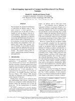
Figure 1A shows a typical FESEM image of initial ZnO
nanoseeds that prepared on the FTO surface via an alco-
holthermal process. As can be noticed from the i mage,
high-density nanoseeds with a relatively uniform parti cle
size of approximately 5 nm and distributed homoge-
nously throughout the surface were obtaine d using this
approach. The bigger background structures are FTO
crystals. After following a growth process in a growth
solution that contains, for example, 0.01 M Zn
(CH
3
COO)
2
and 0.036 M NH
3
(standard reaction), these
nanoseeds grew up to large-scale vertically oriented
quasi-1D-nanostructures and covered the entirity of the
substrate surface (Figure 1B). As revealed in Figure 1B,
such high-density quasi-NRs interestingly produce
considerably highly porous nanostructured-films of ZnO,
a structure that is demanded in photoelectrochemical
devices applications for facilitating an active redox reac-
tion. The cross-se ctional image taken from the same
samples further confirmed that the nanostructures were
1D like structures, which emerge from the initial ZnO
nanoseed part icles (Figure 1C). The lengths of the struc-
tures are approximately 70 nm. However, because of
the limited resolution of our SEM machine (Figure 1C),
a detailed pic ture of the vertic al orientation of ZnO
quasi-NRs that were prepared using this prescription
could not be obtained at the moment. Though, a much
clearer picture of vertical orientation of ZnO quasi-NRs
could be obtained if they were pre pared in a higher zinc
salt concentration which will be discussed later. As
revealed in the higher-magnification FESEM image, the
quasi-NRs have the preference to collide and fuse each
other at the top-end of the structure, producing big and
high contrast particles on the surface. This can be
directly related to the result of surface energy minimisa-
tion process in ZnO nanocrystals that evolved in such
high kinetic activity.
Meanwhile, on the dimension of the quasi-NRs, in spite
of such intense aggregates amongst the nanostructures,
on the basis of available free-standing individual quasi-
NRs (see dotted circles in high-resolution image in Figure
1D); the diameter can be estimated to be approximately
30 nm. It is true that the present quasi-NRs are relatively
inferior in terms of morphology and orientation control
compared to those currently obtained using other syn-
thetic methods. However, the present technique at least
provides an alternative way for a rapid formation of
quasi-1D ZnO nanostructures films directly on the sur-
face. Improved and controlled morphology might be
achieved later if suitable conditions are obtained, for
example via a surfactant modification.
Ali Umar et al . Nanoscale Research Letters 2011, 6:564
/>Page 3 of 12
It is important to note here that the nanoseeds are
necessary for the preparation of quasi-NRs morphology.
If they were absent on the surface, no quasi-NRs pro-
ducts were obtained. Irregular and big nanostructures
sometimes were found on the surface instead. Howev er,
these could be the precipitates that formed in the solu-
tion which then attached onto the surface.
Unlike in the growth of most metaloxide nanostructures
prepared by ammonia [29] or strong base-mediated
decomposition such as in the preparation of CuO nano-
wires [30,31] that produced intermediate metal complexes
byproducts [32], the present technique surprisingly pro-
duced pure ZnO phase only, evident in the XRD result
shown in Figure 2. This definitely could be the result of an
effective olatio n process of Zn-co mplexes on the ZnO
nanoseed surface in the formation of quasi-NRs (will be
discussed later) that efficiently t ransformed them into
the pure ZnO. Thus, no Zn-complexes existed in the
A
B
D
C
ZnO
FTO
Figure 1 ZnO nanoseeds on the FTO surface. (A) FESEM image of initial ZnO nanoseeds on the FTO surface and (B) after being grown for
approximately 5 min in the mixture of 10 mL of 0.01 M Zn(CH
3
COO)
2
and 36 mM ammonia (standard reaction) producing vertically oriented
ZnO quasi-NRs. (C) and (D) are its cross-section and high magnification images, respectively. Dotted circles in Figure 1D indicate available free-
standing individual ZnO quasi-NRs. Scale bar is 100 nm.
Ali Umar et al . Nanoscale Research Letters 2011, 6:564
/>Page 4 of 12
quasi-NRs structures. The result is particularly important
and advantageous because, as for those with the presence
of other phases, an after growth annealing process was
normally required to facilitate complex removal and pro-
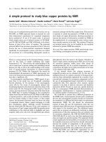
duce high-puri ty ZnO system [29-31]. As can be noticed
in Figure 2c, the XRD profile for the as-prepared samples,
five prominent peaks at 31.7, 34.4, 36.25, 47.5 and 56.5
besides other peaks indicated by asterisks are apparent on
the spectrum. According to the JCPDS (file no. 79-2205),
the spectrum can be indexed as the he xagonal w urtzite
structure (cell constant of a = 3.2501 A and c = 5.2071 A)
of ZnO with peaks corresponding to (100), (002), (101),
(102) and (110) planes, respectively. The peaks with aster-
isks are assigned t o the diffraction peaks from the FTO
crystal substrate (see curve a of Figure 2). As also evident
in Figure 2c, no peaks related to other zinc complexes are
observed, confirming the phase purity of ZnO nanocrys-
tals. A similar spectrum was also obtained for the nano-
seeds as shown in curve b, ascertaining the phase purity of
the nanoseeds from which the quasi-NRs are grown up. In
spite of the fact that the as-prepared quasi-NRs are pure
ZnO, we also examined th e effect of annealing treatment
at 350°C in air on the crystallinity of the samples. How-
ever, interestingly the XRD profile was noticed to be rela-
tively unchanged as judged from the height and the width
of the peaks, inferring that the as-prepared sa mples have
been t hrough a highly pure ZnO phase so that anne aling
treatment will give no effect to the modification of their
crystallinity. Thus, these results further confirmed the cap-
ability of the present technique to produce highly pure
ZnO quasi-NRs immediately from the solution.
On the quasi-NRs crystals growth direction, as is evident
from the XRD results, the preferred growth orientation of
the quasi-NRs might be towards [002] direction judging
from the appearance of relatively higher peaks belonging
to this crystallographic plane on the spectrum. The peak
ratio between this plane and (101) is as high as approxi-
mately 1.5 to 2.0, which is much higher comp ared to the
standard ZnO XRD data (JCPDS 01-079-2205), namely
approximately 0.5. This result agrees well with those
obtained from most ZnO na norods prepared us ing, e.g.
hydrothermal or other techniques [22,23] in which the
[002] is the main cr ystal gro wth orientation of the ZnO
nanorods. It is true that HRTEM analysis is required for
determining the growth orientation of the quasi-NRs.
Since the apparatus is u navailable at the moment, a
detailed analysis on the crystal growth orientation is being
pursued and will be reported in a separate publication.
On the basis of the experimental results, we confirmed
that the present approach has successfully promoted the
(100)
(002)
(101)
(102)
(110)
*
*
*
*
*
*
*
*
*
*
*
*
* = FTO
a
b
c
d
28 33 38 43 48 53 58
2 θ / deg. (°)
Intensity (a.u.)
Figure 2 X-ray diffraction spectra. X-ray diffraction spectrum of the (b) ZnO nanoseeds, (c) the as-prepared ZnO quasi-NRs and (d) ZnO quasi-
NRs after annealed at 350 C. (a) is XRD for FTO background substrate.
Ali Umar et al . Nanoscale Research Letters 2011, 6:564
/>Page 5 of 12
formation of ZnO quasi-NRs from the nanoseed parti-
cles. However, at the moment, the mechanism o f growth
is still not yet well understood. Though, we thought that
the growth characteristic of the present system seems
identical to the formation of CuO nanowires as reported
in [28] . As has been well known, when an aqueous metal
salts solution, such as Zn(CH
3
OO)
2
here, was introduced
to the NH
3
, unstable zinc-ammonium complexes might
be formed at the first instance. They then rapidly trans-
formed into zinc hydroxides, more stable zinc complexes
in solution. I n the pre sence of ZnO nanoseeds on the
surface, as confirmed by the XRD shown in Figure 2,
these complexes might transform into tetragonal ZnO
4
phases that initiates the formation of O-Zn-O bridges
with the nanoseeds via an olation process [31,33]. Thus,
the nanorod structures were projected. In the present
work, unsuccessful coordinated zinc hydroxide com-
plexes might apparently be formed, but remained in bulk
solution in the form of white-bluish suspension. If
attached onto the surface, it can be easily washed out by
rinsing with excessive water.
It needs t o be noted here that to pro duce quasi-NRs
morphology, the stirring process is necessary in this proce-
dure. If there were no stirring, no quasi-NRs growths were
obtained, but a thin f ilms structure composed of quasi-
spherical particles instead. It is typical in the present pro-
cedure that the zinc complexes were rapidly hydrolysed in
the solution upon the addit ion of amm onia (see gro wth
process in section 2.2.). The hydrolysed complexes easily
aggregate on each other forming a bluish colour in solu-
tion and at a certain condition they precipitate down to
the bottom of the vials. In order to maintain the formation
of ZnO quasi-NRs on the surface, the zinc complexes pre-
cursors’ availability near the nanoseed surface should be
sufficient and be controlled. For that reason, the zinc com-
plexes have to be quickly transported to the vicinity of the
nanoseed surface by means o f stirring shortly after being
hydrolysed. Thus, quasi-1D morphology can be formed.
The concentrations of ammonia and zinc salt used in the
reaction were found to noticeably affect the structural
growth (diameter and length) of the ZnO quasi-NRs on
the surface. For the case of the ammonia, firstly, it is noted
that the concentration which prom otes th e formation of
quasi-NRs morphology is in the range of 36 to 360 mM. If
the a mmonia concentration is outside this range, for exam-
ple lower than this value, no quasi-NRs were obtained, but
instead irregular shape particlesfilmformedonthesurface.
This could probably be associated with the limited precur-
sor availability as a result of a weak hydrolysis process
under such low ammonia concentration. Meanwhile, when
the ammonia is higher (>360 mM), no or limited quasi-
NRs growth was obtained. At this condition, highly com-
pact quasi-spherical nanostructures films were obtained.
This could be the result of solution instability under such
high ammonia concentration in which the zinc complexes
extremely formed and agglomerated in solution that in
turn hindered the olation process on the nanoseed surface.
Figure 3 shows typical FESEM images of ZnO quasi-NRs
that were prepared using four different ammonia concen-
trations, namely 36 (standard reaction), 180, 288 and
360 mM, with zinc salt fixed at 10 mM. From the image,
at a certain ammonia concentrati on, it is see n that the
quasi-NRs efficiently grew up to large-scale producing
high-density vertical quasi-NRs array films on the surface.
Further analysis on the surface morphology found inter-
estingly that the quasi-NRs density relatively increased
with the increasing of ammonia concentration. On the
quasi-NRs diameter, to tell the truth, due to extreme
aggregation amongst the quasi-NRs, it is quite difficult to
obtain the diameter of the quasi-NRs. However, judging
from the “grain size” of the nanostructures on the surface
that visibly reduced with the increasing of ammonia, it can
be remarked that the quasi-NRs diameter should also
decrease with the increasing of ammonia. On the basis of
available free-standing quasi-NRs, the di ameter was seen
to decrease from 30 nm to 15 nm for ammonia concentra-
tion increasing from 36 to 360 mM, inferring an essential
effect of ammonia on the structural growth of ZnO quasi-
NRs. Similar to what was obtained in the diameter, the
nanorods length was also significantly modified upon var-
iation of ammonia concentration. From the cross-sectional
analysis, it was revealed that the quasi-NRs length
expanded from 70 to 80 nm when the ammonia concen-
tration was increased from 36 to 360 mM.
In addition, besides modifying the diameter and the
length, the variation of ammonia also significantly alters
the o verall nanorod density on the surface; namely it
improves with the increasing of ammonia concentration.
Unfortunately, contrary to such enhancement in the den-
sity, the augmentation of ammonia induced extreme coa-
lescence amongst the quasi-NRs at their top-end as the
result of surface energy minimisation, generating bigger or
irregular-shaped nanostructures on the surface that hides
the underneath structure of individual quasi-NRs (see
Figure 3).
Similar to what has been obtained in the ammonia con-
centration variation, a substantial modification on the
quasi-NRs morphology was obtained w hen the zinc salt
concentration was altered. I n the typical process, the quasi-
NRs morphology becomes more rounded and “fatter” with
the increasing of zinc salt concentration as can be noticed
in the cross-section image in Figure 4D. Analysis on the
quasi-NRs diameter found that it significantly increases
if the zinc salt concentration was augmented. For example,
the quasi-NRs diameter was approximately 30 nm if pre-
pared using the standard solution (zinc salt concentration
Ali Umar et al . Nanoscale Research Letters 2011, 6:564
/>Page 6 of 12
of 10 mM). It efficiently grew up to approximately 40 nm if
the zinc salt used was augmented to 30 mM. As a conse-
quence of the diameter increase, as seen in the image, the
quasi-NRs array became denser, producing solid film struc-
tures instead of porous morphology as ob tained in those
prepared using the low zinc concentration. Regarding the
quasi-NRs length, it also indicated an effective increase
namely from 80 to 11 0 nm when t he zinc salt was changed
from 10 to 30 mM, correspondingly, suggesting the
controllability of the nanostructure morphology using the
present method.
Up to this stage, the quasi-NRs diameter and de nsity
could more o r less be adjusted via an ammonia and zinc
salt concentration variation. However, frankly, effective
control on the quasi-NRs length via one-step growth pro-
cess was not obtained. We thought that this presumably
was correlated with the nature of the reaction in which
the zinc salt underwent an extreme rapid hydrolysis and
A
DC
B
Figure 3 FESEM and cross-section images of ZnO quasi-NRs.Preparedin10mMofZn(CH
3
COO)
2
with different ammonia concentration,
namely (A) 36 (standard reaction), (B) 180, (C) 288 and (D) 360 mM. Scale bar is 100 nm.
Ali Umar et al . Nanoscale Research Letters 2011, 6:564
/>Page 7 of 12
quickly completed in solution, i.e. only within 4 to 5 min
of the reaction. Thus, sufficient precursors for maintain-
ingthekineticgrowthprocessareprobablyunavailable.
During the injection of ammonia into the reaction, at the
beginning each nanoseed probably quickly projected
small nanorod structures with high density on the sur-
face.Inanidealcase,thenanorodsshouldfurthergrow
until the entire precursors are consumed and prom ote
long nanorod formation on the surface. However, active
hydrolysis of zinc salt drove the formation of massive
zinc complexes (precursors for quasi-NRs) in solution
and aggregated on each other instead of supporting the
olation process on the nanoseed surface. Therefore, the
quasi-NRs growth was stopped earlier and their length
was less developed. However, this could be overcome by
using a multiple growth process to provide sufficient pre-
cursor materials in order to support a longer quasi-NRs
growth.Byusingastandardgrowthsolutionthatcon-
tained 10 mM of zinc salt and 36 mM of ammonia, the
length of the quasi-NRs could be effectively increased
B
C
ZnO
FTO
D
A
Figure 4 FESEM and cross-section images of ZnO quasi-NRs. Prepared in three different Zn(CH
3
COO)
2
, namely (A) 10, (B) 20 and (C) 30 mM
with ammonia concentration was fixed at 36 mM. (D) is a typical cross-section image for the sample (C). Scale bar is 100 nm.
Ali Umar et al . Nanoscale Research Letters 2011, 6:564
/>Page 8 of 12
from approximately 110 nm (under one cycle growth) to
approximately 220 nm if using four cycle’ s growth pro-
cess. The results are shown in Figure 5.
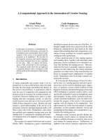
Figure 6 shows typical room-temperature optical absorp-
tion spectra of the as-prepared ZnO quasi-NRs films.
As can be noticed from the figure, one strong and one
small shoulder band at the UV region are recognised from
the spec trum. These two bands could be associated with
two separate excitonic characters of A- and B-excitons of
the ZnO quasi-NRs. The presence of such “clear splitting”
in the excitonic bands is quite surprising to us, since this
normally only appears in the nanocrystals that contain low
defect density; in other words , high-crystallinity [34]. In
nanocrystals with low-crystallinity and high defect density,
these peaks are broad and will overlap each other forming
a single broad absorption band in this region. Therefore,
although high-resolution TEM is not available at the
moment to confirm the real crystallinity of the nanorods,
A
B
C
D
220 nm
175 nm
120 nm
110 nm
FTO
ZnO
Figure 5 Cross-section image of ZnO quasi-NRs prepared using different cycle’s growth (multiple) process. (A) 1, (B) 2, (C) 3 and (D) 4
cycles. The growth solution used contained 10 mM of zinc salts and 36 mM of ammonia. The growth time for each cycle is 4 min. The scale
bars are 100 nm.
Ali Umar et al . Nanoscale Research Letters 2011, 6:564
/>Page 9 of 12
on the basis of this result it is worthwhile to conclude that
the ZnO quasi-NRs prepared using the present approach is
high crystallinity in nature. It is true that the B-e xciton
band obtained here is still relatively small. This could be
ass ociated with the nature of the quasi-NRs crystallinity,
e.g. crystallinity degree or defect content, etc., of the nano-
crystals. In addition to these interesting absorption bands,
two other bands in the visible region, namely at 450 to
550 nm and 600 to 700 nm, are also apparent in the spec-
trum. This result is actually different from those normally
obtained in most ZnO films, in which no absorption band
appeared in this region. Since we used FTO on glass as the
substrate, which normally produces an artificial wave
pattern at the glass-FTO interface due to an internal reflec-
tion, one could have thought that these might come
from the contribution o f this process to the spectrum.
However, since the optical absorption of the sample was
recorded via a double-beam spectrometer in which the
substrate absorption contribution t o the spectrum has
been deducted, we conclude that the obtained spectrum
could be the special characteristics of the optical absorp-
tion of the ZnO sample with the current struct ure. The
bands could be related to several physical processes in the
nanocrystals such as singlet excitation in ionised oxygen
vacancy [35], z inc interstitial [36-38] or antisite oxygen
defect level-related absorption [39]. Even so, a more
detailed analysis on the optical properties of the ZnO
quasi-NRs on FTO substrate is being pursued and will be
reported in a subsequent paper.
Conclusions
An alternative method for the formation of vertically
oriented ZnO quasi-NRs growth on the s urface via 1D
crystal growth of nanoseeds under a rapid hydrolysis of
zinc complexes in the presence of ammonia has been
demonstrated. In a typical process, high-density verti-
cally oriented ZnO quasi -NRs with diameter and length
in the range of approximately 30 and 110 nm, r espec-
tively, was the characteristic of the product s. Quasi-NRs
were found not to freely stand but leant on each other
andcombinedatthetopofthenanarodsprobablyas
the results of coalescing process of several quasi-NRs.
The growth process was very quick; namely in the range
of 4 t o 5 min. The quasi-NRs morphology was influ-
enced by the concentration of ammonia used in the
reaction. In typical results, the quasi-NRs shape
becomes more rounded and fatter with the increasing of
ammonia concentration. Meanwhile, the diameter of the
quasi-NRs decreased with the increasing of ammonia
concentration. The as-prepared quasi-NRs products
300
Wavelength (nm)
Absorbance
400 500 600 700 800
0
0.2
0.4
0.6
0.8
A-exciton
B-exciton
Figure 6 Typical UV-VIS optical absorption spectrum of ZnO quasi-NRs. Two separate excitonic characters, namely A- and B-excitons, were
observed in the spectrum, reflecting the ZnO quasi-NRs are high-crystallinity in nature.
Ali Umar et al . Nanoscale Research Letters 2011, 6:564
/>Page 10 of 12
were pure ZnO phase without the presence of any zinc
complexes and feature a relatively high-crystalli nity
property as confirmed by XRD and optical absorption
spectroscopy results, respectively.
As for the mechanism, the quasi-NRs were projected
from the nanoseeds probably due to an olation process
of zinc complex[31,33], such as zinc hydroxide, on the
surface of ZnO nanoseeds, a process that is similar to
what has been obtained in CuO nanorods [28].
At present, ZnO quasi-NRs with free-standing and a
controlled morphology has not yet been achieved; how-
ever, the present method may become a potential alterna-
tive for the preparation of ZnO nanorods on the surface.
Since the quasi-NRs morphology exhibited a relative
dependence on the ammonia and zinc salt concentrations,
ZnO quasi-NRs with controlled morphology will be rea-
lised if suitable conditions were obtained; for example by
utilising the surfactants. The study o n this effect is in
progress.
Abbreviations
Quasi-1D, quasi-one-dimensional; quasi-NRs, quasi-nanorods.
Acknowledgements
We acknowledge the support from the Universiti Kebangsaan Malaysia and
Ministry of Higher Education of Malaysia under research grant UKM-GUP-
NBT-08-25-086 and UKM-RRR1-07-FRGS0037-2009 and the Universiti Tenaga
Nasional and Ministry of Science and Technology and Innovation Malaysia
under Science Fund 03-02-03-SF0196 project.
Author details
1
Institute of Microengineering and Nanoelectronics (IMEN), Universiti
Kebangsaan Malaysia, 43600, Bangi, Selangor, Malaysia
2
College of
Engineering, Universiti Tenaga Nasional, 43000, Kajang, Selangor, Malaysia
3
Department of Materials Chemistry, Graduate School of Engineering, Kyoto
University, Nishikyo-ku, Kyoto 615-8520 Japan
Authors’ contributions
RT carried out nanostructure preparation and characterisation and drafted
the manuscript. AAU designed the concept and experiment, analysed the
results and revised and finalised the manuscript. MYAR participated in data
analysis and ideas. MMS provided the facilities and discussed the results. MO
provided the concept of the growth process of the nanostructures. All the
authors contributed to the preparation and revision of the manus cript and
approved its final version.
Competing interests
The authors declare that they have no competing interests.
Received: 11 August 2011 Accepted: 25 October 2011
Published: 25 October 2011
References
1. Wei Y, Ding Y, Li C, Xu S, Ryo JH, Dupuis R, Sood AK, Polla DL, Wang ZL:
Growth of vertically aligned ZnO nanobelt arrays on GaN substrate. J
Phy Chem C 2008, 112:18935-18937.
2. Lao JY, Wen JG, Ren ZF: Hierarchical ZnO Nanostructures. Nano Lett 2002,
2:1287-1291.
3. Bae SY, Seo HW, Choi HC, Park J: Heterostructures of ZnO nanorods with
various one-dimensional nanostructures. J Phys Chem B 2004,
108:12318-12326.
4. Gao PX, Ding Y, Wang ZL: Crystallographic orientation-aligned ZnO
nanorods grown by a tin catalyst. Nano Lett 2003, 3:1315-1320.
5. Umar A, Ribeiro C, Al-Hajry A, Masuda Y, Hahn YB: Growth of highly c-axis-
oriented ZnO nanorods on ZnO/glass substrate: growth mechanism,
structural, and optical properties. J Phys Chem C 2009, 113:14715-14720.
6. Yi GC, Wang C, Park WI: ZnO nanorods: synthesis, characterization and
applications. Semicond Sci Technol 2005, 20:S22-S34.
7. Bao J, Zimmler MA, Capasso F, Wang X, Ren ZF: Broadband ZnO single-
nanowire light-emitting diode. Nano Lett 2006, 6:1719-1722.
8. Olson DC, Lee YJ, White MS, Kopidakis N, Shaheen SE, Ginley DS, Voigt JA,
Hsu JWP: Effect of polymer processing on the performance of poly(3-
hexylthiophene)/ZnO nanorod photovoltaic devices. J Phys Chem C 2007,
111:16640-16645.
9. Goldberger J, Sirbuly DJ, Law M, Yang P: ZnO nanowire transistors. J Phys
Chem B 2005, 109:9-14.
10. Sadaf JR, Israr MQ, Kishwar S, Nur O, Willander M: White
electroluminescence using ZnO nanotubes/GaN heterostructure light-
emitting diode. Nanoscale Res Lett 2010, 5:957-960.
11. Fang X, Li J, Zhao D, Shen D, Li B, Wang X: Phosphorus-doped p-Type
ZnO nanorods and ZnO nanorod p-n homojunction LED fabricated by
hydrothermal method. J Phys Chem C 2009, 113:21208-21212.
12. Wang M, Fei GT, Zhang LD: Porous-ZnO-nanobelt film as recyclable
photocatalysts with enhanced photocatalytic activity. Nanoscale Res Lett
2010, 5 :1800-1803.
13. Martinson ABF, Elam JW, Hupp JT, Pellin MJ: ZnO nanotube based dye-
sensitized solar cells. Nano Lett 2007, 7:2183-2187.
14. Luan C, Vaneski A, Susha AS, Xu X, Wang H-E, Chen X, Xu J, Zhang W,
Lee C-S, Rogach AL, Zapien JA: Facile solution growth of vertically aligned
ZnO nanorods sensitized with aqueous CdS and CdSe quantum dots for
photovoltaic applications. Nanoscale Res Lett 2011, 6.
15. Wang X, Summers CJ, Wang ZL: Large-scale hexagonal-patterned growth
of aligned ZnO nanorods for nano-optoelectronics and nanosensor
arrays. Nano
Lett 2004, 4:423-426.
16. Park WI, Yi GC, Kim M, Pennycook SJ: ZnO nanoneedles grown vertically
on Si substrates by non-catalytic vapor-phase epitaxy. Adv Mater 2002,
14:1841-1843.
17. Calestani D, Zha MZ, Zanotti L, Villani M, Zappettini A: Low temperature
thermal evaporation growth of aligned ZnO nanorods on ZnO film: a
growth mechanism promoted by Zn nanoclusters on polar surfaces.
CrystEngComm 2011, 13:1707-1712.
18. Huang MH, Wu Y, Feick H, Tran N, Weber E, Yang P: Catalytic growth of
zinc oxide nanowires by vapor transport. Adv Mater 2001, 13:113-116.
19. Liu X, Wu X, Cao H, Chang RPH: Growth mechanism and properties of
ZnO nanorods synthesized by plasma-enhanced chemical vapor
deposition. J Appl Phys 2004, 95:3141-3147.
20. Wang L, Zhang X, Zhao S, Zhou G, Zhou Y, Qi J: Synthesis of well-aligned
ZnO nanowires by simple physical vapor deposition on c -oriented ZnO
thin films without catalysts or additives. Appl Phys Lett 2005, 86:024108-
024101-024108-024103.
21. Jie J, Wang G, Wang Q, Chen Y, Han X, Wang X, Hou JG: Synthesis and
characterization of aligned ZnO nanorods on porous aluminum oxide
template. J Phys Chem B 2004, 108:11976-11980.
22. Vayssieres L: Growth of arrayed nanorods and nanowires of ZnO from
aqueous solutions. Adv Mater 2003, 15:464-466.
23. Liu B, Zeng HC: Hydrothermal synthesis of ZnO nanorods in the
diameter regime of 50 nm. J Am Chem Soc 2003, 125:4430-4431.
24. Baruah S, Dutta J: Hydrothermal growth of ZnO nanostructures. Sci
Technol Adv Mater 2009, 10.
25. Kim TU, Kim JA, Pawar SM, Moon JH, Kim JH: Creation of nanoscale two-
dimensional patterns of ZnO nanorods using laser interference
lithography followed by hydrothermal synthesis at 90°C. Cryst Growth
Des 2010, 10:4256-4261.
26. Warule SS, Chaudhari NS, Kale BB, More MA: Novel sonochemical assisted
hydrothermal approach towards the controllable synthesis of ZnO
nanorods, nanocups and nanoneedles and their photocatalytic study.
CrystEngComm 2009, 11:2776-2783.
27. Hu Y, Qian H, Liu Y, Du G, Zhang F, Wang L, Hu X: A microwave-assisted
rapid route to synthesize ZnO/ZnS core-shell nanostructures via
controllable surface sulfidation of ZnO nanorods. CrystEngComm 2011,
13:3438-3443.
28. Umar AA, Oyama M: A seed-mediated growth method for vertical array
of single-crystalline CuO nanowires on surfaces. Cryst Growth Des 2007,
7:2404-2409.
Ali Umar et al . Nanoscale Research Letters 2011, 6:564
/>Page 11 of 12
29. Xiao Y, Li L, Li Y, Fang M, Zhang L: Synthesis of mesoporous ZnO
nanowires through a simple in situ precipitation method. Nanotechnology
2005, 16:671-674.
30. Song X, Sun S, Zhang W, Yu H, Fan W: Synthesis of Cu(OH)2 nanowires at
aqueous-organic interfaces. J Phys Chem B 2004, 108:5200-5205.
31. Lu C, Qi L, Yang J, Zhang D, Wu N, Ma J: Simple template-free solution
route for the controlled synthesis of Cu(OH)2 and CuO nanostructures. J
Phys Chem B 2004, 108:17825-17831.
32. Wang ZL, Kong XY, Wen X, Yang S: In situ structure evolution from Cu
(OH)2 nanobelts to copper nanowires. J Phys Chem B 2003,
107:8275-8280.
33. Du GH, Van Tendeloo G: Cu(OH)2 nanowires, CuO nanowires and CuO
nanobelts. Chem Phys Lett 2004, 393:64-69.
34. Lee GH: Optical properties of ZnO thin films on LiNbO3 and LiTaO 3
substrates grown by pulsed laser deposition. Sol Sta Commun 2003,
128:351-354.
35. Vanheusden K, Warren WL, Seager CH, Tallant DR, Voigt JA, Gnade BE:
Mechanisms behind green photoluminescence in ZnO phosphor
powders. J Appl Phys 1996, 79:7983-7990.
36. Liu M, Kitai AH, Mascher P: Point defects and luminescence centres in
zinc oxide and zinc oxide doped with manganese. J Lumin 1992,
54:35-42.
37. Studenikin SA, Cocivera M: Time-resolved luminescence and
photoconductivity of polycrystalline ZnO films. J Appl Phys 2002, 91:5060.
38. Reynolds DC, Look DC, Jogai B: Fine structure on the green band in ZnO.
J Appl Phys 2001, 89:6189-6191.
39. Jiao Y, Zhu HJ, Zhou MJ, Wang XF, Li Q: Suppression of green emission in
ZnO nanorodssa discussion on surface and interior structural quality
Manipulation. J Phys Chem C 2010, 114:208-211.
doi:10.1186/1556-276X-6-564
Cite this article as: Ali Umar et al.: A simple route to vertical array of
quasi-1D ZnO nanofilms on FTO surfaces: 1D-crystal growth of
nanoseeds under ammonia-assisted hydrolysis process. Nanoscale
Research Letters 2011 6:564.
Submit your manuscript to a
journal and benefi t from:
7 Convenient online submission
7 Rigorous peer review
7 Immediate publication on acceptance
7 Open access: articles freely available online
7 High visibility within the fi eld
7 Retaining the copyright to your article
Submit your next manuscript at 7 springeropen.com
Ali Umar et al . Nanoscale Research Letters 2011, 6:564
/>Page 12 of 12