Báo cáo hóa học: " Structural and physical properties of antibacterial Ag-doped nano-hydroxyapatite synthesized at 100°C" pptx
Bạn đang xem bản rút gọn của tài liệu. Xem và tải ngay bản đầy đủ của tài liệu tại đây (712.68 KB, 8 trang )
NANO EXPRESS Open Access
Structural and physical properties of antibacterial
Ag-doped nano-hydroxyapatite synthesized at
100°C
Carmen Steluta Ciobanu
1
, Florian Massuyeau
2
, Liliana Violeta Constantin
3
and Daniela Predoi
1*
Abstract
Synthesis of nanosized particle of Ag-doped hydroxyapatite with anti bacterial properties is in the great interest in
the development of new biomedical applications. In this article, we propose a method for synthesized the Ag-
doped nanocrystalline hydroxyapatite. A silver-doped nanocrystalline hydroxyapatite was synthesized at 100°C in
deionized water. Other phase or impurities were not observed. Silver-doped hydroxyapatite nanoparticles (Ag:HAp)
were performed by setting the atomic ratio of Ag/[Ag + Ca] at 20% and [Ca + Ag]/P as 1.67. The X-ray diffraction
studies demonstrate that powders made by co-precipitation at 100°C exhibit the apatite characteristics with good
crystal structure and no new phase or impurity is found. The scanning electron microscopy (SEM) observations
suggest that these materials present a little different morphology, which reveals a homogeneous aspect of the
synthesized particles for all samples. The presence of calcium (Ca), phosphor (P), oxygen (O), and silver (Ag) in the
Ag:HAp is confirmed by energy dispersive X-ray (EDAX) analysis. FT-IR and FT-Raman spectroscopies revealed that
the presence of the various vibrational modes corresponds to phosphates and hydroxyl groups. The strain of
Staphylococcus aureus was used to evaluate the antibacterial activity of the Ca
10-x
Ag
x
(PO4)6(OH)2 (x = 0 and 0.2). In
vitro bacterial adhesion study indicated a significant difference between HAp (x = 0) and Ag:HAp (x = 0.2). The Ag:
Hap nanopowder showed higher inhibition.
1. Introduction
Inorganic biomaterials based on calcium orthophosphate
have their wide range of applications in medicine [1-4].
Among them, synthetic hydroxyapatite (HAP, Ca
10
(PO
4
)
6
(OH)
2
) is the most promising because of its biocompat-
ibility, bioactivity, and osteoconductivity. Hydroxyapatite
has been used to fill a w ide range of bony defects in
orthopedic and maxillofacial surgeries and dentistry
[5-8]. It has also been widely used as a coating for
metallic prostheses to improve their biological proper-
ties [9-11]. In recent years, the use of inorganic antibac-
terial agents has attracted interest for control of
microbes. The key advantages of inorganic antibacterial
agents are improved safety and stability [12-14]. The
most antibacterial inorganic materials are the ceramics
immobilizing antibacterial metals, such as silver and
copper. Hydroxyapatite (HAp) has widely been used for
bone repair and substitute because of its good biocom-
patibility, and the cation exchange rate of HAp is very
high with silver ions. Silver, known as a disinfectant for
many years, has a broad spectrum of antibacterial activ-
ity and exhibits low toxicity toward mammalian cells
[12]. The most common technique to incorporate Ag
into HAp coatings is via an ion exchange method, in
which the Ca ions in HAp are replaced by Ag ions
while dipping the HAp coatings into AgNO
3
for a per-
iod of time [15,16]. The limitation of the ion exchange
method is that Ag will reside mostly on the outer sur-
face of the c oating and will be quickly depleted in vivo/
in vitro without long-term antibacterial effect. In order
to achieve the continuous release of Ag, HAp coatings
doped with Ag through the entire thickness have been
developed using sol-gel [17,18], co-sputtering [19,20],
and thermal or cold spraying [21,22]. Although Ag in
small percentages can have an antibacterial effect, larger
amounts can be toxic [18], and ther efore optimization
of the Ag concentration in the coating is critical to
* Correspondence:
1
National Institute of Materials Physics, 105 bis Atomistilor, P.O. Box MG 07,
077125, Bucuresti-Magurele, Romania
Full list of author information is available at the end of the article
Ciobanu et al. Nanoscale Research Letters 2011, 6:613
/>© 2011 Ciobanu et al; licensee Springer. This is an Ope n Access article distributed under the ter ms of the Creative Comm ons
Attribution License (http://creativecom mons.org/licenses/by/2.0), which permits unrestricted use, distribu tion, and reproduction in
any medium, provided the original work is properly cited.
guarantee an optimum antibacterial effect without
cytotoxicity.
From the view point of biomedical engineering, the
element silver is well known for its broad spectrum anti-
bacterial effect at very low concentrations [23], and it
possesses many advantages, such as good antibacterial
ability, excellent biocompatibility, and satisfactory stabi-
lity [24,25]. The scientific literature points to the wide
use of silver in numerous applications. It is well estab-
lished that silver nanoparticles are known for their
strong antibacterial effects for a wide array of organisms
(e.g., viruses, bacteria, fungi) [26]. Therefore, silver
nanoparticles are widely used in medical devices and
supplies such as wound dressings, scaffold, skin dona-
tion, recipient sites, and sterilized materials in hospita ls,
medical catheters, contraceptive devices, surgical instru-
ments, bone prostheses, artificial teeth, and bone coat-
ing. One can also observe their wide use in consumer
products such as cosmetics, lotions, creams, toothpastes,
laundry detergents, soaps, surface cleaners, room sprays,
toys, antimicrobial paints, home appliances (e.g., wash-
ing machines, air, and water filters), automotive uphols-
tery, shoe insoles, brooms, food storage containers, and
textiles [27-30].
Previous studies have focused on preparation and
characterization of silver nanoparticles (AgNPs) [31].
The exact antibacterial action of AgNPs is not comple-
tely understood [32]. On the other hand in the litera-
ture, the studies on the preparation and characteriz ation
of the silver-doped hydroxyapatite powders are almost
absent. The antibacterial studies on the Ag:HAp nano-
powder are not presented, too.
In this article, we propose a method for synthesized
the nanocrystalline hydroxyapatite doped with Ag at
100°C. Preparation of Ag-doped hydroxyapatite by co-
precipitation method at 100°C has several advantages
over other techniques. Specifically, it can generate highly
crystalline nanopowder Ag:HAp. The Ag:HAp nanocrys-
talline powders will be used for implantable medical
devices. Ag-doped nanocrystallin e hydroxyapatite pow-
ders are obtained. Other phase or impurities were not
observed. The Ca
10-x
Ag
x
(PO
4
)
6
(OH)
2
with x = 0 and 0.2
was synthesized by co-precipitation method at 100°C.
The Ca
10-x
Ag
x
(PO
4
)
6
(OH)
2
with x = 0.2 was synthesized
by co-precipitation method at 100°C mixing AgNO3, Ca
(NO
3
)
2
·4H
2
O, and (NH
4
)
2
HPO
4
in deionized water.
The structure, morphology, vibrational, and optical
properties of the obtained samples were systematically
characterized by X-ray diffraction (XRD), scanning elec-
tron microscopy (SEM), transmission electron micro-
scopy (TEM), Fourier transform infrared (FT-IR), and
FT-Raman spectroscopies. For reveal the presence of the
silver in the Ag:HAp (x = 0. 2) nanopowder, the X-ray
photoelectron spectroscopy (XPS) results are presented,
too. In addition, the antibacterial activity of the Ca
10-
x
Ag
x
(PO
4
)
6
(OH)
2
with x = 0 and 0.2 is studied.
2. Experimental procedure
2.1. Sample preparation
All the reagents for synthesis including ammonium
dihydrogen phosphate [(NH
4
)
2
HPO
4
], calcium nitrate
[Ca(NO
3
)
2
·4H
2
O], and silver nitrate (AgNO
3
)(Alpha
Aesare) were purchased and used without further
purification.
The Ca
10-x
Ag
x
(PO
4
)
6
(OH)
2
, with x = 0 (HAp), ceramic
powder was prepared (Ca/P molar ratio–1:67) using Ca
(NO
3
)
2
·4H
2
Oand(NH
4
)
2
HPO
4
by co-precipitation. A
designed amount of ammonium dihydrogen phosphate
[(NH
4
)
2
HPO
4
] was di ssolved in deionized water to form
a 0.5-mol/L solution. A designed amount of calcium
nitrate tetrahydrate was also dissolved in deionized
water to form a 1.67-mol/L solution. The mixture was
stirred constantly for 2 h by a mechanical stirrer at 100°
C. The pH was constantly adjusted and kept at 10 dur-
ing the reaction. After the reaction, the deposited mix-
tures were washed several times with deionized water.
The res ulting mate rial (HAp) was dried at 100°C for 72
h in an electrical air oven.
Silver-doped hydroxyapatite nanoparticles, Ca
10-x
Ag
x
(PO
4
)
6
(OH)
2
, with x = 0.2 (Ag:HAp), were performed by
setting the atomic ratio of Ag/[Ag + Ca] at 20% and [Ca
+ Ag]/P as 1.67. The AgNO
3
and Ca(NO
3
)
2
·4H
2
O
were dissolved in deionized water to obtain 300 mL [Ca
+ Ag]-containing so lution. On the other hand, the
(NH
4
)
2
HPO
4
was dissolved in deionized water to make
300 mL P-containing solution. The [Ca + Ag]-contain-
ing solution was put into a Berzelius and stirred at 100°
C for 30 min. Meanwhile, the pH of P-containing solu-
tion was adjusted to 10 with NH
3
and stirred continu-
ously for 30 min. The P-containing solution was added
drop-by-drop into the [Ca + Ag]-containing solution
andstirredfor2handthepHwas constantly adjusted
and kept at 10 during the reaction. After the reaction,
the deposited mixtures were washed several times with
deionized water. The resulting material was dried at
100°C for 72 h.
2.2 Sample characterization
2.2.1. XRD
The XRD was performed on a Bruker D8 Advance dif-
fractometer, with nickel-filtered Cu Kμ (l = 1.5418 Å)
radiation, and a high efficiency one-dimensional detector
(Lynx Eye type) operated in integration mode. The dif-
fraction patterns were collected in the 2θ range 15°-
140°, with a step size of 0.02° and 34 s measuring time
per step. In an attempt to perform a complete XRD
characterization of the nano-powders, the measured
data were processed with the MAUD software, version
Ciobanu et al. Nanoscale Research Letters 2011, 6:613
/>Page 2 of 8
2.26 [33]. The instrumental line broadening has been
evaluated using a heat-treated ceria powder proved to
produce no observable size or strain line broadening.
2.2.2. Scanning electron microscopy
The structur e and morphology of the samples were stu-
died using a HITACHI S2600N-type scanning electron
microscope (SEM), operating at 25 kV in vacuum. The
SEM studies were performed on powder samples. For
the elemental analysis, the electron microscope was
equipped with an energy dispersive X-ray attachment
(EDAX/2001 device).
2.2.4. TEM
TEM studies were carried out using a JEOL 200 CX.
The specimen for TEM imaging was prepared from the
particles suspension in deio nized water. A drop of w ell-
dispersed supernatant was p laced on a carbon-coated
200 mesh copper grid, followed by drying the sample at
ambient conditions before it is attached to the sample
holder on the microscope.
2.2.5. FT-IR spectroscopy
The functional groups present in the prepared powder
and in the powders calcined at different temperatures
were identified by FT-IR (Bruker Vertex 7 Spectro-
meter). For this, 1% of the powder was mixed and
ground with 99% KBr. Tablets of 10 mm diameter for
FTIR measurements were prepared by pressing the pow-
der mixture at a load of 5 tons for 2 min and the spec-
trum was taken in the range of 400-4000 cm
-1
with
resolution 4 and 128 times scanning.
2.2.6. FT-Raman spectroscopy
Raman studies have been carried out at the wavelength
excitation of 1064 nm using an FT Raman Bruker RFS
100 spectrophotometer. The laser was operated at 100
mWandupto100scansat4cm
-1
resolution were
accumulated.
2.2.7. XPS
Soft XPS is one of the most important techniques for
the study of the elemental ratios in the surface region.
The surface sensitivity (typically 40-100 Å) makes this
technique ideal for measurements as oxidation states or
biomaterials powder. In this analysis, we have used a
VG ESCA 3 MK I I XPS installation (E
ka
= 1486.7 eV).
The vacuum analysis chamber pressure was P~3×10
-8
torr. The XPS r ecorded spectrum involved an energy
window w =20eVwiththeresolutionR =50eVwith
256 recording channels. The XPS spectra were pro-
cessed using Spectral Data Processor v 2.3 (SDP)
software.
2.2.8. In vitro antibacterial activity
The strains of bacteria used for this study were the
strain of Stap hylo coccus aureus (ATCC 6538). The sta-
phylococci were grown overnight in Todd-Hewit broth
supplemented with 1% yeast extract at 37°C, followed by
centrifuging. The supernatants were discarded and
pellets were re-suspended in phosphate-buffered saline
(PBS) followed by a second centrifuging and re-suspen-
sion in PBS. The samples to be tested were placed in 50
mL sterilized tubs followed by the addition of 2 mL of
the bacterial suspension. The tubes were incubated at
37°C for 4 h . At t he end of the incubation period, the
samples were gently rinsed three times with PBS. The
non-adherent bacteria were eliminated. After washing,
the samples were then put into a new tube containing 5
mL PBS and vigorousl y vortexed for 30 s to remove the
adhering microorganisms. The viable organisms in the
buffer were quantified by plating serial dilutions on
yeast extract agar plates. Yeast extract agar plates were
incubated for 24 h at 37°C and the colony forming units
were counted visually.
3. Results and discussions
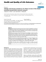
The XRD patterns, presented in Figure 1, show the
characteristic peaks of hydroxyapatite for each sample,
according to ICDD-PDF no. 9-432, represented at the
bottom of the figure, as reference. No other crystalline
phases were detected beside this phase (Figure 1).
We performed whole powder pattern fitting by the
Rietveld method of the as-prepared Ag-HAp structures.
As a prerequisite f or the atomic structure refinement, a
good fit of the diffraction line profiles must be achieved.
Because the peaks’ broadening is related to the micro-
structural characteristics (crystallite size and micro-
strain) a suitable microstructure model is needed. Good
pattern fit has been achieved using MAUD [33] for all
the samples, by applying the Po pa approach for the ani-
sotropic microstructure analysis [34], implemented in
Figure 1 Comparative representation of the experimental XRD
patterns of the Ca
10-x
Ag
x
(PO
4
)
6
(OH)
2
samples synthesized xAg
= 0 (HAp) and xAg = 0.2 (Ag:HAp), and the characteristic lines
of hydroxyapatite according to the ICDD-PDF number 9-432.
Ciobanu et al. Nanoscale Research Letters 2011, 6:613
/>Page 3 of 8
the MAUD code as “Popa rules”. It resulted t hat each
sample is constituted of elongated nanocrystallites
which can be approximated by circular ellipsoids, with
the longer dimension parallel to the c crystallographic
axis of HAp.
For the undoped HAp, Ag:HAp the length of the aver-
age crystallite (the average column size parallel to the c-
axis) is around 43 nm and the width (the average col-
umn size perpendicular to the c-axis) is around 16 nm.
The mean crystallite size averaged over all crystallo-
graphic directions is around 21 nm. For Ag:HAp, the
length is around 38 nm and the width around 14 n m.
The averaged diameter is around 19 nm.
The XRD of HAp and Ag:HAp also demonstrate that
powders made by co-precipitation at 100°C exhibit the
apatite characteristics with good crystal structure and
no new phase or impurity is found.
Figure 2 displays the TEM images of pure HAp ( xAg
= 0) and Ag:HAp (xAg = 0.2) with low resolution. Fig-
ure 2 (left) shows that HAp particles at 100°C are crys-
tallized with a maximum size around 40 nm. In Figure 2
(right), the ellipsoidal-shaped Ag:HAp (xAg = 0.2) parti-
cles about 30 nm are observed after Ca
2+
is partially
substituted by Ag
+
. The substitu tion of Ca by Ag in the
apatite structure leads to slight changes in the shapes of
the nanoparticles. The morphology identifications indi-
cated that the nanoparticles with good crystal structure
could be made at 100°C by co-precipitation method.
SEM (Figure 3) image and EDAX (Figure 4) spectrum
of Ca
10-x
Ag
x
(PO
4
)
6
(OH)
2
, with x = 0 and 0.2, are shown.
The morphology of the nanoparticles of HAp and Ag:
HAp was investigated by SEM. SEM images provide the
direct information about the size and typical shape of
the as-prepared samples. The results suggest that the
doping Ag
+
has little influence on the morphology of
the HAp. The samples prepared at the atomic ratio Ag/
[Ag + Ca] 20% (Ag:HAp) exhibit much smaller particle
size. Elemental maps for the samples prepared at the
atomic ratio Ag/[Ag + Ca] 20% are also shown. The
spectrum and images confirmed the presence of silver
on hydroxyapatite. The EDAX spectrum of Ag:HAp
confirms the presence of calcium (Ca), phosphor (P),
oxygen (O), and silver (Ag) in the samples.
XPS technique has been tested as a useful tool for
qualitatively determining the surface components and
composition of the samples. Figure 5 shows the survey
XPS narrow scan spectra of Ag:HAp (x = 0.2) nanopow-
der obtained at 100°C and XPS narrow scan spectra of
Ag element. In the XPS spectrum of Ag:HAp, the bind-
ing energy of Ca (2p, 347.3 eV), O (1s, 532.1 eV), and P
(2p, 133.09 eV) can obviously be found (Figure 5A). The
peaks of Ag (Ag(3d
5/2
) 368.4 eV and Ag((3d
3/2
) 374.3
eV) agree well with the literature [35]. XPS narrow scan
spectra of Ag element are presented in Figure 5B. XPS
results provide the additional evidence for the successful
doping of Ag
+
, in Ag:HAp.
FT-IR spectroscopy was performed to investigate the
functional groups present in nanohydroxyapatite, Ca
10-
x
Ag
x
(PO
4
)
6
(OH)
2
,withx = 0 and 0.2 obtained at 100°C
by co-precipitation method (Figure 6). These data
clearly revealed that the presence of the various vibra-
tional modes corresponding to phosphates and hydroxyl
groups. For all the samples, the presence of strong OH
-
vibration peak could be noticed. The broad bands in the
Figure 2 TEM images of the Ca
10-x
Ag
x
(PO
4
)
6
(OH)
2
samples with xAg = 0 (HAp) and xAg = 0.2 (Ag:HAp).
Ciobanu et al. Nanoscale Research Letters 2011, 6:613
/>Page 4 of 8
regions 1600-1700 and 3200-3600 cm
-1
correspond to
H-O-H bands of lattice water [36-39]. The large bands
which were attributed to adsorbed water diminished for
the HAp_Ag20 sample. The chang es are attributed to
the substitution of Ag
+
from Ca
2+
into the lattice of
apatite.
Bands’ characteristics of the phosphate and hydrogen
phosphate groups in apatitic environment were
observed: 563, 634, 603, 960, and 1000-1100 cm
-1
for
the PO
4
3-
groups [39,40] and at 875 cm
-1
for the
HPO
4
2-
ions [41]. Moreover, it should be noted that
the HPO
4
2-
band was present in all t he spectra but for
high values of Ag/(Ca+Ag) atomic ratio the band
diminished. The small CO
2-
band was presented in the
spectra with atomic ratio Ag/(Ca + Ag) = 20% at 1384
cm
-1
[41].
Figure 3 SEM images of the Ca
10-x
Ag
x
(PO
4
)
6
(OH)
2
samples with xAg = 0 (HAp) and xAg = 0.2 (Ag:HAp).
Figure 4 EDAX spectrum of the Ag:HAp samples and simultaneous distributions of individual elements based on selected region of
the sample.
Ciobanu et al. Nanoscale Research Letters 2011, 6:613
/>Page 5 of 8
Complementary information can be obtained from
FT-Raman spectroscopy (Figure 7). The internal modes
of the PO
4
3-
tetrahedral ν
1
frequency (960 cm
-1
) corre-
sponds to the symmetric stretching of P-O b onds. The
vibrational bands at 429 cm
-1
(ν
2
), 450 cm
-1
(ν
2
)are
attributed to the O-P-O bending modes. We assigned
the bands present at 1046 cm
-1
(ν
3
) and 1074 cm
-1
(ν
3
)
to asymmetric ν
3
(P-O) stretching. The ν
4
frequency
(589 and 608 cm
-1
) can be addressed mainly to O-P-O
bending character [42].
Bands observed in the FT-IR and FT-Raman spectro-
scopies are characteristic of crystallized apatite phase.
However, the intensity of vibration peak decreases when
the atomic ratio Ag/(Ca + Ag) is 20%. These results are
in agreement with the XRD patterns, evidencing the
crystallized apatitic phase and the apatitic phase is the
only one detected.
Figure 8 shows the results of viable bacteria adhering
to the 5, 15, 25, and 50 μg/mL o f Ca
10-x
Ag
x
(PO
4
)
6
(OH)
2
,(x =0and0.2)whenexposedtoStaphylococcus aur-
eus. Bacterial adhesion were significantly reduced on
sample with x = 0.2 when compared to samples with x
= 0. Howeve r, no significantly difference in Staphylococ-
cus aureus adhesion was observed between the different
concentration of Ag:HAp nanopowder.
Significant differences in bacterial adhesion on HAp (x
= 0) a nd Ag:HAp (x = 0.2) were observed. The Ag:H Ap
nanopowders were observed to have significantly lower
adhesion of Staphylococcus aureus, suggesting that the
Ag:HAp nanopowders were antibacterial. In the future,
the effect of silver-doped hydroxyapatite on other bac-
teria strains will be evaluated and these strains will be
selected depending on the field of applications. The
influence of atomic rat io Ag/[Ca + Ag] on bacteria
strains will be also studied.
Figure 5 XPS general spectrum of Ca
10-x
Ag
x
(PO
4
)
6
(OH)
2
,(x
Ag
=
0.2) powder (A). XPS narrow scan spectra for Ag (B).
Figure 6 Transmittance infrar ed spectra of the Ca
10-x
Ag
x
(PO
4
)
6
(OH)
2
samples with xAg = 0 (HAp) and xAg = 0.2 (Ag:HAp).
Figure 7 FT-Raman spect ra of the Ca
10-x
Ag
x
(PO
4
)
6
(OH)
2
samples with x = 0 (HAp) and x = 0.2 (Ag:HAp).
Figure 8 Adherence of Staphylococcus aureus on different
concentrations of Ca
10-x
Ag
x
(PO
4
)
6
(OH)
2
(x = 0 and 0.2)
nanopowders.
Ciobanu et al. Nanoscale Research Letters 2011, 6:613
/>Page 6 of 8
4. Conclusions
In this article, we have described an easy simple and
low-cost method for obtaining a Ag:HAp nanoparti-
cles powders. Nanocrystalline antibacterial Ag:HAp
with xAg from 0 (HAp) to 0.2 (Ag:HAp) can be made
at 100°C by co-precipitation. The Ag
+
partially substi-
tutes for calcium and enters the structure of
hydroxyapatite.
The XRD studies have shown that the characteristic
peaks of hydroxyapatite in each are presented. The Popa
model for size and microstrain anisotropy used in this
article is a reliable method for crystallite size and micro-
strain measurement. The morphology identifications by
TEM indicated that the nanoparticles with good crystal
structure could be made at 100°C by co-precipitation
method.
In the agreement with the results of XRD and TEM,
the FTIR and FT-Raman spectra of the HAp show the
absorption bands characteristic of hydroxyapatite. XPS
results provide the additional evidence for the successful
doping of Ag
+
, in Ag:HAp.
The inhibition of bacteria containing different concen-
trations of HAp (x = 0) and Ag:Hap (x = 0. 2) nanopow-
ders was investigated in Staphylococcus aureus. T he Ag:
HAp nanopowders show strong antibacterial activity. In
vitro bacterial adhesion study i ndicated a significantly
reduced number of Staphylococcus aureus on different
concentrations of Ag:Hap (x = 0.2) nanopowders. In
conclusion, we have demonstrated a highly facile and
simple methodology for preparing silver-doped hydro-
xyapatite nanopowder.
Abbreviations
EDAX: energy-dispersive X-ray spectroscopy; FT-IR spectroscopy: Fourier
transform infrared spectroscopy; FT-Raman spectroscopy: Fourier transforms
Raman spectroscopy; SEM: scanning electron microscopy; TEM: transmission
electron microscopy; XRD: X-ray diffraction.
Acknowledgements
The authors would like to thank Dr. N. Popa for his constructive discussions
for the XRD analysis. The authors also wish to thank Alina Mihaela Prodan
for assistance with antibacterial assays.
Author details
1
National Institute of Materials Physics, 105 bis Atomistilor, P.O. Box MG 07,
077125, Bucuresti-Magurele, Romania
2
Institut des Matériaux-Jean Rouxel, 02
rue de la Houssinière BP 32 229, 44 322 Nantes, France
3
Faculty of Physics,
University of Bucharest, 405 Atomistilor, CP MG - 1, 077125, Bucuresti-
Magurele, Romania
Authors’ contributions
CSC and DP conceived the study. CSC, LVC, and DP performed the synthesis
of the powders. Characterization of materials was carried out by FM, CSC,
and DP. DP directed the study and wrote the draft paper. All authors
contributed to the interpretation of results, discussion and read, corrected
and approved the final manuscript.
Competing interests
The authors declare that they have no competing interests.
Received: 6 June 2011 Accepted: 3 December 2011
Published: 3 December 2011
References
1. Dorozhkin SV: Calcium orthophosphates in nature, biology and medicine.
Materials 2009, 2:399-498.
2. Vallet-Regí M, González-Calbet JM: Calcium phosphates as substitution of
bone tissues. Progress Solid State Chem 2004, 32:1-31.
3. Dorozhkin SV: Nanodimensional and nanocrystalline apatites and other
calcium orthophosphates in biomedical engineering, biology and
medicine. Materials 2009, 2:1975-2045.
4. Chen F, Zhu Y-J, Zhang K-H, Wu J, Wang K-W, Tang Q-L, Mo X-M:
Europium-doped amorphous calcium phosphate porous nanospheres:
preparation and application as luminescent drug carrier. Nanoscale Res
Lett 2011, 6:6.
5. Hench LL: Bioceramics. J Am Ceram Soc 1998, 81:1705-1728.
6. Friedman CD, Costantino PD, Snyderman CH, Chow LC, Takagi S:
Reconstruction of the frontal sinus and frontofacial skeleton with
hydroxyapatite cement. Arch Facial Plast Surg 2000, 2:124-129.
7. Zakharov NA, Polunina IA, Polunin KE, Rakitina NM, Kochetkova EI,
Sokolova NP, Kalinnikov VT: Calcium hydroxyapatite for medical
applications. Inorg Mater 2004, 40:641-648.
8. Verret DJ, Ducic Y, Oxford L, Smith J: Hydroxyapatite cement in
craniofacial reconstruction. Otolaryngol Head Neck Surg 2005, 133:897-899.
9. Tanaskovic D, Jokic B, Socol G, Popescu A, Mihailescu IN, Petrovic R,
Janackovic D: Synthesis of functionally graded bioactive glass-apatite
multistructures on Ti substrates by pulsed laser deposition. Appl Surf Sci
2007, 254:1279-1282.
10. Hayashi K, Mashima T, Uenoyama K: The effect of hydroxyapatite coating
on bony ingrowth into grooved titanium implants. Biomaterials 1999,
20:111-119.
11. Morris HF, Ocbi S: Hydroxyapatite-coated implants: a case for their use. J
Oral Maxillofac Surg 1998, 56:1303-1313.
12. Park EJ, Lee SW, Bang IC, Park HW: Optimal synthesis and characterization
of Ag nanofluids by electrical explosion of wires in liquids. Nanoscale Res
Lett 2011, 6:223.
13. Chen K, Deng Ja, Zhao F, Cheng G, Zheng R: Fabrication and properties of
Ag-nanoparticles embedded Amorphous Carbon nanowire/CNT.
Nanoscale Res Lett 2010, 5:1449-1455.
14. Zhao G, Stevens SE Jr: Multiple parameters for the comprehensive
evaluation of the susceptibility of Escherichia coli to the silver ion.
BioMetals 1998, 11:27-32.
15. Shirkhanzadeh M, Azadegan M, Liu GQ: Bioactive delivery systems for the
slow-release of antibiotics
–incorporation
of Ag+ ions into micro-porous
hydroxyapatite coatings. Mater Lett 1995, 24:7-12.
16. Feng QL, Kim TN, Wu J, Park ES, Kim JO, Lim DY, Cui FZ: Antibacterial
effects of Ag-HAp thin films on alumina substrates. Thin Solid Films 1998,
335:214-219.
17. Park SS, Lee HJ, Oh IH, Lee BT: Effects of Ag-doping on microstructure
and mechanical properties of hydroxyapatite films. Key Eng Mater 2005,
113:277-279.
18. Chen W, Liu Y, Courtney HS, Bettenga M, Agrawal CM, Bumgardner JD,
Ong JL: In vitro anti-bacterial and biological properties of magnetron co-
sputtered silver-containing hydroxyapatite coating. Biomaterials 2006,
27(32):5512-5517.
19. Chen W, Oh S, Ong AP, Oh N, Liu Y, Courtney HS, Appleford M, Ong JL:
Antibacterial and osteogenic properties hydroxyapatite coatings
produced using of silver-containing a sol gel process. J Biomed Mater Res
A 2007, 82A:899-906.
20. Chung RJ, Hsieh MF, Huang KC, Perng LH, Chou FI, Chin TS: Anti-microbial
hydroxyapatite particles synthesized by a sol-gel route. J Sol-Gel Sci
Technol 2005, 33:229-239.
21. Noda I, Miyaji F, Ando Y, Miyamoto H, Shimazaki T, Yonekura Y, Miyazaki M,
Mawatari M, Hotokebuchi T: Development of novel thermal sprayed
antibacterial coating and evaluation of release properties of silver ions. J
Biomed Mater Res B Appl Biomater 2008, 89B:456-465.
22. Sanpo N, Tan ML, Cheang P, Khor KA: Antibacterial property of cold-
sprayed HA-Ag/PEEK coating. J Thermal Spray Technol 2009, 18:10-15.
23. Melaiye A, Youngs WJ: Silver and its application as an antimicrobial
agent. Expert Opin 2005, 15:125-130.
Ciobanu et al. Nanoscale Research Letters 2011, 6:613
/>Page 7 of 8
24. Zhao L, Chu PK, Zhang Y, Wu Z: Antibacterial coatings on titanium
implants. J Biomed Mater Res B Appl Biomater 2009, 91:470-480.
25. Babu R, Zhang J, Beckman EJ, Virji M, Pasculle WA, Wells A: Antimicrobial
activities of silver used as a polymerization catalyst for a wound-healing
matrix. Biomaterials 2006, 27:4304-4314.
26. Wen H, Lin Y, Jian S, Tseng S, Weng M, Liu Y: Observation of growth of
human fibroblasts on silver nanoparticles. J Phys 2007, 61:445-449.
27. Polizzi S, Meneghetti M: Free silver nanoparticles synthesized by laser
ablation in organic solvents and their easy functionalization. Langmuir
2007, 23:6766-6770.
28. Fernandez EJ, Garcıa-Barrasa J, Laguna A, Lopez-de-Luzuriaga J, Monge M,
Torres C: The preparation of highly active antimicrobial silver
nanoparticles by an organometallic approach. Nanotechnology 2008,
19:1-6.
29. Navaladian S, Viswanathan B, Varadarajan TK, Viswanath RP: Microwave-
assisted rapid synthesis of anisotropic Ag nanoparticles by solid state
transformation. Nanotechnology 2008, 19:1-7.
30. Thomas V, Yallabu MM, Sreedhar B, Bajpai SK: A versatile strategy to
fabricate hydrogel-silver nanocomposites and investigation of their
antimicrobial activity. J Colloid Interface Sci 2007, 315:389-395.
31. Sondi I, S-S Ba: Silver nanoparticles as antimicrobial agent: a case study
on E. coli as a model for gram-negative bacteria. J Colloid interface Sci
2004, 275:177-182.
32. Li X, Li S, Zhang M, Zhang W, Li C: Evaluations of antibacterial activity
and cytotoxicity on Ag nanoparticles. Rare Met Mater Eng 2011,
40(2):0209-0214.
33. Lutterotti L: Total pattern fitting for the combined size-strain-stress-
texture determination in thin film diffraction. Nucl Inst Methods Phys Res B
2010, 268:334-340.
34. Popa NC: The (hkl) dependence of diffraction-line broadening caused by
strain and size for all Laue groups in Rietveld refinement. J Appl Cryst
1998, 31:176-180.
35. Shanmugam S, Wiswanathan B, Varadarajan TK: A novel single step
chemical route for noble metal nanoparticles embedded organic-
inorganic composite films. Mater Chem Phys 2006, 95:51-53.
36. Predoi D, Ghita RV, Ungureanu F, Negrila CC, Vatasescu-Balcan RA,
Costache M: Characteristics of hydroxyapatite thin films. J Optoelectron
Adv Mater 2007, 9(12):3827-3831.
37. Predoi D, Barsan M, Andronescu E, Vatasescu-Balcan RA, Costache M:
Hydroxyapatite–iron oxide bioceramic prepared using nano-size
powders. J Optoelectron Adv Mater 2007, 9(11):3609-3613.
38. Costescu A, Pasuk I, Ungureanu F, Dinischiotu A, Costache M, Huneau F,
Galaup S, Le Coustumer P, Predoi D: Physico-chemical properties of nano-
sized hexagonal hydroxyapatite powder synthesized by sol-gel. Dig J
Nanomater Biostruct 2010, 5:989-1000.
39. Ciobanu CS, Andronescu E, Vasile BS, Valsangiacom CM, Ghita RV, Predoi D:
Looking for new synthesis of hydroxyapatite doped with europium.
Optoelectron Adv Mater Rapid Commun 2010, 4:1515-1519.
40. Bai X, More K, Rouleau CM, Rabiei A: Functionally graded hydroxyapatite
coatings doped with antibacterial components. Acta Biomaterialia 2010,
6:2264-2273.
41. Doat A, Pelle F, Gardant N, Lebugle A: Synthesis of luminescent bioapatite
nanoparticles for utilization as a biological probe. J Solid State Chem
2004, 177:1179-1187.
42. Elliot J: Structural and Chemistry of Apatites and Other Calcium
Ortophosphates Amsterdam: Elsevier; 1994.
doi:10.1186/1556-276X-6-613
Cite this article as: Ciobanu et al.: Structural and physical properties of
antibacterial Ag-doped nano-hydroxyapatite synthesized at 100°C.
Nanoscale Research Letters 2011 6:613.
Submit your manuscript to a
journal and benefi t from:
7 Convenient online submission
7 Rigorous peer review
7 Immediate publication on acceptance
7 Open access: articles freely available online
7 High visibility within the fi eld
7 Retaining the copyright to your article
Submit your next manuscript at 7 springeropen.com
Ciobanu et al. Nanoscale Research Letters 2011, 6:613
/>Page 8 of 8