Báo cáo hóa học: " Changing patterns in diagnostic strategies and the treatment of blunt injury to solid abdominal organs" docx
Bạn đang xem bản rút gọn của tài liệu. Xem và tải ngay bản đầy đủ của tài liệu tại đây (1 MB, 9 trang )
REVIE W Open Access
Changing patterns in diagnostic strategies and
the treatment of blunt injury to solid abdominal
organs
Cornelis H van der Vlies
1
, Dominique C Olthof
2*
, Menno Gaakeer
3
, Kees J Ponsen
4
, Otto M van Delden
5
and
J Carel Goslings
2
Abstract
Background: In recent years there has been increasing interest shown in the nonoperative management (NOM) of
blunt traumatic injury. The growing use of NOM for blunt abdominal organ injury has been made possible because
of the progress made in the quality and availability of the multidetector computed tomography (MDCT) scan and
the development of minimally invasive intervention options such as angioembolization.
Aim: The purpose of this review is to describe the changes that have been made over the past decades in the
management of blunt trauma to the liver, spleen and kidney.
Results: The management of blunt ab dominal injury has changed considerably. Focused assessment with
sonography for trauma (FAST) examination has replaced diagnostic peritoneal lavage as diagnostic modality in the
primary survey. MDCT scanning with intravenous contrast is now the gold standard diagnostic modality in
hemodynamically stable patients with intra-abdominal fluid detected with FAST. One of the current discussions in
the literature is whether a whole body MDCT survey should be implemented in the primary survey.
Conclusions
The progress in imaging techniques has contributed to NOM being currently the treatment of choice for
hemodynamically stable patients. Angioembolization can be used as an adjunct to NOM and has increased the
success rate to 95%. However, to date many controversies exist about the optimum patient selection for NOM, the
proper role of angioembolization in NOM, the best technique and material to use in angioembolization, and the
right follow-up strategy of patients sustaining blunt abdominal injury. Conducting a well-designed prospective
clinical trial or a Delphi study would be preferable.
Introduction
Trauma is the leading cause of death among people who
are younger than 45 years [1]. One of the main cause s
of death after trauma, with number s ranging from 40 to
80%, is exsanguination caused by injuries to the abdom-
inal organs.
The spleen and liver are the most commonly injured
organs as a result of blunt trauma [2]. The kidney is
also commonly injured [2].
Over the past 40 years, many changes in the primary
survey and treatment of patients with blunt abdominal
trauma have occurred. Traditionally, emergent laparot-
omy was the standard of care. Currently, nonoperative
management (NOM) is the most common management
strategy in hemody namically stable patients. The aim of
this review is to describe the shift in management o f
blunt abdominal trauma over the past decades and to
discuss recommendations for the future. We have
focused on the follow ing abdominal organs: the liver,
spleen and kidney.
Results
Primary care
Before the 1970s, the structure of the diagnosis and
treatment of life-threatening injury was very dependent
upon the physician. The turning point of this
* Correspondence:
2
Trauma Unit Dept. of Surgery, Academic Medical Center, Amsterdam, The
Netherlands
Full list of author information is available at the end of the article
van der Vlies et al. International Journal of Emergency Medicine 2011, 4:47
/>© 2011 van der Vlies et al; licensee Springer. This is an Open Access arti cle distributed under the terms of the Cre ative Commons
Attribution License ( which permits unrestricted use, distribution, and reproduction in
any medium, provided the original work is properly cited.
management style came with the introduction of the
Advanced Trauma Life Support (ATLS) principles by
Steiner and Collicott in 1978 [3]. With this ATLS proto-
col, a clear guideline for the optimal primary clinical
survey of patients with life-threatening injury was devel-
oped. The goal of the primary survey is to quickly assess
and stabilize the trauma patient. Structure, simplicity
and a multidisciplinary methodology are essential to this
approach. An important ATLS principle is: ‘ treat first
what kills first.’
Diagnostic strategies
Major changes in the diagnostics of hemodynamically
stable patients with blunt trauma have occurred. Cur-
rently, the primary survey consists of a chest X-ray, X-
rays of the cervical spine and pelvis, blood and urine
samples, and a Focused assessment with sonography for
trauma (FAST).
Diagnostic peritoneal lavage (DPL)
Formerly, diagnostic peritoneal lavage (DPL) was the
procedure of choice for the quick diagnosis of a hemo-
peritoneum in patients with blunt abdominal trauma.
DPL, first d escribed in 1965, resulted in a decrease in
mortality and morbidity following abdominal trauma [4].
In general, FAST examination has replaced the use of
DPL, because DPL is an invasive procedure and provides
no information about which organ is injured, resulting
in a high rate of negative or non-therapeutic laparo-
tomies [5].
FAST
FAST is useful in trauma evaluation to identify intra-
abdominal fluid, a herald of significant organ injury,
with a sensitivity of 90-93% [6,7]. FAST can be per-
formed simultaneously with resu scitation efforts during
the initial trauma management and can be completed
rapidly. FAST is, therefore, also useful in hemodynami-
cally unstable patients [8]. One of the strengths of FAST
in this patient group is that it helps to direct the sur-
geon to the abdomen as a major source of blood loss
when positive, thereby leading to early laparotomy
rather than CT. Despite its efficacy and non-invasive
character, FAST has several important disadvantages.
First, FAST does not accurately detect the extent
(grade) or the exact site of the organ injury. Hemoperi-
toneum detected with FAST in hemodynamically stable
patients should be followed by a CT scan to evaluate
the nature and extent of injury in more detail [9]. Sec-
ond, its sensitivity for direct demonstration of blunt
abdominal injury is relatively low (between 34% and
55%), since the presence of free fluid in sufficient quan-
tity indirectly indicates intraperitoneal injury [10]. Other
limitations of FAST include operato r dependence, lim-
ited retroperitoneal accuracy, and poor scanning results
in obese patients or patients with overlying wounds.
When the FAST i s negative for hemoperitoneum, it is
still debatable whether a c omputed tomogr aphy (CT)
scan is required. Estimates for the presence of intra-
abdomina l injury in the absence of hemoperitoneum on
FAST can be as high as 29% [11]. In a recent study, 13%
of the patients with clinical signs of abdominal injury
and a negative FAST for intra-abdominal fluid were
shown to have significant injury upon CT scanning [12].
Therefore, hemodynamically stable patients with a nega-
tive FAST and a high clinical suspicion of splenic injury,
for example, a seat belt sign or upper abdomi nal pain,
should undergo routine CT scanning [13,14].
CEUS
An increase in the utilization of another radiological
modality, the contrast-enhanced ultrasound (CEUS),
could contribute to the shift towards NOM. CEUS is a
real-life, non-invasive, bedside, radiation-free technique.
Some studies suggest that CEUS is a good alternative to
MDCT scanning for the evaluation of traumatic lesions
in solid abdominal organs, especially in patients w ith
contraindicati ons for CT contrast agents and in hemody-
nam ically compromised patients [15]. The exact place of
CEUS in the diagnostics of patients with blunt abdominal
injury should be further determined in the future.
Computed tomography
The introduction of helical tomography in the 1980s has
improved the de tection and classification of blunt
abdominal injury [16]. Currently, multidetector com-
puted tomography (MDCT) scanning with intravenous
contrast is the gold standard diagnostic modality i n
hemodynamically stable patients with intra-abdominal
fluid detected with FAST. MDCT scanning with intrave-
nous contrast has numerous advantages. First, the detec-
tion of injuries related to the liver, spleen and kidney
can be reliably determined, with a sensitivity of 90-
100%. Second, active bleeding (a contrast b lush), pseu-
doaneurysms and post-traumatic arteriovenous fistulas
can be diagnosed, and the localization of these vascular
injuries can also be established. Third, the MDCT scan
plays a decisive part in the order of treatment if more
than one injury is present [17].
Because of the technical developments that have
resulted in a higher degree of resolution of the CT scan
and in quicker scanning, the effectiveness of conven-
tional radiology (X-rays and FAST) in the clinical ATLS
appr oach has been challenged. One of the main reasons
for this is the lack of any research that proves that the
mortality and disability rates of injured patients decrease
after the implementation of the ATLS concept [18]. One
of the current discussions in the literature is whether a
whole body MDCT survey should be implemented in
the primary survey. Some authors recommend conduct-
ing a whole body MDCT (the so-called imaging s urvey)
as the standard diagnostic tool during the early
van der Vlies et al. International Journal of Emergency Medicine 2011, 4:47
/>Page 2 of 9
resuscitation phase for patients with polytrauma. They
report that a MDCT scan of the chest or abdomen
results in a change of treatment in up to 34% of patients
with blunt trauma [19]. A 30% reduction in mortality
using the whole body MD CT is also reported [20].
Other arguments in favor of an imaging survey are the
reductionintimefromadmissiontointerventionand
the possibility of managing hemodynamically unstable
patients in the same way [21].
It is debatable whether a whole body MDCT survey is
to be recommended considering its disadvantages. The
need for iodine-containing contrast and radiation expo-
sure, especially in the relatively young trauma popula-
tion, is not negligible when one considers the lifetime
risk of cancer [22]. Moreover, whole body MDCT as
part of the primary survey can only be adopted if an
MDCT scan is available in, or very close to, the emer-
gency department [23]. For the moment the benefit of
whole-body MDCT scanning seems particularly high for
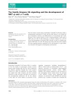
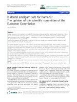
patients with severe injury. The diagnostic algorithm for
abdominal evaluation of hemodynamically stable
patients after blunt trauma is depicted in Figure 1.
Treatment
Historically, surgical management was the preferential
treatment for most blunt abdominal injury, because
NOM was associated with a highmortalityrate[24].
However, many of the laparotomies w ere unnecessary
and non-therapeutic [25]. With the wide availab ility and
improved quality of CT scanning, and the more modern,
less invasive intervention options, such as angioemboli-
zation, NOM has evolved into the treatment of choice
for hemodynamically stable patients [26].
NOM consists of close observation of the patient
completed w ith angioembolization, if necessary. Obser-
vational management involves admission to a unit and
the monitoring of vital signs, with strict bed rest, fre-
quent monitor ing of hemoglobin concentration and
serial abdominal examinations [27].
NOM, with or without angioembolization, is of benefit
to trauma patients because the function in the organ
concerned is preserved. In addition, the possible mor-
bidity that may accompany a laparotomy, such as inci-
sional hernia, abscess formation, pneumonia, wound
infection, multiorgan failure, pan creatitis, bleeding,
thromboembolic events and paralytic ileus, is avoided.
Angioembolization has proven to be a valuable
adjunct to ob servational management and has increased
the success rate of NOM to 95% [28]. The foundation
for angioembolization was laid by Charles Theodore
Dotter (1920-1985). In 1964 he performed the first
transluminal angioplasty in a patient with peripheral
Figure 1 Diagnostic algorithm of patients with blunt abdominal injury.
van der Vlies et al. International Journal of Emergency Medicine 2011, 4:47
/>Page 3 of 9
occlusive disease [29]. Later on, the technique of embo-
lization was introduced. The first application of emboli-
zationoftheinternaliliacarteryinapatientwitha
pelvic fracture occurred in 1972, and from then on, the
role of interventional radiol ogy in the diagnosis and
treatment of traumatic bleeding has increased signifi-
cantly. Research demonstrates that angioembolization is
a well-tolerated and effective t ool in the treatment of
traumatic liver, splenic and kidney injury [30-33].
Determining which patients can benefit the most from
angioembolization is still a controversial subject. CT fea-
tures, such as a high grade of injury (AAST grade 3-5),
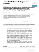
pseudoaneurysm or arteriovenous fistula, contrast extra-
vasation contained within t he spleen (Figure 2), liver or
kidney, and th e presence of a hemoperiton eum, as well
as patient characteristics such as age above 55 years old,
GCS < 8 and male gender, are associated with an
increased failure rate of NOM. Angioembolization can
be advocated to improve the success rate of NOM in
these patients [34-37].
The sing le CT finding that warrants immediate
angioembolization ( or a laparotomy) is a contrast blush
within the peritoneal cavity (Figures 3, 4, and 5).
Liver
The liver is frequently injured after blunt abdominal
trauma [2]. Traditionally, a lesion of the liver was trea-
ted surgically. The major techn iques that have been
used over time are, in consec utive order, selective hepa-
tic artery ligati on and major liver resection using omen-
tal flaps for tamponade.
Ongoing bleeding, infections and the high mortality
rate after operative treatment stimulated the search for
alternative treatments, and, in 1990, NOM was intro-
duced as a treatment for liver injury [ 38]. The high suc-
cess rate (approximately 90%) combined with the lower
mortality and complication rates, in comparison to sur-
gical treatment, make NOM the treatment of choice for
the majority of liver injuries, including high grade liv er
injury [39].
NOM consists of observation , supplemented by endo-
scopic retrograde cholangiopancrea tography with the
placement of a stent, or drainage by percutaneous trans-
hepatic cholangiography if injury to the bile ducts has
taken place. For active bleeds, angioembolization can be
performed. Angioembolization may also be applied to
control the hemorrhaging that may occur after damage-
control operations using perihepatic packing in hemody-
namically unstable patients.
Despite the reduction of mortality that has been
achieved using angioembolization, some studies describe
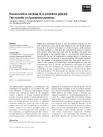
Figure 2 Computed tomography with intravenous contrast
shows small amounts of hemoperitoneum around the spleen
and a contrast ‘blush’ confined to the splenic parenchyma.
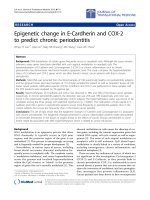
Figure 3 Liver injury with intraperitoneal contrast
extravasation visible on computed tomography scan.
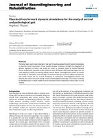
Figure 4 Computed tomography with intravenous contrast
showing hemoperitoneum, a fractured spleen with large
hematoma and extravasation of contrast medium into the
abdominal cavity.
van der Vlies et al. International Journal of Emergency Medicine 2011, 4:47
/>Page 4 of 9
ariseinseverebuttreatablecomplicationssuchas
hepatic necrosis, abscesses or bile leakage [40-42]. Gall-
bladder ischemia, hepatic parenchyma l necrosis and
biloma may also occur, and in patients with a high
grade liver injury (grade 4 an d 5) the incidence of com-
plications can be high [43].
Spleen
The spleen is the most frequently injured organ in blunt
abdominal trauma, and a missed splenic injury is the
most common cause of preventable death in trauma
patients [44]. Formerly, in the early twentie th century, a
splenectomy was nearly always performed. This invasive
management was based on the following two findings:
the first was the belief that the spleen could not heal
spontaneously; the second was called the ‘latent period
of Baudet,’ which refers to the tendency of the spleen to
rupture at a later stage [45].
Changes to this type of management occurred in the
1970s when data about postsplenectomy complications
were published describing the risk of overwhelming
postsplenectomy infection (OPSI) and its high mortality
rate [46]. In less than 10 years, NOM became the treat-
ment of choice for splenic injury.
In 1995, Sclafani described the first successful use of
angioembolization in a patient with a splenic injury [47].
Since the 1990s, angioembolization has been frequently
used to achieve better splenic salvages rates. To date,
there is no c onsensus about the optimal localization of
embolization, either proximal (Figures 6 and 7) or distal
(selective), in the splenic artery.
A recent development is proximal splenic artery
embolization (PSAE). The surgical equivalent of PSAE
for splenic injury was first described in 1979 [48]. PSAE
is predominantly used in cas es with multiple dissemi-
nated hemorrhage sites or when quick intervention is
needed because of the condition of the patie nt. Argu-
ments in favor of proximal embolization are: the low
failure rate, its speed, and the decreased incidence of
splenic abscess or infarction [49,50]. PSAE does not sig-
nificantly influence the splenic anatomy or the immune
function in the long term [51]. A disadvantage of PSAE,
however, could be t hat selective embo lization in case of
Figure 5 Computed tomography with intravenous contrast
demonstrating large hematoma around the right kidney with
contrast extravasation.
Figure 6 Selective digital subtraction angiogram of the celiac
axis showing the intra-peritoneal contrast ‘blush’ in the spleen,
confirming active bleeding.
Figure 7 Selective splenic angiogram immediately post
proximal embolization demonstrating perfusion defects.
Contrast extravasation is no longer present.
van der Vlies et al. International Journal of Emergency Medicine 2011, 4:47
/>Page 5 of 9
rebleeding is difficult, if not impossible, because the
splenic artery cannot be a ccessed. Furthermore, ische-
mia of the pancreas (when embolization is performed
proximally to the main pancreatic artery) and dislodge-
ment of coils resulting in infarction of the spleen have
been reported [52].
Selective embolization, used to stop focal bleeding, has
also proved to be successful in NOM. This technique
achieves hemostasis to the injured parts while preserving
perfusion to the remainder of the spleen [53]. Disadvan-
tages include the possibility of subsequent bleeding out
of vascular injuri es that were unnoticed owing to vasos-
pasm [54] and the higher rate of minor complications
such as infa rctions [52]. However, the clini cal relevance
of these infarctions is questionable.
A recent meta-analysis showed that both techniques
have an equivalent rate of major infarctions and infec-
tions requiring splenectomy [52]. However, the results
regarding major rebleeding, the most common reason
for failure of SAE [52], were inconclusive.
Kidney
The kidneys are affected in nearly 10% of all trauma
patients, whereas blunt trauma is responsible for 90% of
the renal injuries [55]. The switch from operative to
nonoperative management for t he treatment of renal
injuries occurred as a result of critical perceptions.
Researchers noticed that patients who underwent a
laparotom y had a signific antly higher risk of nephrect-
omy than the patients who were treated nonoperatively;
it therefore seemed that maximal renal preservation,
with a minimum of subsequent complications, could be
better achieved with NOM [56].
In 2004, the Renal T rauma Committee and, in 2005,
the European Association of Urology drew up guidelines
for the optimum evaluation of patients with urological
trauma [57,58]. T he decisive factor in the evaluation is
hemodynamic stability. Hemodynamic instability related
to renal ble eding, complete ureteral tears or pelvic avul-
sions or leakage of urine into the peritoneal cavity are
imperative indications for laparotomy. If the patient is
hemodynamically stable, the distinction between gross
or microscopic hematuria determines whether there is
any further need for imaging and what the treatment
options are. In case of gross hematuria, a MDCT scan is
the gold standard for the eva luation of renal injury [58].
Microscopic hematuria does not demand imaging.
Exclusion of coexisting injuries is of overriding impor-
tance in the initiation of NOM. Currently, NOM is used
in up to 90% of renal injuries. This is b ecause of the
particularly high incidence of minor renal injury. Peri-
nephric fluid collections or urinomas can be treated
with percutaneous drainage. Patients with active hemor-
rhages detected on the MDCT scan can be treated with
angioembolization of the renal arteries [33]. Kidney
function can be preserved through recanalization and
stenting even when a transection of the renal artery had
been made (Figures 8 and 9).
Discussion
Even though NOM has proven to be of tremendous
benefit, a couple of controversies regarding the current
management of trauma patients should be discussed.
Advances in CT technology have improved the practi-
tioner’s ability to determine the degree of injury and to
identify patients who are more likely to fail NOM. How-
ever, until now, MDCT scanning has not been able to
differentiate, in a precise manner, among which patients
should be tr eated conservatively, which would benefit
from angioembolization and which would respond best
to a surgical response. The decision for treatment
should always be based on the clinical situation and the
physiological response of the patient to initial
resuscitation.
A determinant of the success of NOM is the level of
coope ration between different specialist s in the hospital.
Good teamwork among the trauma surgeon, the
anesthesiologist and the (interventional) radiologist leads
to a quicker understanding of the underlying injuries
and thus shortens the time between entering the hospi-
tal and the initiation of therapeutic interventions. This
seems obvious in level 1 trauma centers, but can be a
matter of concern, especi ally in level II or II trauma
centers.
Recommendations for the future
The exact position o f angioembolization in the NOM of
blunt abdominal injury is still subject to discussion.
Angioembolization has been shown to be a valuable
adjunct to ob servational management and has increased
Figure 8 Computed tomography with intravenous contrast:
transection of the renal artery without contrast in the left
kidney.
van der Vlies et al. International Journal of Emergency Medicine 2011, 4:47
/>Page 6 of 9
the success rate of NOM in many series of clinical trials.
However, a lot of controversies regarding angioemboli-
zation in patients with blunt abdominal trauma exist.
Neither the optimal technique (proximal, distal or a
combination of both) nor the material to use have been
compared in a prospective trial with regard to out come
(success rate) and complication rate. A recently pub-
lished systematic review and meta-analysis of Schnüriger
et al. [52] is based on retrospective data, and the results
regarding major bleeding, the most important reason for
failure of SAE [52], were inconclusive.
The optimal follow-up strategy of patients sustaining
blunt abdominal injur ies has not been elucidated either.
Up to no w, the length of hospital stay, the need for, fre-
quency of and best modality of follow-up imaging as
well as discharge instructions with regard to resuming
of activities are at the discretion of the physician.
Research shows that practice patterns between physi-
cians are quite variable [59].
Although difficult to conduct because of the nature of
the trauma p opulation, prospective (clinical) trials are
necessary to determine the optimal patient sel ection for
angiography and embolization, the most favorable tech-
nique and material to use for angioembolization, and
the follow-up strategy in patients with traumatic blunt
injury. One way of tackling this issue would be to con-
duct a Delphi study. The Delphi method is a systematic
interactiv e forecasting method for o btaini ng experience-
based agreement from a panel of independent experts.
The process allows anonymous, non-biased consensus
building and has been well validated for systematically
assessing and organizing expert opinion [60]. Although
low in level of evidence, we hold this study d esign
appropriate since many of the controversies regarding
the clinical decision making could be resolved by an
international expert panel, selected on the basis of
extensive clinical and/or research experience. We
recommend a study such as this to be performed.
Furthermore, we advocate the improvement of logistic
factors. If MDCT scans were present and ava ilable in
trauma resuscitation rooms, the ‘ one hour rule’ would
be easier to fulfill. The MDCT scan could also play a
part in the diagnostics of hemodynamically unstable
patients [61]. At present, these patients go straight to
the operating room; however, in the future they might
also be treated with angioembolization.
Conclusion
Over the past several years, major changes in the man-
agement of blunt abdominal injury have occurred.
Because of the progress that has been made in the
quality and wide availability of the M DCT scan com-
bined with minimally invasive intervention options like
angioembolization, NOM has evolved to be the treat-
ment of choice for hemodynamically stable patients.
NOM is a safe treatment f or stable patients with trau-
matic liver, splenic or kidney injuries, and success
rates of up to 95% are described in the literature.
However, to date many controversies exist about the
optimum patient selection for NOM, the proper role
of angioembolization in NOM and the right follow-up
strategy.
List of abbreviations
NOM: nonoperative management; CT: computed tomography; ATLS:
advanced trauma life support; FAST: focused assessment with sonography
for trauma; DPL: diagnostic peritoneal lavage; CEUS: contrast enhanced
ultrasonography; MDCT: multidetector computed tomography; OPSI:
overwhelming postsplenectomy infection; PSAE: proximal splenic artery
embolization
Author details
1
Department of Surgery, Maasstad Ziekenhuis, Rotterdam, The Netherlands
2
Trauma Unit Dept. of Surgery, Academic Medical Center, Amsterdam, The
Netherlands
3
Dept. of Emergency Medicine, Medisch Spectrum Twente,
Enschede, The Netherlands
4
Trauma Unit Dept. of Surgery, Medisch Centrum
Alkmaar, Alkmaar, The Netherlands
5
Dept. of Radiology, Academic Medical
Center, Amsterdam, The Netherlands
Authors’ contributions
CHV was responsible for the manuscript and carried out the writing process.
DCO collected relevant articles, provided a great contribution to the writing
process and took care of the word processing and layout. MG was involved
in drafting the manuscript and created the reference list. KJP participated in
the design of the study and gave valuable additions with respect to the
content. OMD provided the figures and shared his expertise with regard to
the diagnostics strategies. JCG conceived of the study, participated in the
Figure 9 Angio gram of the same patient as in Figure 5 af ter
recanalization and placement of a stent in the renal artery,
resulting in good perfusion of the kidney.
van der Vlies et al. International Journal of Emergency Medicine 2011, 4:47
/>Page 7 of 9
design of the study and revised it critically for important intellectual content.
All authors read and approved the final manuscript.
Competing interests
The authors declare that they have no competing interests.
Received: 16 May 2011 Accepted: 27 July 2011 Published: 27 July 2011
References
1. Sauaia A, Moore FA, Moore EE, Moser KS, Brennan R, Read RA, Pons PT:
Epidemiology of trauma deaths: a reassessment. J Trauma 1995,
38:185-193.
2. Zwingmann J, Schmal H, Sudkamp NP, Strohm PC: Injury severity and
localisations seen in polytraumatised children compared to adults and
the relevance for emergency room management. Zentralbl Chir 2008,
133:68-75.
3. Carmont MR: The Advanced Trauma Life Support course: a history of its
development and review of related literature. Postgrad Med J 2005,
81:87-91.
4. Root HD, Hauser CW, Mckinley CR, Lafave JW, Mendolia RP Jr: Diagnostic
Peritoneal lavage. Surgery 1965, 57:633-637.
5. Gonzalez M, Bucher P, Ris F, Andereggen E, Morel P: Splenic trauma:
predictive factors for failure of non-operative management. J Chir (Paris)
2008, 145:561-567.
6. Bakker J, Genders R, Mali W, Leenen L: Sonography as the primary
screening method in evaluating blunt abdominal trauma. J Clin
Ultrasound 2005, 33:155-163.
7. Stengel D, Bauwens K, Sehouli J, Porzsolt F, Rademacher G, Mutze S,
Ekkernkamp A: Systematic review and meta-analysis of emergency
ultrasonography for blunt abdominal trauma. Br J Surg 2001, 88:901-912.
8. Bode PJ, Edwards MJ, Kruit MC, van Vugt AB: Sonography in a clinical
algorithm for early evaluation of 1671 patients with blunt abdominal
trauma. AJR Am J Roentgenol 1999, 172:905-911.
9. Schnuriger B, Kilz J, Inderbitzin D, Schafer M, Kickuth R, Luginbühl M,
Candinas D, Exadaktylos AK, Zimmermann H: The accuracy of FAST in
relation to grade of solid organ injuries: a retrospective analysis of 226
trauma patients with liver or splenic lesion. BMC Med Imaging 2009, 9:3.
10. Rozycki GS, Ballard RB, Feliciano DV, Schmidt JA, Pennington SD: Surgeon-
performed ultrasound for the assessment of truncal injuries: lessons
from 1540 patients. Ann Surg 1998, 228:557-567.
11. Miller MT, Pasquale MD, Bromberg WJ, Wasser TE, Cox J: Not so FAST. J
Trauma 2003, 54:52-59.
12. Deunk J, Brink M, Dekker HM, Kool DR, Blickman JG, van Vugt AB,
Edwards MJ: Routine versus selective computed tomography of the
abdomen, pelvis, and lumbar spine in blunt trauma: a prospective
evaluation. J Trauma 2009, 66:1108-1117.
13. Deunk J, Brink M, Dekker HM, Kool DR, Blickman JG, van Vugt AB,
Edwards MJ: Predictors for the Selection of Patients for Abdominal CT
After Blunt Trauma: A Proposal for a Diagnostic Algorithm. Ann Surg 2010.
14. Brink M, Deunk J, Dekker HM, Kool DR, Edwards MJ, van Vugt AB,
Blickman JG: Added value of routine chest MDCT after blunt trauma:
evaluation of additional findings and impact on patient management.
AJR Am J Roentgenol 2008, 190:1591-1598.
15. Catalano O, Aiani L, Barozzi L, Bokor D, De Marchi A, Faletti C, Maggioni F,
Montanari N, Orlandi PE, Siani A, Sidhu PS, Thompson PK, Valentino M,
Ziosi
A, Martegani A: CEUS in abdominal trauma: multi-center study.
Abdom Imaging 2009, 34(2):225-34.
16. Mullinix AJ, Foley WD: Multidetector computed tomography and blunt
thoracoabdominal trauma. J Comput Assist Tomogr 2004, 28(Suppl1):S20-7.
17. Miller LA, Shanmuganathan K: Multidetector CT evaluation of abdominal
trauma. Radiol Clin North Am 2005, 43:1079-95, viii.
18. Jayaraman S, Sethi D: Advanced trauma life support training for hospital
staff. Cochrane Database Syst Rev 2009, CD004173.
19. Deunk J, Dekker HM, Brink M, Vugt vR, Edwards MJ, van Vugt AB: The value
of indicated computed tomography scan of the chest and abdomen in
addition to the conventional radiologic work-up for blunt trauma
patients. J Trauma 2007, 63:757-63.
20. Huber-Wagner S, Lefering R, Qvick LM, et al: The value of indicated
computed tomography. Effect of whole-body CT during trauma
resuscitation on survival: a retrospective, multicentre study. Lancet 2009,
373:1455-1461.
21. Chan O: Primary computed tomography survey for major trauma. Br J
Surg 2009, , 96: 1377-1378.
22. Brenner DJ, Hall EJ: Computed tomography – an increasing source of
radiation exposure. N Engl J Med 2007, 357:2277-84.
23. Saltzherr TP, Fung Kon Jin PH, Bakker FC, Ponsen KJ, Luitse JS, Scholing M,
Giannakopoulos GF, Beenen LF, Henny CP, Koole GM, Reitsma HB,
Dijkgraaf MG, Bossuyt PM, Goslings JC: An evaluation of a Shockroom
located CT scanner: a randomized study of early assessment by CT
scanning in trauma patients in the bi-located trauma center North-West
Netherlands (REACT trial). BMC Emerg Med 2008, 8:10.
24. Richardson JD: Changes in the management of injuries to the liver and
spleen. J Am Coll Surg 2005, 200:648-669.
25. Sorkey AJ, Farnell MB, Williams HJ Jr, Mucha P Jr, Ilstrup DM: The
complementary roles of diagnostic peritoneal lavage and computed
tomography in the evaluation of blunt abdominal trauma. Surgery 1989,
106:794-800.
26. Malangoni MA, Cue JI, Fallat ME, Willing SJ, Richardson JD: Evaluation of
splenic injury by computed tomography and its impact on treatment.
Ann Surg 1990, 211:592-597.
27. Pachter HL, Guth AA, Hofstetter SR, Spencer FC: Changing patterns in the
management of splenic trauma: the impact of nonoperative
management. Ann Surg 1998, 227:708-717.
28. Stein DM, Scalea TM: Nonoperative management of spleen and liver
injuries. J Intensive Care Med 2006, 21
:296-304.
29.
Dotter CT, Judkins MP: Transluminal treatment of arteriosclerotic
obstruction. Description of a new technic and preliminary report of its
application. Circulation 1964, 30:654-670.
30. Hagiwara A, Murata A, Matsuda T, Matsuda H, Shimazaki S: The efficacy
and limitations of transarterial embolization for severe hepatic injury. J
Trauma 2002, 52:1091-1096.
31. Brugere C, Arvieux C, Dubuisson V, Guillon F, Sengel C, Bricault I,
Regimbeau JM, Pilleul F, Menegaux F, Letoublon C: [Early embolization in
the nonoperative management of blunt splenic injuries: a retrospective
multicenter study]. J Chir(Paris) 2008, 145:126-132.
32. Nijhof HW, Willemssen FE, Jukema GN: Transcatheter arterial embolization
in a hemodynamically unstable patient with grade IV blunt liver injury:
is nonsurgical management an option? Emerg Radiol 2006, 12:111-115.
33. Krämer SC, Görich J, Rilinger N, Gottfried HW, Mattes R, Aschoff AJ: The
percutaneous transarterial embolization therapy of traumatic kidney
hemorrhages. Rofo 1998, 169(3):297-301.
34. Fang JF, Chen RJ, Wong YC, Lin BC, Hsu YB, Kao JL, Chen MF: Classification
and treatment of pooling of contrast material on computed
tomographic scan of blunt hepatic trauma. J Trauma 2000, 49:1083-1088.
35. Marmery H, Shanmuganathan K, Alexander MT, Mirvis SE: Optimization of
selection for nonoperative management of blunt splenic injury: comparison
of MDCT grading systems. Am J Roentgenol 2007, 189:1421-1427.
36. Schurr MJ, Fabian TC, Gavant M, Croce MA, Kudsk KA, Minard G,
Woodman G, Pritchard FE: Management of blunt splenic trauma:
computed tomographic contrast blush predicts failure of nonoperative
management. J Trauma 1995, 39:507-512.
37. Harbrecht BG, Peitzman AB, Rivera L, Heil B, Croce M, Morris JA Jr,
Enderson BL, Kurek S, Pasquale M, Frykberg ER, Minei JP, Meredith JW,
Young J, Kealey GP, Ross S, Luchette FA, McCarthy M, Davis F, Shatz D,
Tinkoff G, Block EF, Cone JB, Jones LM, Chalifoux T, Federle MB, Clancy KD,
Ochoa JB, Fakhry SM, Townsend R, Bell RM: Contribution of age and gender
to outcome of blunt splenic injury in adults: Multicenter study of the
eastern association for the surgery of trauma. J Trauma 2001, 51:887-895.
38. Knudson MM, Maull KI: Nonoperative management of solid organ
injuries. Past, present, and future. Surg Clin North Am 1999, 79:1357-1371.
39. Buckman RF Jr, Miraliakbari R, Badellino MM: Juxtahepatic venous injuries:
a critical review of reported management strategies. J Trauma 2000,
48:978-984.
40. Dabbs DN, Stein DM, Scalea TM: Major hepatic necrosis: a common
complication after angioembolization for treatment of high-grade liver
injuries. J Trauma 2009, 66:621-627.
41. Kozar RA, Moore JB, Niles SE, Holcomb JB, Moore EE, Cothren CC,
Hartwell E, Moore FA: Complications of nonoperative management of
high-grade blunt hepatic injuries. J Trauma 2005, 59:1066-1071.
42. Mohr AM, Lavery RF, Barone A, Bahramipour P, Magnotti LJ, Osband AJ,
Sifri Z, Livingston DH:
Angiographic embolization for liver injuries: low
mortality,
high morbidity. J Trauma 2003, 55:1077-1081.
van der Vlies et al. International Journal of Emergency Medicine 2011, 4:47
/>Page 8 of 9
43. Misselbeck TS, Teicher EJ, Cipolle MD, Pasquale MD, Shah KT,
Dangleben DA, Badellino MM: Hepatic angioembolization in trauma
patients: indications and complications. J Trauma 2009, 67:769-773.
44. Cales RH, Trunkey DD: Preventable trauma deaths. A review of trauma
care systems development. JAMA 1985, 254:1059-1063.
45. Peitzman AB, Ford HR, Harbrecht BG, Potoka DA, Townsend RN: Injury to
the spleen. Curr Probl Surg 2001, 38:932-1008.
46. Holdsworth RJ, Irving AD, Cuschieri A: Postsplenectomy sepsis and its
mortality rate: actual versus perceived risks. Br J Surg 1991, 78:1031-1038.
47. Sclafani SJ, Shaftan GW, Scalea TM, Patterson LA, Kohl L, Kantor A,
Herskowitz MM, Hoffer EK, Sharon H, Dresner LS, Wetzel W: Nonoperative
salvage of computed tomography-diagnosed splenic injuries: utilization
of angiography for triage and embolization for hemostasis. J Trauma
1995, 39:818-825.
48. Keramidas DC: The ligation of the splenic artery in the treatment of
traumatic rupture of the spleen. Surgery 1979, 85:530-533.
49. Smith HE, Biffl WL, Majercik SD, Jednacz J, Lambiase R, Cioffi WG: Splenic
artery embolization: Have we gone too far? J Trauma 2006, 61:541-544.
50. Bessoud B, Denys A, Calmes JM, Madoff D, Qanadli S, Schnyder P, Doenz F:
Nonoperative management of traumatic splenic injuries: is there a role
for proximal splenic artery embolization? Am J Roentgenol 2006,
186:779-785.
51. Malhotra AK, Carter RF, Lebman DA, Carter DS, Riaz OJ, Aboutanos MB,
Duane TM, Ivatury RR: Presevartion of splenic immunocompetence after
splenic artery angioembolization for blunt splenic injury. J Trauma 2010,
69:1126-1131.
52. Schnuriger B, Inaba K, Konstantinidis A, Lustenberger T, Chan LS,
Demetriades D: Outcomes of proximal versus distal splenic artery
embolization after trauma: a systematic review and meta-analysis. J
Trauma 2011, 70:252-260.
53. Raikhlin A, Baerlocher MO, Asch MR, Myers A: Imaging and transcatheter
arterial embolization for traumatic splenic injuries: review of the
literature. J Can Chir 2008, 61:464-472.
54. Haan JM, Biffl W, Knudson MM, Davis KA, Oka T, Majercik S, Dicker R,
Marder S, Scalea TM: Splenic embolization revisited: a multicenter review.
J Trauma 2004, 56:542-7.
55. Taviloglu K, Yanar H: Current trends in the management of blunt solid
organ injuries. Eur J Trauma Emerg Surg 2009, 35:90-4.
56. Bergen CT, Chan TN, Bodzin JH: Intravenous pyelogram results in
assoication with renal pathology and therapy in trauma patients. J
Trauma
1987, 27:515.
57. Santucci RA, Wessells H, Bartsch G, Descotes J, Heyns CF, McAninch JW,
Nash P, Schmidlin F: Evaluation and management of renal injuries:
consensus statement of the renal trauma subcommittee. BJU Int 2004,
93(7):937-54.
58. Lynch TH, Martínez-Piñeiro L, Plas E, Serafetinides E, Türkeri L, Santucci RA,
Hohenfellner M: European Association of Urology. EAU guidelines on
urological trauma. Eur Urol 2005, 47(1):1-15.
59. Fata P, Robinson L, Fakhry SM: A survey of EAST member practices in
blunt splenic injury: a description of current trends and opportunities
for improvement. J Trauma 2005, 59:836-842.
60. Ludlow J: Delphi enquiries and knowledge utilisation. In The Delphi
method: techniques and applications. Edited by: Linstone HA, Turoff M.
Reading, MA: Addison-Wesley; 1975:102-123.
61. Lin WC, Chen YF, Lin CH, Tzeng YH, Chiang HJ, Ho YJ, Shen WC, Chen JH:
Emergent transcatheter arterial embolization in hemodynamically
unstable patients with blunt splenic injury. Acad Radiol 2008, 15(2):201-8.
doi:10.1186/1865-1380-4-47
Cite this article as: van der Vlies et al.: Changing patterns in diagnostic
strategies and the treatment of blunt injury to solid abdominal organs.
International Journal of Emergency Medicine 2011 4:47.
Submit your manuscript to a
journal and benefi t from:
7 Convenient online submission
7 Rigorous peer review
7 Immediate publication on acceptance
7 Open access: articles freely available online
7 High visibility within the fi eld
7 Retaining the copyright to your article
Submit your next manuscript at 7 springeropen.com
van der Vlies et al. International Journal of Emergency Medicine 2011, 4:47
/>Page 9 of 9