báo cáo hóa học: " “Case files from the University of Florida: When an earache is more than an earache": A case report" potx
Bạn đang xem bản rút gọn của tài liệu. Xem và tải ngay bản đầy đủ của tài liệu tại đây (349.36 KB, 5 trang )
CAS E REP O R T Open Access
“Case files from the University of Florida:
When an earache is more than an earache":
A case report
Bobby K Desai
*
and Thomas Walls
Abstract
Brain abscess is not a common diagnosis as there are only approximately 2000 cases reported each year in the
United States. There are three main routes of access to the brain including contiguous infection from the
oropharynx, direct implantation and hematogenously. We present a case of brain abscess in a child who had
multiple visits for ear pain to various physicians including pediatricians and to emergency departments.
Additionally, the microbiology of brain abscesses is briefly discussed, as is treatment.
Introduction
We present a new series for the Int ernati onal Journal of
Emergency Medicine, “Case Files from the University of
Florida,” in which we will present a case seen by the
residents and faculty of the Emergency Medicine resi-
dency at the University of Florida, Gainesville, and have
you, the reader, consider wha t the diagnostic possibili-
ties are, determine what diagnostic tests are required,
and “run” the case. We hope that these cases are educa-
tionally rewarding for you.
Presentation
Initial Management
Treatment/Resuscitation
Diagnosis/Disposition
“When an earache is more than an earache”
Foreword
Patients with otitis media and related conditions present
nearly 2 million times to the emergency department
every year. The vast majority of these are benign in nat-
ure, and the treatment simply observation versus anti-
biotic therapy. There are occasions, however, where the
simple earache turns i nto something much more. We
present such a case.
Presentation
A 5-year-old child presented to the University of Florida
Emergency Department (ED), brought by the mother,
with complaints of earache, vomiting, and fever for 3
weeks. The mother had brought the child to their pedia-
trician the previous week, and he was subsequently diag-
nosed with dehydration. The parents also brought the
child to another emergency department later in the week,
and she stated the patie nt was given intravenous fluids
for dehydration. He was discharged home, and his par-
ents given instructions to give acetaminophen for fever
and to continue oral rehydration. On this second ED pre-
sentation, the mother stated the child was tolerating oral
liquids, had urinated once that a.m., and his last bowel
movement had been the previous day. The stool was nor-
mal in consistency and not bloody. The maximum tem-
perature the patient had was 102°F. The patient had
vomited two times on the day of presentation, and it con-
sisted of previously eaten food with no blood. Further
history revealed that the child did not attend daycare,
there were no smokers in the household, and the child
had not received any immunizations for religious reasons.
Upon review of syst ems, the mother denied any rashes,
cough, runny nose, complaints o f sore throat, diarrhea,
or abdominal cramping or pain. She did however state
that the patient reported ear pain and facial pain.
Past medical history: None
Past Surgical history: None
Allergies: None
Medications: None
* Correspondence:
University of Florida, Department of Emergency Medicine, PO BOX 100186,
Gainesville, FL, 32610, USA
Desai and Walls International Journal of Emergency Medicine 2011, 4:33
/>© 2011 Desai; licensee Springer. This is an Open Access article distributed under the terms of the Creative Commons Attri bution
License ( which permits unrest ricted use, distribution, and reprodu ction in any medium,
provided the original work is properly cited.
Physical exam
On presentation the patient’s vital signs were: tempera-
ture 36.7°C, pulse 60 beats per minute, respiratory rate
28 breaths per minute, and blood pressure 90/39
mmHg. His weight was 22 kg. The patient was alert and
looked fatigued, but was conversant with the parents
and physician. On eye examination, his pupils were
round and reactive t o light, without corneal injection.
The eyelid exam was normal. The ear exa m revealed
auricular tenderness of both ears, with bulging tympanic
membranes and decreased light reflex. The throat was
normal. The lungs were clear to auscultation bilater ally,
and the heart exam was unremarkable. He had a soft
and non-tender abdomen with normal bowel sounds,
and his guaiaic test was negative. His neurological exam
was normal.
Questions to ponder
1. What do you think of this presentation?
2. What differential diagnosis should be considered
for this patient?
3. Based on this pre sentation, what diagnostic tests
should be considered?
4. Is anything missing from the history or physical
examination?
Emergency physician’s thought process
On initial presentation the patient was afebrile, and his
vital signs were stable. He appeared tired and fatigued,
but did not appear to be septic. The initial differential
diagnosis included an otitis - either media o r externa -
or perhaps a combination of the two, a simple pro-
longed upper respiratory infection, Influenza, and viral
enteritis. The physicians felt that his emergent condition
was due to failed outpatienttherapyforvomitingand
dehydration. They were concerned about his lack of oral
intake, and it was therefore decided to order intravenous
fluids. Laboratory tests were also drawn at this time, and
these included a chemistry panel and complete blood
count. Due to the reported fever, blood cultures were
also drawn.
There was no mention in the initial physical examina-
tion of mucous membrane moisture or skin turgor,
which would be important if the physician was consider-
ing a shock-like state for this patient. Additionally, was
the patient receiving antipyretics? How much and how
often would be important to d ocument. Furthermore,
was there any follow-up with the primary care physician
after the first ED visit?
One could consider the addition of a urinalysis to
evaluate the specific gravity and assess the degree of
dehydration, though a BUN/creatinine ratio of 20:1
could detect a pre-renal azotemia.
Emergency department course
An intravenous line was placed uneventfully, and fluids
were started. Laboratory tests were sent and were all
within normal limits with the exception of a white
blood cell count that was 19,000 cells/mm
3
.
Per physician re-evaluation, the child looked
improved, was tolerating oral fluids without difficulty,
was afebrile, and his vital signs were normal. He was
ambulatory without assistance to the bathroom. How-
ever, the family was concerned that these same events
occurred at their prior ED visit and requested admi ssion
for observation. The ED physician agreed and consulted
the pediatric admission team to evaluate the patient.
After admission was arranged there was a delay in trans-
porting the patient to t he in-patient unit, and he had to
remain in the ED until a bed was available.
Questions to ponder
1. What do you think of this patient’s management?
2. Would you add (or remove) any diagnostic tests?
3. Would you change the treatment in any way?
4. Would you have admitted this child?
Emergency physician’s response
Since he appe ared “ fatigued” based on the physical
examination and sinc e his patient ’soralintakehad
diminished over t he course of prolonged illness, it
seemed reasonable to fluid resuscitate this patient. Since
it appeared that he improved over the course of his stay
in the ED, this management presumably resulted in the
clinical improvement of the patient, since he was now
tolerating oral liquids, and due to the patient ’ sunaided
ambulation to the bathroom, he acted less fatigued.
The question of whether the patient should have been
admitted is a difficult one. The patient seemed to be
improved, and looked an d presumably felt much better.
Based on this clinical gestalt, he did not seem to meet
admission criteria. However, it appeared the parents
were clearly uncomfortable with his being discharged,
and without being privy to the conversation between the
emergency physician and family, it is likely a third party
- namely t he pediatric admitting tea m - we re called to
assess the patient. Ultimately, it is a moot point a s the
admitting team did readily admit the patient, so there
clearly was little or no issue in that regard.
After admission
Four hours after admission, the physician was alerted by
the nursing staff that the patient was less alert and
lethargic. On exam ination, he continued to be afeb rile -
temperature 36.9°C, pulse 72 beats per minute, and
respiratory rate 16 breaths per minute. A blood pressure
was not recorded. His physical examination revealed an
Desai and Walls International Journal of Emergency Medicine 2011, 4:33
/>Page 2 of 5
unchanged cardiovascular, pulmonary, and gastrointest-
inal examination. However, on neurological examina-
tion, he wa s lethargic, and found to have dysarthria and
ataxia . A computed tomography (CT) scan was immedi-
ately ordered
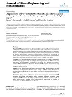
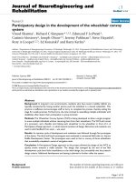
CT scan
Figure 1 - Coronal view of brain
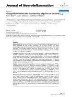
Figure 2 - Transverse view of brain
Questions to ponder
1. What happened?
2. Could this change have been prevented?
3. What does the CT show?
Emergency physician’s response
This clearly was an acute change in the patient’s condition.
This most likely could not have been foreseen based on
the patient’s initial exa mination, tho ugh it can be argued
that due to the extent of the edema, a more thorough neu-
rologic examination could have picked up subtle findings.
On the other hand, it can be argued that the edema pre-
sent on the CT scan could have been caused as a result of
the fluid resuscitation given to the patient.
The CT showed a 6.1 (anteroposterior) × 4.8 (trans-
verse) × 4.1 (craniocaudal) cm thin ring-enhancing
lesion whose epicenter was located in the left low con-
vexit y posterior temporal lobe. The lesion is rim-enhan-
cing with a thin peripheral wall, and associated with
vasogenic edema. There is 0.9 cm of rightward
subfalcine shift, with effacement of the posterior horn of
the left lateral ventricle, thus causing sequestration of
the ipsilateral temporal horn.
Neurosurgery evaluated the patient and recommended
immediate evacuation of the abscess. He was taken to
the operating r oom and had a stereotactic-guided left
temporal craniotomy with excision of t he brain abscess.
He was started on antibiotic therapy and was discharged
in good condition 9 days after admission.
Discussion
Brai n absce ss is a rare diagnosis; there are only 1,500 to
2,500 reported cases each year in the US [1,2]. Factors
that lead to permanent neurologic disability and death
due to brain abscess include: impaired host immunity,
Glasgow Coma Scale score less than 12, delays in hospi-
talization, focal neurologic deficits at admission, and
uncontrolled diabetes [1-6,3-7]. Brain abscess most com-
monly occurs as the result of contiguous spread of
infection from the oropharynx, middle ear, and parana-
sal sinuses [1,2]. Organisms reach the brain by one of
threeknownroutes:hematogenously(onethirdof
cases); from contiguous infections of the middle ear,
sinus, or teeth (one third of cases); or by direct implan-
tation by neurosurgery or penetrating trauma (ap proxi-
mately 10% of cases) [8]. The route is unknown in
approximately 20% of cases. Circumstances that reduce
oxygenation of brain parenchyma are important predis-
posing factors for bacterial invasion. Spread from a con-
tiguous infection usually involves intervening cerebral
thrombophlebitis , with congestive ischemic hypox emia of
Figure 1 Coronal view of brain.
Figure 2 Transverse view of brain.
Desai and Walls International Journal of Emergency Medicine 2011, 4:33
/>Page 3 of 5
the tissue destined to become infected [7,1]. Hematogen-
ous seeding is facilitated by systemic hypoxemia, as in
congenital heart diseases with right-to-left shunt and
chronic pulmonary suppuration. This is demonstrated by
the prominent role of anaerobic bacteria in brain
abscesses. The source of brain abscess should be identi-
fied for the dual purpose of eliminating the source itself
and gaining insight into the probable bacteriologic char-
acteristics of the abscess. Gram-negative rods, especially
Bacteroides, are the usual pathogens in otogenic brain
abscesses, whic h are typically single and located in the
adjacent temporal lobe or cerebellum. Anaerobic and
microaerophilic streptococci are the most common
pathogens in sinogenic and odontogenic abscesses, and
are more typically located in the frontal lobes. Abscesses
formed from hematogenous spread are often multiple
and polymicrobial, with anaerobic and microaerophilic
streptococci commonly represented. Staphylococci are
typical pathogens in abscesses due to direct implantation.
Gram-negative rods are also suspected in cases related to
a neurosurgical procedure. Enteric gram-negative bacilli
can be seen in association with an intraabdominal or gen-
itourinary source. Pseudomonas spp. can be seen in brain
abscesses arising from otitis media or otitis externa [1,2].
In the immunocompromised or elderly patient, oppor-
tunistic pathogens must be considered as a potential
source of infection. Nocardia spp. can be seen from dis-
semination of cutaneous or pulmonary infection; brain
abscesses caused by M. tuberculosis and nontuberculous
mycobacteria have been reported in patients with HIV
infection, while L. monocytogenes may cause brain
abscesses in immunosuppressed individuals [9-11].
Fungal brain abscesses caused by yeast (e.g., Candida
spp., Cryptococcus sp p.), dimorphi c fungi (e.g., Histo-
plasma spp., Coccidioides spp., Blastomyces spp.), and
molds (e.g., Aspergillus spp., Rhizopus) are associated
with immunocompromised states [1,2]. Zygomycosis can
be seen in patients with po orly controlled diabetes [1,2].
Helminths and protozoa can cause parasitic brain
abscesses, but these are rare.
Clinical presentation
Patients with brain abscess may present a myriad of
complaints including headache, mental status c hanges,
focal neurologic deficit, fever, and new-onset seizures.
Headache and mental status changes are found most
frequently, followed by focal neurologic deficits, fever,
and seizures [12,13]. The classic clinical triad of fever,
headache, and focal neurologic deficits was found to be
only 17% sensitive [13,12]. Clinical manifestations are
dependent on the locati on and size of the brain absces s,
host immune status, and the virulence of the causative
microorganism.
Diagnosis
CT with intravenous contrast can show ring-enhancing
lesions, especially in chronic brain abscesses. However,
MRI with gadolinium contrast is more sensitive and spe-
cific than CT scan with contrast study to diagnose brain
abscess [8]. CT-guided stereotactic biopsy with aspiration
of abscesses can reduce the necessity of open craniotomy
and can be both diagnostic and therapeutic [14]. It is
mandatory to perform microbiologic investigation once
the abscess is drained to guide further therapy.
Treatment
Since brain abscesses are frequently polymicrobial, initial
antimicrobial therapy should cover gram-positive, gram-
negative, and anaerobic microorganisms, and should be
later tailored to the specific organism that is identified
[2,3]. The duration of therapy is dependent upon the
organism identified; longer t herapy is indicated for
opportunistic infections, whereas 6-8 weeks of parent-
eral therapy is indicated for bacterial brain abscesses.
Duration of therapy is influenced by causative microor-
ganisms and reduction in the size of the abscess [7,1].
Follow-Up
Subsequent to the patient’s craniotomy and aspiration of
contents that morning, his cultures indicated the abscess
pathogen to be Streptococcus pneumoniae. Blood cul-
tures w ere negative. He was started on 6 weeks of par-
enteral therapy. Follow-up 1 month after surgery
indicated the child had a mild speech impediment, but
was improving. Follow-up 1 year later indicated com-
plete improvement back to his normal neurological
function.
Conclusions
Otitis media and related condit ions are a common pre-
senting complaint to the emergency department with
over two million visits per year. Treatment failures can
potentially occur and the astute clinician must consider
other etiologies of otalgia if multiple visits for the same
complaint occur. Brain abscess is not a common diagno-
sis, though potentially has significant morbidity if left
undiagnosed. Brain absc ess occurs as result of contigu-
ous spread of infection from the oropharynx, middle
ear, and paranasal sinuse s. Organisms r each the brain
hematogenously, contiguous spread from nearby areas
or direct implantation. Patients with brain abscess pre-
sent most commonly with headache and mental status
changes. Other common symptoms and signs include
focal neurologic deficits, fever and seizures. Contrasted
MRI is more sensitive and specific in diagnosing brain
abscess than is computed tomography. Treatment is
broad spectrum initially, but microbiologic investigation
Desai and Walls International Journal of Emergency Medicine 2011, 4:33
/>Page 4 of 5
is necessary in order to tailor therapy to the specific
cause.
Consent
Written informed consent was obtained from the par-
ents of the patient fo r publication of this Case report
and any accompanying images. A copy of the written
consent is available for review by the Editor-in-Chief of
this journal.
Authors’ contributions
TW: Wrote case report. BKD: Formulated questions, answers, and discussion
Competing interests
The author declares that they have no competing interests.
Received: 4 April 2011 Accepted: 21 June 2011 Published: 21 June 2011
References
1. Mathisen GE, Johnson JP: Brain abscess. Clin Infect Dis 1997, 25(4):763-79.
2. Honda H: Central nervous system infections: meningitis and brain
abscess. Infect Dis Clin North Am 23(3):609-23.
3. Mamelak AN, Mampalam TJ, Obana WG, et al: Improved management of
multiple brain abscesses: a combined surgical and medical approach.
Neurosurgery 1995, 36(1):76-85.
4. Seydoux C, Francioli P: Bacterial brain abscesses: factors influencing
mortality and sequelae. Clin Infect Dis 1992, 15(3):394-401.
5. Xiao F, Tseng MY, Teng LJ, et al: Brain abscess: clinical experience and
analysis of prognostic factors. Surg Neurol 2005, 3(5):442-9.
6. Tseng JH, Tseng MY: Brain abscess in 142 patients: factors influencing
outcome and mortality. Surg Neurol 2006, 65(6):557-62.
7. Tonon E, Scotton PG, Gallucci M, et al: Brain abscess: clinical aspects of
100 patients. Int J Infect Dis 2006, 10(2):103-9.
8. Heilpern KL, Lorber B: Focal intracranial infections. Infect Dis Clin North Am
1996, 10(4):879-98.
9. Yang KY, Chang WN, Ho JT, et al: Postneurosurgical nosocomial bacterial
brain abscess in adults. Infection 2006, 34(5):247-51.
10. Farrar DJ, Flanigan TP, Gordon NM, et al: Tuberculous brain abscess in a
patient with HIV infection: case report and review. Am J Med 1997,
102(3):297-301.
11. Mylonakis E, Hohmann EL, Calderwood SB: Central nervous system
infection with Listeria monocytogenes. 33 years’ experience at a general
hospital and review of 776 episodes from the literature. Medicine
(Baltimore) 1998, 77(5):313-36.
12. Tseng JH, Tseng MY: Brain abscess in 142 patients: factors influencing
outcome and mortality. Surg Neurol 2006, 65(6):557-62.
13. Tunkel AR: Brain abscess. In Principles and Practice of Infectious Disease 6
edition. Edited by: Mandel GL, Bennett JE, Dolin R. Philadelphia. Elsevier
Churchill Livingstone; 2005:1154.
14. Mampalam TJ, Rosenblum ML:
Trends in the management of bacterial
brain abscesses: a review of 102 cases over 17 years. Neurosurgery 1988,
23(4):451-8.
doi:10.1186/1865-1380-4-33
Cite this article as: Desai and Walls: “Case files from the University of
Florida: When an earache is more than an earache": A case report.
International Journal of Emergency Medicine 2011 4:33.
Submit your manuscript to a
journal and benefi t from:
7 Convenient online submission
7 Rigorous peer review
7 Immediate publication on acceptance
7 Open access: articles freely available online
7 High visibility within the fi eld
7 Retaining the copyright to your article
Submit your next manuscript at 7 springeropen.com
Desai and Walls International Journal of Emergency Medicine 2011, 4:33
/>Page 5 of 5