Báo cáo hóa học: " Investigation of utilization of nanosuspension formulation to enhance exposure of 1,3dicyclohexylurea in rats: Preparation for PK/PD study via subcutaneous route of nanosuspension drug delivery" doc
Bạn đang xem bản rút gọn của tài liệu. Xem và tải ngay bản đầy đủ của tài liệu tại đây (445.68 KB, 9 trang )
NANO EXPRESS Open Access
Investigation of utilization of nanosuspension
formulation to enhance exposure of 1,3-
dicyclohexylurea in rats: Preparation for PK/PD
study via subcutaneous route of nanosuspension
drug delivery
Po-Chang Chiang
*
, Yingqing Ran, Kang-Jye Chou, Yong Cui and Harvey Wong
Abstract
1,3-Dicyclohexylure a (DCU), a potent soluble epoxide hydrolase (sEH) inhibitor has been reported to lower systemic
blood pressure in spontaneously hypertensive rats. One limitation of continual administration of DCU for in vivo
studies is the compound’s poor oral bioavailability. This phenomenon is mainly attributed to its poor dissolution
rate and low aqueous solubility. Previously, wet-milled DCU nanosuspension has been reported to enhance the
bioavailability of DCU. However, the prosperities and limitations of wet-milled nanosuspension have not been fully
evaluated. Furthermore, the oral pharmacokinetics of DCU in rodent are such that the use of DCU to understand
PK/PD relationships of sEH inhibitors in preclinical efficacy model is less than ideal. In this study, the limitation of
orally delivered DCU nanosuspension was assessed by a surface area sensitive absorption model and
pharmacokinetic modeling. It was found that dosing DCU nanosuspension did not provide the desired plasma
profile needed for PK/PD investigation. Based on the model and in vivo data, a subcutaneous route of delivery of
nanosuspension of DCU was evaluated and demonstrated to be appropriate for future PK/PD studies.
Introduction
In recent years, research ers have demonstrated that var-
ious epoxyeicosatr ienoic acid (EETs) regioisomers cause
either vasodilatation or vasoconstriction in a number of
vascular beds [1-3] and that they hold anti-inflammatory
properties [4]. There is compelling evidence from the
literature that increasing the levels of EETs demon-
strates anti-inflammatory, cardio-protective [5-8] antihy-
pertensive, and renal vascular protective effects during
disease states. These properties make this pathway an
extremely a ttractive target for intervention. Based on
these findings, soluble epoxide hydrolase (sEH) inhibi-
tion is a poten tially attractive pharmacologica l approach
to treat human hypertension. It has been reported that
1,3-dicyclohexyl urea (DCU) is a potent sEH inhibitor
and inhibits human vascular smooth muscle (VSM) cell
proliferation in a dose-dependent manner [9,10].
Because of the anti-inflammatory and antihypertensive
properties of sEH inhibition, DCU can be used as a
model sEH inhibitor to further investigate decreased
VSM cell proliferation, a crucial pathologic mechanism
in the progression from systemic hypertension to the
atherosclerotic state [4,11,12]. However, despite having
high in vitro potency, the utility of DCU to investigate
sEH is limited based both on its short t
1/2
in rats
[13-15] and its low aqueous solubility, which makes oral
deliveryofDCUtomaintainprolongedandconstant
exposure difficult. Such an issue is not DCU specific. It
is well acknowledged in the pharmaceutical industry
today that an increasing number of lipophilic drug can-
didates are providing scientists with the growing chal-
lenge of reaching desired exposures in vivo. Approaches
to deliver poorly soluble molecules have been developed
for both clinical and preclinical activities [14-17]. How-
ever, in the early phase of drug discovery where large
* Correspondence:
Small Molecule Research. Genentech, 1 DNA Way, South San Francisco, CA
94080, USA
Chiang et al. Nanoscale Research Letters 2011, 6:413
/>© 2011 Chiang et al; licensee Springer. This is an Open Access article distributed under the terms of the Creative Commons Attribution
License ( which permits unrestricted use, distribution, and reproduction in any medium,
provide d the original work is properly cited.
numbers of potential candidates are screened, develop-
ment of suitable formulations in time for a drug candi-
date’ s in vivo evaluation remains a big challenge. In
general, formulations made at this early stage need to be
prepared on a small scale using common excipients with
little lead development time and the assurance of reli-
able delivery of target concentration levels.
Recently, nano- and microparticle drug delivery has
been widely used in the pharmaceutical industry as a
tool to overcome exposure issues [17-23]. Previously,
much improved exposures were reported when nanosus-
pension formulations were used to deliver DCU [13-15].
Improvements in oral exposure by a DCU nanosuspen-
sion formulation enabled a dose-dependent efficacy
study in a diseased animal model [14]. Despite the suc-
cess of dem onstrating preclincal efficacy, further utiliza-
tion of DCU as a tool to evaluate target PK/PD
relationships in chronic animal models [24] remains
challenge. A short t
1/2
coupled with a high drug plasma
peak to trough (P/T) ratio was observed when DCU
nanosuspension was dosed orally in rats [14].
In order to have full confidence of chemistry strategy
for drug research, a full understanding of PK/PD rela-
tionships is essential when new targets are explored.
The short apparent oral t
1/2
(2.6 h) [14] and the high
plasma P/T ratio limits the abili ty of dos ing DCU nano-
suspension orally to characterize PK/PD relationships in
detail. In this case, the short t
1/2
of DCU required twice
daily (b.i.d.) to three times daily (t.i.d.) dosing to cover
the target plas ma IC50 and multiples. In addition, th e
high plasma P/T ratio confounds the researcher’sability
to understand IC50 coverage requirements needed for
in vivo efficac y. For exa mple, it is very difficult to deter-
mine if the observed efficacy is driven by maximum
plasma concentration (C
max
) o r minimum plasma con-
centration (C
min
)whensuchasteepdropofDCU
plasma exposure is encountered [14]. Unless full PK/PD
relationships can be determined, the drug target candi-
date profile for first in class targets cannot b e estab-
lished with confidence; consequently, chemistry strategy
cannot be implemented without risks.
In order to overcome this issue, the delivery of DCU
via intravenous (IV) infusion route was explored. Similar
to oral delivery, IV delivery of DCU was limited by the
poor aqueous solubility of DCU. The poor aqueous
solubility of DCU is such that it cannot be formulated
for IV delivery without a high percentage of organic
cosolvents which is incompatible with animal models in
terms of ef ficacy. An alternative IV formulation using
nanosuspension has also been evaluated in rats and
demonstrated as a valuable option [13]. However, due to
the complexity of the setup, such technique is only sui-
table for short term study (i.e., 2-4 h). The tool of
delivering DCU to a chronic model for preclinical PK/
PD still remains unanswered.
In this research, a drug surface area-based in vivo
absorption model was established to evaluate the limita-
tion of oral dosing DCU nanosuspension with respect to
in vivo coverage. Due to the limitation of an inadequate
t
1/2
and a high plasma P/T ratio associated with oral
dosing of DCU nanosuspension, it was concluded that
an adequate and sustained coverage without a high
plasma P/T ratio was not easily achievable by t he oral
route. In this investigation, a subcutaneous (SC) route of
delivery of the nanosuspension of DCU was tested and
was found to be suitable for future PK/PD studies. The
findings confirmed our previous hypothesis and strongly
support the use of SC dosing of DCU nanosuspension
in the disease model (rat) to evaluate PK/PD
relationships.
Materials and methods
HPLC grade acetonitrile was obtained from Burdic k &
Jackson (Honey well Burdick & Jackson, Muskegon, MI,
USA), the reagent g rade formic acid was obtained from
EM Science (Omnisolve, EM Science, Gibbstown, NJ,
USA), and 1,3-dicyclohexyl urea, Tween 80 were pur-
chased from Sigma-Aldrich (Sigma-Aldrich Corp., St.
Louis, MO, USA).
Lead-free glass beads (0.5-0.75 mm) were purchased
from Glen Mill (Glen Mill’ s, Inc., Clifton, NJ, USA) and
were preconditioned in-house. The water purification
system used was a Millipore Milli-Q system (Millipore,
Billerica, MA, USA). The XRPD pattern was recorded at
room temperature with a Rigaku (Rigaku Americas
Corp., The Woodlands, TX, USA) Mini Flex II desktop
X-ray powder diffractometer. Radiation of Cu Ka at 30
kV-15 mA was used with a 2θ increment rate of 3°/min.
The scans ran over a range of 2-40° 2θ with a step size
of 0.02° and a step time of 2 s. The powder samples
were placed on a flat s ilicon zero background sample
holder. The particle size distribution of a regular sus-
pension and na nosuspensi on was measure d by using a
Mictrotrac
®
S3500 (Mictrotrac, Inc., Montgomeryville,
PA, USA) instrument. Triplicates were measured for
each sample, and the average was used for the final par-
ticle size distribution. The particle size distribution was
calculated based on the general purpose (normal sensi-
tivity) analysis model and the following refractive indices
(RI): particle RI, 1.58; absorption, 1.0; and dispersant RI,
1.38.
Formulation
For the particle size reduction, a bench scale wet milling
devise was developed as described by Chiang et al. [13].
To prepare a nanosuspension stock formulation (50 mg/
Chiang et al. Nanoscale Research Letters 2011, 6:413
/>Page 2 of 9
mL), bulk DCU, an appropriate amount of glass beads
(1.5 times weight by weight of the final formulation),
and a vehicle containing 0.5% (w/w) Tween 80 in phos-
phate saline (pH 7.4) were added in a scintillation vial
to the desired volume. The mixture was then stirred on
at 1,200 rpm for a period of 24 h with occasional shak-
ing to prevent a b uildup of the drug around the vial.
The stock formulation was harvested by filtration to
remove the glass beads. The same vehicle (0.5% (w/w)
Tween 80 in phosphate saline pH 7.4) was used to pre-
pare the regular suspension. For the regular suspension,
a formulation was made by directly suspending bulk
DCU in the vehicle. Formulation concentrations were
verified by liquid chromatographic tandem mass spec-
trometric (LC/MS/MS).
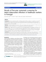
The stability of the DCU formulations (both regular
suspension and nanosuspension) was assessed, and no
issue was found. No particle size, potency, and form
change was observed in a period of 7 days. All samples
were found to be consistent with the previously reported
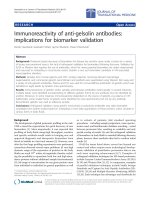
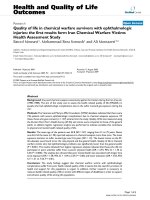
data [13-15]. In general, an analysis of unmilled and
milled DCU particles revealed a mean particle size of
20.2 μm (regular suspension) and 0.8 μm (nanosuspen-
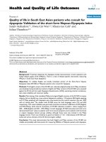
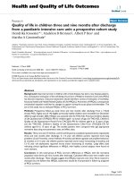
sion), respectively (Figure 1). No form change was
detected by PXRD when compare pre and post milling
sample (Figure 2). The rate of dissolution of the DCU
nanosuspension versus regular suspension is expected to
increase at least 20-folds. To estimate the impact of dis-
solution, the Noyes and Whitney equation was used:
dC/dt = D ∗ S
(
C
s
− C
t
(
t
)
)
/Vh
d
,
where dC/dt is the dissolution rate (R), D is the solute
diffusion coefficient, S is the surface area of solute, C
s
is
the saturation solubility of the solute, C
t
(t)isthebulk
solute concentration, V is the volume of dissolution
medium, and h
d
is the diffusion boundary thickness.
Figure 1 DCU particle size analysis. Regular suspension (top) and nanosuspension (bottom).
Chiang et al. Nanoscale Research Letters 2011, 6:413
/>Page 3 of 9
Animals
Male Sprague-Dawley rats weighing between 290 and
350 g, obtained from Charles Rivers Lab oratories
(Charles Rivers Laboratories, Inc., Wilmington, MA,
USA), were housed in a room with an ambient tempera-
ture of 22°C ± 1°C on a 12-h light/dark cycle. The ani-
mals were allowed 7 days to acclimate a nd were given
ad libitum access to standard rat chow (0.5% N aCl)
(Baxter Healthcare Corporation, Deerfield, IL, USA) and
tap water until the initiation of the experiment [14].
Thecurrentstudywasconductedinaccordancewith
the institutional guidelines for humane treatment of ani-
mals and was approved by the IACUC of Genentech.
For dosing, each group of three male Sprague-Dawley
rats was given either a 30-mg/kg subcutaneous dose o f
DCU fo rmulated as regular suspension or nanosuspen-
sion. Oral dosing followed the same guidelines [14]. At
the initiation of the study, the rats weigh ed from 297 to
329 g. Blood samples (approximately 0.2 mL per sample)
were collected from each anim al via jugular vein cannu-
lae at the following time points: predose; 5, 15, and 30
minpostdose;and1,2,4,8,and24hpostdose.All
samples were collected into tubes containing potassium
ethylenediaminetetraacetic a cid as an anticoagulant.
Blood samples were centrifuged within 30 min of the
collection, and plasma was harvested. Plasma samples
were stored at approximately 70°C un til analysis for
DCU concentrations by a LC/MS/MS assay method.
LC/MS/MS analysis
DCUplasmaconcentrationswerequantifiedbyusing
LC/MS/MS. Briefly, an internal standard (in-house com-
pound) was added to samples followed by protein preci-
pitation involving the addition of acetonitrile.
Chromatography of DCU was achieved using a HALO
Phenyl Hexyl column (2 × 50 mm, 2.7 μM pa rticle size)
(Advanced Materials Technology, Wilmington, DE,
USA). The mobile phase used was 0.1% formic acid (A)
and acetonitrile with 0.1% formic acid (B). A gradient
was used and is described as follows: 10% B at 0 min
and hold for 0.2 min, linear gradient to 95% B at 0.8
min and hold until 1.2 min, back to 10% B at 1.25 min
and hold until 2.0 min. The total run time was 2.0 min,
and the flow rat e was 0.75 mL/min. An AB Sciex
QTRAP 5500 mass spectrometer was used for detection.
The MRM transition monitored for DCU was m/z 225.4
to m/z 100.2. The lower limit of quantitation was 0.013
μM(S/N = 6) in plasma.
Dose simulation
A mod el based on the Wagner-Nelson (W-N) equation
was established in-house and was used to calculate the
drug absorbed to further assess the amount of drug
absorbed as a function of time [25,26]. The utilization of
the W-N equation allows us to obtain all the drug that
is absorbed (including excreted) at different time points.
This allowed us to estimate the relationship and the
impact on the absorption on the surface area changes of
the drug.
dA = V ∗ dC
p
+ V ∗ k ∗ C
p
∗ d
t
A = V ∗ Cp + V ∗ K ∗
t
0
Cp ∗ d
t
where A is the drug absorbed, V is the volume of dis-
tribution, Cp is the plasma concentration, K is the elimi-
nation rate constant, and t is time.
A slight ly simplified gastro transit time equation was
integrated in the model [27] to estimate the amount of
drug entering the small intestine as a function of time.
M = D
e
−Ke(t
)
where M is the mass of the drug remaining in the sto-
mach, D is the drug dosed, Ke is the stomach empting
rate, and t is the time.
A nonpsychological model was used to estimate the
total available surface area of the DCU as a f unction of
time. A linear movement was assumed in the GI [25,26].
Result and discussion
The use of nanoparticles and particle size reduction in
general to increase in vivo exposure for p oorly soluble
drugsiswellpracticed[17-23]. Reducing the particle
size increases the surface area available to the dissolu-
tion media and thus increases the overall apparent drug
dissolution. This can be estimated by the equation
developed by Noyes and Whitney. Despite the under-
standing of surface area impact on the drug dissolution,
Figure 2 PXRD DCU before mil ling (bottom) and post milli ng
(top).
Chiang et al. Nanoscale Research Letters 2011, 6:413
/>Page 4 of 9
the degree of impact on absorption by dosing nanoparti-
cles remains un clear [25]. In theory, the best usage of
utilizing nanoparticles to improve in vivo exposure (dis-
solution) is to dose it within the dissolution control
range. In which the higher surface area of the nanoparti-
cles is translated into a higher in vivo exposure.
An oral dose of DCU nanosuspension has been
reported to greatly improve the in vivo exposure [14].
However, the overall limit of improvement that a nano-
suspension formulation can provide for orally dosed
DCU is not well unders tood [14]. In order to u nder-
stand the degree of improvement provided by an oral
nanosuspension formulation, simulations were per-
formed using a Wagner-Nelson equation-based model
that was established in-house in order to assess the
amount of drug absorbed (dA) as a function of time
[26,27]. In this model, the stomach empting time was
taking into consideration. A log linear gastro transit
model [28] was used to estimate the amount of drug
available (W) in the small intestine for absorption. The
surface area of the DCU was estimated by assuming a
sphere shape particle and a true density (d)of1.3cm
3
/
gm. The total surface area (A) was estimated by first
obtaining the particle volume (V)usingtheequationof
V =3/4π r
3
, and then total particle number (n)using
the equation of n = ((drug weight)/V/d). The total sur-
face area of the dose was estimated by the equation A =
(4 πr
2
)×n. The unit surface area by weight (A/W)was
calculated to estimate the surface area reduction after
the absorption took place, and the total residual surfa ce
area (RA) was calculated for each time point. The
absorption efficiency (AE) was calculated by taking the
ratio of the amount of drug available and was divided by
the RA (AE = W/RA), and the absorption constant (K)
was calculated as AE/δT.
All of the above parameters were obtained by using
the 3-mg/kg rat oral PK data with regular suspension
[14] as the base case and predictions were performed
forhigherdoses(10and30mg/kg) with nanosuspen-
sion formation. Results for 3, 10, and 30 mg/kg are
listed in Table 1. According to theory, this model should
hold within the linear range where absorption efficacy
AE should be very close (amount of drug absorbed is
affected by dissolution hence surface area) if oral
absorption is dissolution rate-limited and should show
deviations when absorption becomes solubility rate-lim-
ited. Within the linear range, an increased surface area
(i.e., due to the nanolized drug) will result in a linear
incre ase of oral absorption. This model was found to be
sufficient to predict the exp osure for dissolution rate-
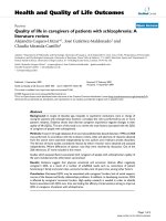
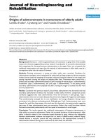
limited absorption at a 10-mg/kg dose. A much bigger
deviation was observed at a 30-mg/kg dose when the
predicted verse observed was compared with the
absorbed amount (Figure 3). According to the model, at
C
max
, a total of 3.0 mg of DCU should be absorbed
whereonly1.1mgwasobservedin vivo (Table 1). A
reduction in absorption efficiency (AE) was observed
particularly between the 10- and 30-mg/kg doses (Table
1). These changes suggested that at a 30-mg/kg dose,
the abso rption is no longer dissolution rate-limited and
most likely solubility rate-limited. The simulations sug-
gest that doses of DCU that are higher than 30 mg/kg
delivered using nanosuspension will not provide signifi-
cantly higher exposure in vivo. Based on the modeling,
doses higher than 30 mg/kg PO were not tested in vivo.
Simulations for oral dosing were performed using the
30-mg/kg oral dose in order to assess the dose fre-
quency required to hit a range of target concentrations.
A prediction of the oral dose amount and frequency
to cover different plasma co ncentrations were based on
maintaining free fraction plasma concentrations of DCU
(3% unbound) above a multiple of the cellular IC50 (6
nM) at the trough levels. Modeling of the pharmacoki-
netic data was performed using in-house model (1 com-
partment, f irst-order elimination), the pharmacokinetic
parameters V
f
(5.8 L), K
01
(absorption rate constant,
26.3 h
-1
)andK
10
(elimination r ate constant, 0.277 h
-1
)
were estimated [15]. Several concentrations were used
as “target coverage” since PK/PD investigations often
require a broad range of target coverage (i.e., from 0.25
× IC50 to 10 × IC50). Based on the simulation (figure
4), oral dosing of 30 mg/kg of DCU nanosuspension
twice a day (b.i.d.) is needed to provide continuous cov-
erage of the plasma concentrations of 0.2 μM (1 × cellu-
lar IC50 corrected for free fraction) and t.i.d. dosing will
be needed to cover 0.6 μM (3 × cellular IC50 corrected
for free fraction). The increase in dosing frequency in
order to cover three time the cellular IC50 is one short-
coming fo r the or al dosing of DCU especially f or
chronic studies. An additional drawback of this design is
Table 1 Predicted dug absorption (at C
max
) versus in vivo data (Wagner-Nelson equation) based on the surface area
model
Dose/Drug absorbed in mg (impact by
surface area only)
In vivo (Wagner-Nelson
equation) mg
Predicted
(mg)
Total surface area of the
drug dose (cm
2
)
Absorption efficiency
(AE) mg/cm
2
3 mg/kg (regular suspension) 0.02 0.02 2.08 E0 9.0E-6
10 mg/kg (nano suspension) 0.84 1.01 1.38 E2 7.2 E-6
30 mg/kg (nano suspension) 1.11 3.03 4.15 E2 2.6E-6
Chiang et al. Nanoscale Research Letters 2011, 6:413
/>Page 5 of 9
the high plasma P/T ratio. Higher than needed exposure
resulting from the high P/T ratio can result in unwanted
side effects and confound the efficacy read out [29].
Thus, oral dosing DCU to obtain the PK/PD relation-
ship remains less than ideal.
The SC route of delivery of the DCU nanosuspension
in rats was investigated as a means to improve the
pharmacokinetic properties of DCU. There are two
potential benefits to investigate the SC dose for DCU.
First, unlike oral absorption where all drug abso rbed
will first go through the liver then the circulation, the
drug absorbed via the SC route will go directly into the
circulation and hence avoid the “first pass” effect and
potentially improve systemic exposure [30]. Secondly,
the SC drug depot should continuously provide a slow
release of drug to the bloodstream providing a longer
and sometimes steady drug supply. Combined, both
effects may result in a d rug plasma profile with a more
sustained drug coverage and lower P/T ratio. Despite
the described advantages, the SC route of dosing is not
free of problems. Drug exposure via SC route of deliv-
ery can be still limited by absorption, stability, dissolu-
tion rate, and solubility of the drug. In order to
overcome these limitations, a suitable formulation was
needed to maximize the potential of DCU in vivo.Sev-
eral formulations strategies for SC dose have been
assessed. Formulations such as emulsions and
cosolvents were quickly found unsuitable since the goal
was to target a formulation that can be directly ap pli ed
to the efficacy model without any interference of excipi-
ents (i.e., high organic). After carefully evaluating all
available options, nanosuspension was found to be the
best option for the purpose. In order to understand the
impact of nanosuspensions on the systemic exposure of
DCU, both nanosuspension and regular suspension
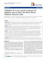
were dosed in vivo to contrast. It was found that when
dosed via the S C route, the DCU nanosuspension
greatly improved the exposure when compared with
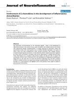
regular suspension. Results of this SC investigation are
illustrated in Figure 5.
The SC dose of DCU with nanosu spension was very
successfully. With DCU nanosuspension, an a pproxi-
mately threefold improvement of an apparent t
1/2
was
observed in SC dosing (10.2 h) in co mparison to oral
dosing (2.6 h). In addition, SC dosing resulted in a
lower plasma P/T (C
max
/C
min 24 h
) ratio of 4. This much
improved plasma P/T ratio was consistent with slower
release. Furthermore, the nanosuspension was found to
greatly improve the exposure and variability of the SC
dose compared to the regular suspension. The regular
suspension demonstrated similar effects on the exposure
profile of DCU nanosuspension; however, a much
reduced absorption rate (δc/δt) and lower exposure was
observed(Figure5).Theexposureobtainedviaregular
D
C
U simulation
0.0001
0.001
0.01
0.1
1
10
0.00 0.25 0.5
0
hr
mg abs
dose 1 in vivo (3 mg/Kg)
calculated data fit dose 1
predicted for dose 2
dose 2 in vivo (10 mg/kg)
predicted for dose 3
dose 3 in vivo (30 mg/kg)
Figure 3 DCU in vivo exposure model fit (SD rat PK).
Chiang et al. Nanoscale Research Letters 2011, 6:413
/>Page 6 of 9
Figure 4 DCU nanosuspension oral dose simulation for PK/PD.
DCU SC PK exposure profile
0.01
0.1
1
10
0 4 8 12 16 20 24 2
8
Time
(
hr
)
Plasma uM
30 mg/kg nanosuspension
SC
30 mg/kg regular
suspension SC
Figure 5 DCU SC PK plasma exposure. Thirty-milligram per kilogram nanosuspension versus regular suspension).
Chiang et al. Nanoscale Research Letters 2011, 6:413
/>Page 7 of 9
suspension at 30 mg/kg dosed was at least fivefold less
when compared with the nanosuspension and results
are listed as Table 2. It is hypothesized that the much
reduced exposure was caused by the slower dissolution
(dissolution rate-limited absorption) of the regular sus-
pension which makes it unsuitable for a PK/PD study
where higher exposures are needed.
Modeling of the pharmacokinetic data was per-
formed using the same in-house model (one compart-
ment, first-order elimination) and revised to fit the in
vivo data for SC dose. Based o n the simulation, SC
dosing of 30 mg/kg DCU nanosuspension once a day
(s.i.d.) can provide continuous coverage of the plasma
concentrations 0.2 μM (1 × cellular IC50 corrected for
free fraction) and b.i.d. dose will cover 0.6 μM(3×
cellular I C50 corrected for free fraction) for target PK/
PD (Figure 6). F or the same coverage, the SC dose of
the nanosuspension enabled a reduced total dose
amount and frequency. This provides a welcomed
advantage for a chronic dosing setting where a reduced
burden to animals and manpower are desired. In addi-
tion, the significantly reduced plasma P/T ratio is less
confounding for the interpretation of PK/PD relation-
ships. When dosed s.i.d. via SC, a DCU plasma P/T
ratio of 4 is expected (compare b.i.d oral P/T ratio of
25). When dosed b.i.d. via SC, a DCU plasma P/T
ratio of less than 2 is expected (compare to t.i.d oral
P/T ratio of >8). Our investig ation with D CU provides
an example of how nanosuspension can serve as a
powerful formulation for the delivery of low solubility
compounds in the preclinical sett ing. Based upon t he
positive results of our investigation, the continued use
of nanosuspension to deliver low solubility compounds
in preclinical PK/PD studies is expected.
Table 2 SC dose exposure comparison (nanosuspension versus regular suspension)
Dose (mg/kg) C
max
(μM) ± STDEV C
min24hr
(μM) ± STDEV AUC
0-t
(h*μM) ± STDEV t
1/2
(h) ± STDEV
30 mg/kg (nanosuspension) 0.82 ± 0.03 0.20 ± 0.02 11.0 ± 0.5 10.2 ± 0.8
30 mg/kg (regular suspension) 0.13 ± 0.02 0.06 ± 0.02 2.0 ± 0.8 34.4 ± 21.4
Figure 6 DCU nanosuspension SC dose plasma exposure simulation. Exposure versus cellular IC50 corrected for free fraction).
Chiang et al. Nanoscale Research Letters 2011, 6:413
/>Page 8 of 9
Conclusion
It is well-known that safety and efficacy are the two major
concerns of any new therapeutic target. Failure to fully
understand either target safety or efficacy in the early
development process often results in a more costly failure
later in the clinic or even postmarketing. For this reason,
the pharmace utical industry spends significant resources
on early target evaluation in order to minimize the risk in
moving forward. However, such a process often relies on
finding a suitable compound to interrogate the target
which may take a considerable time and is not cost effec-
tive. Here, we describe an effort with a less than ideal
model compound, DCU, utilizing nanosuspension formu-
lation and careful evaluation using PK modeling and simu-
lation. This approach helped us identify clear advantages
of using nanosuspension. In addition, we were able to
evaluate SC delivery of DCU which has distinct advantages
when compared to what has been previously described in
literature. We firmly believedthatusingthesystematic
approach will enable earlier “preclinical proof of concept
studies” and ultimately save both time and resources when
investigating new and novel targets. Further research is
needed to continue the development in this area.
Authors’ contributions
PCC conceived, design, and coordination the study. YR, KJC, and YC
contributed to the manuscript. HW carried out the animal study.
Competing interests
The authors declare that they have no competing interests.
Received: 10 March 2011 Accepted: 7 June 2011 Published: 7 June 2011
References
1. Katoh T, Takahashi K, Capdevila J, Karara A, Falck J, Jacobson H, Badr K:
Glomerular stereospecific synthesis and hemodynamic actions of 8,9-
epoxyeicosatrienoic acid in rat kidney. Am J Physiol 1991, 261:578-586.
2. Lin W, Falck J, Wong P: Effect of 14,15-epoxyeicosatrienoic acid infusion
on blood pressure in normal and hypertensive rats. Biochem Biophys Res
Commun 1990, 167:977-981.
3. Imig J, Navar L, Roman R, Reddy K, Falck J: Actions of epoxygenase
metabolites on the preglomerular vasculature. J Am Soc Nephrol 1996,
7:2364-2370.
4. Node K, Huo Y, Ruan X, Yang B, Spiecker M, Ley K, Zeldin D, Liao J: Anti-
inflammatory properties of cytochrome P450 epoxygenase-derived
eicosanoids. Science 1999, 285:1276-1279.
5. Roman R: P-450 metabolites of arachidonic acid in the control of
cardiovascular function. Physiol Rev 2002, 82:131-185.
6. Spector A, Fang X, Snyder G, Weintraub N: Epoxyeicosatrienoic acids
(EETs): metabolism and biochemical function. Prog Lipid Res 2004,
43:55-90.
7. Imig J: Epoxide hydrolase and epoxygenase metabolites as therapeutic
targets for renal diseases. Am J Physiol Renal Physiol 2005, 289:F496-F503.
8. Zhao X, Imig J: Kidney CYP450 enzymes: biological actions beyond drug
metabolism. Current Drug Metabolism 2003, 4:73-84.
9. Yu Z, Xu F, Huse L, Morisseau C, Draper A, Newman J, Parker C, Graham L,
Engler M, Hammock B, Zeldin D, Kroetz D: Soluble epoxide hydrolase
regulates hydrolysis of vasoactive epoxyeicosatrienoic acids. Circ Res
2000, 87:992-998.
10. Davis B, David A, Howard L, Morisseau C, Hammock B, Weiss R: Inhibitors
of soluble epoxide hydrolase attenuate vascular smooth muscle cell
proliferation. PNAS 2002, 99:2222-2227.
11. Ross R: The pathogenesis of atherosclerosis: a perspective for the 1990s.
Nature (London) 1993, 362:801-809.
12. Smith K, Pinkerton K, Watanabe T, Pedersen T, Ma S, Hammock B:
Attenuation of tobacco smoke-induced lung inflammation by treatment
with a soluble epoxide hydrolase inhibitor. PNAS 2005, 102:2186-2191.
13. Chiang P, Wahlstrom J, Selbo J, Zhou S, Wene S, Albin L, Warren C,
Smith M, Roberds S, Ghosh S, Zhang L, Pretzer D: 1,3-Dicyclohexyl urea
nanosuspension for intravenous steady-state delivery in rats. J of Exp
Nano 2006, 2:239-250.
14. Ghosh S, Chiang P, Wahlstrom J, Fujiwara H, Selbo J, Roberds S: Oral
delivery of 1,3-dicyclohexylurea nanosuspension enhances exposure and
lowers blood pressure in hypertensive rats. Basic Clin Pharmacol Toxicol
2008, 102(5):453-458.
15. Wahlstrom J, Chiang P, Ghosh S, Warren C, Wene S, Albin L, Smith M,
Roberds S: Pharmacokinetic evaluation of a 1,3-
dicyclohexylureananosuspension formulation to support early efficacy
assessment. Nanoscale Res Lett 2006, 2:291-296.
16. Bittner B, Mountfield R: Intravenous administration of poorly soluble new
drug entities in early drug discovery: the potential impact of
formulation on pharmacokinetic parameters. Curr Op Drug Discov Devel
2002, 5(1):59-71.
17. Barrett R: Nanosuspensions in drug delivery. Nat Rev Drug Discov 2004,
3:785-796.
18. Chiang P, Hu Y, Thurston A, Sommers C, Guzova J, Kahn L, Lai Y, Blom J:
Pharmacokinetic and pharmacodynamic evaluation of the suitability of
using fluticasone and an acute rat lung inflammation model to
differentiate lung versus systemic efficacy. J Pharm Sci 2009,
98(11):4354-4364.
19. Liu C: Research and Development of Nanopharmaceuticals in China.
Nano Biomed Eng 2009, 1(1):1-18.
20. Liu Y, Miyoshi H, Nakamura M: Nanomedicine for drug delivery and
imaging: a promising avenue for cancer therapy and diagnosis using
targeted functional nanoparticles. Int J Cancer 2007, 120(12):2527-2537.
21. Urisu T, Wei C: America–Japan Nanomedicine Society (AJNS).
Nanomedicine 2006, 2(4):297-298.
22. Liang X, Chen C, Zhao Y, Jia L, Wang P: Biopharmaceutics and therapeutic
potential of engineered nanomaterials. Curr Drug Meta 2008, 9(8):697-709.
23. Si D, Sun Y, Cheng T, Liu C: Biomedical evaluation of nanomedicines.
Asian Journal of Pharmacodynamics and Pharmacokinetics 2007, 7(2):83-97.
24. Badyl D, Lata H, Dadhich A: Animal models of hypertension and effect of
drugs. Indian J Pharmacol 2003, 35:349-362.
25. Lai Y, Chiang P, Li N, Shevlin K, Brayman T, Hu Y, Selbo J, Hu L: Comparison
of in vitro nanoparticles uptake in various cell lines and in vivo
pulmonary cellular transport in intratracheally dosed rat model.
Nonoscale Res Lett 2008, 3:321-329.
26. Chiang P, South S, Wene S: The impact of dosing interval in a novel
tandem oral dosing strategy: enhancing the exposure of low solubility
drug candidates in a pre-clinical setting. J Drug Delivery 2011, Article ID
528284, 9 pages
27. Chiang PC, South SA, Foster KA, Daniels JS, Wene SP, Albin LA,
Thompson DC: Utilizing a novel tandem oral dosing strategy to enhance
exposure of low solubility drug candidates in pre-clinical setting. J
Pharm Sci 2010, 99(7):3132-3140.
28. Oberle R, Chen T, Lloyd C, Barnett J, Owyang C, Meyer J, Amidon G: The
influence of the interdigestive migrating myoelectric complex on the
gastric emptying of liquids. Gastroenterology 1990, 99(5):1275-1282.
29. Chiang P, Kishore N, Thompson D: Combined use of pharmacokinetic
modeling and a steady state delivery approach allow early assessment
of IκB kinase-2 (IKK-2) target safety and efficacy. J Pharm Sci 2010,
99(3):1278-1287.
30. Kerns E, Di L: Drug-like properties: concepts, structure design and methods:
from ADME to toxicity optimization Burlington: Elseveir; 2008.
doi:10.1186/1556-276X-6-413
Cite this article as: Chiang et al.: Investigation of utilization of
nanosuspension formulation to enhance exposure of 1,3-
dicyclohexylurea in rats: Preparation for PK/PD study via subcutaneous
route of nanosuspension drug delivery. Nanoscale Research Letters 2011
6:413.
Chiang et al. Nanoscale Research Letters 2011, 6:413
/>Page 9 of 9