Báo cáo hóa học: " Understanding urine output in critically ill patients" ppt
Bạn đang xem bản rút gọn của tài liệu. Xem và tải ngay bản đầy đủ của tài liệu tại đây (393.25 KB, 8 trang )
REVIEW Open Access
Understanding urine output in critically ill patients
Matthieu Legrand
*
and Didier Payen
Abstract
Urine output often is used as a marker of acute kidney injury but also to guide fluid resuscitation in critically ill
patients. Although decrease of urine output may be associated to a decrease of glomerular filtration rate due to
decrease of renal blood flow or renal perfusion pressure, neurohormonal factors and functional changes may
influence diuresis and natriuresis in critically ill patients. The purpose of this review is to discuss the mechanisms of
diuresis regulation, which may help to interpret the urine output in critically ill patients and the appropriate
treatment to be initiated in case of changes in urine output.
Introduction
Acute renal failure or acute kidney injury (AKI) is
defined by an acute decline of glomerular filtration rate
(GFR). Occurrence of AKI is associ ated with substantial
in-hospital mortality, exceeding 50% when AKI is part
of a multiple organ failure syndrome [1, 2]. Therefore,
early recognition of AKI, better understanding of its
pathogenesis, and development of preventing strategies
appear to be potential areas of improvement of patient’s
prognosis. The decrease of glomerular filtration rate and
urine output in response to a decrease of renal blood
flow is classically referred as pre-renal azotemia, which
can evolve into structural damage if renal hypoperfusion
persists. In this line, urine output often is used as a mar-
ker of AKI but also to guide fluid resuscitation in criti-
cally ill patients. However, both the contribution of
renal hypoperfusion to AKI and the genuine definition
of pre-renal and intra-renal azotemia have been chal-
lenged by several authors [3-5]. The recent international
consensus conference on acute renal failure therefore
recommended the term “ acute kidney insufficiency”
rather than “acute kidney injury” in the light of paucity
of evidence of a relation between tissue damage and
organ failure in human AKI [6]. The purpose of this
review is to discuss the mechanism of diuresis regula-
tion and the interpretation of urine output in critically
ill patients in the light of clinical and physiological
studies.
Why should we wonder about oliguria and AKI?
There is accumulating evidence that critically ill patients
developing AKI have an increa se relative risk of death.
Occurrence of AKI is a marker of severity of the underly-
ing acute illness but also appears as an independent factor
associated with mortality in unselected critically ill patients
[7], in sepsis [8], pneumonia [9], or cardiac surgery [10].
The mechanistic pathways of such an association remain
elusive, with intrication of inflammation, metabolism, and
apoptotic phenomena. Remote organs damage has been
suggested in several experimental studies [11,12].
Ischemic-induced AKI has been found to induce myocar-
dial apoptosis [13], to activate lung inflammatory and
apoptotic pathways, and to increase lung water permeabil-
ity [14]. Surprisingly, even a small increase of serum crea-
tinine after cardiac surge ry or transient (i.e., reversible
within 3 days) AKI has been found to be asso ciated with
an increased risk of death [15]. Although fluid resuscita-
tion and optimization of renal perfusion pressure are cen-
tral to the prevention and treatment of AKI, excessive
fluid resuscitation may be harmful in some critically ill
patients. Payen et al. [16] and Bouchard et al. [17] found,
when analyzing two large cohorts of critically ill patients,
that a positive fluid balance was associated with an
increased risk of death in patients suffering from AKI.
First, aggressive fluid resuscitation, although increasing
renal blood flow, can be ineffective in restoring renal
microvascular oxygena tion due to hemodilution with no
increase in blood-oxygen carriage capacities [18]. Second,
positive fluid balance can deteriorate cell oxygenation and
prolong mechanical ventilation [19]. Finally, fluid overload
may lead to central venous congestion and decrease of
renal perfusion pressure [20], which will promote the
* Correspondence:
Department of Anesthesiology and Critical Care and SAMU, Lariboisière
Hospital, Assistance Publique- Hopitaux de Paris; University of Paris 7 Denis
Diderot, 2 rue Ambroise-Paré, 75475 Paris Cedex 10, France
Legrand and Payen Annals of Intensive Care 2011, 1:13
/>© 2011 Legrand and Payen; licensee Springer. This is an Open Access article distributed under the terms of the Creative Commons
Attribution License ( which permits unrestricted use, distribution, and repro duction in
any me dium, provided the orig inal work is properly cited.
development of AKI in patients with acute heart failure
[21] or sepsis [22]. The type of fluid used also can have a
role with “ renal toxicity” associated with the use of
colloids.
Urine output and definition of acute kidney
injury
In clinical research, more than 30 definitions of acute
renal failure have been used before the release of the
RIFLE criteria by the Acute Dialysis Quality Initiative
group in 2004 [23]. The first merit of this classification
was to introduce a standard and simple definition of AKI
for clinical research purposes but also to stratify the sever-
ity of AKI based on serum creatinine level, creatinine
clearance, or urine output. In 2007, the Acute Kidney
Injury Network classification was published, introducing
subtle modifications to the RIFLE criteria. A part from the
change in nomenclature (Risk, Injury, and Failure were
replaced by stage 1, 2, and 3, the categories Loss and End-
stage disappeared), an absolute increase of serum creati-
nine of 0.3 mg/dl was sufficient to classify patients in stage
1, introducing the notion than only small changes in
serum creatinine are of clinical relevance. Finally, the
AKIN criteria should be applied “after following adequate
resuscitation when applicable” with the purpose of exclud-
ing patients wit h pure renal pre-azotemia. The introduc-
tion of the RIFLE and AKIN definitions were a c rucial
step forward in the development of clinical research and
have since been widely accepted by the medical commu-
nity. Using these classifications, a patient with decrease of
urine output will be classified as “AKI.” However, a non-
sustained decrease of urine output does not necessarily
imply a decrease of glomerular filtration rate but can sim-
ply represent a physiological renal adaptation (i.e., anti-
diuresis and antinatriuresis) to maintain the body volume
and/or electrolytes homeostasis. This would be the case if
decreased urine output is not associated with a decline of
creatinine clearance. Although severe acute renal failure
with oliguria or anuria has been reported to be associated
with a worse outcome compared with patients with pre-
served urine output, the use of urine output as a criterion
to classify AKI severity may be misleading. It was reported
that the combination of creatinine and urinary output for
classifying the patient’s risk of death was more stringent
than urinary output alone for classifying patients [7,24].
One can conclude that patients classified according to the
urine output criterion only might be less severe than those
classified acco rding to the combination of creatinine and
urine output [25]. On the other hand, severe tubular dys-
function can lead to increased urine output despite low
GFR. Urine output therefore seems to be a nonspecific
and poor parameter for classifying of AKI in critically ill
patients.
Glomerular filtration rate as a determinant of
urine output
At constant hydraulic permeability o f the glomerular fil-
tration barrier, the glomerular filtration is driven by the
pressure gradient across the glomerular capillary walls
(Figure 1). The pressure gradient across the glomerular
capillary wall is determined by the opposing forces of the
hydraulic and oncotic pressures gradients between the
capillaries and the Bowman’s space. Because the length
of the afferent and efferent arterioles in the glomerular
capillary network is relatively short and the resistance is
low, the glomerular capillary hydraulic pressure remains
rather constant along the capillaries, whereas the oncotic
pressure along the capillary increases in relation with fil-
tration. Therefore, the limiting factors of GFR are the
renal plasma flow and the plasma protein concentrat ion.
A higher renal plasm a flow will induce a reduction in fil-
tration fraction (i.e., ratio of ultrafiltration to renal
plasma flow) with a lesser increase of capillary plasma
protein concentration along the glomerular capillaries.
Conversely, when the renal plasma flow is reduced, the
glomerular filtration rate decreases but with an increase
in the filtration fracti on. An increase of capil lary hydrau-
lic pressure will cause the ultrafiltrate to be mainly gener-
ated on the first portion of the afferent side of the
capillary network and to cease when hydraulic and onco-
tic pressures become equal along the glomerular capillary
network (Figure 1). Therefore, the oncotic pressure
becomes the limiting factor of glomerular filtration [26].
In this line, the natriuresis and diuresis response to cry s-
talloids infusion are in part mediated by the changes of
intraglomerular oncotic forces following plasma protein
dilution [27,28], an effect that is not observed after
hyperoncotic colloids administration. When hydraulic
permeability is altered (decreased of glomerular surface
area as in chronic kidney disease) glomerular hydraulic
capillary pressure becomes the major determinant of the
glomerular filtration rate (Figure 1) [29].
Relationship between renal blood flow and GFR
Physiologically, the renal blood flow is autoregulated,
which means that it remains unchanged when arterial
blood pressure varies [30]. Such autoregulation is
mediated by a myogenic mechanism, the tubuloglomer-
ular feedback (TGF), and a “third mechanism” not yet
fully identified. The lower autoregulator y threshold of
mammalian kidney occurs at a mean arterial pressure
(MAP) of ~80 mmHg. Below this pressure level, renal
blood flow and glomerular filtration rate decrease along
with the decrease in pressure [31].
In normal kidneys, the total interruption of renal
blood flow for a prolonged period of time (i.e., more
than 30 minutes) followed by reperfusion is always
Legrand and Payen Annals of Intensive Care 2011, 1:13
/>Page 2 of 8
associated with major tubular and microvascular
damage. In this condition, cellular lesions result from a
combination of cellular hypoxia-reperfusion injury and
oxidative stress-associated damage [32]. This situation is
a rare clinical s cenario except during suprarenal aortic
surgery with aortic clamping. Experimental studies have
shown that prolonged period of renal hypoperfusion
would not systematically lead to renal histological
damage and renal failure [33,34]. Saotome et al. reported
that prolonged mechanical reduction of renal blood flow
by 80% for 2 h in conscious sheep did not induce sus-
tained renal function impairment or kidney damage
[33]. In a rat m odel, Johannes et al. have shown that
temporary mechanical reduction of renal blood flow
does not impair microcirculatory oxygenation and renal
function [34]. However, severe renal damage were
observed in rats recovering from an ischemic acute
renal failure induced by intra-arterial infusion of norepi-
nephrine [35], which underwent additional injury by
mild hemorrhage, an effect partially prevente d by renal
denervation. These observations highlight the role of
renal innervation in the induction of renal failure.
Together, these experiments suggest that a severe transi-
ent hypoperfusion is able to reduce GFR and urine out-
put but is not sufficient to induce persistent AKI.
However, this is the superimposition of renal hypoperfu-
sion episodes in relation to other insults, such as sepsis
or ischemia, which may induce renal failure. Because of
Pressure (mmHg)
PGC-PT
πGC
πGC πGC
πGC
Pressure (mmHg)
Pressure (mmHg) Pressure (mmHg)
GFR= 100ml/min
GFR= 100ml/min
GFR= 60ml/min GFR= 90ml/min
A
DC
B
Glomerular capillary lenghtGlomerular capillary lenght
Glomerular capillary lenghtGlomerular capillary lenght
PGC-PT
PGC-PT PGC-PT
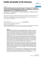
Figure 1 Schematic representation of the glomerular capillary hydraulic and oncotic pressure in normal kidneys (A and B) and
pathologic kidneys with decrease of the total ultrafiltration surface (C and D). The difference between the hydraulic pressure difference
[P
GC
, glomerular capillary hydraulic pressure-P
T
hydraulic pressure in Bowman’s space) and the intracapillary oncotic pressure (∏
GC
) represents the
effective filtration pressure gradient. In normal condition (A), the P
GC
-
PT
slightly decreases along the glomerular capillary axe and the ∏
GC
increases leading to equilibrium between the opposing forces to filtration. If renal perfusion pressure and P
GC
increase (B), the point of
equilibrium is reached earlier along the axe due to increase of filtration fraction. GFR does not change and only increase of renal plasma flow
and decrease of filtration fraction causes the GFR to increase (B). GFR is likely to increase with rise of renal perfusion pressure if the filtration
surface is impaired, the point of equilibrium not being reached (C and D). Note the role of plasma oncotic pressure. Infusion of crystalloid
decreases plasma oncotic pressure due to hemodilution favoring the net filtration pressure while infusion of colloids increases plasma oncotic
pressure therefore reducing GFR. GFR, glomerular filtration rate.
Legrand and Payen Annals of Intensive Care 2011, 1:13
/>Page 3 of 8
the above-mentioned arguments, it is expected that pre-
venting a decrease of renal blood flow may prevent or
limit the occurrence of AKI in ICU patients.
Renal blood flow autoregulation exists at high mean
arterial blood pressure, protecting the glomerular struc-
ture from hypertens ive injury by a decrease of glomerular
capillary pressure [36]. Therefore, one can expect that
increasing renal perfusion pressure when MAP is below
the threshold of renal blood flow autoregulation or if
autoregula tion is impaired could improve GFR and urine
output through an increase of renal blood flow. Sepsis is
the leading c ontributor to AKI in the ICU setting,
accounting for more than 50% of episodes of AKI.
Whereas fluid challenge can improve renal perfusion
pressure and renal perfusion in hypovolemic states, the
sole fluid resuscitation is unlikel y to increase largely the
mean arterial pressure. Vasopressor infusion is therefore
required to improve renal perfusion pressure in condi-
tions with systemic inflammation [37]. Norepinephrine
has been reported to increase renal blood flow, urine out-
put, and creatinine clearance in experimental sepsis [38].
Although norepinephrine also has been found to increase
creatinine clearance in human sepsis [39], clinical studies
in which MAP was increased with norepinephrine have
provided conflicting results. Bourgoin et al. found that
increasing MAP from 65 to 85 mmHg did not further
improve creatinine clearance in patients with septic
shock [40]. In contrast, in a more recent study among
patients with vasodilatory shock after cardiac surgery,
infusing norepinephrine was found to improve renal oxy-
gen delivery, oxygen delivery/consumption balance, and
GFR when MAP was increased from 60 to 75 mmHg
[41]. Infusion of norepinephrine in septic patients titrated
to increase MAP from 65 to 75 mmHg was associated
with a decrease of renal Doppler resistive index, suggest-
ing an increase in renal vascular conductance [42], con-
firming the experimental data. These results are in
accordance with physiological animals studies that
showed that norepinephrine and vasopressin can induce,
in septic states, an increase of renal blood flow through a
combined increase of renal perfusion pressure (i.e., prere-
nal mechanism) and an increase of renal vascular con-
ductance (i.e., intrarenal mechanism) [38,43].
Such an increase of renal blood flow does not necessarily
translate into GFR increase. For example, infusion of low-
dose dopamine (2 μg/kg/min) can increase renal blood
flow, induce renal vasodilatation, and incr ease urine out-
put but with no effect on creatinine clearance [44].
These apparent conflicting findings call for several com-
ments. First, increase of renal blood flow or urine output
does not necessarily translate into increase of creatinine
clearance. The systematic review of human AKI by Prowle
et al. showed that renal plasma flow and GFR were poorly
correlated [45]. In a septic hyperdynamic animal, a fall in
creatinine clearance can occur despite an increase of renal
blood flow [46]. The same group using the same model
found that infusion of angiotensin II could improve creati-
nine clearance while depressing renal blood flow [47].
Ventilation with positive end expiratory pressure always
decreases urine output in correlation with a decreased
renal perfusion pressure (mean arterial blood pressure -
renal venous pressure) and reduced renal blood flow [48].
A nonpharmacologic technique (lower body positive pres-
sure) was used to increase cardiac output and renal blood
flow but with no impact on diuresis [48]. In other words,
increasing renal perfusion pressure can increase urine out-
put and natriuresis independently of changes in total renal
blood flow and GFR. These discrepancies could, in part, be
due to the effect of neurohormonal regulation of vascular
tone between the afferent and efferent glomerular arter-
ioles (Figure 2). As an example, predominant vasodilatation
on efferent arterioles leads to increase renal blood flow
with a steady glomerular capillary pressure and GFR. Con-
versely, a predo minant vasoconstrictio n of the efferent
arterioles, even if renal blood flow remains unchanged,
increases the GFR and urine output, potentially inducing
renal ischemia. Second, renal fluid and sodium excretion
(i.e., diuresis and natriuresis) can exhibit a pressure-depen-
dency response [43,49,50]. Several humoral factors control
sodium excretion through, in part, changes of renal
medulla blood flow and intrarenal redistribution of blood
flow.
Role of intrarenal blood flow distribution in
regulation of diuresis and natriuresis
Whereas normal kidneys receive ~20% of cardiac output,
the medulla receives less than 10% of renal blood flow
[51]. Even with a stable renal blood flow within the range
of autoregulation, the cortical and medulla have different
responses to changes in renal perfusion pressure (RPP). In
contrast to the cortical microcirculation, t he medulla
microcirculation appears to be poorly autoregulated, i.e.,
pressure-dependent. Renal medulla blood flow regulation
is of paramount importance with respect of the regulation
of diuretics and natriuresis and, therefore, the response of
the kidney to the body fluid composition and volume sta-
tus (Figure 2). In fact, in mammalians kidneys, the ability
of the medulla circulati on to regulate its own blood flow
depends largely on the body volume status. In euvolem ic
dogs, when a RPP is decreased from 153 to 114 mmHg
within the range of RBF autoregulation (i.e., with no
change of renal blood flow), flow in the inner medulla
decreases with no redistribution of flow within the renal
cortex [50]. In contrast, both renal cortical and medulla
are well autoregulated in hydropenic rats. Because the des-
cending vasa recta provide blood flow to the medulla
emerge from efferent arterioles of juxtamedullary glomer-
ules, these data suggest t hat changes in resistance in the
Legrand and Payen Annals of Intensive Care 2011, 1:13
/>Page 4 of 8
postglomerular circulation of juxtamedullary nephrons
might be responsible for the lack of autoregulation of
medullary blood flow in volume expended animals [51].
Increase in renal medullary blood flow decreases the
outer-inner medullar osmotic gradient and increases renal
interstitial hydrostat ic pressure, which both impair the
ability to concentrate urine and participate in the natriur-
esis response to hypertension in well-hydrated mamma-
lians. In hydropenic animals, this response is blunte d
preventing further loss of water and sodium. The tubular
sodium handling may be mediated more by the angioten-
sin II and paracrine effects of NO rather than the increase
in RPP per se. In the absence of angiotensin II, volume
expansion with no increase in MAP induces natriuresis,
whereas the increase in MAP by angiotensin II infusion
did not induce a natriuresis response [52]. Increase of
plasma vasopressin concentration (independently of any
incr ease of systemic art erial pressure) also influences the
pressure-natriuresis/diuresis relationship in decreasing the
medullary blood flow through receptor V1a [43]. Binding
to the V2-receptors in the inner medullary collecting
ducts activates the UT-A1 molecules, which increases the
urea permeability of collecting duct and increase the abil-
ity to concentrate urine. Increased vascular response of
the renal microcirculation to vasoconstrictors has been
proposed to elicit intense renal vasoconstriction in sepsis-
induced AKI [53]. Although this hypothesis warrants
further exploration, it is possible in sepsis that endogenous
vasoconstrictors, including angiotensin II, could both
decrease GFR due to decrease in renal blood flow but also
blunt the natriuresis response after the renal perfusion
pressure has been restored. Endotoxemia also can increase
urine output and water clearance despite decrease in GFR
due to tubular aquaporin-2 dysfunction [54].
The adaptation of medullary blood flow to the Na
+
con-
centration in the tubular lumen adds another level of com-
plexity to the regulation of regional blood flow and sodium
handling. The glomerular filtration rate will decrease due
to vasoconstriction of the afferent glomerular arteriole in
response to increase of the filtrated Na
+
reaching the
macula densa, a mechanism called the tubuloglomerular
feedback (TGF, Figure 2). Tubular salt sensing by the
macula densa i nvolves the Na
+
/K
+
/2Cl
-
cotransporter
(NKCC2). The mechanism of TGF consists in an increase
of the glomerular afferent arteriole vascular tone, mainly
mediated by adenosine release, in response to a raise of
the [NaCl] concentrations in the tubular fluid. The juxta-
glomerular apparatus also mediates renin-release signal s
Diuresis
N
a
tri
u
r
es
i
s
1
6
2
1
8
3
G
FR regulation
1
Renal blood flow
and perfusion pressure
Afferent and efferent
glomerular arteriole tone
Balance
Tubulo-glomerular feedbac
k
Plasma oncotic pressure
Bowman’s capsule
hydraustatic pressure
4
Water and Na
+
handling
Intra-Renal blood flow
Distribution
Increase renal interstitial
Hydrostatic pressure
conformational changes of
tubule Na
+
/H
+
exchanger,
urea and chloride channels
Aquaporin-2 expression
2
3
4
5
6
7
8
5
7
Figure 2 Schematic view of regulating facto rs of diuresis and natriuresis . Renal blood flow, renal perfusion pressure, and plasma oncotic
pressure influence the effective filtration pressure gradient. Afferent and efferent glomerular arteriole tone can further influence the glomerular
capillary hydraulic pressure while tubular cast accumulation increases the Bowman’s hydrostatic pressure decreasing the effective filtration
pressure gradient. Finally intrarenal blood flow distribution, conformational changes of tubule Na+/H+ exchanger, urea, and chloride channels
and aquaporin-2 expression regulate water and sodium (Na
+
) handling of the ultrafiltrate (see text for further details). GFR, glomerular filtration
rate.
Legrand and Payen Annals of Intensive Care 2011, 1:13
/>Page 5 of 8
through prostaglandins (i.e., PGI2 and PGE2) and nitric
oxide release. The TGF response to increa se of Na
+
con-
centration in the tubular f luid operates within a few sec-
onds but is not sustained. Prolonged stimulation of the
TGF will induce the TGF to reset within 30-60 m inutes,
increasing the renal blood flow without restoring the GFR
[55]. Activation of the TGF has long been proposed by
Thureau et al. as an adaptative mechanism to tubular dys-
function and referred as an “acute renal success” in acute
renal failure [56]. In theo ry, TGF respon se could prevent
the rapid loss of water and electrolytes in conditions of
tubular dysfunction-associated decrease of Na
+
reabsorp-
tion. Na
+
-tubular reabsorptive work constitutes a major
part of renal oxygen consumption in the healthy kidney.
As a consequence, decrease of GFR or inhibition of Na
+
tubular reabsorption can decrease renal oxygen consump-
tion [57]. However, in ischemic-induced AKI there is a
diversion of oxygen co nsumption from Na
+
reabsorptio n
to other oxygen-consuming pathways illustrated by an
increase of the ratio oxygen consumption/Na
+
reabsorp-
tion [58]. Redfors et al. have recently shown in an elegant
physiological study in patients developing AKI after car-
diac surgery that total renal oxygen consumption increases
despite a decrease of Na+ reabsorptive work [59]. The
oxygen consumption to absorptive work mismatch is not
well understood and may result from: 1) higher produc-
tion of reactive oxygen species by infiltrative immune cells
[60]; 2) high level of NO, which regulates the renal oxygen
consumptio n [58]. This may partially explain why stra te-
gies designed to inhibit renal oxygen consumption (e.g.,
loops diuretics) have failed to improve the prognosis of
patients suffering from AKI [61].
Urine output, urine biochemistry, and mechanism
of AKI
Medical textbooks provide urine biochemistry profiles to
differentiate prerenal causes from intra renal causes of
AKI in oliguric patients. Although very popular among
clinicians, the ability of urinary indices, such as urinary Na
+
(UNa) and excretion fraction of Na
+
(FeNa), to separate
prerenal from intrarenal causes of AKI is questionable.
First, these urinary markers have been poorly studied
among critically ill patients. Recent reviews of experimen-
tal and human sepsis have highlighted the paucity of avail-
able studies and their design heterogeneity regarding
urinary findings in septic AKI [62, 63]. Most importantly,
the re is no evid ence th at these urinary biochemical find-
ings can predict the response to hemodynamic optimiza-
tion in terms of renal injury and renal function. Although
a low UNa or FeNa (e.g., FeNa <1%) sugg est a preserved
renal tubular reabsorptive capacity, there is no evidence
for a correlation between urinary biochemical modifica-
tions and tissue damage. Inflammation mediators can
induce tubular cell dysfunction with conformational
changes of tubule Na
+
/H
+
exchanger, urea, or chloride
channels that will influence urine composition indepen-
dently of any structural damage [14,64,65]. As mentioned,
the control of urinary Na
+
excretion results from a com-
plex neurohumoral regulation and is influenced by fluid
resuscitation, arterial pressure, or infusion of diuretics. A
fractional excretion of urea (FeU) of 35% or less has been
proposed to differentiate prerenal AKI from intrarenal
causes independen tly of the use of diuretics. However,
mechanically ventilated patients with transient AKI (resol-
ving within 3 days) exhibited higher FeU than patients
with persistent AKI in a recently published cohort [66].
To summarize, sensitivity and specificity of traditional
urinary biochemicals showed significant disparities among
clinical studies such that their value to classify AKI
remains doubtful. There is much more expectation in the
use of new biomarkers (i.e., NGAL, KIM1) to make an
early diagnosis o f tubular damage during the course o f
AKI and therefore to differentiate prerenal from intrarenal
AKI in oliguric patients. Only a f ew studies are available
regarding the association between plasma and/or urine
levels of those biomarkers and the reversibility of AKI.
Bagshaw et al. reported that plasma NGAL had an area
under the ROC curve of 0.71 (95% confidence interval
(CI), 0.55-0.88) for predicting AKI progression and of 0.78
(95% CI, 0.61-0.95) for need for renal replacement therapy.
Cruz et al. reported an area under the ROC curve of 0.82
(95% CI, 0.7-0.95) for predicting the use of renal replace-
ment therapy [67]. Nickolas et al. reported that urine
NGAL remained low in patients admitted in the emer-
gency department with prerenal azotemia versus AKI [68].
Conclusions
Decrease urine output is common among critically ill
patients and c an mirror a decrease in creatinine cle ar-
ance. Although a decrease in renal blood flow and/or a
decrease in renal perfusion pressure is a major determi-
nant of GFR, plasma oncotic pressure appears to be cen-
tral in the glomerular hydrodynamic forces. In
hypovolemic states, prompt fluid resuscitation is needed
to prevent further det erioration of renal function. The
choice of the type of fluid also seems to be crucial,
because colloids increase the oncotic pressure and may
reduce filtration rate. Fluid administration may be found
inappropriate and even harmful in numerous situations
due to the inconstant relati onship between renal blood
flow or renal perfusion pressure and diuresis/natriur esis
due to complex neurohormonal control. Furthermore,
systemic inflammation can induce natriuresis and diur-
esis changes due to functional changes unrelated to
hypoperfusion, histological, or tubular damage. Experi-
mental and clinical researc h is needed to determine
appropriate therapeutic response to oliguria in critically
ill patients.
Legrand and Payen Annals of Intensive Care 2011, 1:13
/>Page 6 of 8
Authors’ contributions
ML and DP wrote and approved the final manuscript.
Competing interests
The authors declare that they have no competing interests.
Received: 23 March 2011 Accepted: 24 May 2011
Published: 24 May 2011
References
1. Chen YC, Jenq CC, Tian YC, Chang MY, Lin CY, Chang CC, Lin HC, Fang JT,
Yang CW, Lin SM: Rifle classification for predicting in-hospital mortality in
critically ill sepsis patients. Shock 2009, 31:139-145.
2. Morgera S, Schneider M, Neumayer HH: Long-term outcomes after acute
kidney injury. Crit Care Med 2008, 36:S193-197.
3. Bouchard J, Mehta RL: Fluid accumulation and acute kidney injury:
consequence or cause. Curr Opin Crit Care 2009, 15:509-513.
4. Bellomo R, Bagshaw S, Langenberg C, Ronco C: Pre-renal azotemia: a
flawed paradigm in critically ill septic patients? Contrib Nephrol 2007,
156:1-9.
5. Langenberg C, Bellomo R, May C, Wan L, Egi M, Morgera S: Renal blood
flow in sepsis. Crit Care 2005, 9:363-374.
6. Brochard L, Abroug F, Brenner M, Broccard AF, Danner RL, Ferrer M, Laghi F,
Magder S, Papazian L, Pelosi P, Polderman KH: An Official ATS/ERS/ESICM/
SCCM/SRLF Statement: Prevention and Management of Acute Renal
Failure in the ICU Patient: an international consensus conference in
intensive care medicine. Am J Respir Crit Care Med 2010, 181:1128-1155.
7. Ricci Z, Cruz D, Ronco C: The RIFLE criteria and mortality in acute kidney
injury: A systematic review. Kidney Int 2008, 73:538-546.
8. Bagshaw SM, George C, Bellomo R: Early acute kidney injury and sepsis: a
multicentre evaluation. Crit Care 2008, 12:R47.
9. Murugan R, Karajala-Subramanyam V, Lee M, Yende S, Kong L, Carter M,
Angus DC, Kellum JA: Acute kidney injury in non-severe pneumonia is
associated with an increased immune response and lower survival.
Kidney Int 2010, 77:527-535.
10. Dasta JF, Kane-Gill SL, Durtschi AJ, Pathak DS, Kellum JA: Costs and
outcomes of acute kidney injury (AKI) following cardiac surgery. Nephrol
Dial Transplant 2008, 23:1970-1974.
11. Grigoryev DN, Liu M, Hassoun HT, Cheadle C, Barnes KC, Rabb H: The local
and systemic inflammatory transcriptome after acute kidney injury. JAm
Soc Nephrol 2008, 19:547-558.
12. Golab F, Kadkhodaee M, Zahmatkesh M, Hedayati M, Arab H, Schuster R,
Zahedi K, Lentsch AB, Soleimani M: Ischemic and non-ischemic acute
kidney injury cause hepatic damage. Kidney Int 2009, 75:783-792.
13. Kelly KJ: Distant effects of experimental renal ischemia/reperfusion injury.
J Am Soc Nephrol 2003, 14:1549-1558.
14. Rabb H, Wang Z, Nemoto T, Hotchkiss J, Yokota N, Soleimani M: Acute
renal failure leads to dysregulation of lung salt and water channels.
Kidney Int 2003, 63
:600-606.
15.
Uchino S, Bellomo R, Bagshaw SM, Goldsmith D: Transient azotaemia is
associated with a high risk of death in hospitalized patients. Nephrol Dial
Transplant 2010, 25:1833-1839.
16. Payen D, de Pont AC, Sakr Y, Spies C, Reinhart K, Vincent JL: A positive
fluid balance is associated with a worse outcome in patients with acute
renal failure. Crit Care 2008, 12:R74.
17. Bouchard J, Soroko SB, Chertow GM, Himmelfarb J, Ikizler TA, Paganini EP,
Mehta RL: Fluid accumulation, survival and recovery of kidney function
in critically ill patients with acute kidney injury. Kidney Int 2009,
76:422-427.
18. Legrand M, Mik EG, Balestra GM, Lutter R, Pirracchio R, Payen D, Ince C:
Fluid resuscitation does not improve renal oxygenation during
hemorrhagic shock in rats. Anesthesiology 2010, 112:119-127.
19. Wiedemann HP, Wheeler AP, Bernard GR, Thompson BT, Hayden D,
deBoisblanc B, Connors AF Jr, Hite RD, Harabin AL: Comparison of two
fluid-management strategies in acute lung injury. N Engl J Med 2006,
354:2564-2575.
20. Kastner PR, Hall JE, Guyton AC: Renal hemodynamic responses to
increased renal venous pressure: role of angiotensin II. Am J Physiol 1982,
243:260-264.
21. Damman K, van Deursen VM, Navis G, Voors AA, van Veldhuisen DJ,
Hillege HL: Increased central venous pressure is associated with impaired
renal function and mortality in a broad spectrum of patients with
cardiovascular disease. J Am Coll Cardiol 2009, 53:582-588.
22. Van Biesen W, Yegenaga I, Vanholder R, Verbeke F, Hoste E, Colardyn F,
Lameire N: Relationship between fluid status and its management on
acute renal failure (ARF) in intensive care unit (ICU) patients with sepsis:
a prospective analysis. J Nephrol 2005, 18:54-60.
23. Bellomo R, Kellum JA, Ronco C: Defining and classifying acute renal
failure: from advocacy to consensus and validation of the RIFLE criteria.
Intensive Care Med 2007, 33:409-413.
24. Cruz DN, Bolgan I, Perazella MA, Bonello M, de Cal M, Corradi V, Polanco N,
Ocampo C, Nalesso F, Piccinni P, Ronco C: North East Italian Prospective
Hospital Renal Outcome Survey on Acute Kidney Injury (NEiPHROS-AKI):
targeting the problem with the RIFLE Criteria. Clin J Am Soc Nephrol 2007,
2:418-425.
25. Hoste EA, Kellum JA: Incidence, classification, and outcomes of acute
kidney injury. Contrib Nephrol 2007, 156:32-38.
26. Moran M, Kapsner C: Acute renal failure associated with elevated plasma
oncotic pressure. N Engl J Med 1987, 317:150-153.
27. Cowley AW, Skelton MM: Dominance of colloid osmotic pressure in renal
excretion after isotonic volume expansion. Am J Physiol 1991,
261:1214-1225.
28. Fleming SJ, Dallemagne CR, Endre ZH, Yesberg NE, Cross RB: Acute
lowering
of plasma oncotic pressure increases filtration fraction and
sodium excretion in conscious sheep. Ren Physiol Biochem 1992,
15:334-340.
29. Ramaswamy D, Corrigan G, Polhemus C, Boothroyd D, Scandling J,
Sommer FG, Alfrey E, Higgins J, Deen WM, Olshen R, Myers BD:
Maintenance and recovery stages of postischemic acute renal failure in
humans. Am J Physiol Renal Physiol 2002, 282:271-280.
30. Cupples WA: Interactions contributing to kidney blood flow
autoregulation. Curr Opin Nephrol Hypertens 2007, 16:39-45.
31. Bellomo R, Giantomasso DD: Noradrenaline and the kidney: friends or
foes? Crit Care 2001, 5:294-298.
32. Legrand M, Mik EG, Johannes T, Payen D, Ince C: Renal hypoxia and
dysoxia after reperfusion of the ischemic kidney. Mol Med 2008,
14:502-516.
33. Saotome T, Ishikawa K, May CN, Birchall IE, Bellomo R: The impact of
experimental hypoperfusion on subsequent kidney function. Intensive
Care Med 2010, 36:533-540.
34. Johannes T, Mik EG, Ince C: Nonresuscitated endotoxemia induces
microcirculatory hypoxic areas in the renal cortex in the rat. Shock 2009,
31:97-103.
35. Kelleher SP, Robinette JB, Miller F, Conger JD: Effect of hemorrhagic
reduction in blood pressure on recovery from acute renal failure. Kidney
Int 1987, 31:725-730.
36. Cupples WA, Braam B: Assessment of renal autoregulation. Am J Physiol
Renal Physiol 2007, 292 :1105-1123.
37. Losser MR, Forget AP, Payen D: Nitric oxide involvement in the
hemodynamic response to fluid resuscitation in endotoxic shock in rats.
Crit Care Med 2006, 34:2426-2431.
38. Bellomo R, Kellum JA, Wisniewski SR, Pinsky MR: Effects of norepinephrine
on the renal vasculature in normal and endotoxemic dogs. Am J Respir
Crit Care Med 1999, 159:1186-1192.
39. Albanese J, Leone M, Garnier F, Bourgoin A, Antonini F, Martin C: Renal
effects of norepinephrine in septic and nonseptic patients. Chest 2004,
126:534-539.
40. Bourgoin A, Leone M, Delmas A, Garnier F, Albanese J, Martin C: Increasing
mean arterial pressure in patients with septic shock: effects on oxygen
variables and renal function. Crit Care Med 2005, 33:780-786.
41. Redfors B, Bragadottir G, Sellgren J, Sward K, Ricksten SE: Effects of
norepinephrine on renal perfusion, filtration and oxygenation in
vasodilatory shock and acute kidney injury. Intensive Care Med 2010,
37:60-67.
42. Deruddre S, Cheisson G, Mazoit JX, Vicaut E, Benhamou D, Duranteau J:
Renal arterial resistance in septic shock: effects of increasing mean
arterial pressure with norepinephrine on the renal resistive index
assessed
with Doppler ultrasonography. Intensive Care Med 2007,
33:1557-1562.
43. Albert M, Losser MR, Hayon D, Faivre V, Payen D: Systemic and renal
macro- and microcirculatory responses to arginine vasopressin in
endotoxic rabbits. Crit Care Med 2004, 32:1891-1898.
Legrand and Payen Annals of Intensive Care 2011, 1:13
/>Page 7 of 8
44. Di Giantomasso D, Morimatsu H, May CN, Bellomo R: Increasing renal
blood flow: low-dose dopamine or medium-dose norepinephrine. Chest
2004, 125:2260-2267.
45. Prowle JR, Ishikawa K, May CN, Bellomo R: Renal plasma flow and
glomerular filtration rate during acute kidney injury in man. Ren Fail
2010, 32:349-355.
46. Langenberg C, Wan L, Egi M, May CN, Bellomo R: Renal blood flow in
experimental septic acute renal failure. Kidney Int 2006, 69:1996-2002.
47. Wan L, Langenberg C, Bellomo R, May CN: Angiotensin II in experimental
hyperdynamic sepsis. Crit Care 2009, 13:R190.
48. Farge D, De la Coussaye JE, Beloucif S, Fratacci MD, Payen DM: Interactions
between hemodynamic and hormonal modifications during PEEP-
induced antidiuresis and antinatriuresis. Chest 1995, 107:1095-1100.
49. McDonough AA: Mechanisms of proximal tubule sodium transport
regulation that link extracellular fluid volume and blood pressure. Am J
Physiol Regul Integr Comp Physiol 2009, 298:R851-861.
50. Lerman LO, Bentley MD, Fiksen-Olsen MJ, Strick DM, Ritman EL, Romero JC:
Pressure dependency of canine intrarenal blood flow within the range
of autoregulation. Am J Physiol 1995, 268:F404-409.
51. Mattson DL: Importance of the renal medullary circulation in the control
of sodium excretion and blood pressure. Am J Physiol Regul Integr Comp
Physiol 2003, 284:R13-27.
52. Andersen JL, Andersen LJ, Sandgaard NC, Bie P: Volume expansion
natriuresis during servo control of systemic blood pressure in conscious
dogs. Am J Physiol Regul Integr Comp Physiol 2000, 278:R19-27.
53. Boffa JJ, Arendshorst WJ: Maintenance of renal vascular reactivity
contributes to acute renal failure during endotoxemic shock. J Am Soc
Nephrol 2005, 16:117-124.
54. Chagnon F, Vaidya VS, Plante GE, Bonventre JV, Bernard A, Guindi C,
Lesur O: Modulation of aquaporin-2/vasopressin2 receptor kidney
expression and tubular injury after endotoxin (lipopolysaccharide)
challenge. Crit Care Med 2008, 36:3054-3061.
55. Deng A, Hammes JS, Thomson SC: Hemodynamics of early
tubuloglomerular feedback resetting during reduced proximal
reabsorption. Kidney Int 2002, 62:2136-2143.
56. Thurau K, Boylan JW: Acute renal success. The unexpected logic of
oliguria in acute renal failure. Am J Med 1976, 61:308-315.
57. Brezis M, Rosen S: Hypoxia of the renal medulla–
its implications for
disease. N Engl J Med 1995, 332:647-655.
58. Legrand M, Almac E, Mik EG, Johannes T, Kandil A, Bezemer R, Payen D,
Ince C: L-NIL prevents renal microvascular hypoxia and increase of renal
oxygen consumption after ischemia-reperfusion in rats. Am J Physiol
Renal Physiol 2009, 296 :F1109-1117.
59. Redfors B, Bragadottir G, Sellgren J, Sward K, Ricksten SE: Acute renal
failure is NOT an “acute renal success"–a clinical study on the renal
oxygen supply/demand relationship in acute kidney injury. Crit Care Med
2010, 38:1695-1701.
60. Belikova I, Lukaszewicz AC, Ass VF, Damoisel C, Singer M, Payen D: Oxygen
consumption of human peripheral blood mononuclear cells in severe
human sepsis. Crit Care Med 2007, 35:2702-8.
61. Sampath S, Moran JL, Graham PL, Rockliff S, Bersten AD, Abrams KR: The
efficacy of loop diuretics in acute renal failure: assessment using
Bayesian evidence synthesis techniques. Crit Care Med 2007, 35:2516-24.
62. Bagshaw SM, Langenberg C, Wan L, May CN, Bellomo R: A systematic
review of urinary findings in experimental septic acute renal failure. Crit
Care Med 2007, 35:1592-8.
63. Langenberg C, Wan L, Bagshaw SM, Egi M, May CN, Bellomo R: Urinary
biochemistry in experimental septic acute renal failure. Nephrol Dial
Transplant 2006, 21:3389-97.
64. Schmidt C, Hocherl K, Schweda F, Bucher M: Proinflammatory cytokines
cause down-regulation of renal chloride entry pathways during sepsis.
Crit Care Med 2007, 35:2110-2119.
65. Schmidt C, Hocherl K, Schweda F, Kurtz A, Bucher M: Regulation of renal
sodium transporters during severe inflammation. J Am Soc Nephrol 2007,
18:1072-1083.
66. Darmon M, Schortgen F, Vargas F, Liazydi A, Schlemmer B, Brun-Buisson C,
Brochard L: Diagnostic accuracy of Doppler renal resistive index for
reversibility of acute kidney injury in critically ill patients. Intensive Care
Med 2010, 37:68-76.
67. Cruz DN, Soni S, Ronco C: NGAL and cardiac surgery-associated acute
kidney injury. Am J Kidney Dis 2009, 53:565-566.
68. Nickolas TL, O’Rourke MJ, Yang J, Sise ME, Canetta PA, Barasch N, Buchen C,
Khan F, Mori K, Giglio J, Devarajan P, Barasch J: Sensitivity and specificity
of a single emergency department measurement of urinary neutrophil
gelatinase-associated lipocalin for diagnosing acute kidney injury. Ann
Intern Med 2008, 148:810-819.
doi:10.1186/2110-5820-1-13
Cite this article as: Legrand and Payen: Understanding urine output in
critically ill patients. Annals of Intensive Care 2011 1:13.
Submit your manuscript to a
journal and benefi t from:
7 Convenient online submission
7 Rigorous peer review
7 Immediate publication on acceptance
7 Open access: articles freely available online
7 High visibility within the fi eld
7 Retaining the copyright to your article
Submit your next manuscript at 7 springeropen.com
Legrand and Payen Annals of Intensive Care 2011, 1:13
/>Page 8 of 8