Báo cáo hóa học: " Noninvasive positive pressure ventilation for acute respiratory failure in children: a concise review" potx
Bạn đang xem bản rút gọn của tài liệu. Xem và tải ngay bản đầy đủ của tài liệu tại đây (272.45 KB, 10 trang )
REVIEW Open Access
Noninvasive positive pressure ventilation for
acute respiratory failure in children: a concise
review
Abolfazl Najaf-Zadeh
1,2
and Francis Leclerc
1,3*
Abstract
Noninvasive positive pressure ventilation (NPPV) refers to the delivery of mechanical respiratory support without
the use of endotracheal intubation (ETI). The present review focused on the effectiveness of NPPV in children > 1
month of age with acute respiratory failure (ARF) due to different conditions. ARF is the most common cause of
cardiac arrest in children. Therefore, prompt recognition and treatment of pediatric patients with pending
respiratory failure can be lifesaving. Mechanical respiratory support is a critical intervention in many cases of ARF. In
recent years, NPPV has been proposed as a valuable alternative to invasive mechanical ventilation (IMV) in this
acute setting. Recent physiological studies have demonstrated benefici al effects of NPPV in children with ARF.
Several pediatric clinical studies, the majority of which were noncontrolled or case series and of small size, have
suggested the effectiveness of NPPV in the treatment of ARF due to acute airway (upper or lower) obstruction or
certain primary parenchymal lung disease, and in specific circumstances, such as postoperative or postextubation
ARF, immunocompromised patients with ARF, or as a means to facilitate extubation. NPPV was well tolerated with
rare major complications and was associated with improved gas exchange, decreased work of breathing, and ETI
avoidance in 22-100% of patients. High FiO
2
needs or high PaCO
2
level on admission or within the first hours after
starting NPPV appeared to be the best independent predictive factors for the NPPV failur e in children with ARF.
However, many important issues, such as the identification of the patient, the right time for NPPV application, and
the appropriate setting, are still lacking. Further randomized, controlled trials that address these issues in children
with ARF are recommended.
Introduction
Breathing dif ficultie s are common symptoms in children
and common reason for v isits to the emergency depart-
ment [1]. In United Kingdo m, respiratory illnesses (both
acut e and chronic) accounted for 20% of weekly general
practitioner consultations, 15% of hospital admissions,
and 8% of deaths in childhood in 2001 [2]. Although the
great majority of cases are benign and self-limited,
requiring no intervention, some patients will require a
higher level of respiratory support. Invasive mechanical
ventilation (IMV) is a critical intervention in many cases
of acute respiratory failure (ARF), but there are definite
risks associated w ith endotracheal intubation (ETI) [3].
By providing respiratory support without ETI, non-
invasive positive pressure ventilation (NPPV) may be, in
appropriately selected patients, an extremely valuable
alternative to IMV. It i s generally much safer than IMV
and has been shown to decrease resource utilization and
to avoid the myriad of complications associated with
ETI, including upper airway trauma, laryngeal swelling,
postextubation vocal cord dysfunction, and nosocomial
infections [3]. NPPV usually refers to continuous posi-
tive airway pressure (CPAP) or bilevel respiratory su p-
port, including expiratory positive airway pressure
(EPAP) and inspiratory positive airway pressure (IPAP),
i.e., biphasic positive airway pressure (BIPAP) and bile-
vel positive airway pressure (BiPAP), delivered through
nasal prongs, facemas ks, or helmets. Although there is
high-level evidence in the literature to support the use
of NPPV for the treatment of ARF due to different
causes , such as exacerbation of chronic obstructive pul-
monary disease [4] and acute cardiogenic pulmonary
* Correspondence:
1
Univ Lille Nord de France, UDSL, EA 2694, F-59000 Lille, France
Full list of author information is available at the end of the article
Najaf-Zadeh and Leclerc Annals of Intensive Care 2011, 1:15
/>© 2011 Najaf-Zadeh and Leclerc; licensee Springer. This is an Open Access article distributed under the terms of the Creative
Commons Attribution License (http://creative commons.org/licenses/by/2.0), which permits unrestricted use, distribution, and
reproduction in any medium, provided the original work is properly c ited.
edema [5] in adults, there are few reports about its use
in this acute setting in children. So far, case series con-
stitute the vast majority of the available knowledge in
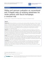
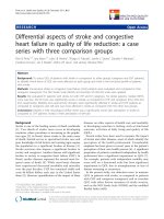
this a ge group. However, there is an increasing interest
in the use of NPPV as a therapeutic tool for children
with respiratory distress that is clear from the increasing
number of published studies over time (Figure 1); a
research of studies on the use of NPPV in children > 1
month of age, published before December 30, 2010
(database: MEDLINE via PubMed; keywords: noninva-
sive ventilation, non-invasive ventilation, noninvasive
positive pressure ventilation, non-invasive positive pres-
sure ventilation, bipap, c ontinuous positive airway pres-
sure; age limits: children from 1 month to 18 years old)
identified 332 relevant articles, of which 48% were pub-
lished during the past 5 years. This concise review is
designed to focus on the effectiveness of NPPV i n chil-
dren > 1 month of age with ARF (excluding patients
with neurologic or chronic lung disease).
Acute respiratory failure in children
The frequency of ARF is higher in infants and young
children than in adults. This difference can be explained
by defining anatomic compartments and their develop-
mental differences in pediatric patients that influence
susceptibility to ARF [6]. In addition, respiratory failure
often precedes cardiopulmonary arrest in children,
unlike in adults where primary cardiac disease often is
responsible. Therefore, prompt recognition and treat-
men t of pediatric patients with pending respiratory fail-
ure can be lifesaving [6].
Respiratory fai lure is a syndrome in which the respira-
tory system fails in one or both of its gas exchange func-
tions: oxygenation and carbon dioxide elimination. In
general, patients with respiratory failure may be classified
into two groups, depending on the component of the
respiratory system that is involved: hypoxemic respiratory
failure and hypercapnic respiratory failure [7].
Hypoxemic respiratory failure (known as type I)
Hypoxemic respiratory fail ure (type I) can be associated
with virtually all acute diseases of the lung , such as sta-
tus asthmaticus, bronchiolitis, pneumonia, and pulmon-
ary edema, which interfere with the normal function of
the lung and airway. The predominan t mechanism in
type I failure is uneven or mismatched ventilation and
perfusion (intrapulmonary shunt) in regional lung units.
This is the most common form of respiratory failure,
characterized by a PaO
2
<60mmHgwithanormalor
low PaCO
2
. The primary treatment of type I respiratory
failure in children is to administer supplemental oxyge n
at a level sufficient to increase the arterial oxygen
saturation (SaO
2
) to greater than 94%. In situations
whenafractionofoxygenininspiredgas(FiO
2
)of
greater than 0.5 is necessary to achieve this goal, this
often is referred to as “acute hypoxemic respiratory fail-
ure” [7]. In this setting, NPPV may be considered.
Hypercapnic respiratory failure (known as type II)
Hypercapnic respiratory failure (type II) is a conse-
quence of ventilatory failure and can occur in conditions
that affect the respiratory pump, such as depressed
0
10
20
30
40
50
60
70
80
90
100
110
< 1993 1993-1995 1996-1998 1999-2001 2002-2004 2005-2007 2008-2010
Time years
References (n)
Figure 1 Time course of published references on noninvasive mechanical ventilation in children aged 1 month to 18 years.
Najaf-Zadeh and Leclerc Annals of Intensive Care 2011, 1:15
/>Page 2 of 10
neural ventilatory drive, acute or chronic upper airway
obstruction, neuromuscular weakness, marked obesity,
and rib-cage abnormalities. Alveolar hypoventilation is
character ized by a PaCO
2
> 50 mmHg [7]. The onset of
type II failure may be insidious and may develop when
respiratory muscle fatigue complicates preexisting disor-
ders, such as pneumonia or status asthmaticus, which
present initially with hypox emia without hypoventila-
tion. Aministration of oxygen alone is not an appropri-
ate treatment for hypercapnic respiratory failure and can
result in the patient retaining even more carbon dioxide,
especially in situations where t he child has adapted to
chronic hypercapnia and is relati vely dependent on oxy-
gen-sensitive peripheral chemoreceptors to maintain
ventilatory drive. In addi tion to supplemental oxygen,
therapies t o reduce the load on the respiratory muscles
and increase the level of alveolar ventilation should be
instituted in children with type II respiratory failure.
When to use NPPV for acute respiratory failure?
When the cause of ARF is reversible, medical treatment
works to maximize lung functi on and reverse the preci-
pitating cause, whereas the goal of ventilatory support is
to “gain time” by unloading respiratory muscles, increas-
ing ventilation, and thus reducing dyspnea and respira-
tory rate and improving gas exchange. Two recent
physiologica l studies have demonstrated these beneficial
effects of NPPV in children with ARF [8,9]. NPPV is
increasingly used for trea tment of ARF in children.
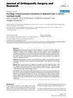
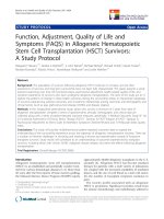
Tables 1 and 2 summarize the studies reporting the
effectiveness of NPPV in children with ARF of various
etiologies [8,10-36]. However, the determinants of suc-
cess of NPPV relate more pr ominently to the pr imary
diagnosis as discussed below.
NPPV in pediatric ARF from primary respiratory
disease
Acute lower airway obstruction
Lower airway disease is a common cause of ARF.
Asthma accounts for the largest percentage of this
group, but infections, such as viral bronchiolitis, also are
common and predominantly impact the small airways.
Physicians caring for acutely ill children are regularly
faced with this condition. Both non-invasive and inva-
sive ventil ation may be options when m edical treatment
fails to prevent respiratory failure. ETI and positive pres-
sure ventilation in children with lower airway obstruc-
tion may increase bronchoconstriction, increase the risk
of airway leakage, and has disadvantageous effects on
circulation and cardiac output. Therefore, ETI should be
avoided unless resp iratory failure is imminent despite
adequate institution of all available treatment measures.
NPPV can be an attractive alternative to IMV for these
patients. Clinical trials in children with acute lower
respiratory airway obstruction have suggested that
NPPV may improve symptoms and ventilation without
significant adverse events and reduce the need for IMV
[10-20]. NPPV theoretically improves the respiratory
status of patient s with lower respiratory airway obstruc-
tion by several mechanisms [37]. During acute bronch-
ospastic episodes, p atients have an increase in airway
resistance and expiratory time constant. The combina-
tion of prolonged expiratory time constant and prema-
ture closure of inflamed airways during exhalation
results in dynamic hyperinflation, which causes
increased positive pressure in the alve oli at end-expira-
tion (auto-PEEP). Because the alveolar pressure must be
reduced to subatmospheric levels to initiate the next
breath, this auto-PEEP increases the inspiratory load
and induces respiratory muscle fatigue. The EPAP deliv-
ered by NPPV may help to decrease dynamic hyperinfla-
tion by maintaining small airway patency and may
reduce the patient’s work of breathing by decreasing the
drop in alveolar pressure needed to initiate a breath. In
addition, inspiratory support, i.e., IPAP delivered by
NPPV, helps to support fatigued respiratory muscles,
thereby improving dyspnea and gas exchange. Needle-
man et al., in a physiological study, found that the
NPPV use in children with status asthmaticus was asso-
ciated with a decrease in respiratory rate and fractional
inspired time and an improvement of tho racoabdominal
synchrony in 80% of patients [12]. A few clinical studies
of small size (3-73 patients) reported the use of NPPV
for treatment of status asthmaticus in children (Table 1)
[10,11,13,14]. NPPV was well tolerated with no major
complications and was associated with an improvement
of gas exchange and respiratory effort (Table 1).
Viral bronchi olitis, mainly due t o respiratory syncytial
virus, represents the largest cohort of children treated
with NPPV [15-20]. Use of NPPV in infant with severe
bronchiolitis was associated with improved respiratory
rate [15,19] and PaCO
2
[16,19,20], decreased work of
breathing [17], and ETI avoidance in 67-100% of
patients (Table 1) [17,18].
Acute upper airway obstruction
In children, dynamic upper airway obstruction can pre-
sent as an acute life-threatening condition and leads to
severe alveolar hypoventilation. In 2006, a survey of
French PICU group found that 67% of pediatric intensi-
vists applied frequently or systematically NPPV in the
management of dynamic u pper airway obstruction in
children [38]. However, there is a paucity of literature
on the use of NPPV in the acute setting of upper airway
obstruction in children. NPPV was associated with a sig-
nificant decrease in respiratory effort [21] and a sus-
tained improvement in gas exchange [22] in children
with dynamic upper airway obstruction (Table 1).
Najaf-Zadeh and Leclerc Annals of Intensive Care 2011, 1:15
/>Page 3 of 10
Table 1 NPPV in pediatric ARF from different causes
Study Cause of ARF (n) Location,
Patients
(n)
Age
(yr)
NPPV
type,
Interface
Avoided
ETI (%)
Other reported outcomes
ARF due to acute airway obstruction
Beers et al. [10]
retrospective
Status asthmaticus ED, 73 2-17
a
BiPAP
Nasal mask
97 Improved RR, SaO
2
Avoided PICU admission: 22%
Major complication: 0%
Carroll et al. [11]
retrospective
Status asthmaticus PICU, 5 9.6
b
BiPAP
Nasal mask
100 Improved RR, MPIS
Major complication: 0%
Needleman et al. [12]
prospective, physiological
Status asthmaticus PICU, 15 8-21
a
BiPAP
Nasal mask
- Improved RR, thoracoabdominal
synchrony, fractional inspired time:
80%
Akingbola et al. [13]
case reports
Status asthmaticus PICU, 3 9-15
a
BIPAP
Nasal mask
100 Improved RR, PaCO
2
,pH
Major complication: 0%
Till et al. [14]
prospective, randomized, crossover
Acute lower airway
obstruction
PICU, 16 4 (0.2-
14)
a,c
BiPAP
Nasal or
facial mask
- Improved RR, CAS, O
2
requirement
Major complication: 0%
Yanez et al. [15]
multicentric, prospective,
randomized, controlled (NPPV
subgroup)
Bronchiolitis-
pneumonia (18), asthma
(4), pneumonia (3)
PICU, 25 1.3
(0.1-
13)
a,c
BIPAP,
BiPAP
Facial mask
72 Improved RR, HR, PaO
2
/FiO
2
at 1 hr
Major complication: 4% (interstitial
emphysema)
Thia et al. [16]
d
prospective, randomized, crossover
Bronchiolitis PICU, 29 0.2
(0.1-
0.4)
c,e
CPAP
Nasal
prongs
- Improved PaCO
2
Major complication: 0%
Cambonie et al. [17]
d
prospective, physiological
Bronchiolitis PICU, 12 0.1
b
CPAP
Nasal mask
100 Improved HR, P
tc
CO
2
,O
2
requirement, respiratory distress
score, MABP at 1 hr
Major complication: 0%
Javouhey et al. [18]
d
retrospective
(NPPV subgroup)
Bronchiolitis PICU, 15 0.1
c
BiPAP,
CPAP
Nasal mask
67 Major complication: 7% (bacterial
pulmonary coinfections)
Larrar et al. [19]
d
prospective, noncontrolled (NPPV
subgroup)
Bronchiolitis PICU, 53 0.1
(0.01-
1)
a,b
CPAP
Nasal
prongs
75 Improved RR, PaCO
2
at 2 hrs
Death: 0%
Major complication: 0%
Campion et al. [20]
d,f
prospective, noncontrolled (NPPV
subgroup)
Bronchiolitis-pneumonia PICU, 69 0.1
(0.03-
1)
a,c
BIPAP,
CPAP
Nasal
prongs,
facial mask
83 Improved PaCO
2
, pH at 2 hrs
Death: 0%
Major complication: 0%
Essouri et al. [21]
prospective, randomized,
controlled
Laryngomalacia (5),
tracheomalacia (3),
others (2)
PICU, 10 0.8
(0.2-
1.5)
a,c
BiPAP,
CPAP
Nasal mask
- Improved RR, respiratory effort in
both types of NPPV
Patient-ventilator asynchrony with
BiPAP
Padman et al. [22]
f
prospective, noncontrolled (upper
airway obstruction subgroup)
Inspiratory stridor PICU, 3 13
b
BiPAP
Nasal mask
100 Improved RR, HR, gas exchange,
serum HCO
3
, dyspnea score at 72
hrs
Major complication: 0%
ARF due to parenchymal lung disease
Munoz-Bonet et al. [23]]
f
prospective, noncontrolled
(pneumonia subgroup)
Pneumonia PICU, 13 0.2-
15.8
a
BIPAP
Facial mask
100 Improved RR, HR, PaCO
2
, SaO
2
, pH,
clinical score within the first 6 hrs
Death: 0%
Major complication: 0%
Bernet et al. [24]
d
prospective, noncontrolled
(pneumonia subgroup)
Pneumonia PICU, 14 2.4
(0.01-
18)
g
BIPAP,
CPAP
Nasal or
facial mask
50 Improved RR, HR, PaCO
2
, serum
HCO
3
within the first 8 hrs
Death: 0%
Fortenberry et al. [25]
f
retrospective, (pneumonia subgroup)
Pneumonia PICU, 21 0.7-17
a
BiPAP
Nasal mask
90 Improved RR, PaCO
2
, PaO
2
, pH, SaO
2
,
PaO
2
/FiO
2
at 1 hr
Death: 5%
Major complication: 0%
Joshi et al. [26]
retrospective (primary
parenchymal lung disease subgroup)
Pneumonia, ARDS PICU, 29 13
c
BiPAP
Facial mask
62 Improved RR, PaCO
2
,O
2
requirement
Major complication: 0%
Najaf-Zadeh and Leclerc Annals of Intensive Care 2011, 1:15
/>Page 4 of 10
Parenchymal lung disease
The main goa ls of NPPV in patients with parenchymal
lung disease, such as pneumonia, acute lung injury
(ALI), and acute respiratory distress syndrome (ARDS),
are to improve oxygenation, to unload the respiratory
muscles, and to relieve dyspnea. The first goal can
usually be achieved by using EPAP to recruit and stabi-
lize previously collapsed lung tissue [39]. Unloading of
the respiratory muscles during N PPV with IPAP has
been reported by L’Her et al. in adult patients with ALI
[39]. The authors concluded that adding IPAP to EPAP
may b e indispensable in patients with ALI treated with
NPPV [39]. Indeed, IPAP allows a better respiratory sys-
tem muscle unloading, alveolar recruitment, oxygena-
tion, and CO
2
washout improvement.
Although NPPV seems disappointing in ARF owing to
pneumonia in adult patients, with failure rates of up to
66% [40], several noncontrolled trials have suggested
that NPPV could improve symptoms and ventilation
without significant adverse events and reduce the need
for IMV in children with ARF due to pneumonia
[22-27]. Use of NPPV in this acute setting in children
was associated with reduct ion in ETI rates ranging from
50-100% (Table 1) [23,24].
The most challenging application of NPPV may be in
patients with ARDS. Studies of NPPV for the treatment
of ARDS in adult population have reported failure rates
of 50-80% [40]. A meta-analysis of the topic in adult
population concluded t hat NPPV w as unlikely to have
any significant benefit [41]. In children, the use of
NPP V for the treatm ent of ARDS was assoc iated with a
failure rate of 78%, and 22% of them died (Table 1) [27].
Therefore, NPPV use in such a patient group is rarely
justified. However, if a trial of NPPV is initiated, patients
should be closely monitored and promptly intubated if
their conditions deteriorate, so that inordinate delays in
needed interventions are avoided.
Acute chest syndrome (ACS) is one of the leading
caus es of death and hospitalization among patients with
sickle cell disease [42]. Approximately 70% of patients
(adults or children) with ACS are hypoxic [43]. Indeed,
patients with sickle cell disease are prone to infarctive
crises. Thoracic bone infarction (usually in the ribs) in
such patients leads to pain, splinting, h ypoventilation,
and the clinical signs of ACS. In situ red blood cell sick-
ling in the lung vasculature is possibly a consequenc e of
hypoventilation with subsequent infarction of lung par-
enchyma. NPPV has been proposed as a therapeutic
option for patients with ACS. By improving patient oxy-
gen ation, NPPV could prevent progression from painful
crisis to ACS, and ultimately to ARDS. Three retrospec-
tive studies reported favorable outcomes in children
with ACS treated with NPPV (Table 1) [22,27,28].
NPPV in specific circumstances
Postoperative respiratory failure
Postoperative pulmonary complications are a major
cause of morbidity, mortality, prolonged hos pital stay,
and increased cost of care [44]. It has been reported
that 5-10% of al l surgical adult patients exper ience post-
operative pulmonary complications [45]. Atelectasis,
postoperative pneumonia, ARDS, and postoperative
respiratory failure have all been classified as postopera-
tive p ulmonary complications. Postoperative respiratory
Table 1 NPPV in pediatric ARF from different causes (Continued)
Essouri et al. [27]
retrospective (primary
parenchymal lung disease subgroup)
CAP (23), ARDS (9), ACS
(9)
PICU, 41 8 (0.2-
16)
a,b
BIPAP
Nasal or
facial mask
87 (CAP)
22
(ARDS)
100 (ACS)
Improved RR, PaCO
2
at 2 hr
Death: 4% (CAP), 22% (ARDS), 0%
(ACS)
Major complication: 0%
Padman et al. [22]
f
prospective, noncontrolled (primary
parenchymal lung disease subgroup)
Pneumonia (13), ACS (5
episodes)
PICU, 17 10.6
b
BiPAP
Nasal mask
85 (CAP)
80 (ACS)
Improved RR, HR, gas exchange,
serum HCO
3
, dyspnea score at 72
hrs
Major complication: 0%
Padman et al. [28]
retrospective
ACS (25 episodes) Inpatient
ward, 9
11.8
(4-20)
a,
b
BiPAP
Nasal mask
100 Improved RR, HR, SaO
2
,O
2
requirement
Avoided PICU admission: 44%
ACS, acute chest syndrome; ARDS, acute respiratory distress syndrome; ARF, acute respiratory failure; BiPAP, bilevel positive airway pressure; BIPAP, biphasic
positive airway pressure; CAS, clinical asthma score; CAP, community-acquired pneumonia; CPAP, continuous positive airway pressure; ED, emergency
department; ETI, endotracheal intubation; FiO
2
, fraction of oxygen in inspired gas; HR, heart rate; MABP, mean arterial blood pressure; MPIS, modified pulmonary
index score; NPPV, noninvasive positive pressure ventilation; PICU, pediatric intensive care unit; PaCO
2
, arterial partial pressure of carbon dioxide; PaO
2
, arterial
partial pressure of oxygen; P
tc
CO
2
, transcutaneous PCO
2
; RR, respiratory rate; SaO
2
, arterial oxygen saturation.
a
Range.
b
Mean.
c
Median.
d
Neonatal cases also were included in the study.
e
Interquartile range.
f
Certain patients included in the study had underlying neurologic or chronic lung disease.
g
The numbers represent the median (range) age of the patients (n = 42) with ARF of various cause s included in the study.
Najaf-Zadeh and Leclerc Annals of Intensive Care 2011, 1:15
/>Page 5 of 10
Table 2 NPPV in specific circumstances
Study Cause of ARF (n) Location,
Patients
(n)
Age (yr) NPPV type,
Interface
Avoided ETI
(%)
Other reported outcomes
NPPV in postoperative ARF
Stucki et al. [8]
a
prospective, crossover
(cardiac surgery)
Interstitial
pulmonary oedema
PICU, 6 0.4 (0.04-0.6)
b,c
BIPAP
Nasal mask
100 Improved RR, PTPes, dPes, dyspnea
score
Death: 0%
Bernet et al. [24]
a
prospective,
noncontrolled (cardiac
surgery subgroup)
ND PICU, 11 2.4 (0.01-18)
d
BIPAP,
CPAP
Nasal or facial
mask
64 Improved RR, HR, PaCO
2
, pH, serum
HCO
3
within the first 8 hrs
Death: 0%
Joshi et al. [26]
retrospective
(postoperative
subgroup)
Atelectasis PICU, 16 12
b
BiPAP
Facial mask
94 Improved RR, PaCO
2
,O
2
requirement, SaO
2
Major complication: 0%
Essouri et al. [27]
a
retrospective
(postextubation
subgroup)
e
ND PICU, 61 3.2 (0.04-
15)
c,f
BIPAP
Nasal or facial
mask
67 Improved RR, PaCO
2
at 2 hrs
Death: 11%
Major complication: 0%
Kovacikova et al. [29]
case reports (cardiac
surgery)
Bilateral diaphragm
paralysis
PICU, 2 0.9-3.5
c
BIPAP
Facial mask,
Nasopharyngeal
tube
100 Improved RR, gas exchange
Major complication: 100%
(respiratory tract infection)
Chin et al. [30]
retrospective (liver
transplantation)
Atelectasis,
hypercapnia
+/-hypoxemia,
pleural effusion,
pneumonia
PICU, 15 0.2-14
c
BiPAP
Nasal or facial
mask
87 Improved PaCO
2
, SaO
2
, atelectasis
Death: 13%
NPPV for facilitation of ventilation weaning/rescue of failed extubation (not postoperatively)
Lum et al. [31]
a
prospective,
noncontrolled (prior
IMV subgroup)
Post-extubation
failure (51), weaning
facilitation (98)
PICU, 149 0.5 (0.1- 2)
b,g
BiPAP
Nasal or facial
mask
75 (failure
group), 86
(weaning
group)
Improved RR, HR, FiO
2
within the
first 24 hrs
Death: 5%
Major complication: 11%
(pneumonia)
Mayordomo-Colunga
et al. [32]
a,h
prospective,
noncontrolled
Post-extubation
failure (20),
weaning facilitation
(21)
PICU, 36
(41
episodes)
1.7 (0.04-
17)
b,c
BiPAP,
CPAP
Nasal or facial
mask, helmet
50 (failure
group), 81
(weaning
group)
Death: 5%
Major complication: 5%
(hypercapnia), 12% (upper airway
obstruction), 7% (apnea), 10% (other)
NPPV in immunocompromised patients
Munoz-Bonet et al.
[23]
prospective,
noncontrolled
(immunocompromised
subgroup)
Pnemonia (3), ARDS
(5)
PICU, 8 1.5-13.8
c
BIPAP
Facial mask
100
(pneumonia),
40 (ARDS)
Improved RR, HR, PaCO
2
, SaO
2
, pH,
clinical score within the first 6 hrs
Death: 0%
Major complication: 0%
Essouri et al. [27]
retrospective
(immunocompromised
subgroup)
ND PICU, 12 8 (3-16)
c,f
BIPAP
Nasal or facial
mask
92 Improved RR, PaCO
2
at 2 hrs
Death: 8%,
Major complication: 0%
Schiller et al. [33]
retrospective
Pneumonia (5),
ARDS (10),
pulmonary mass (1)
PICU, 14
(16
episodes)
13.3
f
BiPAP
Facial mask
80 Improved RR, PaO
2
at 1 hr
Death: 20%
Major complication: 0%
Piastra et al. [34]
prospective,
noncontrolled
ARDS PICU, 23 10.2
f
BIPAP
Facial mask,
Helmet
54 Improved gas exchange at 1
hr (82%), sustained (74%)
Death: 35%
Major complication: 0%
Desprez et al. [35]
case reports
Pneumonia (1),
ARDS (1)
PICU, 2 13-14
c
BIPAP
Facial mask
100 Death: 0%
Major complication: 50% (upper and
lower digestive hemorrhage)
Najaf-Zadeh and Leclerc Annals of Intensive Care 2011, 1:15
/>Page 6 of 10
failureismostcommonlydefined as the inability to be
extubated 48 hours after surgery [46], although some
investigators have used 5 days [47]. NPPV has been suc-
cessfully used to treat postoperative respiratory failure
in both pediatric and adult patients. Compared with
standard treatment, NPPV used after major abdominal
surgery improved hypoxemia and reduced the need for
ETI in adult population [48]. NPPV application in chil-
dren with postoperative respiratory failure was asso-
ciated with improved respiratory effort, gas exchange,
oxygen saturation, and reduced the need for ETI (Table
1) [8,24,26,27,29,30].
Facilitation of ventilation weaning/rescue of failed
extubation
The need for reintubation after failed extubation is asso-
ciated with increased morbidity and high mortali ty [49].
NPPV has been proposed as a means of “facilitating”
weaning from IMV, and as a “ curative” treatment for
postextubation respiratory failure. Although seve ral stu-
dies have shown the efficacy of NPPV in weaning from
IMV in adult population [50], its applicati on for postex-
tubation respiratory failure is not supported by rando-
mized, contro lled trials [51] . In children, two
noncontrolled trials assessed the efficacy of NPPV in
these settings: the application of NPPV as a means of
“facilita ting” ventilation weaning, and as “curative” treat-
ment for postextubation respiratory failure was asso-
ciated with success rates of 81-86% and 50-75%,
respectively [31,32].
Immunocompromised children
ARF in immunocompromised patients most often
results from infections, pulmonary localization of the
primary d isease, or even postchemotherapy cardiogenic
pulmonary edema. Treatment of such patients often
requires intubation and mechanical ventilation. Avoid-
ance of the infectious complications associated with
IMV is particularly attractive in these high-risk patients,
in whom this could be devastating, if not fatal. Results
of randomized, controlled trials have proven the benefi-
cial effects of NPPV in immunocompromised adult
patients [52,53]. Some case series reported the use of
NPPV in the treatment of respiratory failure in immu-
nocompromised children (Table 2) [23,27,33-36]. The
likelihood of NPPV success in immunocompromised
children seems to be related rather to the type of pul-
monary disease: the ETI avoidance rates varied from
40% for ARDS to 100% for pneumonia (Table 2).
Are there predictive factors of NPPV failure in
children with ARF?
It is not always apparent which patients will initially
benefit from NPPV; some patients do not obtain ade-
quate ventilation with NPPV. The NPPV failure rate
may be fairly consistent for certain diseases, and NPPV
failure eventually requires intubation. Inability to early
identify patients who will fail NPPV can cause inap-
propriate delay of intubation, which can cause clinical
deterioration and increase morbidity and mortality.
Knowing the predictors of NPPV failure in patient with
ARF is therefore crucial in deciding if and when to
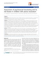
apply this ventilatory technique. Several authors have
identified different predictive factors of NPPV failure in
children with ARF: the results of studies are given in
Table 3[20, 24,26,27,31,54,55]. The best predictive factors
for the NPPV failure in ARF appear to be the level of
FiO
2
and PaCO
2
on admission or within the first hours
after starting NPPV (Table 3).
Conclusions
During recent years, there has been an increasing inter-
est in the use of NPPV for children with ARF. There are
some promising studies supporting its use in this acute
setting. NPPV was well t olerated with rare major com-
plications and was associated with improved gas
Table 2 NPPV in specific circumstances (Continued)
Pancera et al. [36]
retrospective (NPPV
subgroup)
ND PICU, 120 9
i
BIPAP
Nasal mask
74 Death: 22.5%
ARDS, acute respiratory distress syndrome; ARF, acute respiratory failure; BiPAP, bilevel positive airway pressure; BIPAP, biphasic positive airway pressure; CPAP,
continuous positive airway pressure; dPes, oesophageal inspiratory pressure swing; ETI, endotracheal intubation; HR, heart rate ; IMV, invasive mechanical
ventilation; ND, not determined; NPPV, noninvasive positive pressure ventilation; PaCO
2
, arterial partial pressure of carbon dioxide; PaO
2
, arterial partial pressure
of oxygen; PICU, pediatric intensive care unit; PTPes, oesophageal pressure-time product; RR, respiratory rate; SaO
2
, arterial oxygen saturation.
a
Neonatal cases also were included in the study.
b
Median.
c
Range.
d
Numbers represent the median (range) age of the patients (n = 42) with ARF of various causes included in the study.
e
32 patients were intubated for liver transplantation, 11 for other abdominal surgery, and 18 for respiratory distress.
f
Mean.
g
Interquartile range.
h
Certain patients included in the study had underlying neurologic disease.
i
Number represent the mean age of the patients (n = 239) included in the study, of which 120 had NPPV.
Najaf-Zadeh and Leclerc Annals of Intensive Care 2011, 1:15
/>Page 7 of 10
exchange, decreased work of breathing, and decreased
need for ETI. Both critical care ventilators and portable
ventilators have been used for NPPV. However, the vast
majority of the available knowl edge in this acute setting
results from noncontrolled trials and case series of small
size. As such, many important issues, such as the identi-
ficatio n of the patient, the right time for NPPV applica-
tion, and the appropriate setting, are still lacking.
Further randomized, controlled trials addressing t hese
issues in children with ARF are needed to define better
the patients who are likely to benefit from this alterna-
tive method of respiratory support. Also, the respective
place of NPPV and high flow oxygen therapy in children
with ARF due to different conditions has to be deter-
mined [56].
Author details
1
Univ Lille Nord de France, UDSL, EA 2694, F-59000 Lille, France
2
Pediatric
Emergency and Infectious Diseases Unit, Roger-Salengro Hospital, Rue E
Laine, CHU Lille, F-59037 Lille, France
3
Paediatric Intensive Care Unit, Jeanne-
de-Flandre Hospital, CHU Lille, Avenue E Avinée, F-59037 Lille, France
Authors’ contributions
AN and FL contributed to query the literature and to draft the manuscript.
They approved the final version.
Competing interests
The authors declare that they have no competing interests.
Received: 27 April 2011 Accepted: 26 May 2011 Published: 26 May 2011
References
1. Armon K, Stephenson T, Gabriel V, MacFaul R, Eccleston P, Werneke U,
Smith S: Determining the common medical presenting problems to an
accident and emergency department. Arch Dis Child 2001, 84:390-392.
2. The burden of respiratory disease: Department of public health sciences
St George’s Hospital Medical School 2003.[ />factsheets/953.pdf].
3. Antonelli M, Conti G, Rocco M, Bufi M, De Blasi RA, Vivino G, Gasparetto A,
Meduri GU: A comparison of noninvasive positive-pressure ventilation
and conventional mechanical ventilation in patients with acute
respiratory failure. N Engl J Med 1998, 339:429-435.
4. Lightowler JV, Wedzicha JA, Elliott MW, Ram FS: Non-invasive positive
pressure ventilation to treat respiratory failure resulting from
exacerbations of chronic obstructive pulmonary disease: Cochrane
systematic review and meta-analysis. BMJ 2003, 326:185.
5. Weng CL, Zhao YT, Liu QH, Fu CJ, Sun F, Ma YL, Chen YW, He QY: Meta-
analysis: Noninvasive ventilation in acute cardiogenic pulmonary edema.
Ann Intern Med 2010, 152:590-600.
Table 3 Predictive factors for the outcome of NPPV in children with ARF
Study Population (n) Age (yr) Success
rate (%)
a
Predictors of failure
Campion et al. [20]
b,c
prospective noncontrolled
Bronchiolitis (69) 0.1 (0.03-
1)
d,e
83 Apnea
Higher PaCO
2
on admission
Higher PRISM score at 24
hrs
Bernet et al. [24]
b
prospective noncontrolled
Pneumonia (14), bronchiolitis (4), postoperative ARF
(11), other (13)
2.4 (0.01-
18)
d,e
57 FiO
2
> 0.8 at 1 hr
Joshi et al. [26]
retrospective (primary parenchymal
lung disease subgroup)
Pneumonia or ARDS (29) 13
d
62 Age ≤ 6yr
FiO
2
> 0.6 within the first
24 hrs
PaCO
2
≥ 55 mmHg within
the first 24 hrs
Essouri et al. [27]
b
retrospective
CAP (23), ARDS (9), ACS (9), immune deficiency
(12), postextubation ARF (61)
5.3 (0.04-
16)
e,f
73 ARDS
High PELOD score
Lum et al. [31]
b
prospective noncontrolled
Pulmonary diseases (129), postextubation ARF (149) 0.7 (0.3-
2.8)
d,g
76 Higher FiO
2
needs at start
of NPPV
Higher PRISM score on
admission
Sepsis at start of NPPV
Munoz-Bonet et al. [54]
prospective noncontrolled
Pneumonia (20), ARDS (10), postextubation ARF
(11), other (6)
7.1 (0.1-
16)
e,f
81 MAP > 11.5 cm H
2
O
FiO
2
> 0.6
Mayordomo-Colunga et al. [55]
b
prospective noncontrolled
Type I ARF (38), type II ARF (78) 0.9 (0.05-
14)
d,e
84 Lower RR decrease at 1 hr
and 6 hrs
Higher PRISM score at start
and at 1 hr
Type I ARF
ACS, acute chest syndrome; ARDS, acute respiratory distress syndrome; ARF, acute respiratory failure; CAP, community-acquired pneumonia; FiO
2
, fraction of
oxygen in inspired gas; MAP, mean airway pressure; NPPV, noninvasive positive pressure ventilation; PaCO
2
, arterial partial pressure of carbon dioxide; PELOD,
pediatric logistic organ dysfunction; PRISM, pediatric risk of mortality; RR, respiratory rate.
a
NPPV was considered successful when endotracheal intubation was avoided.
b
Neonatal cases also were included in the study.
c
Certain patients included in the study had underlying neurologic or chronic lung disease.
d
Median.
e
Range.
f
Mean.
g
Interquartile range.
Najaf-Zadeh and Leclerc Annals of Intensive Care 2011, 1:15
/>Page 8 of 10
6. Rotta AT, Wiryawan B: Respiratory emergencies in children. Respir Care
2003, 48:248-258, discussion 258-260.
7. Teague WG: Noninvasive ventilation in the pediatric intensive care unit
for children with acute respiratory failure. Pediatr Pulmonol 2003,
35:418-426.
8. Stucki P, Perez MH, Scalfaro P, de Halleux Q, Vermeulen F, Cotting J:
Feasibility of non-invasive pressure support ventilation in infants with
respiratory failure after extubation: a pilot study. Intensive Care Med 2009,
35:1623-1627.
9. Essouri S, Durand P, Chevret L, Haas V, Perot C, Clement A, Devictor D,
Fauroux B: Physiological effects of noninvasive positive ventilation
during acute moderate hypercapnic respiratory insufficiency in children.
Intensive Care Med 2008, 34:2248-2255.
10. Beers SL, Abramo TJ, Bracken A, Wiebe RA: Bilevel positive airway pressure
in the treatment of status asthmaticus in pediatrics. Am J Emerg Med
2007, 25:6-9.
11. Carroll CL, Schramm CM: Noninvasive positive pressure ventilation for the
treatment of status asthmaticus in children. Ann Allergy Asthma Immunol
2006, 96:454-459.
12. Needleman J, Sykes J, Schroeder S, Singer L: Noninvasive Positive Pressure
Ventilation in the Treatment of Pediatric Status Asthmaticus. Pediatric
Asthma, Allergy & Immunology 2004, 17:272-277.
13. Akingbola OA, Simakajornboon N, Hadley EF Jr, Hopkins RL: Noninvasive
positive-pressure ventilation in pediatric status asthmaticus. Pediatr Crit
Care Med 2002, 3:181-184.
14. Thill PJ, McGuire JK, Baden HP, Green TP, Checchia PA: Noninvasive
positive-pressure ventilation in children with lower airway obstruction.
Pediatr Crit Care Med 2004, 5:337-342.
15. Yanez LJ, Yunge M, Emilfork M, Lapadula M, Alcantara A, Fernandez C,
Lozano J, Contreras M, Conto L, Arevalo C, et al: A prospective,
randomized, controlled trial of noninvasive ventilation in pediatric acute
respiratory failure. Pediatr Crit Care Med 2008, 9:484-489.
16. Thia LP, McKenzie SA, Blyth TP, Minasian CC, Kozlowska WJ, Carr SB:
Randomised controlled trial of nasal continuous positive airways
pressure (CPAP) in bronchiolitis. Arch Dis Child 2008, 93:45-47.
17. Cambonie G, Milesi C, Jaber S, Amsallem F, Barbotte E, Picaud JC, Matecki S:
Nasal continuous positive airway pressure decreases respiratory muscles
overload in young infants with severe acute viral bronchiolitis. Intensive
Care Med 2008, 34:1865-1872.
18. Javouhey E, Barats A, Richard N, Stamm D, Floret D: Non-invasive
ventilation as primary ventilatory support for infants with severe
bronchiolitis. Intensive Care Med 2008, 34:1608-1614.
19. Larrar S, Essouri S, Durand P, Chevret L, Haas V, Chabernaud JL,
Leyronnas D, Devictor D: Effects of nasal continuous positive airway
pressure ventilation in infants with severe acute bronchiolitis. Arch
Pediatr
2006, 13:1397-1403.
20. Campion A, Huvenne H, Leteurtre S, Noizet O, Binoche A, Diependaele JF,
Cremer R, Fourier C, Sadik A, Leclerc F: Non-invasive ventilation in infants
with severe infection presumably due to respiratory syncytial virus:
feasibility and failure criteria. Arch Pediatr 2006, 13:1404-1409.
21. Essouri S, Nicot F, Clement A, Garabedian EN, Roger G, Lofaso F, Fauroux B:
Noninvasive positive pressure ventilation in infants with upper airway
obstruction: comparison of continuous and bilevel positive pressure.
Intensive Care Med 2005, 31:574-580.
22. Padman R, Lawless ST, Kettrick RG: Noninvasive ventilation via bilevel
positive airway pressure support in pediatric practice. Crit Care Med 1998,
26:169-173.
23. Munoz-Bonet JI, Flor-Macian EM, Rosello PM, Llopis MC, Lizondo A, Lopez-
Prats JL, Brines J: Noninvasive ventilation in pediatric acute respiratory
failure by means of a conventional volumetric ventilator. World J Pediatr
2010, 6:323-330.
24. Bernet V, Hug MI, Frey B: Predictive factors for the success of noninvasive
mask ventilation in infants and children with acute respiratory failure.
Pediatr Crit Care Med 2005, 6:660-664.
25. Fortenberry JD, Del Toro J, Jefferson LS, Evey L, Haase D: Management of
pediatric acute hypoxemic respiratory insufficiency with bilevel
positiv e pressure (BiPAP) nasal mask ventilation. Chest 199 5,
108:1059-1064.
26. Joshi G, Tobias JD: A five-year experience with the use of BiPAP in a
pediatric intensive care unit population. J Intensive Care Med 2007,
22:38-43.
27. Essouri S, Chevret L, Durand P, Haas V, Fauroux B, Devictor D: Noninvasive
positive pressure ventilation: five years of experience in a pediatric
intensive care unit. Pediatr Crit Care Med 2006, 7:329-334.
28. Padman R, Henry M: The use of bilevel positive airway pressure for the
treatment of acute chest syndrome of sickle cell disease. Del Med J 2004,
76:199-203.
29. Kovacikova L, Dobos D, Zahorec M: Non-invasive positive pressure
ventilation for bilateral diaphragm paralysis after pediatric cardiac
surgery. Interact Cardiovasc Thorac Surg 2009, 8:171-172.
30. Chin K, Uemoto S, Takahashi K, Egawa H, Kasahara M, Fujimoto Y, Sumi K,
Mishima M, Sullivan CE, Tanaka K: Noninvasive ventilation for pediatric
patients including those under 1-year-old undergoing liver
transplantation. Liver Transpl 2005, 11:188-195.
31. Lum LC, Abdel-Latif ME, de Bruyne JA, Nathan AM, Gan CS: Noninvasive
ventilation in a tertiary pediatric intensive care unit in a middle-income
country. Pediatr Crit Care Med 2011, 12:e7-13.
32. Mayordomo-Colunga J, Medina A, Rey C, Concha A, Menendez S, Los
Arcos M, Garcia I: Non invasive ventilation after extubation in paediatric
patients: a preliminary study. BMC Pediatr 2010, 10:29.
33. Schiller O, Schonfeld T, Yaniv I, Stein J, Kadmon G, Nahum E: Bi-level
positive airway pressure ventilation in pediatric oncology patients with
acute respiratory failure. J Intensive Care Med
2009, 24:383-388.
34. Piastra M, De Luca D, Pietrini D, Pulitano S, D’Arrigo S, Mancino A, Conti G:
Noninvasive pressure-support ventilation in immunocompromised
children with ARDS: a feasibility study. Intensive Care Med 2009,
35:1420-1427.
35. Desprez P, Ribstein AL, Didier C, Barats A, Scheib C, Defaix A, Lutz P,
Astruc D: Non invasive ventilation for acute respiratory distress with
febrile aplastic anemia. Arch Pediatr 2009, 16:750-751.
36. Pancera CF, Hayashi M, Fregnani JH, Negri EM, Deheinzelin D, de
Camargo B: Noninvasive ventilation in immunocompromised pediatric
patients: eight years of experience in a pediatric oncology intensive care
unit. J Pediatr Hematol Oncol 2008, 30:533-538.
37. Carroll L: Noninvasive Ventilation for the Treatment of Acute Lower
Respiratory Tract Diseases in Children. Clinical Pediatric Emergency
Medicine 2009, 10:90-94.
38. Pouyau R, Javouhey E: Enquête de prévalence et de pratique de
ventilation non invasive en aigu dans les services de réanimation
pédiatrique francophone [abstract]. Réanimation 2007, 16:s32.
39. L’Her E, Deye N, Lellouche F, Taille S, Demoule A, Fraticelli A, Mancebo J,
Brochard L: Physiologic effects of noninvasive ventilation during acute
lung injury. Am J Respir Crit Care Med 2005, 172:1112-1118.
40. Ambrosino N, Vagheggini G: Noninvasive positive pressure ventilation in
the acute care setting: where are we? Eur Respir J 2008, 31:874-886.
41. Agarwal R, Reddy C, Aggarwal AN, Gupta D: Is there a role for noninvasive
ventilation in acute respiratory distress syndrome? A meta-analysis.
Respir Med 2006, 100:2235-2238.
42. Platt OS, Brambilla DJ, Rosse WF, Milner PF, Castro O, Steinberg MH,
Klug PP: Mortality in sickle cell disease. Life expectancy and risk factors
for early death. N Engl J Med 1994, 330:1639-1644.
43. Quinn CT, Buchanan GR: The acute chest syndrome of sickle cell disease.
J Pediatr 1999, 135:416-422.
44. Lawrence VA, Hilsenbeck SG, Mulrow CD, Dhanda R, Sapp J, Page CP:
Incidence and hospital stay for cardiac and pulmonary complications
after abdominal surgery. J Gen Intern Med 1995, 10:671-678.
45. Wong DH, Weber EC, Schell MJ, Wong AB, Anderson CT, Barker SJ: Factors
associated with postoperative pulmonary complications in patients with
severe chronic obstructive pulmonary disease. Anesth Analg 1995,
80:276-284.
46. Svensson LG, Hess KR, Coselli JS, Safi HJ, Crawford ES: A prospective study
of respiratory failure after high-risk surgery on the thoracoabdominal
aorta. J Vasc Surg 1991, 14
:271-282.
47. Money SR, Rice K, Crockett D, Becker M, Abdoh A, Wisselink W, Kazmier F,
Hollier L: Risk of respiratory failure after repair of thoracoabdominal
aortic aneurysms. Am J Surg 1994, 168:152-155.
48. Squadrone V, Coha M, Cerutti E, Schellino MM, Biolino P, Occella P,
Belloni G, Vilianis G, Fiore G, Cavallo F, Ranieri VM: Continuous positive
airway pressure for treatment of postoperative hypoxemia: a
randomized controlled trial. JAMA 2005, 293:589-595.
49. Epstein SK, Ciubotaru RL, Wong JB: Effect of failed extubation on the
outcome of mechanical ventilation. Chest 1997, 112:186-192.
Najaf-Zadeh and Leclerc Annals of Intensive Care 2011, 1:15
/>Page 9 of 10
50. Burns KE, Adhikari NK, Keenan SP, Meade M: Use of non-invasive
ventilation to wean critically ill adults off invasive ventilation: meta-
analysis and systematic review. BMJ 2009, 338:b1574.
51. Keenan SP, Powers C, McCormack DG, Block G: Noninvasive positive-
pressure ventilation for postextubation respiratory distress: a
randomized controlled trial. JAMA 2002, 287:3238-3244.
52. Antonelli M, Conti G, Bufi M, Costa MG, Lappa A, Rocco M, Gasparetto A,
Meduri GU: Noninvasive ventilation for treatment of acute respiratory
failure in patients undergoing solid organ transplantation: a randomized
trial. JAMA 2000, 283:235-241.
53. Hilbert G, Gruson D, Vargas F, Valentino R, Gbikpi-Benissan G, Dupon M,
Reiffers J, Cardinaud JP: Noninvasive ventilation in immunosuppressed
patients with pulmonary infiltrates, fever, and acute respiratory failure. N
Engl J Med 2001, 344:481-487.
54. Munoz-Bonet JI, Flor-Macian EM, Brines J, Rosello-Millet PM, Cruz Llopis M,
Lopez-Prats JL, Castillo S: Predictive factors for the outcome of
noninvasive ventilation in pediatric acute respiratory failure. Pediatr Crit
Care Med 2010, 11:675-680.
55. Mayordomo-Colunga J, Medina A, Rey C, Diaz JJ, Concha A, Los Arcos M,
Menendez S: Predictive factors of non invasive ventilation failure in
critically ill children: a prospective epidemiological study. Intensive Care
Med 2009, 35:527-536.
56. Schibler A, Pham TM, Dunster KR, Foster K, Barlow A, Gibbons K, Hough JL:
Reduced intubation rates for infants after introduction of high-flow
nasal prong oxygen delivery. Intensive Care Med 2011, 37(5):847-52.
doi:10.1186/2110-5820-1-15
Cite this article as: Najaf-Zadeh and Leclerc: Noninvasive positive
pressure ventilation for acute respiratory failure in children: a concise
review. Annals of Intensive Care 2011 1:15.
Submit your manuscript to a
journal and benefi t from:
7 Convenient online submission
7 Rigorous peer review
7 Immediate publication on acceptance
7 Open access: articles freely available online
7 High visibility within the fi eld
7 Retaining the copyright to your article
Submit your next manuscript at 7 springeropen.com
Najaf-Zadeh and Leclerc Annals of Intensive Care 2011, 1:15
/>Page 10 of 10