Báo cáo hóa học: "An antibacterial coating based on a polymer/solgel hybrid matrix loaded with silver nanoparticles" pdf
Bạn đang xem bản rút gọn của tài liệu. Xem và tải ngay bản đầy đủ của tài liệu tại đây (2.29 MB, 7 trang )
NANO EXPRESS Open Access
An antibacterial coating based on a polymer/sol-
gel hybrid matrix loaded with silver nanoparticles
Pedro José Rivero
*
, Aitor Urrutia, Javier Goicoechea, Carlos Ruiz Zamarreño, Francisco Javier Arregui and
Ignacio Raúl Matías
Abstract
In this work a novel antibacterial surface composed of an organic-inorganic hybrid matrix of tetraorthosilicate and
a polyelectrolyte is presented. A precursor solution of tetraethoxysilane (TEOS) and poly(acrylic acid sodium salt)
(PAA) was prepared and subsequently thin films were fabricated by the dip-coating technique using glass slides as
substrates. This hybrid matrix coating is further loaded with silver nanoparticles using an in situ synthesis route. The
morphology and composition of the coatings have been studied using UV-VIS spectrosc opy and atomic force
microscopy (AFM). Energy dispersive X-ray (EDX) was also used to confirm the presence of the resulting silver
nanoparticles within the thin films. Finally the coatings have been tested in bacterial cultures of genus Lactobacillus
plantarum to observe their antibacterial properties. It has been experimentally demonstrated that these silver
loaded organic-inorganic hybrid films have a very good antimicrobial behavior against this type of bacteria.
Background
Microbes and bacteria are the most abundant of all liv-
ing organisms in our planet and a large of them are
pathogens. Because of that, there is an enormous inter-
est in the research of highly efficient and low cost anti-
bacterial surface treatments and coatings to avoid the
apparition of these microorganisms in instrumentals,
devices, laboratories, operating rooms, etc. [1,2].
Silver ions show a notorious broad spectrum biocide
effect. There are several known mechanisms where the
utilization of silver has led to an extraordinary toxicity
for bacteria [3-6]. Moreover, silver is particula rly attrac-
tive because it combines the high toxicity for bacteria
with a low toxicity for humans [7-9]. Its disinfectant
properties for hygienic and medicinal purposes are
known since ancient times, and for exampl e it has been
extensively used to prevent wound infection since
World War I [10].
Most of the approaches for achieving antibacterial sur-
faces are based on doping some elements with silver
particles which act as silver ion source, for example, in
textiles [11,12], surgical instruments [13], and other sur-
faces [14]. Some authors have reported how silver
nanoparticles [15], nanorods [16], or nanotubes [17] are
especially efficient antibacterial agents because of their
large surface to volume ratio. Up to now such silver
nanoparticles have been immobilized on inorganic por-
ous hosts such as zeolites, calcium phosphate, and car-
bon fiber [18-20]. Moreover, silv er-supported silica
materials, such as silica glass [21], silica thin films [22],
and silica nanoparticles [23], are also good candidates
for antibacterial materials due to their fine chemical
durability and high antibacterial activity. Moreover, a
surface can obtain contact bacteria-killing capacity
through chemical modification with tethered bactericidal
functionalities such as quaternary amine compounds
[24,25], phosphonium salts [26], and titanium oxide par-
ticles [27], which are able to kill bacteria upon contact.
However, the biocide efficiency of such coatings
depends on the ability of the trapped silver to release
ions. Consequently, silver particles with a high specific
area show more efficient ion release mechanisms and
the refore the antibacterial effect is enhanced. There is a
wide variety of coating techniques that have been used
for fabricating antibacterial coatings, such as PVD [28],
spin-coating [29], or electrospinning [30,31]. In this
work, we have developed a facile method to produce an
organic-inorganic hybrid matrix by the sol-gel process
using the dip-coating technique onto glass substrates.
This approach allows to fabricate biocide films in a fast
* Correspondence:
Nanostructured Optical Devices Laboratory, Electric and Electronic
Engineering Department, Public University of Navarra, Edif. Los Tejos,
Campus Arrosadía, 31006, Pamplona, Spain
Rivero et al. Nanoscale Research Letters 2011, 6:305
/>© 2011 Rivero et al; licensee Springer. This is an Open Access article distributed under the terms of the Creative Commons Attribution
License (http://cre ativeco mmons.org/l icenses/by/2.0), which permits unrestricted use, distribution, and reproduction in any medium,
provided the origin al work is properly cited.
and simple way compared to other fabrication techni-
ques [32].
Polymer-silica hybrid materials have drawn the atten-
tion of many researchers recently because of their com-
patibility with living matter and their promising
applications in the medical field [33,34]. Organic poly-
mers in general enjoy a high flexibility, low density,
toughness, and easy formability whereas ceramic materi-
als possess other excellent mechanical properties such
as a high hardness, combined with a good resistance to
high temperature or strong solvents. In this work, these
new class of materials are employed to obtain a porous
silica surface containing uniformly distributed silver
nanoparticl es inside the coating. To our knowledge, this
is the first time that TEOS/PAA hybrid matrices loaded
with silver nanoparticles are fabricated. In addi tion,
their possible antibacterial behavior is also studied.
Experimental section
Materials
In this work, the hybrid (organic/inorganic) precursor
solution was prepared mixing a water-based solution
of poly(acrylic acid sodium salt) (PAA), tetraethoxysi-
lane (TEOS), and ethanol (EtOH). The PAA solution
was prepared using ultrapure water (18.2 MΩcm)
and its concentration was varied throughout the
experiment, from 10
-3
to 20·10
-3
M respect to the
repetitive unit. Silver nanoparticles were further in
situ synthesized from silver nitrate and borane
dimethylamine complex (DMAB). All chemicals were
purchased from Sigma-Aldrich and used without any
further purification.
For the bacterial cultures, MRS broth and MRS agar
were provided from Fluka. Lactobacillus plantarum
were obtained f rom CECT (The Spanish Type Culture
Collection University of Valencia). These bacteria are
gram-positive, rod, aerotolerant and belong to risk
group I.
Fabrication of the thin films
A starting solution was prepared by mixing together
TEOS, EtOH, and the water-based solution of PAA in
the following weigh ratio (0.11:0.77:0.12). The pH of the
solution was adjusted to 8 by adding NaOH dropwise.
The chemicals were mixed under vigorous stirring and
the final solution was aged for 30 min. Then the coat-
ings were created by dip-coating. Glass slide substrates
were immersed into the starting solution for 15 s and
then the substrates were lifted from the solution at a
speed of 0.4 mm/s. In order to evaporate very gently the
remaining solvents and to allow the consolidation of the
coating, samples were stored at room conditions for 3 h.
Using this method, high quality transparent coatings
were obtained.
Then, the hybrid coating was used as host for in situ
silver nanoparticle synthesis. Silver ions were immobi-
lized into the hybrid matrix by ion interchange by sim-
ply i mmersing the coated samples into a AgNO
3
solution (10 × 10
-3
M). During this loading immersion
silver cations (Ag
+
) formed electrostatic pairs with some
of the carboxylate groups from PAA. This loading step
was carried out for 5 min. Afterwards the silver loaded
into the coatings were reduced by immersing the sam-
ples into a 0.1 M dimethylamine borane (DMAB) solu-
tion which act as reducing agent. Therefore the
carboxylate-bonded Ag
+
ions were reduced to produce
zero-valent silver (Ag
0
) particles. Between each loa ding
and reduction step the samples were thoroughly rinsed
in ultrapure water. This loading/reduction immersion
cycle can be repeated as many times as desired to
induce a growth of the silver nanoparticles [35].
Characterization of the coating film
Atomic force microscopy (AFM) was used to character-
ize the roughness and the surface morphology of the
coating. The samples were scanned using a Veeco
Innova AFM, in tapping mode. The optical properties of
the antibacterial coatings were characte rized by UV-VIS
spectroscopy with a Jasco V-630 spectrophotometer.
The samples were placed perpendicularly to the light
beam during measurement, and a bare glass slide was
taken as the reference for the measurements.
EDX spectra were obtained from an INCA X-ray
microanalysis system from Oxford Instruments.
Bacteriologic test method
The bacteriologic tests were carried out using the stan-
dard test method [36] which is described in the follow-
ing paragraphs. The antibacterial coatings were tested in
Lactobacillus planta rum (CECT # 4005) cultures to
observe their antibacterial activities. Their optimal
growth conditions are 37°C, 24 h, Tryptic Soy Broth
(TSB). L. plantarum stock culture was o btained from
CECT (The Spanish Type Culture Collection, University
of Valencia) and it was maintained by inoculating a loop
ontoaTrypticSoyAgarslantandincubatingat37°C
for 48 h before storing at 4-10°C.
Test samples were prepared by cutting the coated sub-
strates into 3.5 × 3.5 cm pieces. Three separate pieces of
each substrate were prepared for each bacterial strain to
be eval uated. Bare glass slides were also cut and
prepared following the same procedure. They were dis-
infected by dipping in 70% isoprop yl alcohol and drying
in air.
Afterwards, sterile flasks containing TSB were inocu-
lated with the stock culture and incubated for 18 h at
37°C while shaking. From the stock culture 0.2 ml was
removed and dispersed in 20 ml of sterile phosphate
Rivero et al. Nanoscale Research Letters 2011, 6:305
/>Page 2 of 7
buffer (50 mM, pH 7.0); vortex well. Using a spectro-
photometer zeroed with phosphate buffer at 600 nm,
the Optical Density (OD) of the bacterial solution was
read. This OD was compared with a previously devel-
oped standard curve of OD versus number of viable
cells/ml to obtain the approximate number of viable
cells. Then the bacterial solution was adjusted by dilu-
tion into phosphate buffer to obtain ca. 5 × 10⁵ viable
cells/ml. The obtained solution was intended to be
exposed to the samples. Additionally, in order to deter-
mine the number of viable o f organisms in each of the
exposure solution, it was made a total of six serial 1:10
dilutions of the cell suspension (10
-1
,10
-2
,10
-3
,10
-4
,10
-5
,
10
-6
dilution), and then it was plated 0.5 ml of the last
three dilutions: 10
-4
,10
-5
, and 10
-6
. They were incubated
face down for 24 h at 37°C. After the incubation, it was
counted the colony formation units (CFUs) and calcu-
lated the CFU/ml in all cases.
Finally, 300 μl of the adjusted bacterial suspension was
applied to the plaque sample. Using sterile forceps the
bacterial suspension was covered with the coated sam-
ples and the bare glass as the control sample, and care-
fully pressed down to ensure that the liquid spreads to
all over the samples, avoiding air bubbles in theirs.
Then the samples were introduced into an incubator to
>90% relative humidity at 37°C for 24 h.
In order to measure the bacterial killing efficiency of
the samples, the remaining bacteria were collected again
using the following protocol. The samples were lifted up
with forceps and TSB was repeatedly pipetted over the
exposed area of the culture medium to suspend as many
cells as possible. 0.5 ml of the solution was plated, and
three serial 1:10 dilutions, (100, 10
-1
,10
-2
,10
-3
)onto
TSA plates. Then the inoculated TSA plates were incu-
bated for each sample, face down, at 37°C f or 18 h.
Finally, after the incubation, the plates was counted for
the calculation of the CFU and the CFU/ml.
To measure the effect of an antimicrobial compound,
the percentage of cell reduction is calculated between
the control sample and the test sample:
Cell reduction(%)=
1 −
Test Sample(CFU/ml)
Control
(
CFU/ ml
)
× 10
0
A sample is considered biocide if the cell reduction is
higher than 99% [36].
Results and discussion
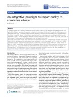
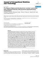
Firstly the morphology of the thin films was studied by
AFM. The resultant coating was uniform and homoge-
neous, showing a slightly porous surface with an average
roughness of 28.3 nm (rms) (Figure 1). It has been
reported in Ref. [37] that the basic pH of the TEOS pre-
cursor solution gives macroporous aggregates that can
be assembled into a film with the dip-coating technique.
This matrix shows the advantages of the inorganic
materials such as mechanical strength and chemical sta-
bility, and at the same time its porosity allows the ion
interchange with the external medium, a fundamental
aspect when an efficient silver-based antibacterial coat-
ing is desired.
Other important aspect of the hybrid matrix is that
both organic and inorganic materials should not show
any phase separation, in order to get a maximum homo-
geneity. Other authors have reported how some poly-
mers can interact with metal alkoxides [38], yielding
even covalent bonding between the polymers and the
inorg anic material. In this work the polyelectrolyte PAA
was specially selected bec ause under certain conditions,
their carboxylic groups could be eventually hydrolyzed
and further covalently bonded to tetraorthosilicate parti-
cles in a sol-gel process. In a detailed AFM analysis, the
phase images o f the samples did not show regions with
different mechanical stiffness, therefore there were not
found any evidence of phase separation in the matrix.
As it has been commented in the previous section, all
the coatings showed a highly transparent appearance
immediately after the dip-coating fabrication step. The
composition of such coatings consisted of an inorganic
matrix with a small proportion of an anionic polyelec-
trolyte embedded within. Such polymeric chains have
carboxylic functional groups that act as host sites for
the silver cations during the loading immersion step by
ion-interchange. Other authors have reported similar
Figure 1 AFM i mage topography of one of the samples.AFM
topography of one of the samples (40 × 40 μm).
Rivero et al. Nanoscale Research Letters 2011, 6:305
/>Page 3 of 7
approaches to load polymeric thin films with silver ions
[35]. The visual aspect of the hybrid thin films remained
unaltered after the silver loading immersion, a lthough
the silver ions were already present in the coating. How-
ever, the thin films showed a dramatic color change
when they were immersed into the DMAB reducing
solution. Then the samples turned almost instanta-
neously into a golden-yel lowish color. The visual aspect
of the film remained highly transparent and smooth, but
with the cited color chan ge. Such alteration of the visi-
ble absorption spectrum of the samples change is
directly related with the Surface Plasmon Resonance
(SPR) phenomenon [39,40] typical of gold and silver
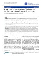
nanoparticles. In fact, the UV-VIS absorbance spectrum
confirms the existence of an absorption peak near
410 nm (Figure 2).
Such narrow absorption bands are due to the SPR
phenomenon of the silver nanoparticles synthesized
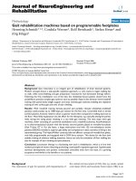
inside the coating. Furthermore, in order to get an addi-
tional evidence of the presence of silver within the
hybrid coatings an elemental analysis was obtained
using the EDX technique (Figure 3). This EDX spectrum
shows a peak at 3 keV that confirms the presence of
silver within the coating. The rest of the lines of the
EDX spectrum correspond to other elements present in
the coating (mainly Si, O, a nd Na) and also present in
the glass slide substrate, as far as the coatings were very
thin (150 nm approx.). The EDX analysis together with
the SPR absorption bands of the UV-VIS spectra make
possible to confirm the reduction of silver ions to ele-
mental silver to form nanoparticles inside the organic/
inorganic hybrid coating.
One of the most attractive aspects of the approach
proposed in this work is that simple variations in the
fabrication process can help the designer to tune the
overall properties of the coating. In this work two
different variations of the initial fabrication process were
studied; on one hand the impact of the polyelectrolyte/
TEOS ratio, and on the other hand, the number of dip/
reduction cycles. The intensity of the SPR absorption
band was t aken as an indicator of the total amount of
synthesized silver nanoparticles.
When the samples are immersed into several consecu-
tive loading/reduction cycles the size and amount of the
nanoparticles is increased [35]. Figure 2 shows the direct
relation between the number of load/reduction cycles
and the incre asing of the SPR absorption band around
410 nm, proportional to the amount of Ag nanoparticles
trapped into the thin film. Moreover, if the concentra-
tion of the polymeric solution is varied, the silver synth-
esis conditions are modified. Figur e 4 shows the growth
of the intensity of the SPR absorption band as the num-
ber of loading/reduction cycles is increased, for two
samples with different PAA molar ratio.
The results exposed in Figure 4 confirms the hypoth-
esis that lower polyelectrolyte concentrations results in
less host sites (carboxylic functional groups) for the Ag
+
cations during the loading step, and after the reduction
Figure 2 UV-VIS absorption. UV-VIS absorption spectra of the
coating with different number of loading/reduction.
Figure 3 EDX image. EDX image of the coating with 4 dip/
reduction cycles (PAA 20 mM).
Figure 4 UV-VIS absorption. UV-VIS absorption spectra of the
coating film with different PAA concentration.
Rivero et al. Nanoscale Research Letters 2011, 6:305
/>Page 4 of 7
with DMAB, the amount of silver nanoparticles is signif-
icantly lower. When the same loading/reduction proto-
col was carried out with TEOS only coatings (without
polyelectrolyte) significant absorption band was
observed in the UV-VIS analysis.
Moreover, the samples were thermally treated at
450°C for 2 h to increase their mechanica l stabil ity. The
resulting coatings showed a sharp improvement of the
mechanical strength, and a further AFM analysis showed
that there was no significant alteration in the morphol-
ogy of the films. Nevertheless, the UV-VI S absorption
spectra of the samples were dramatically modified. After
the thermal treatment the SPR absorption peak was sig-
nificantly narrowed and increased in intensity, as it is
shown in Figure 5. This is consistent with the evidences
found by other authors in other works [41].
Finally the antibacteri al activities against Lactobacillus
plantarum of the coatings were characterized using the
method described in the “Experimental section”. Figure 6
shows the results of two samples placed on agar slabs
after 24 h. The first o ne (Figure 6a) shows a reference
substrate (a bare glass slide) and it is clearly seen that a
high number of Lactobacillus plantarum colonies grow n
up randomly throughout the whole agar slab. The sec-
ond sample (Figure 6b) has a silver-loaded hybrid coated
area where there is no growth of colonies. The behavior
is different in the uncoated area of the s ubstrate and in
the rest of th e agar slab where the growth of colonies is
high as in the reference sample (Figure 6a).
All the experiments were performed in triplicate and
the treated surfaces reached more than 99.9% of kill effi-
ciency on the growth of Lactobac illus plantarum. These
results confirm the high antibacterial behavior of the
coatings based on silver nanoparticles polymer/sol-gel
hybrid matrix.
Conclusions
In t his work, it has been demonstrated that hybrid
organic-inorganic coating matrices can be used for in
situ silver nanoparticles synthesis, showing excellent
antibacterial behavior against La ctobacillus plantarum.
This approach is a simple and cost-effective method to
get coatings with high antibacterial performance, which
have most of the advantages of the inorganic coatings,
like high mechanical resistance, chemical stability, etc.
At the same time, the organic fraction present within
the inorganic coating provides the functionality to
Figure 5 UV-VIS absor ptio n. UV-VIS absorbance spectra before
and after 450°C thermal treatment.
A
B
Figure 6 Bacteria growth. Bacteria growth on culture plates after
24 h in the (a) reference substrate, (b) coated substrate. The coated
area is clearly visible, as far as it inhibits completely the bacteria
growth.
Rivero et al. Nanoscale Research Letters 2011, 6:305
/>Page 5 of 7
synthesize the silver nanoparticles that gives the highly
efficient antibacterial properties. This gives the ability to
tune the overall properties of the film; mechanical, opti-
cal, and antibacterial.
The UV-VIS absorbance spectrum confirms the exis-
tence of silver nanoparticles inside the coating due to the
presence of an absorption peak near 410 nm. Such narrow
absorption bands are typical of silver nanopart icles and
they are originated by the SPR phenomenon. EDX analysis
also confirmed the presence of silver inside the coatings.
Moreover, the impact of the organic-inorganic ratio and
the number of dip-reduction cycles on the total amount of
synthesized silver nanoparticles has been studied. Mechan-
ical resistance of the coatings has been significantly
improved using a thermal treatment at 450°C. This pro-
cess for obtaining antibacterial surfaces can be used for
different applications in a wide range of fields like in build-
ings, pharmaceutical tools, and other instrumental devices.
Abbreviations
AFM: atomic force microscopy; CFUs: colony formation units; DMAB:
dimethylamine borane; EDX: energy dispersive X-ray; PAA: poly(acrylic acid
sodium salt); SPR: Surface Plasmon Resonance; TEOS: tetraethoxysilane; TSB:
Tryptic Soy Broth.
Acknowledgements
This work was supported in part by the Spanish Ministry of Education and
Science CICYT-FEDER TEC 2009-09210 Research Grant. Thanks to the FIDENA
foundation for the EDX measurements. Very special thanks to Paula Aldaz for
her help with the bacteriologic tests.
Authors’ contributions
PJR carried out the main part of the experimental work, including the
fabrication of the thin films, nanoparticles synthesis, and bacteriological
characterization. He participated in the design of the study and in the draft
of the manuscript. AU carried the main part of the experimental work,
including the fabrication of the thin films, nanoparticles synthesis, and
bacteriological characterization. JG participated in the experimental work,
carried out the AFM characterization of the films, and contributed with the
draft of the manuscript. CRZ participated in the experimental work and
carried out the EDX analysis. FJA and IRM participated in the design of the
study and helped with the draft of the manuscript.
Competing interests
I declare that the authors have no competing interests or other interests
that might be perceived to influence the results and discussion reported in
this paper.
Received: 15 October 2010 Accepted: 7 April 2011
Published: 7 April 2011
References
1. Mcdonnell G, Russell AD: Antiseptics and disinfectants: activity, action,
and resistance. Clin Microbiol Rev 1999, 12:147-179.
2. Russell AD: Biocide use and antibiotic resistance: the relevance of
laboratory findings to clinical and environmental situations. Lancet Infect
Diseases 2003, 3:794-803.
3. Klasen HJ: Historical review of the use of silver in the treatment of burns.
I. Early uses. Burns 2000, 26:117-130.
4. Lansdown AB: Silver. I: Its antibacterial properties and mechanism of
action. J Wound Care 2002, 11:125-130.
5. Li W-R, Xie X-B, Shi Q-S, Zeng H-Y, Ou-Yang Y-S, Chen Y-B: Antibacterial
activity and mechanism of silver nanoparticles on Escherichia coli. Appl
Microbiol Biotechnol 2010, 85:1115-1122.
6. Bragg PD, Rainnie DJ: The effect of silver ions on the respiratory chain of
Escherichia coli. Can J Microbiol 1974, 20:883-889.
7. Panáček A, Kolář M, Večeřová R: Antifungal activity of silver nanoparticles
against Candida spp. Biomaterials 2009, 30:6333-6340.
8. Travan A, Pelillo C, Donati I: Non-cytotoxic silver nanoparticle-
polysaccharide nanocomposites with antimicrobial activity.
Biomacromolecules 2009, 10:1429-1435.
9. Greulich C, Kittler S, Epple M, Muhr G, Köller M: Studies on the
biocompatibility and the interaction of silver nanoparticles with human
mesenchymal stem cells (hMSCs). Langenbeck’s Arch Surg 2009,
394:495-502.
10. Chen X, Schluesener HJ: Nanosilver: a nanoproduct in medical
application. Toxicol Lett 2008, 176:1-12.
11. Yuranova T, Rincon AG, Bozzi A: Antibacterial textiles prepared by RF-
plasma and vacuum-UV mediated deposition of silver. J Photochem
Photobiol A 2003, 161:27-34.
12. Lee HY, Park HK, Lee YM, Kim K, Park SB: A practical procedure for
producing silver nanocoated fabric and its antibacterial evaluation for
biomedical applications. Chem Commun 2007, 2959-2961.
13. Gao Y, Cranston R: Recent
advances in antimicrobial treatments of
textiles. Text Res J 2008, 78:60-72.
14. Blaker JJ, Nazhat SN, Boccaccini AR: Development and characterisation of
silver-doped bioactive glass-coated sutures for tissue engineering and
wound healing applications. Biomaterials 2004, 25:1319-1329.
15. Sharma VK, Yngard RA, Lin Y: Silver nanoparticles: green synthesis and
their antimicrobial activities. Adv Colloid Interface Sci 2009, 145:83-96.
16. Sharma J, Imae T: Recent advances in fabrication of anisotropic metallic
nanostructures. J Nanosci Nanotechnol 2009, 9:19-40.
17. Wang J-X, Wen L-X, Wang Z-H, Chen J-F: Immobilization of silver on
hollow silica nanospheres and nanotubes and their antibacterial effects.
Mater Chem Phys 2006, 96:90-97.
18. Kawashita M, Toda S, Kim H-M, Kokubo T, Masuda N: Preparation of
antibacterial silver-doped silica glass microspheres. J Biomed Mater Res A
2003, 66:266-274.
19. Rivera-Garza M, Olguín MT, García-Sosa I, Alcántara D, Rodríguez-Fuentes G:
Silver supported on natural Mexican zeolite as an antibacterial material.
Microporous Mesoporous Mater 2000, 39:431-444.
20. Park S-J, Jang Y-S: Preparation and characterization of activated carbon
fibers supported with silver metal for antibacterial behavior. J Colloid
Interface Sci 2003, 261:238-243.
21. Kawashita M, Tsuneyama S, Miyaji F, Kokubo T, Kozuka H, Yamamoto K:
Antibacterial silver-containing silica glass prepared by sol-gel method.
Biomaterials 2000, 21:393-398.
22. Jeon H-J, Yi S-C, Oh S-G: Preparation and antibacterial effects of Ag-SiO
2
thin films by sol-gel method. Biomaterials 2003, 24:4921-4928.
23. Bravo J, Zhai L, Wu Z, Cohen RE, Rubner MF: Transparent superhydrophobic
films based on silica nanoparticles. Langmuir 2007, 23:7293-7298.
24. Grapski JA, Cooper SL: Synthesis and characterization of non-leaching
biocidal polyurethanes. Biomaterials 2001, 22:2239-2246.
25. Tiller JC, Lee SB, Lewis K, Klibanov AM: Polymer surfaces derivatized with
poly(vinyl-N-hexylpyridinium) kill airborne and waterborne bacteria.
Biotechnol Bioeng 2002, 79:465-471.
26. Popa A, Davidescu CM, T rif R, Ilia G, Iliescu S, Dehelean G: Study of
quaternary ‘onium’ salts grafted on polymers: antibacterial
activity of quat ernary phosphonium salts grafted on ‘ gel-type’
styrene-divinylbenzene copolymers. React Funct Polym 2003,
55:151-158.
27. Sunada K, Watanabe T, Hashimoto K: Studies on photokilling of bacteria
on TiO
2
thin film. J Photochem Photobiol A 2003, 156:227-233.
28. Daniel A, Le Pen C, Archambeau C, Reniers F: Use of a PECVD-PVD process
for the deposition of copper containing organosilicon thin films on
steel. Appl Surf Sci 2009, 256:S82-S85.
29. Li J, Zivanovic S, Davidson PM, Kit K: Production and characterization of
thick, thin and ultra-thin chitosan/PEO films. Carbohydr Polym 2011,
83:375-382.
30. Yang QB, Li DM, Hong YL: Preparation and characterization of a PAN
nanofibre containing Ag nanoparticles via electrospinning. Synth Met
2003, 137:973-974.
31. Jin W-J, Lee HK, Jeong RH, Park WH, Youk JH: Preparation of polymer
nanofibers containing silver nanoparticles by using poly(N-
vinylpyrrolidone). Macromol Rapid Commun 2005, 26:1903-1907.
Rivero et al. Nanoscale Research Letters 2011, 6:305
/>Page 6 of 7
32. Marini M, De Niederhausern S, Iseppi R: Antibacterial activity of plastics
coated with silver-doped organic-inorganic hybrid coatings prepared by
sol-gel processes. Biomacromolecules 2007, 8:1246-1254.
33. Schuleit M, Luisi PL: Enzyme immobilization in silica-hardened
organogels. Biotechnol Bioeng 2001, 72:249-253.
34. Schultze C, Cordes A, Schmidt W, Sternberg K, Behrend D, Schmitz K-P:
Hybrid polymers as implant material for medical devices. IFMBE Proc
2009, 25:164-167.
35. Li Z, Lee D, Sheng X, Cohen RE, Rubner MF: Two-level antibacterial
coating with both release-killing and contact-killing capabilities.
Langmuir 2006, 22:9820-9823.
36. JIS Z 2801: Japanese Industrial Standard, Japanese Standard Association.
2000.
37. Boonamnuayvitaya V, Tayamanon C, Sae-Ung S, Tanthapanichakoon W:
Synthesis and characterization of porous media produced by a sol-gel
method. Chem Eng Sci 2006, 61:1686-1691.
38. Ichinose I, Kawakami T, Kunitake T: Alternate molecular layers of metal
oxides and hydroxyl polymers prepared by the surface sol-gel process.
Adv Mater 1998, 10:535-539.
39. Wang TC, Cohen RE, Rubner MF: Metallodielectric photonic structures
based on polyelectrolyte multilayers. Adv Mater 2002, 14:1534-1537.
40. Nolte AJ, Rubner MF, Cohen RE: Creating effective refractive index
gradients within polyelectrolyte multilayer films: molecularly assembled
rugate filters. Langmuir 2004, 20:3304-3310.
41. Patel AC, Li S, Wang C, Zhang W, Wei Y: Electrospinning of porous silica
nanofibers containing silver nanoparticles for catalytic applications.
Chem Mater 2007, 19:1231-1238.
doi:10.1186/1556-276X-6-305
Cite this article as: Rivero et al.: An antibacterial coating based on a
polymer/sol-gel hybrid matrix loaded with silver nanoparticles.
Nanoscale Research Letters 2011 6 :305.
Submit your manuscript to a
journal and benefi t from:
7 Convenient online submission
7 Rigorous peer review
7 Immediate publication on acceptance
7 Open access: articles freely available online
7 High visibility within the fi eld
7 Retaining the copyright to your article
Submit your next manuscript at 7 springeropen.com
Rivero et al. Nanoscale Research Letters 2011, 6:305
/>Page 7 of 7