Báo cáo hóa học: " A combination of hard and soft templating for the fabrication of silica hollow microcoils with nanostructured walls" pptx
Bạn đang xem bản rút gọn của tài liệu. Xem và tải ngay bản đầy đủ của tài liệu tại đây (1.95 MB, 7 trang )
NANO EXPRESS Open Access
A combination of hard and soft templating for
the fabrication of silica hollow microcoils with
nanostructured walls
Carlos Rodriguez-Abreu
1*
, Neus Vilanova
2
, Conxita Solans
2
, Masaki Ujihara
3
, Toyoko Imae
3
, Arturo López-Quintela
4
and Seiji Motojima
5
Abstract
Hollow silica microcoils have been prepared by using functionalized carbon microcoils as hard templates and
surfactant or amphiphilic dye aggregates as soft templates. The obtained materials have been characterized by
electron and optical microscopy, nitrogen sorption and small angl e X-ray scattering. The obtained hollow
microcoils resemble the original hard templates in shape and size. Moreover, they have mesoporous walls (pore
size ≈ 3 nm) with some domains where pores are ordered in a hexagonal array, originated from surfactant
micelles. The obtained silica microcoils also show prefer ential adsorption of cationic fluorescent dyes. A mechanism
for the formation of silica micro coils is proposed.
Introduction
The use of templates or scaffolds is one of the main
strategies for the fabrication of advanced materials with
new structures at the nano and micro scales that have
attracted considerable research effort over the past dec-
ades. Templates can be classified as ‘hard’ an d ‘soft’ .
Hard templates are usually solid-state materials with
particular structure and morphology, whereas soft tem-
plates are generally in a fluid-like state.
Hard templating is a conceptually straightforward and
highly effective method to prepare hollow structures that
mimic and/or complement the original shape of the tem-
plates [1,2] , which usually consis ts of the follow ing steps:
1. Preparation of hard templates; 2. Functionalization/
modification of template surface; 3. Coating the templates
with the target shell material; and 4. Selective removal of
the templates to obtain hollow structures. Silica particl es
and polymer latex colloids belong to the group of materi-
als commonly employed as hard templates.
On the other hand, soft templates such as supramole-
cular self-assemblies are a powerful tool for th e bottom-
up synthesis of nanomaterials [3-6], particularly
mesoporous inorganic solids. In this appr oach, there is a
cooperativ e interaction between self-assemblies and
inorganic species that lead to structuration.
Carbon microcoils (CMCs) [7], with coil diameters in
the order of micrometers, are a class of carbon materials
with singular properties, such as mechanical elasticity
[8], high hydrogen sorption [9] and electromagnetic
wave absorption [10]. Carbon microcoils also help to
improve material properties when incorporated in
hybrid composites [11], or as template for other materi-
als [12,13].The formation of a coiled structure is attribu-
ted to the fact that the crystal faces of the catalyst used
for the synthesis show different activity (i.e. catalytic ani-
sotropy) in terms of carbon growth [7].
Although there is some literature on the use of carbon
nanotubes (CNT) as scaffolds for the preparation of
silica nanotubes with different morphologies [14-16],
carbon materials with a peculiar structure such as CMC
has not been used in a combined hard and soft templat-
ing strategy to produce hierarchically ordered materials.
In this context, we report the results on the use of such
a method to fabricate nanoporous hollow silica micro-
coils and discuss the c haracterization of the obtained
materials. The combination of soft and hard templating
provides versatility for the preparation of materials with
properties deriving from structuration at different scales.
* Correspondence:
1
International Iberian Nanotechnology Laboratory (INL), Av. Mestre José
Veiga, Braga, 4715-310, Portugal
Full list of author information is available at the end of the article
Rodriguez-Abreu et al. Nanoscale Research Letters 2011, 6:330
/>© 2011 Rodriguez-Abreu et al; licensee Springer. This is an Open Access article distributed under the terms of the Creative Commons
Attribution License ( 2.0), which permits unrestricted use, distribution, and reprod uctio n in
any med ium, provided the or iginal work is properly cited.
Experimental
Materials
Carbon microco ils were synthesized according to a pre-
vious publication [7]. Hexadecyltrimethylammonium
bromide (CTAB), tetraethylorthosilicate (TEOS), rhoda-
mine B and fluorescein were supplied by Sigma-Aldrich
(USA). A Perylenebis(dicarboximide) dye (referred
herein as PDI) was synthesized according to the litera-
ture [17].Ultrapure water (resistivity = 18.2 MΩ/cm)
was used in the experiments. All chemicals were used
without further purification.
Preparation of silica samples
Surface functionalization of CMCs was carried out by
oxidation following a method already reported [18].
Functionalized CMCs with -COOH groups are referred
herein as CMC-COOH. In a typical preparation of silica
hollow coils by sol- gel reaction, CTAB or PDI is dis-
solved in NH
3
(aq., 25%). Then, CMC-COOHs are dis-
persed in the mixture by ultrasonication. Finally, TEOS
is added and the mixture is stirred with a magnetic stir-
ring bar for 3 h at 70° C. The resulting precipitate is
washed, filtered, dried and calcined in air for 6 h at 600°
C (heating rate = 1°C/min), above the decomposition
temperature of CMC-COOHs, as determined by ther-
mogravimetric analysis.
Characterization
Scanning electron microscopic (SEM) images were col-
lected with a Hitachi TM-1000 (Japan) and with a
Zeiss UltraPlus FESEM instrument (Germany). Trans-
mission electron microscopic (TEM) images were
taken with a Hitachi H-7000(Japan). Specimens were
deposited on copper grids from ethanol dispersions.
Fluorescence microscopic images were collected with a
Nikon Eclipse TE2000-U(Japan); for the observation,
samples were immersed in an aqueous dye solution for
1 h and t hen rinsed tho roughly to remove the non-
adsorbed dye. Small angle X-ray scattering (SAXS)
measurements were performedinaninstrument
equipped with a Kratky camera and a linear position
sensitive detector, OED 50 M, both from MBraun
(Austria). Measurements were carried out at 0.5 kW
with radiation coming from a Siemens generator,
model Krystalloflex 760 (Germany). Nitrogen sorption
isotherms were determined using a Micromeritics
TriStar 3000 instrument (USA). Samples were degassed
at 200°C, and weighed prior to sorption experiments.
The pore size distribution was determined by the Bar-
ret-Joyner-Halenda (BJH) method [19].
Results and discussion
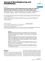
As can be seen in Figure 1a,b, the CMC-COOHs used
as templates are polydisperse in diameter, pitch and
length; some of them are hundreds of micrometers in
length. During the sol-gel reaction, the surface of the
CMC-COOHs is covered by a silica deposit. After calci-
nation, i.e. after removal of CMC-COOHs, silica coils
areleft(seeFigure1c).Somesectionsofthecoilsseem
more transparent, due to their very t hin silica walls,
while coil size and morphology appear similar to th at of
original CMC-COOHs. Silica particles can also be
observed on the surface of the coi ls. The dif ferences in
contrast of the two specimens (CMC-COOHs and silica
coils) can also be clearly observed by the optical micro-
scope (Figure 2). The images are a proof that the CMC-
COOHs used as templates have been burnt off and silica
coils are left ov er, although also n on-CMC-templated
silica particles are obtained mixed with the coils, as iso-
lated silica particles can also form in the bulk solution
Figure 1 SEM images (a, b) CMC-COOH before silica coating, (c) microcoil after silica-coating and calcination. The initial weight ratios for
the silica coating process were CTAB/NH
3
(aq.)/CMC-COOH/TEOS = 13.9/70.4/0.6/15.1. The black arrows indicate silica particles adhered to the
coils whereas white arrows indicate translucid coil sections.
Rodriguez-Abreu et al. Nanoscale Research Letters 2011, 6:330
/>Page 2 of 7
during the sol-gel reaction. The amount of non-CMC-
templated silica decreases by reducing the CTAB/CMC-
COOH ratios, so that most of CTAB is adsorbed on the
CMC-COOH surface.
TEM images of silica hollow microcoils are presented
in Figure 3. The photographs at low magnification
show unambiguously the hollow nature of the speci-
mens; the very thin silica walls allow the transmission
oftheelectronbeam.Thefinestructureofthewalls
can be imaged at higher magnification. Arranged chan-
nels of about 3-nm width were observed in some sec-
tions of the microcoil walls. The cross section of those
channels is circular, namely, the walls contain cylindri-
cal mesopores, some of which ordered in a hexagonal
fashion. However, it should be pointed out that meso-
pores with disordered domains (worm-hole morphol-
ogy) also exist in the microcoil walls. The existence of
mesopores was confirmed by nitrogen so rption experi-
ments, which gav e a relatively narrow pore size distri-
bution with a maximum at 2.3 nm, in agreement with
TEM observations (see Figure S 1 in Additional file 1).
Samples of silica coils were also analyzed by SAXS.
Patterns showed a strong peak corresponding to a
Bragg spacing d of 3.8 nm (see Figure S2 in Additional
file 1); the lattice parameter for a hexagonal array (a =
2d/√3) was calculated as 4.4 nm, which is similar to
that of MCM-41 silica [20].
The dye adsorption properties of silica hollow micro-
coils were tested. As can be seen in Figure 4, the micro-
coils pre-soaked in a cationic dye (rhodamine B)
solution are fluorescent namely, the dye strongly adsorbs
on the surface of silica microcoil, indicating that the
surface is negatively charged, as expected from the high-
pH synthesis conditions. On the other hand, no
fluorescence was emitted from a sample pre-soaked with
an anionic dye (e.g. fluorescein), since there is no charge
matching and hence no adsorption of the anionic dye
on the microcoils.
We also found that PDI (see Figure 5a), which is
known to self-assemble in water [17], can be used as a
soft template for the silica coating of CMC-COOHs.
PDI has n-type semiconductor properties, with possible
applications in organic field effect transistors and photo-
voltaics. Moreover, PDI molecules adsorb strongly to
the CMC-COOH surface, as confirmed by UV-vis spec-
troscopy (see Figure S3 in Additional file 1). In the
absence of CMC-COOH, PDI induces the f ormation of
elongated silica particles in the sol-gel reaction mixture,
which is attributed to the templating effect of cylindrical
self-assemblies of the dye in solution [17]. At high PDI/
CMC-COOH ratios, those elongated silica particles, pre-
cipitated from the solution, coat the surface of CMC-
COOHs (see Figure 5b). When the TEOS/CMC-COOH
ratio is decreased, hollow silica microcoils with
smoother surface are obtained, suggesting that silica for-
mation on the CMC-COOH surface is favoured over
that occurring in the bulk solution (see Figure 5c,d).
The yield of silica microcoils relative to amorphous
silica also increases when the PDI/CMC-COOH ratios
are decreased. It is to be noted here that when PDI is
used as template, the walls of silica coils are probably
microporous rather than mesoporous as suggested by
preliminary nitrogen sorption measurements, which
gave isotherms with features typical of microporous
solids.
The fluorescence emission properties of the PDI
adsorbed on CMC-COOH were preserved after silica
coating. Dispersions of the PDI-silica coils in ethanol
Figure 2 Optical microscopic images of (a) CMC-COOH and (b) hollow silica microcoil prepared using CTAB.
Rodriguez-Abreu et al. Nanoscale Research Letters 2011, 6:330
/>Page 3 of 7
gave two emission bands at 540 and 575 nm (see Figu re
S4 in Additional file 1). However, a change in the UV-
vis spectrum was observed (see Figure S5 in Additional
file 1). Neat PDI solutions in ethanol show two absorp-
tion maxima at 530 and 590 nm, whereas the disper-
sions of PDI-silica coils exhibited only one maximum at
520 nm and a shoulder at about 580 nm.
Based upon the experimental evidence, a mechanism
for the formation of silica hollow microcoils with meso-
porous walls can be proposed (see Figure 6). Cationic
aggregates, which are expected to be elongated at the
used concentrations of amphiphile and electrolyte
[17,21,22], adsorb on the surface of negatively-charged
CMC-COOHs. When TEOS is added at high pH, the
silica coating is built up through anchoring of silica via
electrostatic interaction of siloxy ions with ammonium
ions of the amphiphilic molecules adsorbed on CMCs.
During the sol-gel reaction, free cationic aggregates are
also cooperatively incorporated as porogens in the poly-
siloxane gel. At this stage, there might be some prefer-
ential orientation of aggregates in the silica layers.
Depending on amphiphile concentration, excess silica
particles with inner mesostructure forms in the bulk
solution and some of those particles also adhere to the
silica layers on the surface of CMCs. Finally, upon calci-
nation, the CMCs (hard templates) and amphiphilic
molecules (soft templates) are burnt off, and silica hol-
low microcoils with porous walls are obtained.
Figure 3 TEM images of different sections of silica hollow microcoils. The initial weight ratios for the silica coating process were CTAB/NH
3
(aq.)/CMC-COOH/TEOS = 13.7/69.4/2.0/14.9.
Rodriguez-Abreu et al. Nanoscale Research Letters 2011, 6:330
/>Page 4 of 7
Figure 4 Visible and fluorescent microscopic images of hollow silica microcoils prepared using CTAB after soaking them in an
aqueous solution of rhodamine B.
Figure 5 Silica microcoils prepared using PDI. (a) Molecular structure of PDI. (b) SEM image of silica microcoils at PDI/CMC-COOH ratio = 3;
the arrows indicate elongated particles adhered to the coils. (c, d) SEM image of silica microcoils at PDI/CMC-COOH ratio = 0.2. The other initial
weight ratios for the silica coating process were NH
3
(aq.)/CMC-COOH/TEOS = 300/0.37/10.1.
Rodriguez-Abreu et al. Nanoscale Research Letters 2011, 6:330
/>Page 5 of 7
Summary
Hollow silica micro coils with mesostructured walls were
prepared by using carbon microcoils and amphiphilic
molecules as hard and soft temp lates, respectively, and
both serve as porogens upon calc ination. Cationic aggre-
gates adsorb on functionalized CMCs and behave both as
an anchor and porogen of silica. The mesopores originated
from surfactant aggregates were either ordered hexagon-
ally or had a disordered, worm-hole morphology. Since
the obtained hollow silica microcoils have a negatively
charged surface (as a result of synthesis conditions), they
show advantages for preferentially trapping cationic mole-
cules. The method described here can be used to prepare
hollow microcoils of other oxides via sol-gel reaction.
Additional material
Additional file 1: Figure S1. Pore size distribution of silica hollow
microcoils. The initial weight ratios for preparation were CTAB/NH
3
(aq.)/
CMC-COOH/TEOS = 13.9/70.4/0.6/15.1. The distribution is estimated from
nitrogen sorption measurements using the Barret-Joyner-Halenda (BJH)
method. Figure S2. SAXS spectra of (a) CMC-COOH, (b) hollow silica
microcoils prepared with initial CTAB/NH
3
(aq.)/CMC-COOH/TEOS weight
ratios of 13.9/70.4/0.6/15.1. The numbers indicate the peak position ratios
corresponding to a hexagonal lattice. (c) Hollow silica microcoils
prepared with initial CTAB/NH
3
(aq.)/CMC-COOH/TEOS weight ratios of
8.1/85.0/2.4/4.5. Figure S3. UV-vis spectra of PDI aqueous solutions
before contact (continuous line) and after 1 h contact (dashed line) with
CMC-COOHs. The decrease in absorbance is due to adsorption of PDI
molecules on the surface of CMC-COOHs. Figure S4. Fluorescence
emission spectrum of of PDI on CMC-COOH after silica coating. The
spectrum corresponds to a dispersion in ethanol measured in a 1-cm
path length cuvette. Figure S5. UV-vis absorption spectra of neat PDI
(continuous line) and PDI on CMC-COOH after silica coating (dashed
line). Spectra correspond to dispersions in ethanol measured in 1-cm
path length cuvettes.
Abbreviations
CMCs: carbon microcoils; CNT: carbon nanotubes; CTAB:
hexadecyltrimethylammonium bromide; PDI: Perylenebis(dicarboximide);
SAXS: small angle X-ray scattering; SEM: scanning electron microscopic; TEM:
Transmission electron microscopic; TEOS: tetraethylorthosilicate.
Acknowledgements
Authors are grateful to Consejo Superior de Investigaciones Científicas (CSIC,
Spain) and National Science Council (NSC, Taiwan) for research funding
within the frame of the bilateral cooperation program (2008TW006,
2009TW0031). C.R-A. is also grateful to to the Ministerio de Ciencia e
Innovación, Spain (Project CTQ2008-01979/BQU) for financial support.
Authors thank Lucia Casal (Universitat de Barcelona, Spain) for his help in
the synthesis of PDI dye, and Prof. Po-Da Hong and Prof. Shawn D. Lin
(National Taiwan University of Science and Technology) for experimental
support.
Author details
1
International Iberian Nanotechnology Laboratory (INL), Av. Mestre José
Veiga, Braga, 4715-310, Portugal
2
Instituto de Química Avanzada de Cataluña.
Consejo Superior de Investigaciones Científicas (IQAC-CSIC), Jordi Girona 18-
26, Barcelona, 08034, Spain
3
Graduate Institute of Engineering, National
Taiwan University of Science and Technology, 43 Keelung Road, Section 4,
Taipei, Taiwan
4
Departamento de Química Física, Facultad de Química,
Universidad de Santiago de Compostela, Santiago de Compostela, 15782,
Spain
5
Toyota Physical & Chemical Research Institute, Nagakute, Aichi, 480-
1192, Japan
Authors’ contributions
CR-A conceived the study and participated in its design and coordination, as
well as in sample preparation and characterization by TEM, SEM and SAXS.
NV participated in TEM observations and in spectroscopic measurements. CS,
AL-Q and TI participated in the preparation and revision of the manuscript
as well as in giving access to SAXS, SEM and TEM facilities. MU participated
in electronic and optical microscopy experiments. SM carried out the
synthesis of carbon microcoils. All authors read and approved the final
manuscript.
Competing interests
The authors declare that they have no competing interests.
Received: 26 October 2010 Accepted: 13 April 2011
Published: 13 April 2011
Figure 6 Proposed scheme for the mechanism of formation of hollow silica microcoils.
Rodriguez-Abreu et al. Nanoscale Research Letters 2011, 6:330
/>Page 6 of 7
References
1. Lou XW, Archer LA, Yang Z: Hollow Micro-/Nanostructures: Synthesis and
Applications. Adv Mater 2008, 20: 3987-4019.
2. Zhang Q, Wang W, Goebl J, Yin Y: Self-Templated Synthesis of Hollow
Nanostructures. Nano Today 2009, 4: 494-507.
3. Wan Y, Zhao D: On the Controllable Soft-Templating Approach to
Mesoporous Silicates. Chem Rev 2007, 107: 2821-2860.
4. Lu AH, Schüth F: Nanocasting: A Versatile Strategy for Creating
Nanostructured Porous Materials. Adv Mater 2006, 18: 1793-1805.
5. Hamley IW: Nanotechnology with Soft Materials. Angew Chem Int Ed 42:
1692-1712.
6. Lazzari M, Rodríguez C, Rivas J, López-Quintela A: Self-assembly: a
minimalist route to the fabrication of nanomaterials. J Nanosci
Nanotechnol 2006, 6: 892-905.
7. Motojima S, Chen X: Preparation and Characterization of Carbon
Microcoils (CMCs). Bull Chem Soc Jpn 2007, 80: 449-455.
8. Motojima S, Chen X, Yang S, Hasegawa M: Properties and potential
applications of carbon microcoils/nanocoils. Diam Relat Mater 2004, 13:
1989-1992.
9. Furuya Y, Hashishin T, Iwanaga H, Motojima S, Hishikawa Y: Interaction of
hydrogen with carbon coils at low temperature. Carbon 2004, 42:
331-335.
10. Motojima S, Hoshiya S, Hishikawa Y: Electromagnetic wave absorption
properties of carbon microcoils/PMMA composite beads in W bands.
Carbon 2003, 41: 2658-2660.
11. Adhikari PD, Ujihara M, Imae T, Hong PD, Motojima S: Reinforcement on
Properties of Poly(vinyl alcohol) Films by Embedding Functionalized
Carbon Micro Coils. J Nanosci Nanotechnol 2011, 11: 1004-1012.
12. Motojima S, Suzuki T, Noda Y, Hiraga A, Iwanaga H, Hashishin T,
Ishikawa Y, Yang S, Chen X: Preparation of TiO2 microcoils from carbon
microcoil templates using a sol-gel process. Chem Phys Lett 2003, 378:
111-116.
13. Motojima S, Suzuki T, Hishikawa Y, Chen X: TiO
2
/C Composite Microcoils
and TiO
2
Hollow Microcoils with High Photocatalytic Activities and
Electromagnetic (EM) Wave Absorption Abilities. Jpn J Appl Phys 2 Lett
2003, 42: L938-L940.
14. Bian SW, Ma Z, Zhang LS, Niu F, Song WG: Silica nanotubes with
mesoporous walls and various internal morphologies using hard/soft
dual templates. Chem Commun 2009, 1261-1263.
15. Kim M, Hong J, Lee J, Hong CK, Shim SE: Fabrication of silica nanotubes
using silica coated multi-walled carbon nanotubes as the template. J
Colloid Interface Sci 2008, 322: 321-326.
16. Ding K, Hu B, Xie Y, An G, Tao R, Zhang H, Liu Z: A simple route to coat
mesoporous SiO2 layer on carbon nanotubes. J Mater Chem 2009, 19:
3725-3731.
17. Tam-Chang SW, Helbley J, Iverson IK: A Study of the Structural Effects on
the Liquid-Crystalline Properties of Ionic Perylenebis(dicarboximide)s
Using UV-Vis Spectroscopy, Polarized Light Microscopy, and NMR
Spectroscopy. Langmuir 2008, 24: 2133-2139.
18. Adhikari PD, Tai Y, Ujihara M, Chu CC, Imae T, Motojima S: Surface
Functionalization of Carbon Micro Coils and Their Selective
Immobilization on Surface-Modified Silicon Substrates. J Nanosci
Nanotechnol 2010, 10: 833-839.
19. Barret EP, Joyner JG, Halenda PP: The Determination of Pore Volume and
Area Distributions in Porous Substances. I. Computations from Nitrogen
Isotherms. J Am Chem Soc 1951, 73: 373-380.
20. Di Renzo F, Cambon H, Dutartre R: A 28-year-old synthesis of micelle-
templated mesoporous silica. Micropor Mater 1997, 10: 283-286.
21. Imae T, Ikeda S: Sphere-rod transition of micelles of
tetradecyltrimethylammonium halides in aqueous sodium halide
solutions and flexibility and entanglement of long rodlike micelles. J
Phys Chem 1986, 90: 5216-5223.
22. Kunieda H, Rodríguez C, Tanaka Y, Kabir MH, Ishitobi M: Effects of added
nonionic surfactant and inorganic salt on the rheology of sugar
surfactant and CTAB aqueous solutions. Colloid Surf B Biointerfaces 2004,
38: 127-130.
doi:10.1186/1556-276X-6-330
Cite this article as: Rodriguez-Abreu et al.: A combination of hard and
soft templating for the fabrication of silica hollow microcoils with
nanostructured walls. Nanoscale Research Letters 2011 6:330.
Submit your manuscript to a
journal and benefi t from:
7 Convenient online submission
7 Rigorous peer review
7 Immediate publication on acceptance
7 Open access: articles freely available online
7 High visibility within the fi eld
7 Retaining the copyright to your article
Submit your next manuscript at 7 springeropen.com
Rodriguez-Abreu et al. Nanoscale Research Letters 2011, 6:330
/>Page 7 of 7