Báo cáo hóa học: " Synthesis of Bio-Compatible SPION–based Aqueous Ferrofluids and Evaluation of RadioFrequency Power Loss for Magnetic Hyperthermia" docx
Bạn đang xem bản rút gọn của tài liệu. Xem và tải ngay bản đầy đủ của tài liệu tại đây (423.3 KB, 6 trang )
NANO IDEAS
Synthesis of Bio-Compatible SPION–based Aqueous Ferrofluids
and Evaluation of RadioFrequency Power Loss for Magnetic
Hyperthermia
A. P. Reena Mary
•
T. N. Narayanan
•
Vijutha Sunny
•
D. Sakthikumar
•
Yasuhiko Yoshida
•
P. A. Joy
•
M. R. Anantharaman
Received: 9 May 2010 / Accepted: 2 August 2010 / Published online: 15 August 2010
Ó The Author(s) 2010. This article is published with open access at Springerlink.com
Abstract Bio-compatible magnetic fluids having high
saturation magnetization find immense applications in
various biomedical fields. Aqueous ferrofluids of super-
paramagnetic iron oxide nanoparticles with narrow size
distribution, high shelf life and good stability is realized by
controlled chemical co-precipitation process. The crystal
structure is verified by X-ray diffraction technique. Particle
sizes are evaluated by employing Transmission electron
microscopy. Room temperature and low-temperature mag-
netic measurements were carried out with Superconducting
Quantum Interference Device. The fluid exhibits good
magnetic response even at very high dilution (6.28 mg/cc).
This is an advantage for biomedical applications, since only
a small amount of iron is to be metabolised by body organs.
Magnetic field induced transmission measurements carried
out at photon energy of diode laser (670 nm) exhibited
excellent linear dichroism. Based on the structural and
magnetic measurements, the power loss for the magnetic
nanoparticles under study is evaluated over a range of
radiofrequencies.
Keywords Superparamagnetism Á Magnetic heating Á
Power loss Á Magnetic relaxation Á Magnetic hyperthermia
Introduction
Colloidal suspensions of ultrafine magnetic particles (fer-
rofluids) have widespread applications in fields of both
engineering [1–3] and biomedicine [3–6]. Ferrofluids are
used in loudspeakers as coolants and dampers, in dynamic
sealing, smart Ferrogel preparation [7] for controlled
delivery of drugs and as contrast enhancing agents [8].
Ferrofluids are synthesized by dispersing nanosized mag-
netic particles in carrier liquids with suitable surfactants
and proper stabilization techniques. The biocompatibility
and the ease with which it can be dispersed in water qualify
iron oxide–based ferrofluid a competent candidate for
membrane separation, intraocular retinal repair, early
diagnosing, imaging and magnetic hyperthermia [9] for
cancer therapy, enzyme immobilization of cell targeting
and targeted drug delivery [10]. The surface area, size and
shape of the nanoparticles decide the physical and chemical
properties of these particles to a great extent, which in turn
decide the performance in various applications [11]. The
particle size, and its distribution, the magnetic and flow
properties of the fluid influence the application parameters
especially in biomedicine. The spherical shape and
monodispersibility of SPIONs are often a prerequisite for
application in living tissues [12]. So, optimization of the
synthesis of nanoparticles and their conjugation with
organic molecules onto the surface becomes very much
essential.
A. P. Reena Mary Á T. N. Narayanan Á V. Sunny Á
M. R. Anantharaman (&)
Department of Physics, Cochin University of Science
and Technology, Cochin 682022, India
e-mail:
D. Sakthikumar Á Y. Yoshida
Bio-Nano Electronics Research Centre, Department
of Applied Chemistry, Toyo University, Tokyo, Japan
P. A. Joy
National Chemical Laboratories, Pune, India
Present Address:
T. N. Narayanan
Department of Mechanical Engineering and Materials Science,
Rice University, Houston, TX, USA
123
Nanoscale Res Lett (2010) 5:1706–1711
DOI 10.1007/s11671-010-9729-4
Magnetic hyperthermia surmounts other techniques of
hyperthermia for cancer treatment because of reduced side
effects such as damage to healthy tissues [13]. Magnetic
hyperthermia or magneto-thermo cytolysis refers to heating
of cells attached to magnetic particle by an external AC
magnetic field. The increase in temperature is caused due
to hysteresis loss. In the case of superparamagnetic parti-
cles, the loss is caused by the relaxation processes [14].
The heat dissipated when subjected to an alternating
magnetic field depends on the fluid properties such as
viscosity, the ratio of relaxation frequency to the applied
frequency, size distribution of the magnetic component,
domain magnetization and density and the specific heat
capacity of the magnetic constituent [15]. There are reports
of reduction in magnetization with decrease in particle size
in the case of oxide magnetic materials [16]. This is due to
finite size effect. Magnetic nanoparticles–drug conjugate
attached to an antibody or hormone can be magnetically
guided to the tumour site and could specifically bind to it.
This provides a platform for optimum dosage of drug.
The stability of the fluid against sedimentation is deci-
ded collectively by competing interactions [1, 3, 17, 18]
such as van der waals interactions, dipolar interactions,
viscous force of the carrier liquid, and the electrostatic and
steric repulsion of the surfactant. Surfacted ferrofluid have
a long chain of organic molecule around the surface and
mainly, steric repulsion provides stabilization. In ionic
fluids, the electrostatic repulsion provides stabilization.
Hence, the pH of such fluids may vary considerably (from
3 to 9) from basic to acidic depending on the treatment of
the nanoparticles after precipitation. Citric acid, a bio-
compatible surfactant, presents both electrostatic and steric
effects and could easily get conjugated to iron oxide par-
ticles. Iron oxide is most recommended because of its
higher magnetization values, lesser toxicity [19] and the
ease of metabolism by the liver. In the present study, we
report the synthesis of highly stable water-based iron oxide
fluid with narrow particle size distribution at neutral pH,
and the evaluation of magnetic properties for hyperthermia
application. The cell viability test conducted with these
fluids on He La cells was promising (not included). To
study the relaxation of the fluid in an external magnetic
field, the magneto-optic linear dichroism measurement is
presented. The power loss spectrum of these nanoparticles
in an external alternating magnetic field is simulated to
investigate the possibility of applying in AC magnetic
heating.
Experimental
Monodispersed iron oxide particles of average size
9.5 nm were synthesized through controlled chemical
co-precipitation method. For this, analytical grade anhy-
drous ferric chloride (FeCl
3
) and ferrous sulphate hepta-
hydrate (FeSO
4
.7H
2
O from Merk) in the molar ratio of 2:1
each in 500 ml of distilled water were taken as the starting
solution. To the solution, 12% of aqueous ammonia was
added while stirring at room temperature to supersaturate
for the precipitation of the oxide. The rate of reaction was
controlled by allowing one drop of ammonia per second to
react with this solution until a pH of 10, to get a thick dark
precipitate. Five grams of citric acid crystals dissolved in
10 ml water was added to this wet precipitate and allowed
for further reaction at an elevated temperature of 80°C
while stirring for another 90 min. This sample was then
washed with distilled water several times for the removal
of water soluble byproducts. This is then suspended in
distilled water by ultrasound treatment. The obtained fluid
was kept for gravity settling of any bare nanoparticles and
was then centrifuged at a rotation speed of 3500 rpm to
remove any particles that may sediment. The supernatant
fluid is extracted for further analysis. The concentration of
the magnetic particles is estimated to be 6.28 mg/cc.
The structural characterization was carried out using
X-ray diffraction (XRD) technique (Rigaku D Max) at Cu
Ka. The particles were analysed with Transmission elec-
tron microscopy (TEM). The samples for the above-men-
tioned experiments were prepared by evaporating the
moisture content from the fluid. Room temperature and low
temperature magnetic measurements were performed in a
Superconducting Quantum Interference Device (SQUID)
magnetometer (MPMS Quantum Design). The magneto-
optical linear dichroism measurements were carried out on
the fluid taken in a 1-cm cuvette at the photon energy of
diode laser (670 nm). The simulation of the power loss
spectrum of the sample was performed for applied field
strength of 500Oe in the range 100–900 kHz.
Results and Discussion
The fluid synthesized exhibited good shelf life and stability
against sedimentation under gravitational and magnetic
fields. Fluid when an external magnetic field is applied
normal to the free surface exhibited good spiking.
Structural and Morphological Analysis
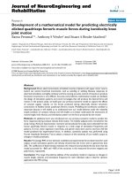
The X-ray diffraction pattern (Fig. 1) shows that the iron
oxide particles have crystallized in the inverse spinel
structure with a lattice constant of 8.41A
˚
. All the major
crystallographic planes corresponding to inverse spinel are
identified [ICDD PDF No.750449]. It is hard to differen-
tiate between maghemite (c Fe
2
O
3)
and magnetite (Fe
3
O
4
)
using X-ray diffraction analysis alone, since both represent
Nanoscale Res Lett (2010) 5:1706–1711 1707
123
an inverse spinel structure and the (hkl) planes are similar.
However, from XRD analysis, it is seen that the compound
contains no traces of nonmagnetic haematite (a-Fe
2
O
3
).
There is significant broadening of peaks due to the size
reduction in particles. The particle size is calculated from
the line broadening applying Scherer’s formula [20] and is
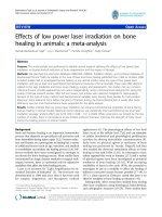
found to be 9.5 nm. The TEM images (Fig. 2a) show that
the particles are nearly spherical. Statistical analysis
(Fig. 2b) of the images revealed a normal distribution of
particles with a mean size of 10 nm and a width of 3 nm.
This is in fair agreement with the particle size obtained
from X-ray diffraction measurements.
The narrow size distribution is an advantage while
considering the magnetic hyperthermia applications or for
targeted drug delivery. The hydrodynamic length of a
single citric acid molecule is calculated to be nearly
0.7 nm. This is the thickness of the surfactant monolayer.
So, there is a minimum spacing of 1.4 nm between the iron
oxide nanoparticles.
Magnetic Characterization
For magnetization measurements, 3 micro litres of the fluid
was dropped over to a quartz substrate and the base liquid
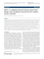
was allowed to evaporate. Magnetic Hysteresis loops of
SPIONs were measured at room temperature and at a low
temperature of 6 K and are depicted in Fig. 3. Field-cooled
(FC) and zero field–cooled (ZFC) magnetization mea-
surements were carried out in an applied field of 30 mT
from 6 to 300 K.
The room temperature M-H loop exhibits negligible
coercivity (9Oe) and remanence, which signifies the
superparamagnetic nature of particles. This is further ver-
ified by fitting experimental curve with the modified
Langevin function [21] for the particles with normal size
distribution (Fig. 4).
M=M
s
¼ LðaÞ; where a ¼ mH=k
B
T ð1Þ
LðaÞ¼CothðaÞÀ
1
=
a
ð2Þ
with normal distribution of particles having a width ‘‘b’’
the function get modified to
Fig. 1 XRD pattern of the fluid particles
Fig. 2 a Transmission electron
micrograph (TEM), b particle
size distribution
Fig. 3 Magnetic hysteresis curves of SPIONs at 300 and 6 K (inset):
enlarged loop under low Fields
1708 Nanoscale Res Lett (2010) 5:1706–1711
123
LðaÞ¼ 1=2baðÞln
1 ÀbðÞsinh a 1 þbðÞðÞ
1 þbðÞsinh a 1 ÀbðÞðÞ
ð3Þ
Where M is the magnetic moment for an applied field H,
M
s
is the spontaneous magnetization, k
B
is the Boltzmann
constant and T the temperature.
At 6 K, the coercivity was 125Oe and where the parti-
cles are in a thermodynamically blocked state. This is
evident from ZFC measurement also. The saturation
magnetization at room temperature calculated by extrapo-
lating the linear portion of Magnetization versus inverse of
the applied field at higher field values is 0.418 emu/cc
(Fig. 4 (inset). For a concentration of 6.288 mg/cc, the
specific magnetization of the magnetic particles is 67emu/
g. It is clearly seen that the magnetic moment is saturated at
low applied fields and there is no further variation of
moment even at high applied fields. If there are any traces
of haematite, the moment even at high applied fields would
not have been saturated. This is yet another evidence for
the nonoccurrence of nonmagnetic iron oxide phase.
The specific magnetization of 67 emu/g for SPIONs is
reasonably a good value and is sufficient for applications.
The ZFC and FC moments were measured at an applied
field of 30mT (Fig. 5). The ZFC shows a broad blocking
behaviour with a maximum moment at 140 K, and above
this temperature, it decreases gradually. This signifies the
distribution of energy barriers present in the sample, and
that they act as an ensemble of interacting fine particles.
This may be collectively due to the randomly oriented
surface spins, the size distribution [22, 23] and the inter-
particle interactions. However, the presence of surfactant
may eliminate the surface anisotropy as is reported by Roca
et al. [24]. The hydrodynamic length of citric acid is nearly
0.7 nm that gives a physical separation of 1.4 nm between
particles’ surfaces, since the base liquid has been dried off
before subjecting to magnetization measurements. Thus,
there could exist among particles strong dipolar interaction
that causes an increase in the effective energy and
enhanced magnetic volume. This explains the increased
value for the blocking temperature. It is also seen that both
the FC and ZFC curves show almost little decrease with
temperature which establishes the inter-particle interaction
that competes the thermal fluctuation [25].
The interacting nature and hence the enhanced magnetic
volume will spoil the performance in biomedical applica-
tions. So, to study the actual behaviour of the fluid in an
applied magnetic field, magnetic dichroism measurements
were carried out.
Magneto-Optical Characterization
Linear magnetic dichroism measurements were carried out
on the ferrofluid samples. The linear transmittance was
fixed at 10%. The field dependant absorbance of parallel
and perpendicular polarized light through magnetized
medium is depicted in Fig. 6. Here, the relaxation process
has contributions from both Brownian and Neel type.
The absorbance of the light polarized parallel to the
applied field increases while that for the perpendicular
polarized light decreases, when the field is applied per-
pendicular to the light propagation. This is expected for a
fluid with no preexisting aggregates [26]. Also, it is
observed that the transmission relaxes back rapidly to the
zero field value as soon as the field is removed. This
emphasizes the noninteracting nature of the particles while
in the fluid. Citric acid, being ionic in nature, may also
contribute to this rapid relaxation since the ions which are
diffusing through the system can redistribute the energy
faster.
Fig. 4 Theoretical fitting of normalized moment with Langevin
function. (Inset) Magnetization–H
-1
plot for Iron Oxide nanoparticles
Fig. 5 Zero Field Cooled (ZFC) and Field Cooled (FC) magnetiza-
tion at 30mT
Nanoscale Res Lett (2010) 5:1706–1711 1709
123
The superparamagnetic nature, narrow size distribution,
noninteracting nature along with high-specific magnetization
make this sample a promising candidate for bio-applications.
The possibility of this particular sample for magnetic
hyperthermia application is analysed theoretically by eval-
uating the specific power loss in an alternating magnetic field.
Theoretical Analysis for Magnetic Heating
Since the hysteresis is almost negligible as is concluded
from M-H loop, the power dissipation is due to the relax-
ation processes. The power loss produced in an applied AC
magnetic field as a function of the relaxation time is given
by [27, 28]
P ¼
mHxsðÞ
2
2sVk
B
T 1 þx
2
s
2
ðÞ
ð4Þ
where m is the magnetic moment of the particle, V the
particle volume, s the relaxation time, H is the strength of
the magnetic field and x is the angular frequency of the
applied AC magnetic field. The relaxation time s is given
by the equation
1
s
¼
1
s
N
þ
1
s
B
ð5Þ
the Neel relaxation time s
N
is [28]
s
N
¼
ffiffiffi
p
p
s
o
exp KV=k
B
TðÞ
2
ffiffiffiffiffiffiffiffiffiffiffiffiffiffiffiffiffi
KV=k
B
T
p
ð6Þ
and the Brownian relation time s
B
is
s
B
¼
4pgr
3
k
B
T
ð7Þ
where s
o
is the characteristic time constant *10
-9
s, K the
anisotropy constant, g is the viscosity of the medium and r
the hydrodynamic radius of the particle. Since the viscosity
of living tissue is very high, the Brownian relaxation time
becomes very large. So, Neel relaxation dominates when
the particle is functionalized and introduced for hyper-
thermia application. So, the power loss and hence the heat
generated becomes a function of domain magnetization,
anisotropy and volume of the particle for an AC field of
fixed strength and frequency.
The anisotropy constant K is calculated from the relation
KV ¼ 25 k
B
T
B
ð8Þ
where T
B
is the blocking temperature obtained from the
ZFC measurements. The anisotropic constant calculated is of
the same order as that of bulk iron oxide (1.1 9 10
5
erg/cc)
[29] and is closer to the values reported in literature for iron
oxide suspended in water [30, 31].
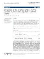
The power loss (also known as specific absorption rate
SAR) of the prepared fluid particles, simulated as function
of AC frequency in the range of 100–900 kHz, is plotted
and is presented in Fig. 7. The results obtained are con-
sistent with the earlier calculations carried out by Okawa
et al. [27], where the power loss for varied sizes is evalu-
ated at a noninvasive frequency 120 kHz. Zhang et.al [32]
has reported the SAR variation with particle size at still
lower frequency of 55 kHz. The applied frequency for
maximum power loss depends on the magnetic diameter
where the Neel mechanism alone is considered. It is
reported that the optimum size for noninvasive frequencies
lies around 12–14 nm [27]. Recent simulations [33] show
that the power dissipation at an applied frequency of
800 kHz is 80 W/g and for 200 kHz, 10 W/g for an applied
field strength of 200 Oe. In this study, the corresponding
values of power loss are 330 W/g and 20 W/g, respec-
tively. Li et al. [31] studied the variation of SAR with the
Fig. 6 Field induced optical absorbance for the aqueous ferrofluid in
two different polarizations
Fig. 7 Power loss spectrum as a function of AC frequency for the
ferrofluid
1710 Nanoscale Res Lett (2010) 5:1706–1711
123
viscosity of the fluid in which an increase in SAR with
viscosity was reported till twice the viscosity of water.
However, at 55 kHz and 200 Oe, for water suspended
fluids, they obtained a power loss of 57 W/g. It is seen
from Fig. 7 that the power loss increases with frequency.
The optimum frequency for required heat generation could
be selected on the basis of the actual experimental condi-
tion where magnetic hyperthermia needs to be performed.
Recent studies on cytotoxicity of these ferrofluids indi-
cate that they are highly biocompatible. The cell viability
test conducted via well-established 3-(4,5-dimethylthia-
zole-2-yl)-2,5-diphenyltetrazolium chloride MTT assay
[34] on He La cells in vitro shows viability up to a loading
of 10 micrograms per ml of cell.
Conclusions
Highly stable aqueous ferrofluid of SPIONs with citric acid
as surfactant has been successfully synthesized by con-
trolled chemical co-precipitation method. The structural
investigation by XRD and TEM shows good toning in
respect of the particle size. The magnetic analysis shows
that the nanoparticles are superparamagnetic in nature. The
fluid as such is typically a noninteracting ensemble of
nanoparticles which is evident from the magneto-optical
measurement. The saturation magnetization of the prepared
fluid is suitable for various biomedical applications espe-
cially for magneto hyperthermia. Based on the magnetic
measurements, the power dissipation in an alternating
magnetic field of 500 gauss as a function of applied fre-
quency is calculated.
Acknowledgments Authors thank Dr. Ildico Guhr and Prof.
G. Schatz University of Konstanz for SQUID measurements, and
DST-DAAD personnel exchange programme. MRA thanks AICTE
sponsored project ‘‘Center for Ferrofluids’’. RMAP (ID.379) and TNN
(ID: 434) acknowledge CSIR and VS thanks UGC RFSMS for
research fellowships.
Open Access This article is distributed under the terms of the
Creative Commons Attribution Noncommercial License which per-
mits any noncommercial use, distribution, and reproduction in any
medium, provided the original author(s) and source are credited.
References
1. R.E. Rosensweig, Ferrohydrodynamics (Cambridge University
Press, New York, 1985)
2. R.P. Castillejos, J.A. Plaza, J. Esteve, P. Losantos, M.C. Acero,
C. Cane, F. Serra-Mestres, Sens Actuators 84, 176 (2000)
3. C. Scherer, A. Figero, M. Neto, Braz. J. Phys 35(3), 718 (2005)
4. D.K. Kim, M. Mikhaylova, F.H. Wang, J. Kehr, B. Bjelke, Y.
Zhang, T. Tsakalakos, M. Muhammed, Chem. Mater. 15, 4343
(2003)
5. N.A. Brusnetsov, V.V. Gogosov, T.N. Brusnetsova, A.V. Sergeev,
N.Y. Jurchenko, A. Kuznetsov, O.A. Kuznetsov, L.I. Shumakov,
J. Magn. Magn. Mater. 225, 113 (2001)
6. D.L. Holligan, G.T. Gillies, J.P. Dailey, Nanotechnology 14, 661
(2003)
7. T.Y. Liu, S.H. Hu, T.Y. Liu, D. Liu, S.Y. Chen, Langmuir 22,
5974 (2006)
8. J.F. Lutz, S. Stiller, A. Hoth, L. Kaufner, U. Pison, R. Cartier,
Biomacromolecules 7, 3132 (2006)
9. C.C. Berry, A.S.G. Curtis, J. Phys. D Appl. Phys. 36, R198
(2003)
10. M. Babincova, V. Altanerova, C. Altaner, C. Bergeman,
P. Babinec, IEEE Trans. Nanobioscience 7, 15 (2008)
11. P. Tartaj, M.P. Morales, S. Veintemillas-Verdaguer, T. Gonzalez-
Carreno, C.J. Serna, J. Phys. D Appl. Phys. 36, R182 (2003)
12. A. Petri-Fink, H. Hofmann, IEEE Trans. Nanobioscience 6(4),
289 (2007)
13. Q.A. Pankhurst, J. Connolly, S.K. Jones, J. Dobson, J. Phys.
D Appl. Phys. 36, R167 (2003)
14. R. Hiergeist, W. Andra
¨
, N. Buske, R. Hergt, I. Hilger, U. Richter,
W. Kaiser, J Magn.Magn.Mater. 201, 420 (1999)
15. J. Giri, P. Pradhan, T. Sriharsha, D. Bahadur, J. Appl. Phys 97,
10Q916 (2005)
16. Y. Jun, J. Seo, J. Cheon, Acc. Chem. Res. 41, 179 (2008)
17. E. Dubois, V. Cabuil, F. Boue, R. Perzynski, J. Chem. Phys. 111,
7147 (2001)
18. J. de Vincente, A.V. Delgado, R.C. Plaza, J.D.G. Duran,
F. Gonsalez-Caballero, Langmuir 16, 7954 (2000)
19. L.F. Pavon, O.K. Okamoto, Einstein 5, 74 (2007)
20. B.D. Cullity, S.R. Stock, Elements of X-ray diffraction (Prentice
Hall, New Jersey, 2001)
21. T.N. Narayanan, A.P. Reena Mary, M.M. Shajumon, L. Ci, P.M.
Ajayan, M.R. Anantharaman, Nanotechnology 20, 055607 (2009)
22. S.J. Lee, J.R. Jeong, S.C. Shin, J.C. Kim, J.D. Kim, J. Magn.
Magn. Mater. 282, 147 (2004)
23. X. Battle, A. Labarta, J. Phys. D Appl. Phys. 35, R15 (2002)
24. A.G. Roca, M.P. Morales, K. O’Grandy, C.J. Serna, Nanotech-
nology 17, 2783 (2006)
25. J. Dai, J.Q. Wang, C. Sangregorio, J. Fang, E. Carpenter, J. Tang,
J. Appl. Phys. 87, 7397 (2000)
26. Q. Zhang, J. Wang, H. Zhu, J.Appl.Phys. 78
, 3999 (1995)
27. K. Okawa, M. Sekine, M. Maeda, M. Tada, M. Abe, N. Mats-
ushida, K. Nishio, H. Handa, J. Appl. Phys 99, 08102 (2006)
28. R.E. Rosenswieg, J. Magn. Magn. Mater 252, 370 (2002)
29. B.D. Cullity, Introduction to Magnetic materials Philippines
(Addison Wesley, London, 1972)
30. Y.Z. Li, C.G. Hong, M.W. Xu, J. Magn. Magn. Mater 311,
228–233 (2007)
31. M. Ma, Y. Wu, J. Zhou, Y. Sun, Y. Zhang, N. Gu, J. Magn.
Magn. Mater. 268, 33 (2004)
32. L.Y. Zhang, Y.H. Dou, L. Zhang, H.C. Gu, Chin. Phys. Lett. 24,
483 (2007)
33. S. Purushotham, R.V. Ramanujan, J. Appl. Phys. 107, 114701
(2010)
34. R. Gopikrishnan, K. Zhang, P. Ravichandran, S. Baluchamy, V.
Ramesh, S. Biradar, P. Ramesh, J. Pradhan, J.C. Hall, A.K.
Pradhan, G.T. Ramesh, Nano-Micro Lett. 2, 27–30 (2010)
Nanoscale Res Lett (2010) 5:1706–1711 1711
123