Báo cáo hóa học: " One-Pot Synthesis of Biocompatible CdSe/CdS Quantum Dots and Their Applications as Fluorescent Biological Labels" doc
Bạn đang xem bản rút gọn của tài liệu. Xem và tải ngay bản đầy đủ của tài liệu tại đây (330.54 KB, 5 trang )
NANO EXPRESS Open Access
One-Pot Synthesis of Biocompatible CdSe/CdS
Quantum Dots and Their Applications as
Fluorescent Biological Labels
Chuanxin Zhai
1
, Hui Zhang
1
, Ning Du
1
, Bingdi Chen
1
, Hai Huang
2
, Yulian Wu
2
, Deren Yang
1*
Abstract
We developed a novel one-pot polyol approach for the synthesis of biocompatible CdSe quantum dots (QDs)
using poly(acrylic acid) (PAA) as a capping ligand at 240°C. The morphological and structural characterization
confirmed the formation of biocompatible and monodisperse CdSe QDs with several nanometers in size. The
encapsulation of CdS thin layers on the surface of CdSe QDs (CdSe/CdS core–shell QDs) was used for passivating
the defect emission (650 nm) and enhancing the fluorescent quantum yields up to 30% of band-to-band emission
(530–600 nm). Moreover, the PL emission peak of CdSe/CdS core–shell QDs could be tuned from 530 to 600 nm
by the size of CdSe core. The as-prepared CdSe/CdS core–shell QDs with small size, well water solubility, good
monodispersity, and bright PL emission showed high performance as fluorescent cell labels in vitro. The viability of
QDs-labeled 293T cells was evaluated using a 3-(4,5-dimethylthiazol)-2-diphenyltertrazolium bromide (MTT) assay.
The results showed the satisfactory (>80%) biocompatibility of as-synthesized PAA-capped QDs at the Cd
concentration of 15 μg/ml.
Introduction
Fluorescent semiconductor nanocrystals, also known a s
one kind of quantum dots (QDs), are of conside rable
interest and under intensive research as b iological labels
either in vitro or in vivo, not only because of their
bright, photostable fluorescence but also because of the
broad excitation spectrum and narrow, size-controlled
emission, which allows multi-color imaging [1,2].
Among them, cadmium selenide (CdSe) QDs have
become one family of the most extensively studied fluor-
escent semiconductor nanocrystals due to their suitable
and tunable band gap throughout the visible spectrum
[3]. The high-temperature chemica l reaction was a well-
known approach for the synthesis of highly crystalline
and monodisperse C dSe QDs with bright fluorescence
using organometallic or chelated cadmium and phos-
phine-coordinated selenium as precursors [4-6]. How-
ever, besides the use of expensive, toxic chemicals, the
as-received QDs were usually hydrophobic and must be
converted into water-soluble nanocrystals through sur-
face ligand exchanges [7] or encapsulat ions of polymers
[8] and thin silica l ayers [9] for biological applications.
The possible weight loss and decrease in quantum yields
are always unavoidable during the conversion [7].
Synthesis directly in water-soluble solvent has been
considered to be an alternative approach for circum-
venting the above-mentioned disadvantages. Recently,
great efforts have been employed to focus on the synth-
esis of hydrophilic CdSe QDs directly in water or
inverse micelles [10,11]. However, the crystal quality
and quantum yields of the as-synthesized QDs w ere
often limited, mainly due to the low reaction tempera-
ture [11]. The polyol method provided a promising
high-temperature hydrophilic system for one-pot synth-
esis of biocompatible QDs, which combined the advan-
tages of the two above-mentioned methods [12]. In last
two decades, it has been widely applied to fabricate
water-soluble particles of various materials with sub-
micrometer size includi ng metals [13], alloys [14], metal
oxi des [15], and metal sulfides [16]. However, obtaini ng
biocompatible QDs with a very small size, high crystal
quality, and quantum yields by polyol approach still
remains a tremendous challenge [12].
* Correspondence:
1
State Key Lab of Silicon Materials and Department of Materials Science and
Engineering, Zhejiang University, 310027, Hangzhou, People’s Republic of
China.
Full list of author information is available at the end of the article
Zhai et al. Nanoscale Res Lett 2011, 6:31
/>© 2010 Zhai et al. This is an Open Acce ss article distributed under the terms of the Creative Commons Attribution License
(http://creativecomm ons.org/licenses/by/2.0), which permits unrestricted use, distribution, and reproduction in any medium,
provided the original work is properly cited.
Herein, we have developed a novel one-pot pol yol
approach for the synthesis of water-soluble CdSe and
CdSe/CdS type-I core–shell QDs with several nan-
ometers in size. The one-pot method can provide high-
quality biocompatible quantum dots without using
expensive phosphines and complicated surface modifica-
tion, which takes the advantages of simpleness, low cost,
and green precursor. Moreover, the as-received QDs
show the tunable and bright PL emission with high
quantum yields and high performance as fluorescent
biological labels in vitro.
Experimental Section
Synthesis of CdSe QDs
In a typical synthesis, 1 g poly(acrylic acid) (PAA, MW =
1,800) and 0.5 mmol cadmium acetate (Cd(AC)
2
)were
subsequently dissolved into 20 ml triethylene glycol
(TREG), which were then heated to 200°C under Ar flow.
After 30 min, the solution was cooled to room t empera-
ture, and 19 mg of Se powder was added. Finally, the
mixture was heated to 240°C and kept for a certain
period of time such as 1, 5, 60, and 120 min.
Synthesis of CdSe/CdS QDs
As sulfur source, 19 mg thiourea was added into the
above-mentioned CdSe precursor solution. The redundant
Cd(AC)
2
in the CdSe precursor solution was used as cad-
mium source. Subsequently, the mixture was heated to
160°C in 1 h. After the reaction for 2 h, the soluti on was
quickly cooled to room temperature and precipitated by
ethyl acetate. The re sultant solid products were further
purified by dialysis and ultrafiltration for cell imaging.
In Vitro Cell Viability and Cell Imaging
Human embryonal kidney cell line 293T cells (ATCC
CRL-11268, American Type Culture Collection,
Manassas, VA) were cultured in a high-glucose Dulbec-
co’s modified Eagle’s medium (H-DMEM; Gibco, Grand
Island, NY) containing 10% f etal bovine serum (FBS;
Gibco) and 1% penicillin/streptomycin (Gibco) at 37°C
under 5% CO
2
condition. The cells were subcultured
every 3 days. Viability of QDs-labeled 293T cells was
evaluated using an MTT assay (SIGMA, St. Louis, MO).
Cells were seeded in 96-well tissue culture plates at a
density of 8 × 10
4
cells/well. After 24 h, the culture med-
ium was repl aced with 200 μL of the as-synthesized QDs
containing differen t concentrations of nanoparticles.
After 24-h labeling and washing, 20 μL of a solution of
MTT (5 mg/mL in PBS) was added to each well, and
assay was performed at specific time intervals. The absor-
bance of the formazen product was then measured at a
wavelength of 570 nm. Four groups of MTT tests were
done for each quantum dots concentration. The values of
MTT assay of labeled cells were expressed as the
percentage of corresponding control cells. For cell ima-
ging, 293T cells (2 × 10
4
cells/24-well plates) were grown
on coverslips for 24 h and then incubated with PAA-
capped CdSe/CdS QDs (Cd concentration of 5 μg/mL,
measured by atomic absorption spectrophotomet er), at
5% CO
2
at 37°C for 4 h. The cells were washed thrice
with PBS and analyzed with confocal microscopy
afterward.
Characterization
The products were characterized by X-ray powder diffrac-
tion (XRD) using a Rigaku D/max-ga X-ray diffractometer
with graphite monochromatized CuKa radiation (l =
1.54178Å) . The transmission electron microscopy (TEM)
with energy-dispersive X-ray (EDX) and high-resolution
transmission electron microscopy (HRTEM) was applied
to determine the morphology and structure. The photolu-
minescence (PL) examination was performed on a detec-
tor PMT and ACTON SpectraPro 2500i using a He–Cd
laser with a 325-nm wavelength as the excitation source.
The confocal fluorescence images were obtained with a
laser scanning confocal microscope (LEICA TCS SP2).
Results and Discussion
In the present study, Cd(Ac)
2
and Se powder are selected
as source. Triethylene glycol (TREG) is used as the sol-
vent due to its good hydrophilic feature and hig h bo iling
point (288°C). A water-soluble and biocompatible poly-
mer with carboxylic f unctional groups, PAA, is selected
as a capping ligand for controlling the crystal quality o f
QDs such as size, size distribution, and crystallinity by
the formation of the chelated cadmium precursors.
Moreover, since PAA is considered as a biocompatible
polymer [8], we believe that the PAA could absorb on
the surface of the QDs through the synthetic process,
which may be advantageous for improving the hydrophi-
licity and biocompatibility as fluorescent biological labels.
Due to the lo w solubility of selenium powder in TREG,
no reaction had been observed at low temperature.
When the temperature rose around the melting point of
selenium powder (221°C), it was quickly reduced in the
polyol system with reductive h ydroxide groups and
reacted with the carboxylate precursors forming numer-
ous of nuclei. The explosive nucleation brings a narrow
size distribution and also reduces the tendency of Ost-
wald ripening [17]. The nanocrystals grow larger as the
extension of reaction time, causing the redshift of both
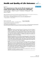
absorption and emission spectra. F igure 1a shows the
ultraviolet–visible (UV–vis) absorption and photolumi-
nescence (PL) emission spectra of the PAA-capped CdSe
QDs as a function of reaction time. With the extension
of the reaction time, the CdSe QDs gradually grow up,
and their PL emission peak can be tuned from 520 to
586 nm. The full width at half maximum (FWHM) of the
Zhai et al. Nanoscale Res Lett 2011, 6:31
/>Page 2 of 5
PL spectra is around 50 nm. Figure 1b shows the typical
TEM image of the as-synthesized PAA-capped CdSe QDs
with the absorption peak around 540 nm. The average
core size of as-prepared CdSe QDs calculated from the
statistical results was about 2.8 nm. The HRTEM image
(Figure 1c) and SAED pattern (Figure 1d) confirm the for-
mation of the c ubic CdSe QDs by PAA-assisted polyol
appr oach . T he XRD pattern of as-synthesized CdSe QD s
shown in Figure 2a also shares same crystal structure with
zinc-blende CdSe (JCPDS file No. 19-0191).
InthePLspectraoftheCdSeQDs,thereisabroad
emission band originating from the surface trap sites
besides the band-to-band emission, especially in the
samples with smaller size, which decreases not only the
monochromaticity of the fluorescence but also the
quantum yields o f the QDs. In our case, the quantum
yields of the as-synthesized CdSe cores are around
2–3%. In order to passivate their surface trap sites and
enhance the quantum yields, a consequent polyol
approach was developed to fabricate type-I CdSe/CdS
core–shell QDs by subsequently growing a thin CdS
layer on the surface of the CdSe QDs using thiourea as
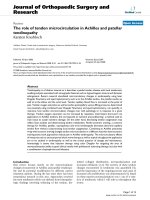
sulfur source at 160°C. The XRD and EDX analysis were
used to reveal the f ormation of CdSe/CdS core–shell
QDs (Figure 2). In comparison with the XRD patte rn of
the CdSe QDs, the three characteristic diffraction peaks
of the CdSe/CdS core–shell QDs (Figure 2a) only shift
to larger angles and locate between those of the CdSe
and CdS cubic pha se, which demons trate the formation
oftheCdSshellonthesurfaceoftheCdSeQDs[18].
The formation of the CdS shell is further supported by
the EDX analysis (Figure 2b). The strong peaks f or S,
Se, and Cd elements in the spectrum confirm the for-
mation of the CdSe/CdS core–shell QDs.
Figure 3a shows a comparison of the PL spectra of the as-
prepared CdSe and CdSe/CdS core–shell QDs. As
observed, the PL emission at 650 nm originating from trap
sites was completely inhibited by coating a thin CdS layer
on the surface of CdSe QDs due to the surface passivation
[19]. Meanwhile, the brighter luminescence was achieved.
Moreover, the PL emission originating from the band to
band of the CdSe/CdS core–shell QDs can be tuned from
531 to 590 nm by the size of CdSe (Figure 3b) with FWHM
of 40–60 nm and a quantum yield of about 30% compared
with Rhodamine B [20], which has been significantly
improved comparing with the CdSe cores. The PAA-
capped QDs are stable for several months without precipi-
tation in aqueous dispersion. Therefore, the PAA-capped
CdSe/CdS core–shell QDs with small size, well water solu-
bility, good monodispersity, and bright PL emission show
the promising applications as fluorescent b iological labels.
Human embryonal kidney cell line is chosen as typical
kind of human cells to demonstrate the promising appli-
cations as fluorescent biological labels. MTT assays were
performed to evalu ate the cytoto xicity corresponding to
the biocompatibility of PAA-capped QDs on 293T cells.
Four groups of MTT tests were done for each quantum
dots concentration. In Figure 4, the cell viability shows
Figure 1 a Temporal evolution of UV–vis absorption (dash) and
PL (solid) spectra of the as-prepared PAA-capped CdSe QDs
dispersed in water; b TEM and size distribution histogram;
c HRTEM; and d SAED images of the as-synthesized PAA-capped
CdSe nanocrystals (the absorption peak around 540 nm).
Figure 2 a XRD patterns of plain CdSe and CdSe/CdS core/shell
nanocrystals. b EDX spectrum of the CdSe/CdS core/shell
nanocrystals prepared on a copper grid.
Figure 3 a PL spectra of PAA-capped CdSe (dash)(4timeof
original intensity) and CdSe/CdS (solid) nanocrystals. b
Normalized fluorescence emission spectrum of CdSe/CdS QDs with
various size.
Zhai et al. Nanoscale Res Lett 2011, 6:31
/>Page 3 of 5
the average cell viability of four tests, while the e rror
bars show the standard deviations. Satisfactory (>80%)
biocompatibility of as-synthesized PAA-capped QDs is
achieved at a particle concentration below 15 μg Cd/mL
in 293T cell lines. No statistical difference in viability is
evident with PAA-QDs-labeled cells and untreated cells
for 24 h at the concentration of 7 μg Cd/mL. It is well
known that without proper surface modification the Cd-
related quantum dots will cause severe cell damage after
24 h. MTT analysis showed that the cell viability of
MCF-7 cells was bel ow 50% after 24-h e xposure to QDs
(10 mg mL
-1
) capped by mercaptopropionic acid [21].
Uncapped QDs were even more toxic [22]. As a result,
further surface modification processes such a s PEGyla-
tion are often taken place to enhance the biocompa tibil-
ity [23]. In our case, PAA absorbed on the surface
improves the hydrophilicity and biocompatibility of the
nanoparticles. The cytotoxicity tests indicate that, with-
out further surface modification, the as-synthesized
PAA-capped QDs show good biocompatibility as biologi-
cal lab els, which i s comparable with PEGylated nanopar-
ticles [23]. It turns out that the PAA modification during
the “ one-pot ” synthesis is both simple and effective.
For their in vitro cell labe ling studies, the cultured
human embryonal kidney cell line 293T cells were
incubated with the PAA-capped CdSe/CdS core–shell
QDs (l
em max
= 559 nm , about 3 nm in size) with the
concentration o f 5 μg/mL for 4 h at 37°C. After 4 h, the
cells were washed thrice with PBS to remove extra nano-
particles that were not uptaken b y the cells and imaged
using a laser scanning confocal microscope. Figure 5
shows t he typical labeling ima ges of 293T cells with
PAA-capped CdSe/CdS core–shell QDs. From these
images, the bright green optical signal can be clearly
observed from the cell interior. The result demonstrated
that the as-synthesized quantum dots can be quickly
uptaken by the 293T cells within 4 h. Moreover, we did
not observe any signs of morphological damage to the
cells after the treatment with PAA-capped CdSe/CdS
core–shell QDs. This preliminary result indicates that the
as-prepared QDs had promising applications as fluores-
cent biological labels.
Conclusions
In summary, we have developed a novel, cost-effective,
and environment friendly polyol approach for the one-
pot synthesis of biocompatible CdSe and CdSe/CdS
core–shell QDs with several nanometers in size, good
biocompatibility, good monodispersity, strong, and
tunable fluorescent emission. The as-synthesized PAA-
capped CdSe/CdS core–shell QDs exhibited high perfor-
mance as f luorescent cell labels i n vitro and thus
promising applications.
Acknowledgements
The authors would like to appreciate the financial supports from 973 Project
(No. 2007CB613403), NSFC (No. 50802086, 30672072), ZiJin Project, ZJPNSFC
(Y407138), the Doctoral Program of the Ministry of Education of China (No.
20070335014), Zhejiang Innovation Program for Graduates (2008022), and
the foundation of 2008DFR50250.
Author details
1
State Key Lab of Silicon Materials and Department of Materials Science and
Engineering, Zhejiang University, 310027, Hangzhou, People’s Republic of
China.
2
Department of Surgery, the Second Affiliated Hospital, School of
Medicine, Zhejiang University, 310027, Hangzhou, People’s Republic of China.
Figure 4 Cell viability of Human embryonal kidney cell line
293T cells labeled with different concentration of QDs (mg Cd
per mL) for 24 h at 37°C as measured by an MTT assay. The
error bars show the standard deviations.
Figure 5 Confocal microscopic visualization of Human embryonal kid ney cell line 293T cells treated with PAA-capped green-emitting
CdSe/CdS QDs (l
em max
= 559 nm, about 3 nm in size) with the concentration of 5 μg/mL for 4 h at 37°C. From left to right, the panels
show the transmission image, luminescence image, and an overlay of the two.
Zhai et al. Nanoscale Res Lett 2011, 6:31
/>Page 4 of 5
Received: 23 July 2010 Accepted: 23 August 2010
Published: 17 September 2010
References
1. Chan WCW, Nie S: Science 1998, 281:2016.
2. Alivisatos AP: Nat Biotechnol 2004, 22:47.
3. Alivisatos AP: Science 1996, 271:933.
4. Murray CB, Noms DJ, Bawendi MG: J Am Chem Soc 1993, 115:8706.
5. Peng X, Wickham J, Alivisatos AP: J Am Chem Soc 1998, 120:5343.
6. Peng ZA, Peng X: J Am Chem Soc 2001, 123:183.
7. Wang Q, Xu Y, Zhao X, Chang Y, Liu Y, Jiang L, Sharma J, Seo D, Yan H:
J Am Chem Soc 2007, 129:6380.
8. Zhang T, Ge J, Hu Y, Yin Y: Nano Lett 2007, 7:3203.
9. Yong K, Roy I, Pudavar HE, Bergey EJ, Tramposch KM, Swihart MT,
Prasad PN: Adv Mater 2008, 20:1.
10. Weller H: Adv Mater 1993, 5:88.
11. Rogach AL, Kornowski A, Eychmüller A, Weller H: J Phys Chem B 1998,
102:8360.
12. Feldmann C: Solid State Sci 2005, 7:868.
13. Sun Y, Xia Y: Adv Mater 2002, 14:833.
14. Roychowdhury C, Matsumoto F, Mutolo PF, Abruna HD, DiSalvo FJ: Chem
Mater 2005, 17:5871.
15. Orel ZC, Anzlovar A, Drazic G, Zigon M: Cryst Growth Des 2007, 7:453.
16. Feldmann C, Jungk H: Angew Chem Int Ed 2001, 40:359.
17. Yang AY, Wu H, Williams RK, Cao CY: Angew Chem Int Ed 2005, 44:6712.
18. Talapin DV, Rogach AL, Kornowski A, Haase M, Weller H: Nano Lett 2007,
1:207.
19. Peng X, Schlamp MC, Kadavanich AV, Alivisatos AP: J Am Chem Soc 1997,
119:7019.
20. Pradhan N, Peng X: J Am Chem Soc 2007, 129
:3339.
21. Lovri J, Cho SJ, Winnik FM, Maysinger D: Chem Biol 2005, 12:1227.
22. Li KG, Chen JT, Bai SS, Wen X, Song SY, Yu Q, Li J, Wang YQ: Toxicol In Vitro
2009, 23:1007.
23. Dif A, Boulmedais F, Pinot M, Roullier V, Baudy-Floch M, Coquelle FM,
Clarke S, Neveu P, Vignaux F, Borgne RL, Dahan M, Gueroui Z, Marchi-
Artzner V: J Am Chem Soc 2009, 131:14738.
doi:10.1007/s11671-010-9774-z
Cite this article as: Zhai et al.: One-Pot Synthesis of Biocompatible
CdSe/CdS Quantum Dots and Their Applications as Fluorescent
Biological Labels. Nanoscale Res Lett 2011 6:31.
Submit your manuscript to a
journal and benefi t from:
7 Convenient online submission
7 Rigorous peer review
7 Immediate publication on acceptance
7 Open access: articles freely available online
7 High visibility within the fi eld
7 Retaining the copyright to your article
Submit your next manuscript at 7 springeropen.com
Zhai et al. Nanoscale Res Lett 2011, 6:31
/>Page 5 of 5