Báo cáo hóa học: " Nanospiral Formation by Droplet Drying: One Molecule at a Time" ppt
Bạn đang xem bản rút gọn của tài liệu. Xem và tải ngay bản đầy đủ của tài liệu tại đây (1.04 MB, 8 trang )
NANO EXPRESS Open Access
Nanospiral Formation by Droplet Drying:
One Molecule at a Time
Lei Wan, Li Li, Guangzhao Mao
*
Abstract
We have created nanospirals by self-assembly during droplet evaporation. The nanospirals, 60–70 nm in diameter,
formed when solvent mixtures of methanol and m-cresol were used. In contrast, spin coating using only methanol
as the solvent produced epitaxial films of stripe nanopatterns and using only m-cresol disordered structure. Due to
the disparity in vapor pressure between the two solvents, droplets of m-cresol solution remaining on the substrate
serve as templates for the self-assembly of carboxylic acid molecules, which in turn allows the visualization of
solution droplet evaporation one molecule at a time.
Introduction
Patterns formed by s olvent evaporation are relevant to
various coating processes as well as patterning technol-
ogy. In capturing the molecular process of an evaporat-
ing droplet, this work demonstrates the possibility to
further modulate dewetting patterns by amphiphiles
capable of self-assembly. Self-assembly as an alternative
to lithography has the potential to generate reconfigur-
able nanostructures [1-3]. Surfactants/amphiphiles are
the simplest molecules to self-assemble into complex yet
often predictable structures and phases. An interface
perturbs and sometimes dominates the self-assembling
behavior of amphiphiles. A well-known example of sub-
strate-dominated self-assembly is the epitaxial stripe
nanopatterns formed by alkanes and alkane derivatives
on highly oriented pyrolytic graphite (HOPG) [4-10].
The 1,3-methylene group distance, 0.251 nm, of all-
trans alkyl chains matches the distance of the next near-
est nei ghbor of the HOPG lattice, 0.246 nm, along, e.g.,
the [11
2
0] crystallographic direction. The he ad-to -head
arrangement gives rise to the stripe nanopattern whose
periodicity is 1 × or 2 × the molecular chain length.
Such nanopatterns serve as model templates for the
study of site-specific adsorption, alignment, assembly,
and reaction of small molecules [8,9,11,12] as well as
macromolecules [13-16].
In an earlier example, we disrupted the stripe nano-
pattern of eicosanoic acid (C
20
A) using mercaptounde-
canoic acid cap ped cadmium sulfide nano particles. C
20
A
nanorods with 1.0 nm in thickness and 5.4 nm in width
are nucleated directly on the nanoparticle to produce
nanoparticle/nanorod hybr id structure [17]. Here, we
present another method to perturb the epitaxial interac-
tion between long-chain carboxylic acids and HOPG
and to create spiral nanopatterns by adding a co-so lvent
to the spin coating solution. We propose that the curved
nanostructure is formed at the receding solid/liquid/
vapor contact line of an evaporating solution droplet,
and it traces the entire droplet evaporation process at
the molecular scale.
Recently, a number of methods have been reported for
making circular nanostructures. Nanorings have been
generated by lithography (microcontact printing [18],
electron beam [19], and AFM tips [20]), template-based
synthesis (using droplets [21], viruses [22], and DNA
[23]), self-assembly [24-27], selective dewetting on pat-
terned surfaces [28-30], and evaporation-driven dewet-
ting [27,31-33]. There have been fewer reports on
nanospirals [34-37]. The scientific interests for nanor-
ings range from quantum rings, whose connected geo-
metry at the nanoscale can trap “persistent currents”
[38-41], to biomimetic light-harvesting complexes
[31,42,43] and DNA microarrays for high -throughput
DNA mapping [44,45]. The nanoring structure is also
interesting because of its resemblance of the toroid
structure of condensed DNA [26].
* Correspondence:
Department of Chemical Engineering and Materials Science, Wayne State
University, Detroit, Michigan 48202, USA.
Wan et al. Nanoscale Res Lett 2011, 6:49
/>© 2010 Wan et al. This is an Open Access article distributed under the terms of the Creative Commons Attribution License
(http://creativecommons.o rg/licenses/by/2.0), which permits unrestricted use, distribu tion, and reproduction in any medium, provided
the ori ginal work is properly cited.
Experimental Section
Materials
Long-chain carboxylic acids including hexadecanoic acid
(C
16
A, Aldrich, 99%), octadecanoic acid (C
18
A,
Fluka, ≥ 99.5%), eicosanoic acid (C
20
A, Sigma, ≥99% ),
docosanoic acid (C
22
A, Aldrich, 99%), tetracosanoic acid
(C
24
A, Fluka, ≥99.0%), and hexacosanoic acid (C
26
A,
Sigma, ≥95%) were used. Solvents used were m-cresol
(Aldrich, 97%), methanol (Mallinckrodt Chemicals,
100%), ethanol (Pharmco, 100%), iso-propanol (Fisher
Scientific, 100%), and sec-butanol (Fisher Scientific,
99.3%). HOPG (grade ZYB) was purchased from Mikro-
Masch. All chemicals were used as received.
Sample Preparation
Carboxylic acids were dissolved in a primary alcoholic
solvent or a binary solvent of alcohol and m-cresol to
yield a final concentration of 0.2–0.4 mM. HOPG
was freshly cle aved by adhesive tapes. The spin coating
(PM101DT-R485 photoresist spinner, Headway
Research) was conducted at room temperature in ambi-
ent air with relati ve humidit y < 40%. A volume of
100 μL of the solution was dispensed onto HOPG and
spun at 3,000 rpm for 60 s. The samples were dried in
air for 20 min or longer.
AFM Characterization
The spin-coated samples were imaged using Nano-
scope III Multimode AFM equipped with a piezoelec-
tric scanner with a maximum scan range of 10 μm
( x and y)and2.5μm(z) from VEECO/Digital Instru-
ments. Height, amplitude, and phase images were
obtained in Tapping Mode (oscillation frequency ~
250–300 kHz) in ambient atmosphere using etched
silicon probes (ACT, NanoScience) with nominal
radius of curvature <10 nm. The scan rate was
1–3 Hz. Integral and proportional gains were approxi-
mately 0.4 and 0.8, respectively. Only flattened height
images were shown. The films were u sually imaged
within minutes of film preparation. However, the
nanostructures were unchanged for at least 1 month
afterward when stored in ambient environment. The
contour length of the stripe was determined using the
WSxM 4.0 software.
Contact Angle Measurement
The contact angle was measured by an NRL contact
angle goniometer (Model 100, Rame-Hart) in the
laboratory atmosphere. One m-cresol droplet of 5 μL
was placed on the substrate and contact angles were
read on both sides of the droplet. Five droplets were
placed at various spots near the center of the sub-
strate, and contact angles were averaged with an error
of ±3°.
Results and Discussion
The spin-coated samples of long-chain n-carboxylic
acids including hexadecanoic acid (C
16
A), octadecanoic
acid (C
18
A), eicosanoic acid (C
20
A), docosanoic acid
(C
22
A), tetracosanoic acid (C
24
A), and hexacosanoic acid
(C
26
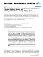
A) were imaged by AFM. When the carboxylic acids
were spin coated on HOPG from alcoholic solvents
including methanol, ethanol, iso-propanol, and sec-
butanol, only epitaxial stripe nanopatterns were formed
(Figure 1). The periodicity of the nanopatterns is 4.5 nm
75 nm
(a) C
16
A
75 nm
(b) C
18
A
75 nm
(c)
C
20
A
75 nm
(d) C
22
A
75 nm75 nm
(e) C
24
A
(f) C
26
A
(g)
A single H-
bonded
dimer stripe
Figure 1 a–f AFM height images of carbox ylic acid monolayers
spin coated from alcoholic solvents. The z range is 2 nm for a–c
and 3 nm for e–f. g Molecular packing in 2-D stripe nanopattern of
carboxylic acid monolayer on HOPG. The structure is based on C
18
A
B-form crystal viewed along the a axis. Monoclinic P2
1
/a crystal
structure with a = 5.591 Å, b = 7.704 Å, c = 43.990 Å, and b = 94.6°.
Wan et al. Nanoscale Res Lett 2011, 6:49
/>Page 2 of 8
for C
16
A, 5.1 nm for C
18
A, 5.6 nm for C
20
A, 6.1 nm for
C
22
A, 6.6 nm for C
24
A, and 7.0 nm for C
26
A. The peri-
odicity is slightly larger than 2 × molecular chain length.
The molecular chain length of saturated carboxylic acids
on HOPG can be calculated by the following formula:
1
2
1
2
number of C atoms per chain number of Oatoms per carboxyl group+
(
))
× 0 246.nm
.
The stripe thickness, 0.3 ± 0.1 nm, is consistent with the
coplanar packing model in which the carbon skeleton
plane of the carboxylic acid molecule lies parallel to the
HOPG basal plane. The orthogonal stripe domains dis-
played the threefold symmetry of the graphite lattice.
It is concluded that the carboxylic acids adopt the
persistent epitaxial arrangement on H OPG [4,7,46-49]
during spin coating, whose packing structure is illu-
strated by Figure 1g.
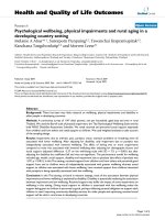
When m-cresol was used as the solvent, largely amor-
phous carboxylic acid films were formed (Figure 2).
A closer examination of the AFM images showed
ordered domains of C
20
A molecules interspersed in the
amorphous film. Clearly, m-cresol does not favor car-
boxylic acid self-assembly either because it is a poor
recrystallization solvent for carboxylic acids or because
it competes for the adsorption sites on HOPG due to its
aromatic group.
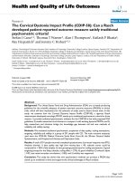
When m-cresol was gradually added to methanol, we
obtained new nanostructures in the spin-coated films.
Figure 3 shows the typical C
20
A film structures at differ-
ent methanol to m-cresol volume ratios: 25, 10, 5, 2, and
1, respectively. With increasing m-cresol content, the
film structure changed from highly ordered stripe nano-
patterns associated with methanol to circular nanostruc-
tures and to disordered phase associated with m-cresol.
The film coverage increased with increasing m-cresol
amount. With trace amount of m-cresol, t he stripe
phase was modified by the presence of isolated curved
stripes, or partial spirals, that were located either at the
edge or on top of the stripe nanopattern (Figure 3a).
These spirals mark the locations of partitioned m-cresol-
rich phase upon solvent evaporation. The curved feature
became more prominent with increasing m-cresol
amount (Figure 3b, b’). The circular stripes are on top
of the straight ones. Increasing coverage of the circular
feature was obtained with increasing m-cresol content
(Figure 3c, c’). The circles are uniform in size with an
average outer diameter of ~70 nm. In addition to the
circles, a straight fiber-like feature is present whose
orientation is in registry with HOPG. Each fiber consists
ofbundlesofstripeswithheightof0.8±0.1nm.The
straight fiber structure resembles ribbons preceding dro-
plet formation upon reaching the Rayleigh instability
limit during dewetting [50,51]. As the ratio decreases to
2, the film became disordered with traces of circular
lines (Figure 3d, d’). More m-cresol resulted in thicker
amorphous f ilms (>1 nm) (Figure 3e, e’). At the edge of
the amorphous film, curves were observed as pointed by
the arrows in Figure 3e.
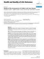
The circular nanopattern was observed on C
18
Aand
C
22
A (Figure 4) but not on longer chains. Less-defined
spirals were formed when ethanol, iso-propanol, or sec-
butanol instead of methanol was used as the primary
solvent (Figure 5). The boundary of the s piral became
less circular and more orthogonal. This is a result of
two completing templates— the droplet edge versus
HOPG basal plane. Less volatile solvents favor epitaxial
interaction between the alkyl chain and HOPG lattice.
AFM images at higher resolution using methanol to
m-cresol ratio of 10 reveal mole cular packing structure
in the circular nanopattern. Figure 6 provides examples
of spirals in inward clockwise (Figure 6a) and
R=5.6 nm
300 nm 50 nm
(a) (b)
Figure 2 AFM height images of carboxylic acid spin coated from m-cresol (a) C
20
A. b Selected area in (a). The periodicity was determined
by the corresponding 2-D FFT images. The z range is 5 nm for both images.
Wan et al. Nanoscale Res Lett 2011, 6:49
/>Page 3 of 8
counterclockwise rotations (Figure 6b). The arrows mark
the beginning and end of each spiral. We found roughly
equal numbers of clockwise and counterclockwise spir-
als. Self-assembled spirals usually involve chiral mole-
cules. Amphiphilic molecules with chiral centers are
capable of self-assembly into spirals in Langmuir
monolayers. The direction of the spirals depends on the
chir ality of the amphiphiles. In one study [52], intermo-
lecular H-bonds caused the neigh boring aromatic head-
groups to tilt and resulted in s piral formation from
achiral amphiphilic molecules in Langmuir monolayers.
Here, the chirality of the spirals is dictate d by the direc-
tion of unidirectional solvent evaporation.
Figure 6c–e shows multiple C
20
A spirals, partial spir-
als, and coexisting straight stripes. The spirals of C
18
A,
C
20
A, and C
22
A display a center-to-center distance of
5.1, 5.6, and 6.2 nm, respectively, which indicates that
the spiral is made of the same head-to-head dimer
arrangement as in the epitaxial stripes on HOPG. The
secti onal height analysis indi cates that the spirals have a
uniform height of 0.8 ± 0.1 nm. The straight stripes out-
side the spiral have the same height as the spirals while
thoseinsidetendtohavealowerheightof0.2–0.4 nm.
The lower height value suggests that the structure is
templatedonlybyHOPGinwhichthecarboxylicacid
carbon plane faces HOPG [4,53]. The higher height
value is consistent with crystalline structure that is not
templated by HOPG.
The spiral nanopattern with a bilayer periodicity sug-
gests that it is templated by precipitation crystallization
of carboxylic acids along the receding solid/liquid/vapor
interface of an evaporating droplet (Figure 7). In the
case of volatile fluid wetting the HOPG substrate, after
the outward flow to produce a smooth film, the last
stage of spin coating is dominated by solvent evapora-
tion [54,55]. The film thickness is a function of spin
speed f,initialviscosityν
0
, and evaporation rate e:
hf e∝
−23
0
13
13/
/
/
[54]. In our case, the high spin speed
combined with low s olution concentration resulted in
ultrathin films. When pure solvents were used, the AFM
images pointed to uniform thinning of the wetting film
until the complete removal of the solvent. The substrate
was covered by a uniform carboxylic acid film eithe r in
an ordered state from alcoholic solvents or disordered
state from m-cresol. When the mixed solvent was used,
dewetting occurred. Dewetting is believed to start from
holes followed by interconnected cellular rims and the
breakup of the rims into droplets [51]. Since methanol
has higher equilibrium vapor pressure (= 128 mmHg)
than m-creso l (<1 mmHg) at 25°C, methanol evaporates
much faster to yield the stripe layer on HOPG.
’
μ
μ
μ
μ
(a)
(b)
(c)
(d)
(f)
(b )
’
(c )
’
(d )
’
(f )
Figure 3 AFM height images of C
20
A film structures spin
coated from methanol and m-cresol with different methanol to
m-cresol volume ratios. The image on the right is an image with
higher resolution than the one to the left. The z range is 5 nm for
a–e and 4 nm for b’–e’.
Wan et al. Nanoscale Res Lett 2011, 6:49
/>Page 4 of 8
Figure 4 AFM height images of C
18
A(left)andC
22
A(right) film structures spin coated from methanol and m-cresol mixed solvent
(methanol: m-cresol = 10). The z range is 5 nm for both images.
Figure 5 AFM height images of C
20
A film structures spin coated from ethanol (a), iso-propanol (b), and sec-butanol (c) with ~10 vol%
m-cresol. The z range is 3 nm for (a) and (b) and 5 nm for (c).
Wan et al. Nanoscale Res Lett 2011, 6:49
/>Page 5 of 8
The remaining m-cresol breaks up into small droplets
and evaporates at a slower rate enabling molecular self-
assembly to proceed. The increase of spiral coverage with
increasing m-cresol content is consistent with the spiral
feature being associated with m-cr esol. Our results point
to the formation of very small and fairly uniform m-cre-
sol-rich droplets in the range of 60–70 nm in outermost
diameter (Figure 8). The uniform size of the spirals
points to a critical film thickness below which the film
breaks up into droplets. A rough estimate based on the
size of the nanospirals gives a critical film rupture thick-
ness of 4.3 ± 0.3 nm (the contact angle of saturated C
20
A
m-cresol droplets on HOPG covered by C
20
A nanostripes
is 15°).
The drying of solution droplets is described by the cof-
fee-stain mechanism [51,56-59]. The higher evaporation
rate at the pinned sessile convex droplet contact edge
causes convective capillary flow and precipitation of
solute at the edge. The capillary flow goes from the bulk
solution to the edge of the droplet in order to maintain
the spherical shape to counter evaporative losses [57].
0.7nm
0.4nm
0.7nm
100 nm
(e)
(d)(c)
(b)(a)
(f)
(f)
Figure 6 AFM height images of C
20
A spirals (a–e). The z range is
4 nm. f Sectional height analysis of the stripe height along the
dashed line.
m
-Cresol droplet
(b) Top view
60 nm
Droplet evaporation/
spiral growth direction
=
b = 0.77 nm
(a) Side view
(c) Side view
60 nm
HOPG
Figure 7 Schema tic mechanism of spiral formation. a m-Cresol
droplets as templates for the nanospiral pattern. b The
counterclockwise inward rotating spiral is made of self-assembled
carboxylic acid dimers along the evaporating liquid/solid/vapor
contact line. c Molecular orientation in the spiral on HOPG as
represented by the unit cell structure of the B-form C
18
A crystal
structure (viewed along the a axis). The height of the spiral is close
to the unit cell dimension along the b axis.
Wan et al. Nanoscale Res Lett 2011, 6:49
/>Page 6 of 8
The flow results in solute accumulation at the pinned
contact edge as a solid ring. Pinning of the contact line is
a “self-pinning” process, which means that th e accumula-
tion of the solute at the contact line perpetuates the pin-
ningofthecontactline[58].Multipleringscanresult
from the solute deposit. An incompl ete transfer of solute
results in material left inside the ring. Our results show
the sequence of this solute deposition for the first time at
the molecular scal e. The results show t hat the pinned
contact l ine moves unidirectionally by either a clockwise
or counterclockwise inward rotating motion. The process
starts with one precipitating H-bonded carboxyl dimer
(some spirals have a thicker starting point indicating that
sometimes evaporation may start from a cluster of
dimers), grows by a crystallization process along a direc-
tion normal to the carbon chain and parallel the triple
contact line, and terminates with the depletion of either
the solute (partial spiral) or solvent (excess deposit of
solute as dots inside the spiral).
The length of the spirals provides a measure of dro-
plet concentration at the beginning of droplet evapora-
tion. For example, the total cont our length of the spiral
in Figure 6b is 272 nm, which corresponds to a total
spiral volume of 1.22 × 10
3
nm
3
assuming width and
height of 5.6 and 0.8 nm, respectively. The B-form C
20
A
unit cell size is 1.97 nm
3
with 4 molecules per unit cell
(a = 0.549 nm, b = 0.740 nm, c =4.855nm,andb =
90°) [60]. Therefore, the total number of molecules in
this spiral is 2.48 × 10
3
. Given an outer diameter of the
spiral of 56.5 nm, the droplet volume is 4.7 × 10
-21
L
(using 15° contact angle). The C
20
A concentration in the
droplet is therefore 0.88 M, a supersaturation of ~60 (the
C
20
A solubility in m-cresol is determined to be ~0.015 M
at the room temperature).
The molecular packing structure in the spiral is visua-
lized based on the most stable B-form carboxylic acid
crystal structure (C
18
A is used here) [61]. The B-form
n-carboxylic acid crystal is described as tablet-shaped
plate terminated by (001) and (110) faces with interpla-
nar angle of 75° [61-64]. The spiral width direction cor-
responds to the [001] direction with an interplanar
spacing same as 2 × chain length. A likely orientation of
the spiral face parallel to HOPG is the (110) face whose
interplanar spacing is 0.452 nm. The spiral thickness as
determined by AFM is larger, which may mean that the
crystalline plane of the spiral face is tilted toward the
b axis as indicated by the scheme in Figure 7c.
Conclusions
The unique combination of the binary solvent system
and the self-assembling tendency of the carboxylic acids
at the interface allow the droplet evaporation process to
be captured at the molecular scale. The solid/liquid/
vapor interface of m-cresol solution droplets serve as
templates for the carboxylic acid molecules to self-
assemble, which in turn allows the visualization of solu-
tion droplet evaporation one molecule at a time. The
AFM images show that the pinned contact line moves
unidirectionally by either a clockwise or counterclock-
wise inward rotating motion. The droplet evaporation
contributes a new method for the nanospiral formation.
Acknowledgements
The authors acknowledge partial support from the National Science
Foundation (CBET-0553533 and CBET-0755654).
Received: 29 July 2010 Accepted: 9 September 2010
Published: 30 September 2010
References
1. Hamley IW: Angewandte Chemie Int Edn 2003, 42:1692.
2. Xia Y, Rogers JA, Paul KE, Whitesides GM: Chem Rev 1999, 99:1823.
3. Mendes PM, Preece JA: Curr Opin Colloid Interface Sci 2004, 9:236.
4. Rabe JP, Buchholz S: Science 1991, 253:424.
5. Kuroda R, Kishi E, Yamano A, Hatanaka K, Matsuda H, Eguchi K, Nakagiri T:
J Vac Sci Techn B 1991, 9:1180.
6. Hibino M, Sumi A, Hatta I: Jpn J Appl Phys Part 1 Regular Papers Short Notes
Rev Papers 1995, 34:610.
7. Hibino M, Sumi A, Tsuchiya H, Hatta I: J Phys Chem B 1998, 102:4544.
8. Mao GZ, Dong WF, Kurth DG, Mohwald H: Nano Lett 2004, 4:249.
9. Dong WF, Wang RM, Mao GZ, Mohwald H: Soft Matter 2006, 2:686.
10. Tao F, Bernasek SL: Langmuir 2007, 23:3513.
11. Mao GZ, Chen DZ, Handa H, Dong WF, Kurth DG, Mohwald H: Langmuir
2005, 21:578.
12. Lu J, Lei SB, Zeng QD, Kang SZ, Wang C, Wan LJ, Bai CL: J Phys Chem B
2004, 108:5161.
13. Hoeppener S, Wonnemann J, Chi LF, Erker G, Fuchs H: Chem Phys Chem
2003, 4:490.
14. Severin N, Rabe JP, Kurth DG: J Am Chem Soc 2004, 126:3696.
15. Adamcik J, Tobenas S, Di Santo G, Klinov D, Dietler G: Langmuir 2009,
25:3159.
16. Severin N, Okhapkin IM, Khokhlov AR, Rabe JP: Nano Lett 2006, 6:1018.
17. Chen D, Wang R, Arachchige I, Mao G, Brock SL: J Am Chem Soc 2004,
126:16290.
18. Guo QJ, Teng XW, Yang H: Nano Lett 2004, 4:1657.
19. Brands M, Carl A, Dumpich G: Superlattices Microstruct 2005, 37:388.
20. Garcia R, Martinez RV, Martinez J: Chem Soc Rev 2006, 35:29.
21. Mano T, Kuroda T, Sanuginetti S, Ochiai T, Tateno T, Kim J, Noda T,
Kawabe M, Sakoda K, Kido G, Koguchi N: Nano Lett 2005, 5:425.
Figure 8 The size histogram of C
20
A n anospirals (population ~ 30).
Wan et al. Nanoscale Res Lett 2011, 6:49
/>Page 7 of 8
22. Nam KT, Peelle BR, Lee SW, Belcher AM: Nano Lett 2004, 4:23.
23. Zinchenko KYDBAA: Adv Mater 2005, 17:2820.
24. Zhu J, Liao Y, Jiang W: Langmuir 2004, 20:3809.
25. Flores H, Menchaca JL, Tristan F, Gergely C, Perez E, Cuisinier FJG:
Macromolecules 2005, 38:521.
26. You YZ, Yu ZQ, Cui MM, Hong CY: Angewandte Chemie Int Edn 2010,
49:1099.
27. Tripp SL, Pusztay SV, Ribbe AE, Wei A: J Am Chem Soc 2002, 124:7914.
28. Xia Y, Qin D, Yin Y: Curr Opin Colloid Interface Sci 2001, 6:54.
29. Lu G, Li W, Yao JM, Zhang G, Yang B, Shen JC: Adv Mater 2002, 14:1049.
30. Li W, Nie YR, Zhang JH, Wang Z, Zhu DF, Lin Q, Yang B, Wang Y: J Mater
Chem 2006, 16:2135.
31. Schenning APHJ, Benneker FBG, Geurts HPM, Liu XY, Nolte RJM: J Am Chem
Soc 1996, 118:8549.
32. Cheyne RB, Moffitt MG: Langmuir 2006, 22:8387.
33. Diaz-Ayala R, Fachini ER, Raptis R, Cabrera CR: Langmuir 2006, 22:10185.
34. Jin L, Wang JB, Choy WCH: Cryst Growth Des 2008, 8:3829.
35. Chen PL, Ma XG, Zhang YQ, Hu KM, Liu MH: Langmuir 2007, 23:11100.
36. Gao H, Ji H, Zhang X, Lu H, Liang Y: J Vac Sci Techn B 2008, 26:585.
37. Chen W, Abeysinghe DC, Nelson RL, Zhan Q: Nano Lett 2010, 10:2075.
38. Buttiker M, Imry Y, Landauer R: Phys Lett A 1983, 96:365.
39. Lorke A, Luyken RJ, Govorov AO, Kotthaus JP, Garcia JM, Petroff PM: Phys
Rev Lett 2000, 84:2223.
40. Fuhrer A, Luescher S, Ihn T, Heinzel T, Ensslin K, Wegscheider W, Bichler M:
Nature 2001, 413:822.
41. Viefers S, Koskinen P, Singha Deo P, Manninen M: Physica E Low Dimens Syst
Nanostruct
2004, 21:1.
42. Bahatyrova S, Frese RN, Siebert CA, Olsen JD, van der Werf KO, van
Grondelle R, Niederman RA, Bullough PA, Otto C, Hunter CN: Nature 2004,
430:1058.
43. Lensen MC, Takazawa K, Elemans JAAW, Jeukens CRLPN, Christianen PCM,
Maan JC, Rowan AE, Nolte RIM: Chem Eur J 2004, 10:831.
44. Blossey R, Bosio A: Langmuir 2002, 18:2952.
45. Dugas V, Broutin J, Souteyrand E: Langmuir 2005, 21:9130.
46. Fang HB, Giancarlo LC, Flynn GW: J Phys Chem B 1999, 103:5712.
47. Tao F, Goswami J, Bernasek SL: J Phys Chem B 2006, 110:19562.
48. Yang T, Berber S, Liu JF, Miller GP, Tomanek D: J Chem Phys 2008, 128.
49. Hatta I, Nishino J, Sumi A, Hibino M: Jpn J Appl Phys Part 1-Regular Papers
Short Notes Rev Papers 1995, 34:3930.
50. Thiele U, Mertig M, Pompe W: Phys Rev Lett 1998, 80:2869.
51. Reiter G: Phys Rev Lett 1992, 68:75.
52. Huangfu D, Maehr R, Guo W, Eijkelenboom A, Snitow M, Chen AE,
Melton DA: Nat Biotechnol 2008, 26:795.
53. Lim R, Li J, Li SFY, Feng Z, Valiyaveettil S: Langmuir 2000, 16:7023.
54. Meyerhofer D: J Appl Phys 1978, 49:3993.
55. Bornside DE, Macosko CW, Scriven LE: J Appl Phys 1989, 66:5185.
56. Redon C, Brochardwyart F, Rondelez F: Phys Rev Lett 1991, 66:715.
57. Deegan RD, Bakajin O, Dupont TF, Huber G, Nagel SR, Witten TA: Nature
1997, 389:827.
58. Deegan RD: Phys Rev E 2000, 61:475.
59. An L, Li W, Nie Y, Xie B, Li Z, Zhang J, Yang B: J Colloid Interf Sci 2005,
288:503.
60. Bailey AV, Mitcham D, French AD, Sumrell G: J Am Oil Chemists Soc 1975,
52:196.
61. Larsson K, Von Sydow E: Acta Chem Scand 1966, 20:1203.
62. Marta V, Celotti G, Zanetti R, Martelli AF: J Chem Soc B 1971, 548.
63. Garti N, Sato K, (eds): Crystallization Processes in Fats and Lipid Systems
Marcel Dekker, New York; 2001.
64. Kaneko F, Sakashita H, Kobayashi M, Suzuki M: J Phys Chem 1994, 98:3801.
doi:10.1007/s11671-010-9793-9
Cite this article as: Wan et al.: Nanospiral Formation by Droplet Drying:
One Molecule at a Time. Nanoscale Res Lett 2011 6:49.
Submit your manuscript to a
journal and benefi t from:
7 Convenient online submission
7 Rigorous peer review
7 Immediate publication on acceptance
7 Open access: articles freely available online
7 High visibility within the fi eld
7 Retaining the copyright to your article
Submit your next manuscript at 7 springeropen.com
Wan et al. Nanoscale Res Lett 2011, 6:49
/>Page 8 of 8