Báo cáo hóa học: " Synthesis of ZnGa2O4 Hierarchical Nanostructure by Au Catalysts Induced Thermal Evaporation" pdf
Bạn đang xem bản rút gọn của tài liệu. Xem và tải ngay bản đầy đủ của tài liệu tại đây (560.53 KB, 6 trang )
NANO EXPRESS
Synthesis of ZnGa
2
O
4
Hierarchical Nanostructure by Au
Catalysts Induced Thermal Evaporation
Xiao Meng Chen
•
Guang Tao Fei
•
Jian Yan
•
Yan Qing Zhu
•
Li De Zhang
Received: 14 March 2010 / Accepted: 16 April 2010 / Published online: 6 July 2010
Ó The Author(s) 2010. This article is published with open access at Springerlink.com
Abstract In this paper, ZnGa
2
O
4
hierarchical nano-
structures with comb-like morphology are fabricated by a
simple two-step chemical vapor deposition (CVD) method:
first, the Ga
2
O
3
nanowires were synthesized and employed
as templates for the growth of ZnGa
2
O
4
nanocombs; then,
the as-prepared Ga
2
O
3
nanowires were reacted with ZnO
vapor to form ZnGa
2
O
4
nanocombs. Before the reaction,
the Au nanoparticles were deposited on the surfaces of
Ga
2
O
3
nanowires and used as catalysts to control the teeth
growth of ZnGa
2
O
4
nanocombs. The as-prepared ZnGa
2
O
4
nanocombs were highly crystallized with cubic spinel
structure. From the photoluminescence (PL) spectrum, a
broad band emission in the visible light region was
observed of as-prepared ZnGa
2
O
4
nanocombs, which make
it promising application as an optical material.
Keywords ZnGa
2
O
4
Á Hierarchical nanostructure Á
Chemical vapor deposition Á Au catalyst
Introduction
With the development of nanotechnology, low dimensional
nanostructures are desired nanobuilding blocks for the
assembly of various electronic and optical nanodevices to
realize their potential applications [1–4]. So far, many
kinds of low dimensional nanostructures, such as 1-D
nanowires, nanorods, nanobelts, or 2-D nanosheets have
been synthesized and studied. For low dimensional nano-
structures, the morphology, structure, and size may sensi-
tively affect the properties of nanostructures, so it is of high
importance to fabricate nanostructures with designed
morphology and size in a controlled way.
ZnGa
2
O
4
is an important semiconducting material for
applications in flat-panel displays as a blue phosphor, for
its good cathode luminescence characteristics at low driv-
ing voltage and with more stability in high vacuum than
sulfide-based phosphors [5–12]. Moreover, since ZnGa
2
O
4
has a low resistivity at room temperature [6], it is also a
promising transparent conducting oxide (TCO) when trans-
parency through the violet to near UV region is desired.
ZnGa
2
O
4
are recently proven to be a promising photocata-
lyst for environmental purification of air and water polluted
by organic compounds due to its photo-electrochemical
properties [13, 14], it may have potential use in the envi-
ronmental purification field.
In the past few years, ZnGa
2
O
4
nanowires and thin films
have been synthesized using various methods such as solid-
state reaction [7, 15, 16], sputtering [8], sol–gel processing
[17], electrophoresis [18], pulsed laser deposition [19],
thermal evaporation [20, 21], and chemical vapor deposi-
tion [22–26]. However, the synthesis of hierarchical
ZnGa
2
O
4
nanostructures has not been investigated yet. As
is known, the hierarchical nanostructures will improve the
performance of materials in the field of optics, electronics,
and catalysis [27–29]. In this paper, we present a novel
route for the synthesis of ZnGa
2
O
4
nanocombs in a con-
trolled way by a simple CVD method. The optical prop-
erties of ZnGa
2
O
4
nanocombs have been studied by the
room-temperature PL, a broad band emission with the full
wavelength at half maximum of about 175 nm in visible
light region can be observed.
X. M. Chen Á G. T. Fei (&) Á J. Yan Á Y. Q. Zhu Á L. De Zhang
Key Laboratory of Materials Physics and Anhui Key Laboratory
of Nanomaterials and Nanotechnology, Institute of Solid State
Physics, Hefei Institutes of Physical Science, Chinese Academy
of Sciences, 230031 Hefei, People’s Republic of China
e-mail:
123
Nanoscale Res Lett (2010) 5:1387–1392
DOI 10.1007/s11671-010-9615-0
Experimental Section
The synthesis of the ZnGa
2
O
4
nanocombs was carried out
in a conventional horizontal furnace in two steps, and the
Ga
2
O
3
nanowires were first synthesized as the templates
for the following growth of ZnGa
2
O
4
nanocombs. In brief,
an alumina tube (outer diameter: 25 mm; length: 80 cm)
was mounted horizontally inside a single-zone high tem-
perature resistance furnace. For the synthesis of Ga
2
O
3
nanowires, a mixture of Ga
2
O
3
and active carbon powders
(molar ratio 1:2) was put in an alumina boat that was
located at the center of the furnace tube, and a silicon wafer
coated with *3-nm Au film was placed downstream at a
distance of 4 cm. Before heating, the system was purged
with 100-sccm (standard cubic centimeter per minute)
high-purity argon (Ar, 99.999%) for 1 h. The furnace was
heated up to 1,000°C and kept at this temperature for
30 min. After the furnace cooled down to room tempera-
ture, a layer of white products was deposited on the Si
wafer.
The as-prepared Ga
2
O
3
nanowires on Si substrate were
coated with *2-nm Au film through an ion coater Eiko-IB-
3 (Vacuum: 0.2 Torr, electricity current: 6 mA for 10 s),
and then annealed at 1,000° C for 30 min under the high-
purity Ar gas surrounding. After annealing, Au parti-
cles formed from the congregation of Au film were well
arranged on the side surface of the Ga
2
O
3
nanowires, and
they act as the secondary catalysts guiding the teeth
growth.
Then, one gram of ZnO and active carbon powders
(molar ratio 1:2) was put in an alumina boat placed at the
center of an alumina tube. The Ga
2
O
3
nanowires with the
Au nanoparticles on its side were placed downstream at a
distance of 4 cm. Before heating, a carrying gas (100 sccm
Ar) was introduced into the tube for about 30 min. Under
the constant flow of Ar, the furnace was rapidly raised to
850°C in 10 min and kept at this temperature for 10 min.
After reaction, white products on the Si substrate were
obtained.
The as-prepared samples were characterized using an
X-ray diffraction (XRD, Philips X’pert PRO) with Cu K
a
radiation, field-emission scanning electron microscopy
(FE-SEM, Sirion 200), high-resolution transmission elec-
tron microscopy (HRTEM, JEOL-2010), and photolumi-
nescence (PL) spectrometer (JY Fluogolog-3-TAU, Xe
lamp) at room temperature.
Results and Discussion
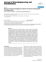
Figure 1 is the characterization of the Ga
2
O
3
nanowires
synthesized in the first step. From Fig. 1a, it can be seen
that the diameter of nanowires is about 80–100 nm and the
length of nanowires is up to several tens of micrometers.
Au particle can be found on the top of each nanowire, and
the diameter of the nanowire is consistent with the size of
Au particle, which indicates that the growth of Ga
2
O
3
nanowires follows the VLS mechanism [30]. The XRD
pattern in Fig. 1b indicates that all the diffraction peaks
except the peak from the Si (111) substrate can be indexed
as monoclinic structure b-Ga
2
O
3
(JCPDS: 11-0370) with
lattice constants of a = 5.80 A
˚
, b = 3.04 A
˚
, c = 12.23 A
˚
,
and b = 103.7°.
Fig. 1 a FE-SEM image of the Ga
2
O
3
nanowires; b XRD pattern of
as-prepared Ga
2
O
3
nanowires; c FE-SEM image of Au nanoparticles
on Ga
2
O
3
nanowires after annealing at 1,000°C for 30 min
1388 Nanoscale Res Lett (2010) 5:1387–1392
123
In recent reports, Ga
2
O
3
nanowires are employed to
synthesis ZnGa
2
O
4
nanowires through high temperature
reaction with ZnO vapor [16, 21]. Inspired by this, here in
this paper, Ga
2
O
3
nanowires were used as templates for the
growth of ZnGa
2
O
4
nanocombs. Catalyst induced growth is
well known as a powerful method to control the growth of
1-D nanostructures. In order to guide the growth of the
teeth of nanocombs, Au nanoparticles are introduced in our
experiment. A thin layer of Au was deposited on the sur-
face of as-grown Ga
2
O
3
nanowires. After annealed at
1,000°C for 30 min, Au layer congregate into nanoparti-
cles. As shown in Fig. 1c, Au nanoparticles arrange regu-
larly on the side surface of Ga
2
O
3
nanowires, which may
derive from the difference of surface energy of Ga
2
O
3
crystal planes. Orderly arrange of Au nanoparticles on the
specific plane of Ga
2
O
3
nanowire may have low energy and
remain stable. This phenomenon is used to obtain the
controlled growth of the teeth of ZnGa
2
O
4
nanocombs. In
addition, the annealing process is very important to get
comb-like Ga
2
O
3
nanostructures. We will discuss in the
following.
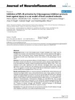
Figure 2 shows the morphologies and crystalline struc-
ture characterization of the as-synthesized ZnGa
2
O
4
prod-
ucts using FE-SEM and XRD. Figures 2a and b show the
low- and high-magnification FE-SEM images of as-pre-
pared products, which reveal that a large amount hierar-
chical nanostructures with comb-like morphologies are
formed. The nanoteeth on the backbones in our fabricated
samples are not as densely aligned as that in other nano-
comb materials. The nanocombs are several tens of
micrometers long with the teeth about 200 nm in length,
and the teeth are orderly arranged on one side of nano-
comb. The FE-SEM image shows clearly that there is a
nanoparticle on the tip of each tooth. The particle was
confirmed to be Au by EDS spectra in the following TEM
analysis. Figure 2c shows the corresponding XRD pattern,
most of the main diffraction peaks can be indexed to cubic
spinel structure ZnGa
2
O
4
(JCPDS: 38-1240) with lattice
constant of a = 8.334 A
˚
, except the peaks from Si(111)
substrate and Au nanoparticles (111) diffraction.
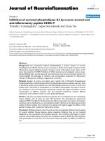
Figure 3a shows a typical TEM image of a single-
ZnGa
2
O
4
nanocomb, indicating that the diameter of the
tooth gradually increasing from the tip to the bottom.
Figure 3b–e are the EDS spectra acquired from the marked
regions 1 to 4 in Fig. 3a. The copper and carbon signals in
EDS spectra are caused by the copper grids used in TEM
observation. The EDS spectrum of region 1 confirms that
the particle on the tip of the tooth is Au catalyst. Appar-
ently, the growth of the teeth is induced by the Au catalyst.
From the EDS spectra of region 2 to 4, it can be found
that the atomic ratio of Zn:Ga in all these regions are close
to 1:2. These results further confirm that the nanocomb
is zcomposed of ZnGa
2
O
4
. The high-resolution TEM
(HRTEM) images and the corresponding selected-area
electron diffraction (SAED) of region 2 to 4 in Fig. 3a are
shown in Fig. 3f–h and the inset to them, respectively,
which reveal that the comb-like ZnGa
2
O
4
nanostructures
are highly crystallized. In Fig. 3f, it can be seen that the
marked interplanar spacing is 0.48 nm which corresponds
to the (111) lattice plane of ZnGa
2
O
4
, indicating the
dominant growth direction of the tooth along the [111]
direction. And no extended defects were found in the whole
tooth. Fig. 3g is the HRTEM image and the corresponding
SAED of junction region between the stem and the tooth
Fig. 2 Characterization of comb-like ZnGa
2
O
4
nanostructures: a
low-magnification FE-SEM image; b high-magnification FE-SEM
image; c XRD pattern of as-prepared ZnGa
2
O
4
nanostructures
Nanoscale Res Lett (2010) 5:1387–1392 1389
123
depicts that the interface is very smooth without planar
defects like other semiconductor comb-like nanostructures
[31, 32]. It is of advantage to be used in the future appli-
cation. Figure 3h is the HRTEM image and the corre-
sponding SAED of stem. The marked interplanar spacings
are 0.48 and 0.29 nm corresponding to the (111) plane and
ð
"
220Þ plane, respectively. It can be inferred that the initial
Ga
2
O
3
nanowires were totally transformed into highly pure
and single-crystalline ZnGa
2
O
4
nanowires after reacting
with Zn and/or ZnO
x
vapors at high temperature.
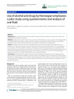
The growth of comb-like ZnGa
2
O
4
nanostructures fol-
lows the VLS process (as shown in Fig. 4). During the
previous annealing process, Au nanoparticles have arran-
ged on the side surface of Ga
2
O
3
nanowires (Stage I in
Fig. 4). During the heating process, the starting material
ZnO powder reacted with carbon to form Zn and/or ZnO
x
vapors at 850°C. The vapors were carried downstream to
the Ga
2
O
3
nanowires by Ar gas (stage II in Fig. 4). These
vapors deposited on the surface of the Ga
2
O
3
nanowires
and ZnGa
2
O
4
stem formed through reaction (stage III in
Fig. 4). The orderly arranged Au nanoparticles on the side
surface of Ga
2
O
3
nanowires seem to be the preferred sites
for the growth of ZnGa
2
O
4
nanoteeth. In the growth pro-
cess, Zn and/or ZnO
x
vapor and the remained O
2
may be
absorbed by Au nanoparticles, and Ga in nanowires may
also diffuse to these sites. When the alloy droplets got
supersaturated, ZnGa
2
O
4
may nucleate to form the teeth
(stage III in Fig. 4). During the heating process, the grad-
ually reaction of Ga
2
O
3
nanowires with Zn and/or ZnO
x
vapor causes the continuous consumption of Ga source,
which results in that the nanoteeth can not growth too long.
At the same time, since the mass diffusion of reactant
adatoms on the side surface, continuous growth resulting in
the formation of tapered teeth (stage IV in Fig. 4). The
orderly arranged Au particles on the side surface of Ga
2
O
3
nanowires are very important to the growth of ZnGa
2
O
4
nanocombs. If the annealing process is canceled, no
ZnGa
2
O
4
nanocombs can be obtained as shown in Fig. 5.
Under this circumstance, Au first exists as a thin layer on
the surface of Ga
2
O
3
nanowires. Thus, there are no pre-
ferred sites for the growth of teeth. Zn and/or ZnO
x
vapors
directly reacted with Ga
2
O
3
and formed ZnGa
2
O
4
nano-
wires. It can be seen that controlling the state and position
of Au nanoparticle on the Ga
2
O
3
nanowires is the key to
obtain ZnGa
2
O
4
nanostructures with desired comb-like
morphology.
The room-temperature PL spectrum of the ZnGa
2
O
4
nanocomb was presented in Fig. 6. The excitation wave-
length is 260 nm. Two emission bands can be observed,
which were centered at the 450 and 501 nm, respectively.
Fig. 3 a Typical TEM image of a single-ZnGa
2
O
4
nanocomb. b–e are the EDS spectra acquired from marked regions 1 to 4 in a. The HRTEM
images and the corresponding SAED of marked regions 2 to 4 in a are shown in f–h and inset to them, respectively
1390 Nanoscale Res Lett (2010) 5:1387–1392
123
The PL spectrum features that the two emission peaks
centered at the 450 and 501 nm merged to form a broad
band emission ranged from 400 to 575 nm. The PL prop-
erties of the ZnGa
2
O
4
films and nanowires had been
investigated intensively [7–9, 22]. From these studies, it
can be suggested that the emission band centered at
450 nm may be originated from the self-activation center
of the octahedral Ga–O group [7], and the emission band
centered at 501 nm may be originated from the electronic
transitions of localized Ga
3?
ion in the octahedral Ga–O
group [23]. By comparing with the previous investigation
[7–9], it can be suggested the broad band emission in the
visible light region of as-prepared ZnGa
2
O
4
nanocombs
may be related to the two-step synthesis method and its
hierarchical morphology.
Conclusions
In summary, we present an easy route to synthesize comb-
like ZnGa
2
O
4
nanostructures in a controllable way. The
Ga
2
O
3
nanowires were used as templates for the following
growth of comb-like ZnGa
2
O
4
nanostructures through the
reaction with Zn and/or ZnO
x
vapor at high temperature.
By annealing Ga
2
O
3
nanowires coated with a thin layer of
Au film at high temperature, the congregation of Au par-
ticles from Au film is the key to the formation of ZnGa
2
O
4
nanoteeth via VLS mechanism. PL spectra for ZnGa
2
O
4
nanocombs show a broad band emission in the visible light
region from 400 to 575 nm at room temperature. This
method can be easily applied to hierarchical nanostructure
growth of other materials to enrich the family of low
Fig. 4 Schematic illustration of growth process for ZnGa
2
O
4
nano-
combs. Stage I: arranged Au nanoparticles on the Ga
2
O
3
nanowires
after annealing; Stage II–III: during the heating process, Au provides
preferential site for nucleation and 1-D growth; Stage IV: due to the
gradually consumption of Ga source and the mass diffusion of
reactant adatoms on the side surface, continuous growth resulting in
the formation of tapered teeth
Fig. 5 Low-magnification (a) and high-magnification (b) SEM images of products obtained without annealing
Fig. 6 The room-temperature PL spectrum of comb-like ZnGa
2
O
4
nanostructure. The excitation wavelength is 260 nm
Nanoscale Res Lett (2010) 5:1387–1392 1391
123
dimensional nanobuilding blocks and may find potential
applications in nanotechnology.
Acknowledgment This work was supported by the National Natural
Science Foundation of China (No.50671099, 50172048, 10374090
and 10274085), the Ministry of Science and Technology of China
(No.2005CB623603), and the Hundred Talent Program of Chinese
Academy of Sciences.
Open Access This article is distributed under the terms of the
Creative Commons Attribution Noncommercial License which per-
mits any noncommercial use, distribution, and reproduction in any
medium, provided the original author(s) and source are credited.
References
1. Y. Cui, C.M. Lieber, Science 291, 851 (2001)
2. X.F. Duan, Y. Huang, R. Agarwal, C.M. Lieber, Nature 421, 241
(2003)
3. Z.L. Wang, J.H. Song, Science 312, 242 (2006)
4. Y.N. Xia, P.D. Yang, Y.G. Sun, Y.Y. Wu, B. Mayers, B. Gates,
Y.D. Yin, F. Kim, Y.Q. Yan, Adv. Mater. 15, 353 (2003)
5. S. Itoh, H. Toki, Y. Sato, K. Morimoto, T. Kishino, J. Electro-
chem. Soc. 138, 1509 (1991)
6. T. Omata, N. Ueda, K. Ueda, H. Kawazoe, Appl. Phys. Lett. 64,
1077 (1994)
7. J.S. Kim, H.I. Kang, W.N. Kim, J.I. Kim, J.C. Choi, H.L. Park,
G.C. Kim, T.W. Kim, Y.H. Hwang, S.I. Mho, M.C. Jung, M.
Han, Appl. Phys. Lett. 82, 2029 (2003)
8. I.J. Hsieh, K.T. Chu, C.F. Yu, M.S. Feng, J. Appl. Phys. 76, 3735
(1994)
9. Y.E. Lee, D.P. Norta, C. Park, C.M. Roulean, J. Appl. Phys. 89,
1653 (2001)
10. I.K. Jeong, H.L. Park, S.I. Mho, Solid State Commun. 108, 823
(1998)
11. S.H.M. Poort, D. Cetin, A. Meijerink, G.J. Blasse, Electrochem.
Soc. 144, 2179 (1997)
12. L.E. Shea, R.K. Datta, J.J. Brown, J. Electrochem. Soc. 141, 2198
(1994)
13. X.N. Zhang, J.H. Huang, K.N. Ding, Y.D. Hou, X.C. Wang, X.Z.
Fu, Environ. Sci. Technol. 43, 5947 (2009)
14. W.W. Zhang, J.Y. Zhang, Z.Y. Chen, T.M. Wang, Catal. Com-
mun. 10, 1781 (2009)
15. O. Maksimov, Mater. Lett. 62, 3969 (2008)
16. K.W. Chang, J.J. Wu, J. Phys. Chem. B 109, 13572 (2005)
17. S.H. Wu, H.C. Cheng, J. Electrochem. Soc. 151, 159 (2004)
18. S.H. Yang, J. Electrochem. Soc. 150, 250 (2003)
19. Y.E. Lee, D.P. Norton, J.D. Budai, Appl. Phys. Lett. 74, 3155
(1999)
20. S.Y. Bae, J.Y. Lee, H.S. Jung, J.H. Park, J.P. Ahn, J. Am. Chem.
Soc. 127, 10802 (2005)
21. Z. Yu, H. Chen, Z.W. Li, Z.M. Yang, H.B. Song, Y.L. Gao, Y.S.
Zhang, Y. Jin, Z.F. Jiao, M. Gong, J.G. Zhu, X.S. Sun, Mater.
Lett. 63, 37 (2009)
22. S.Y. Bae, H.W. Seo, W.C. Na, J. Park, Chem. Commun. 16, 1834
(2004)
23. L. Xu, Y. Su, Q.T. Zhou, S. Li, Y.Q. Chen, Y. Feng, Cryst.
Growth Des. 7, 810 (2007)
24. P. Feng, J.Y. Zhang, Q. Wan, T.H. Wang, J. Appl. Phys. 102,
074309 (2007)
25. Y.J. Li, M.Y. Lu, C.W. Wang, K.M. Li, L.J. Chen, Appl. Phys.
Lett. 88, 143102 (2006)
26. H.J. Fan, Y. Yang, M. Zacharias, J. Mater. Chem. 19, 885 (2009)
27. G. Shen, Y. Bando, C.J. Lee, J. Phys. Chem. B 109, 10779 (2005)
28. G.W. Meng, Y.J. Jung, A.Y. Cao, R. Vajtai, P.M. Ajayan, Proc.
Natl. Acad. Sci. 102, 7074 (2005)
29. M. Misono, Chem. Commun. 13, 1141 (2001)
30. R.S. Wagner, W.C. Ellis, Appl. Phys. Lett. 4, 89 (1964)
31. C. Borchers, D. Stichtenoth, S. Muller, D. Schwen, C. Ronning,
Nanotech. 17, 1067 (2006)
32. Y.Q. Wang, U. Philipose, H. Ruda, K.L. Kavanagh, J Mater. Sci.
Mater. Electron. 17, 1065 (2006)
1392 Nanoscale Res Lett (2010) 5:1387–1392
123