Báo cáo hóa học: "Computed tomographic pulmonary angiography and pulmonary embolism: predictive value of a d-dimer assay" docx
Bạn đang xem bản rút gọn của tài liệu. Xem và tải ngay bản đầy đủ của tài liệu tại đây (192.85 KB, 13 trang )
This Provisional PDF corresponds to the article as it appeared upon acceptance. Fully formatted
PDF and full text (HTML) versions will be made available soon.
Computed tomographic pulmonary angiography and pulmonary embolism:
predictive value of a d-dimer assay
BMC Research Notes 2012, 5:104 doi:10.1186/1756-0500-5-104
Patricia Deonarine ()
Carl de Wet ()
Alistair McGhee ()
ISSN 1756-0500
Article type Research article
Submission date 24 November 2011
Acceptance date 17 February 2012
Publication date 17 February 2012
Article URL />This peer-reviewed article was published immediately upon acceptance. It can be downloaded,
printed and distributed freely for any purposes (see copyright notice below).
Articles in BMC Research Notes are listed in PubMed and archived at PubMed Central.
For information about publishing your research in BMC Research Notes or any BioMed Central
journal, go to
/>BMC Research Notes
© 2012 Deonarine et al. ; licensee BioMed Central Ltd.
This is an open access article distributed under the terms of the Creative Commons Attribution License ( />which permits unrestricted use, distribution, and reproduction in any medium, provided the original work is properly cited.
Computed tomographic pulmonary
angiography and pulmonary embolism:
predictive value of a d-dimer assay
ArticleCategory :
Research Article
ArticleHistory :
Received: 24-Nov-2011; Accepted: 23-Feb-2012
ArticleCopyright
:
© 2012 Deonarine et al; BioMed Central Ltd. This is an Open Access
article distributed under the terms of the Creative Commons Attribution
License ( which permits
unrestricted use, distribution, and reproduction in any medium,
provided the original work is properly cited.
Patricia Deonarine,
Aff1
Corresponding Affiliation: Aff1
Phone: +44-0141-211-4000
Email:
Carl de Wet,
Aff2
Email:
Alistair McGhee,
Aff3
Email:
Aff1
Radiology Department, Glasgow Royal Infirmary, 84 Castle Street,
Glasgow G4 0SF, UK
Aff2
General practitioner and research fellow, NHS Education for
Scotland, Glasgow, UK
Aff3
Radiology consultant, Monklands district general hospital,
Lanarkshire, UK
Abstract
Background
Computed tomographic pulmonary angiography (CTPA) is increasingly being used as first
investigation for suspected pulmonary embolism (PE). The investigation has high predictive
value, but is resource and time intensive and exposes patients to considerable radiation. Our
aim was to assess the potential value of a negative d-dimer assay to exclude pulmonary
emboli and reduce the number of performed CTPAs.
Methods
All CTPAs performed in a Scottish secondary care hospital for a fourteen month period were
retrospectively reviewed. Collected data included the presence or absence of PE, d-dimer
results and patient demographics. PE positive CTPAs were reviewed by a specialist panel.
Results
Pulmonary embolisms were reported for 66/405 (16.3%) CTPAs and d-dimer tests were
performed for 216 (53%). 186/216 (86%) patients had a positive and 30 (14%) a negative d-
dimer result. The panel agreed 5/66 (7.6%) false positive examinations. The d-dimer assay’s
negative predictive value was 93.3% (95% CI = 76.5%–98.8%) based on the original number
of positive CTPAs and 100% (95% CI = 85.9%–100%) based on expert review. Significant
non-PE intrapulmonary pathology was reported for 312/405 (77.0) CTPAs, including 13 new
diagnoses of carcinoma.
Conclusions
We found that a low d-dimer score excluded all pulmonary embolisms, after a further
specialist panel review identified initial false positive reports. However, current evidence-
based guidelines still recommend that clinicians combine a d-dimer result with a validated
clinical risk score when selecting suitable patients for CTPA. This may result in better use of
limited resources, prevent patients being exposed to unnecessary irradiation and prevent
potential complications as a result of iodinated contrast.
Keywords
Pulmonary embolism, D-dimer, CTPA (tomography)
Background
Pulmonary embolism (PE) is associated with substantial morbidity and mortality. In the US
more than 500 000 patients per year are diagnosed with pulmonary emboli, resulting in
approximately 200 000 deaths [1,2]. In England and Wales there are around 65 000 cases of
pulmonary emboli annually amongst hospitalized patients. The prevalence of unsuspected
pulmonary embolism at post-mortem is 3–8%, figures that have changed little over three
decades. The implication is that the true number of cases may be substantially higher than is
currently being diagnosed [3].
The presentation, symptoms and clinical signs of pulmonary embolism varies widely between
patients [4]. Clinical suspicion invariably requires further investigation to confirm or exclude
PE. In Scotland, computed tomographic pulmonary angiography (CTPA) is increasingly used
as the first and only investigation for this purpose [5]. This is because of greater availability
and reported overall sensitivity (89–100%) of helical CT, but exposes patients to substantial
ionizing radiation [6-8]. A recent review by Davies et al. found that iatrogenic radiation
exposure has significant risks which are often overlooked, while it was estimated that 30% of
computed tomography tests may be unnecessary [9].
D-dimer assays have low specificity, but high sensitivity and negative predictive value in
most patients with suspected thromboembolism, and may be an alternative first investigation
to CTPA [10]. D-dimers are degradation products of cross linked fibrin and are considered
the best laboratory markers of coagulation activation [11,12]. They are commonly elevated in
patients with recent surgery, malignancy or infection [3,13]. As a result the diagnostic value
of d-dimer assays is higher for ambulatory patients compared with those in hospital [14].
Our main aim was to assess the potential value of a negative d-dimer assay to exclude
pulmonary emboli and reduce the number of performed CTPAs. A further aim was to
describe incidental but significant intrapulmonary CTPA findings that may have accounted
for patients’ clinical presentations.
Results
A total of 416 CTPAs were performed during the study period. 11/416 (2.6%) CTPAs were
reported as indeterminate or inconclusive and excluded from further analysis. ‘Technical
factors’, for example inadequate contrast opacification of the pulmonary arterial tree, were
stated as the reason for inconclusive studies. Of the remaining 405 CTPA studies, 226
(55.8%) were performed for female and 179 (44.2%) for male patients. The mean age of all
patients was 63 years (range 20–95 and standard deviation ±17.2).
A diagnosis of pulmonary embolism was reported for 66 (16.3%) of the remaining 405
CTPAs. The expert panel unanimously agreed that five (7.6%) of these were false positive
examinations. The positive predictive value of CTPA for pulmonary embolism in this study
was 92.4% (95% CI = 82.5%–97.2%).
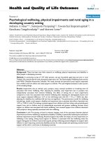
The numbers of performed d-dimer assays, d-dimer results and CTPAs positive for
pulmonary embolism are shown in Figure 1. Of the 216 (53.3%) patients who had a d-dimer
assay performed, 186 (86%) had a positive and 30 (14%) a negative d-dimer result. There
were initially two positive CTPAs with negative d-dimer results. Both of these were
unanimously judged to be negative CTPAs (false positives) by the panel. The d-dimer assay’s
negative predictive value was 93.3% (95% CI = 76.5%–98.8%) based on the original number
of positive CTPA reports and 100% (95% CI = 85.9%–100%) based on the panel’s review.
The d-dimer assay and CTPA sensitivity, specificity and predictive values are shown in Table
1. The complete data set is available as an additional file 1 (see CTPA ddimer.xls).
Figure 1 Number of computed tomography pulmonary angiogram (CTPA) studies, d-
dimer assays, and diagnoses of pulmonary embolism (PE)
Table 1 D-dimer assay and CTPA sensitivity, specificity and predictive values (95%
confidence intervals)
D-dimer assay:
Results based on number of positive CTPA results
CTPA
N = 41
(initial results)
N = 36
(results after specialist review)
N = 405
Sensitivity 95.1 (82.2–99.2) 100 (88.0–100) x
Specificity 16.0 (11.1–22.5) 16.7 (11.7–23.1) 98.5 (96.4–99.5)
Positive predictive value 21.0 (15.5–27.7) 19.4 (14.1–25.9) 92.4 (82.5–97.2)
Negative predictive value 93.3 (76.5–98.8) 100 (85.9–100) x
The main anatomical sites of pulmonary embolism are shown in Table 2. Sub segmental
embolisms were least common, being described in 4 (6%) of cases. The main findings of the
CTPAs considered for analysis are shown in Table 3. Significant non-PE intrapulmonary
pathology was reported for 312/405 (77.0) CTPAs. The most common reported abnormalities
were pleural effusion (16.5%), bronchiectasis (10.6%) and consolidation (7.9%). There were
13 (3.2) new diagnoses of unsuspected carcinoma. Of the 66/405 (16.3) CTPAs reported as
pulmonary embolism, 52 patients had significant additional pulmonary pathology.
Table 2 Classification of pulmonary emboli according to anatomical site
No* (%)
Large main trunk 21 (32)
Lobar artery 20 (30)
Segmental 21 (32)
Sub-segmental 4 (6)
Total 66 (100)
* Each PE positive case was included only once in the classification according to the largest
thrombosed vessel reported
Table 3 The main findings of 405 CTPAs performed between 01/06/2008 and 31/07/2009
that met inclusion criteria
Main CTPA finding No (%)* No (%)*
No pathology 103 (25.4)
Pulmonary embolism (PE) 66 (16.3)
• PE only
14 (3.5)
• PE with additional abnormal finding
(included below)
52 (12.8)
Significant non-PE intrapulmonary pathology 312 (77.0)
• Pleural effusion
67 (16.5)
• Consolidation
32 (7.9)
• Effusion and consolidation
27 (6.7)
• Carcinoma
13 (3.2)
• Lobar collapse
22 (5.4)
• Lymphadenopathy
13 (3.2)
• Bronchiectasis
43 (10.6)
• Pulmonary fibrosis
7 (1.7)
• Other (for example atelectasis)
88 (21.7)
*Some CTPAs had more than one main finding reported. The number and percentage is the
proportion of the 405 CTPAs with that specific finding
Discussion
Main findings
The study’s main aim was to assess the potential value of a negative d-dimer assay to exclude
pulmonary emboli and reduce the number of performed CTPAs. We found a low (negative)
d-dimer score to have a very high negative predictive value, but that it did not exclude all
pulmonary emboli based on the initial CTPA reports. However, a low d-dimer score did
exclude all pulmonary emboli after a further specialist panel review identified false positive
reports.
Comparison with existing literature
Our findings are comparable to other studies in various health care settings. Dunn et al.
reported a negative predictive value for d-dimer assay of 99.6% (95% CI = 98.7–>99.9%) and
suggested that negative results could help to reduce the number of performed CTPAs [15].
More recently, Eng et al. and Hirai et al. concluded that a d-dimer test alone was suitable for
screening patients with a clinical suspicion of PE [16,17]. However, a number of case reports
have questioned whether a negative d-dimer result alone is sufficient to exclude pulmonary
embolisms [18]. There is compelling evidence that a negative d-dimer result can effectively
exclude a PE when it is combined with a low pretest clinical probability score. Current best
practice clinical guidance advises combining the d-dimer result with a validated tool—for
example the Wells or Geneva rule—which allow risk to be quantified in a structured manner
[19-22].
A further study aim was to describe incidental but significant intrapulmonary CTPA findings
that may have accounted for patients’ clinical presentations. Significant pathological findings
were reported for the vast majority of CTPA studies. The number of CTPA reports which
described previously undiagnosed malignancies was substantially higher than reported by
Kino et al. [23]. These significant non-PE findings had clinical relevance for some patients
and may subconsciously lead clinicians to rationalize CTPA requests. However, CTPA has
certain technical limitations that reduce its potential value in assessing non-PE pathology and
cannot be considered a screening tool.
The anatomical distribution of pulmonary emboli we found was comparable to that reported
by Sohns et al. [24]. The majority of thrombi were diffusely distributed between the main
pulmonary trunk, lobar and segmental arteries with only a small minority involving the sub-
segmental vessels. All of the false positive CTPAs were initially reported as ‘small, sub-
segmental pulmonary embolism’. The expert panel’s opinion was that in these cases small
lymph nodes or veins adjacent to sub-segmental arteries had typically been misinterpreted as
filling defects. It is possible that a substantial number of sub-segmental pulmonary emboli
may be false positive as a result of CTPAs being interpreted in a single plane.
Strengths and limitations
Our findings are based on a substantial sample and an additional independent panel that
reviewed CTPA studies to identify false positive results. We also identified the anatomical
distribution of emboli and additional intrapulmonary pathology that may have accounted for
the patients’ symptoms. The study has a number of limitations: CTPA findings were not
linked with clinical outcomes; validated tool such as the Geneva or Wells rule was not used
or recorded by clinicians requesting CTPAs; other imaging, for example doppler ultrasound,
chest x-rays and ventilation perfusion scans were not considered; and more modern
alternatives to the 16-slice CT scanner used in our study may have improved diagnostic
accuracy.
Implication and future research
There are clinical guidelines for health care workers investigating a patient suspected of
having a PE [19-22]. It is recommended that a clinical probability assessment and d-dimer
value should be combined and used to quantify the patient’s risk of PE as low, moderate or
high. CTPAs are only indicated for those patients judged to be at moderate or high risk. This
approach is seldom used in practice, resulting in unnecessary CTPAs being performed. This
is an inefficient use of limited time and resources and expose patients to avoidable irradiation
and potential complications of iodinated contrast [15,18]. Further research is required to
better understand the challenges in promoting and implementing the routine use of clinical
risk stratification for ambulatory patients with suspected PE.
Conclusions
A low d-dimer score had a very high negative predictive value, but did not exclude all
pulmonary embolisms based on the initial CTPA reports. However, a low d-dimer score did
exclude all pulmonary embolisms after a further specialist panel review identified false
positive reports. A practical and evidence-based approach is to combine a d-dimer result with
a validated clinical risk score to help select suitable patients for CTPA. This may result in
better use of limited resources, prevent patients being exposed to unnecessary irradiation and
prevent potential complications as a result of iodinated contrast.
Availability of supporting data
The data set supporting the results of this article is included within the article and its
additional file 1.
Methods
All CTPAs that had been performed at a District General Hospital (DGH) in Lanarkshire,
Scotland, in the fourteen month period from 1
st
June 2008 to 31
st
July 2009 were identified
and retrospectively reviewed on the Hospital Information System (HIS). This sample
included CTPAs requested for hospitalized and ambulatory patients. Patients were considered
ambulatory if they had been referred from the accident and emergency department or from
medical, surgical and oncology out-patient units. An indeterminate or inconclusive CTPA
report was the only exclusion criterion.
All PE positive studies were reviewed independently by a panel consisting of three
radiologists with an interest in this area who interpreted the CTPAs using axial, coronal and
sagittal reformats. The initial CTPA reports were judged to be false positive only if all three
panel members agreed that the study did not show sufficient evidence of PE. Data were
collected for presence or absence of PE and the type of PE, whether a d-dimer assay was
performed and the d-dimer result if applicable. Patients’ age and gender and other reported
pathological intra thoracic findings were also collected. Patient identifiers were removed and
data were entered in an Excel spreadsheet. The data were exported to SPSS version 17.0 for
calculation of descriptive statistics.
The d-dimer assay and CTPA sensitivity, specificity and positive and negative predictive
values were calculated with 95% confidence intervals. The d-dimer assay values were
calculated twice, using the initial number of positive CTPA results, and then the revised
number of positive CTPA results as determined by the specialist panel. Pulmonary emboli
were classified according to their anatomical distribution. Each case was included only once
and grouped according to the largest thrombosed vessel reported.
CTPA
All CTPA studies were performed using a Toshiba Aquillon 16 slice CT scanner with slice
thickness set at 1 mL.
D-dimer
Four different types of d-dimer assay formats are currently available: enzyme linked
immunosorbent assay (ELISA), whole blood erythrocyte agglutination assay (SimpliRED),
semiquantitative latex agglutination assays (Accuclot, Trinity Biotech, Bray) and
immunochromatographic/quantitative immunoturbidimetric assays. ELISA is considered the
gold standard for the determination of d-dimer concentration. It is a highly sensitive test but
is time consuming and not suitable for individual patient testing. The Accuclot d-dimer assay
is less sensitive, but suitable for individual patient testing [25-27]. The Trinity Amax
Accuclot d-dimer assay—a semi quantitative latex agglutination assay—was used during the
study period. A d-dimer value ≥190 ng/mL was considered positive (high) and <190 ng/mL
negative (low) in accordance with local guidelines and the recommendation of the Accuclot
d-dimer assay manufacturer.
Competing interests
None.
Authors’ contributions
PD helped to design the study, collected the data and helped to prepare the manuscript. CdW
coded and analyzed the data and helped to prepare the manuscript. AM had the original idea
for the study, was one of the ‘expert’ reviewers and reviewed the final manuscript. All
authors read and approved the final manuscript.
Acknowledgements
We would like to thank the PACS team at Monklands DGH Lanarkshire, the Biochemistry
department for access to laboratory records to review d-dimer values and Drs. Julien Guse
and Tamas Schilzer.
References
1. Dismuke S, Wagner E: Pulmonary embolism as a cause of death. The changing
mortality in hospitalized patients. JAMA 1986, 255:2039–2042.
2. Horlander K, Minnino D, Leeper K: Pulmonary embolism mortality in United States,
1979–1998: an analysis using multiple-cause mortality data. Arch Intern Med 2003,
163(1711):1717.
3. Torbicki A, Perrier A, Konstantinides S, Agnelli G, Galie N, Pruszczyk P, et al:
Guidelines on the diagnosis and management of acute pulmonary embolism. Eur Heart J
2008, 29:2276–2315.
4. Courtney DM, Kline JA, Kabrhel C, Moore CL, Smithline HA, Nordenholz KE, et al:
Clinical features from the history and physical examination that predict the presence or
absence of pulmonary embolism in symptomatic emergency department patients:
results of a prospective, multicenter study. Ann Emerg Med 2010, 55(4):307–315.e1.
5. O’Neill JM, Wright L, Murchison JT: Helical CTPA in the investigation of pulmonary
embolism: a 6-year review. Clin Radiol 2004, 59(9):819–825.
6. Rathbun S, Raskob G, Whitsett T: Sensitivity and specificity of helical computed
tomography in the diagnosis of pulmonary embolism: a systematc review. Ann Intern
Med 2000, 32:227–232.
7. de Monye W, Pattynama P: Contrast enhanced spiral computed tomography of the
pulmonary arteries: an overview. Semin Thromb Hemost 2001, 27:33–39.
8. O'Neill J, Murchison JT, Wright L, Williams J: Effect of the introduction of helical CT
on radiation dose in the investigation of pulmonary embolism. Br J Radiol 2005,
78(925):46–50.
9. Davies HE, Wathen CG, Gleeson FV: Risks of exposure to radiological imaging and
how to minimise them. BMJ 2011, 342:589–593.
10. Ginsberg JS, Wells PS, Kearon C, Anderson D, Crowther M, Weitz JI, et al: Sensitivity
and specificity of a rapid whole blood assay for D dimer in the diagnosis of pulmonary
embolism. Ann Intern Med 1998, 129:1006–1011.
11. Kroneman H, Nieuwenhuizen W, Knot EA: Monoclonal antibody-based plasma assays
for fibrin (ogen) and derivates, and their clinical relevance. Blood Coagul Fibrinolysis
1990, 1:91–111.
12. Sie P: The value of laboratory tests in the diagnosis of venous thromboembolism.
Haematologica 1995, 80(2 Suppl):57–60.
13. Goldhaber SZ, Simons GR, Elliot CG, Haire WD, Toltzis R, Blacklow SC, et al:
Quantitative plasma D dimer levels among patients undergoing pulmonary angiography
for suspected pulmonary embolism. JAMA 1993, 270:2819–2822.
14. Schrecengost JE, LeGallo RD, Boyd JC, Moons K, Gonias SL, Rose CE, et al:
Comparison of diagnostic accuracies in outpatients and hospitalized patients of D-dimer
testing for the evaluation of suspected pulmonary embolism. Clin Chem 2003, 49:1483–
1490.
15. Dunn KL, Wolf JP, Dorfman DM, Fitzpatrick P, Baker JL, Goldhaber SZ: Normal D-
dimer levels in emergency department patients suspected of acute pulmonary embolism.
J Am Coll Cardiol 2002, 40(8):1475–1478.
16. Eng CW, Wansaicheong G, Goh SK, Earnest A, Sum C: Exclusion of acute pulmonary
embolism: computed tomography pulmonary angiogram or D-dimer? Singapore Med J
2009, 50(4):403–406.
17. Hirai LK, Takahashi JM, Yoon HC: A prospective evaluation of a quantitative D-
dimer assay in the evaluation of acute pulmonary embolism. J Vasc Interv Radiol 2007,
18(8):970–974.
18. Breen ME, Dorfman M, Chan SB: Pulmonary embolism despite negative ELISA D-
dimer: a case report. J Emerg Med 2009, 37(3):290–292.
19. Wells PS, Anderson DR, Rodger M, Stiell I, Dreyer JF, Barnes D, et al: Excluding
pulmonary embolism at the bedside without diagnostic imaging: management of
patients with suspected pulmonary embolism presenting to the emergency department
by using a simple clinical model and D dimer. Ann Intern Med 2001, 135:98–107.
20. Subramaniam RM, Chou T, Swarbrick M, Karalus N: Pulmonary embolism: accuracy
and safety of a negative CT pulmonary angiogram and value of a negative D-dimer
assay to exclude CT pulmonary angiogram-detectable pulmonary embolism. Australas
Radiol 2006, 50(5):424–428.
21. Gupta RT, Kakarla RK, Kirshenbaum KJ, Tapson VF: D-dimers and efficacy of clinical
risk estimation algorithms: sensitivity in evaluation of acute pulmonary embolism. AJR
Am J Roentgenol 2009, 193(2):425–430.
22. Gibson NS, Sohne M, Kruip MJ, Tick LW, Gerdes VE, Bossuyt PM, et al: Further
validation and simplification of the Wells clinical decision rule in pulmonary embolism.
Thromb Haemost 2008, 99(1):229–234.
23. Kino A, Boiselle PM, Raptopoulos V, Hatabu H: Lung cancer detected in patients
presenting to the Emergency Department studies for suspected pulmonary embolism on
computed tomography pulmonary angiography. Eur J Radiol 2006, 58(1):119–123.
24. Sohns C, Amarteifio E, Sossalla S, Heuser M, Obenauer S: 64-Multidetector-row spiral
CT in pulmonary embolism with emphasis on incidental findings. Clin Imaging 2008,
32(5):335–341.
25. Lee AY, Ginsberg JS: The role of D-dimer in the diagnosis of venous
thromboembolism. Curr Opin Pulm Med 1997, 3(275):279.
26. Perrier A, Desmarais S, Miron M, de Moerloose P, Lepage R, Slosman D, et al: Non-
invasive diagnosis of venous thromboembolism in outpatinets. Lancet 1999,
353(9148):190–195.
27. Bernardi E, Prandoni P, Lensing AWA, Agnelli G, Guazzaloca G, Scannapieco G, et al:
D-dimer testing as an adjunct to ultrasonography in patients with clinically suspected
deep vein thrombosis: prospective cohort study. BMJ 1998, 317:1037–1040.
Additional files
Additional_file_1 as XLS
Additional file 1: The complete data set is available as a Microsoft Excel spreadsheet and
can be downloaded as an additional file (CTPA ddimer.xls).
1
Figure 1
Additional files provided with this submission:
Additional file 1: ctpa ddmer.xls, 60K
/>