Báo cáo hóa học: " Combination of Polymer Technology and Carbon Nanotube Array for the Development of an Effective Drug Delivery System at Cellular Leve" pptx
Bạn đang xem bản rút gọn của tài liệu. Xem và tải ngay bản đầy đủ của tài liệu tại đây (464.73 KB, 6 trang )
NANO EXPRESS
Combination of Polymer Technology and Carbon Nanotube Array
for the Development of an Effective Drug Delivery System
at Cellular Level
Cristina Riggio Æ Gianni Ciofani Æ Vittoria Raffa Æ
Alfred Cuschieri Æ Silvestro Micera
Received: 19 January 2009 / Accepted: 5 March 2009 / Published online: 25 March 2009
Ó to the authors 2009
Abstract In this article, a carbon nanotube (CNT) array-
based system combined with a polymer thin film is pro-
posed as an effective drug release device directly at cellular
level. The polymeric film embedded in the CNT array is
described and characterized in terms of release kinetics,
while in vitro assays on PC12 cell line have been per-
formed in order to assess the efficiency and functionality of
the entrapped agent (neural growth factor, NGF). PC12 cell
differentiation, following incubation on the CNT array
embedding the alginate delivery film, demonstrated the
effectiveness of the proposed solution. The achieved results
indicate that polymeric technology could be efficiently
embedded in CNT array acting as drug delivery system at
cellular level. The implication of this study opens several
perspectives in particular in the field of neurointerfaces,
combining several functions into a single platform.
Keywords Vertically aligned carbon nanotubes Á
Drug delivery Á Alginate Á NGF Á PC12 cells
Introduction
Despite advances in understanding of the mechanisms
involved in the evolution of neurodegenerative disorders
and neuroactive agents, drug delivery to the nervous sys-
tem remains problematic, especially as accessibility to the
central nervous system (CNS) is limited by the blood–brain
barrier. In addition, the systemic administration of neuro-
active biomolecules in order to stimulate neuronal
regeneration has several limitations including toxicity and
poor stability associated with many bioactive factors [1].
The purpose behind controlling the drug delivery is to
achieve more effective therapies while eliminating the
potential for both under- and overdosing. In recent years,
controlled drug delivery formulations and polymers used in
these systems have become much more sophisticated [2].
In addition, materials have been developed, which should
lead to targeted delivery systems, in which a particular
formulation can be directed to the specific cell, tissue, or
site where the drug is to be delivered. Among the proposed
solutions, micro- and nano-scale drug delivery systems are
ideal breakthrough therapeutic approaches [3]. In this
article, a carbon nanotube (CNT) array-based system,
combined with a polymer thin film, is proposed as an
effective drug release device directly at cellular level.
Recently, the use of carbon nanotubes [4] attracted
significant attention of several groups for the development
of novel neuronal interfaces [5–7]. More specifically, the
electrical properties of vertically aligned carbon nanofiber
(VACNFs)––a form of carbon quite similar to multi-wall
CNT (MWNT)––arrays have been investigated. Two
applications of this nano-device were proposed: electrical
stimulation and electro-chemical sensing. In the former
case, the device is configured as a forest-like VACNF array
that exhibits extremely low impedance; in the latter case,
C. Riggio Á G. Ciofani Á V. Raffa Á A. Cuschieri Á S. Micera
Scuola Superiore Sant’Anna, Piazza Martiri della Liberta
`
,
33, 56127 Pisa, Italy
C. Riggio (&)
CRIM & ARTS Lab - Scuola Superiore Sant’Anna,
Viale Rinaldo Piaggio, 34, 56025 Pontedera, PI, Italy
e-mail:
S. Micera
Swiss Federal Institute of Technology (ETH),
Zurich, Switzerland
123
Nanoscale Res Lett (2009) 4:668–673
DOI 10.1007/s11671-009-9291-0
the system is designed such that the CNFs are embedded in
a dielectric material (SiO
2
) which should have ideal
properties (low detection limits and high temporal resolu-
tion) for capturing neural signalling events.
Nguyen and collaborator also found that PC12 cells
cultured on PPy-coated CNF arrays (treated with a thin
layer of collagen to promote cell adhesion) can form
extended neural network upon differentiation [5]. In this
study, we propose a combination of drug delivery system
with such CNT array, exploiting a thin film of calcium
alginate as drug reservoir embedded into the platform.
Among polymers, alginate has several unique properties
that have allowed it to be used as a matrix for the entrap-
ment and/or delivery of a variety of biological agents [8].
Alginate is a co-polymer extracted from some types of
brown algae and it is made up of two uronic acids:
D-mannuronic acid and L-guluronic acid. Polyvalent cations
are responsible for interchain and intrachain reticulations
because they are tied to the polymer when two guluronic
acid residuals are close [9]. The reticulation process con-
sists of the simple substitution of sodium ions with calcium
ions [10]. The relatively mild gelation process has enabled
not only proteins [11], but also cells [12] and DNA [13]to
be incorporated into alginate matrices with retention of full
biological activity.
The polymeric film embedded in the CNT array is
described and characterized in terms of release kinetics
using bovine serum albumin as drug model, while in vitro
assays on PC12 cell line have been performed in order to
assess the efficiency and functionality of the entrapped
agent (neural growth factor, NGF). PC12 cells differenti-
ation following incubation on the CNT array embedding
the alginate delivery film demonstrated the effectiveness of
the proposed solution.
Materials and Methods
CNT Array: Properties, Imaging and Coating
Vertically aligned CNT arrays were provided from Nano-
Lab, Inc. (Newton, MA, USA). They were grown by
plasma-enhanced chemical vapour deposition (PECVD)
using Ni catalyst deposited on a 200-nm thick Cr film
covering a Si wafer. The average diameter of the individual
CNT is 80 ± 10 nm and the height is approximately 7 lm,
as specified by the supplier. The CNTs are randomly dis-
tributed in the array (1 cm 9 1 cm) with a density of
8 ± 1 9 10
8
/cm
2
. All the samples were pre-treated in
1.0 M HNO
3
for 30 min to remove the metal catalyst, and
then thoroughly rinsed with deionized water. The sample
was allowed to dry in air and sterilized with UV exposition
before cell culture experiments.
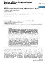
Figure 1 shows a focused ion beam (FIB) image of an
as-grown CNT array used in this study. The FIB system
used in the present study is a FEI 200 (Focused Ion Beam
Localized milling and deposition) delivering a 30-keV
beam of gallium ions (Ga
?
).
Due to the high aspect ratio ([70:1), the as-grown CNT
array is not stable when treated in liquid environments:
during the drying process, CNTs irreversibly stick together
to form microbundles, driven by the capillary force of
water droplets. In order to prevent the CNT sticking in a
liquid environment, and to improve mechanical features of
CNTs, a thin layer of SiO
2
is deposited onto the array [5].
SiO
2
film was deposited via sputtering at a sputtering rate
of 1 nm/min for 45 min (RF Sputtering Sistec, model DCC
150, operating at a constant pressure of 1 Pa, using 99.99%
pure SiO
2
target and 99.999% pure argon as sputtering
gas).
Alginate Thin Film Design, Production
and Characterization
The CNT array owns a forest-like structure that could be
exploited for the deposition of a polymeric thin film acting
as drug delivery device.
For drug release kinetics investigation, bovine serum
albumin (BSA, A3156 from Sigma, MW = 66,430 g/mol)
was added to an alginate solution at a final concentration of
200 lg/mL. BSA was used as ‘‘protein model’’, as its
molecular weight is similar to that one of NGF (N1408
from Sigma, reconstituted in a 0.1% BSA solution in PBS)
and its concentration can be much more easily quantified
[14]. For release kinetics investigation, the alginate solu-
tion (200 lg/mL of sodium alginate and 200 lg/mL of
Fig. 1 FIB image of the as-grown CNT array
Nanoscale Res Lett (2009) 4:668–673 669
123
BSA) was deposited onto a polystyrene clean surface at a
concentration of 130 lL/cm
2
and the sample was allowed
to dry under laminar flux for 12 h until the film was
completely dried. Crosslinking was thus performed with a
30% CaCl
2
solution at a concentration of 130 lL/cm
2
,
gently stirred and quickly removed [15]. Three ml of dis-
tilled water was added on the polymeric film as release
bulk. BSA concentration was thereafter assessed in the
release bulk via spectrophotometry (with a LIBRA S12
Spectrophotometer UV/Vis/NIR, Biochrom) at 280 nm
[16]. All the experiments were performed in triplicate.
Fitting of experimental data was performed with
Matlab
Ò
Curve fitting toolbox, with a non-linear least
square method adopting Gauss–Newton algorithm.
Cell Culture and In Vitro Testing
In vitro experiments were carried out on PC12 cells (ATCC
CRL-1721), a cell line derived from a transplantable rat
pheochromocytoma that responds reversibly to NGF by
inducing a neuronal phenotype. In its presence, these cells
undergo a dramatic change in phenotype whereby they
acquire most of the characteristic properties of sympathetic
neurons. Other salient responses to NGF include cessation
of proliferation, generation of long neurites, acquisition of
electrical excitability, hypertrophy and a number of chan-
ges in composition associated with acquisition of a
neuronal phenotype [17].
PC12 cells were cultured in Dulbecco’s modified Eagle’s
medium with 10% horse serum, 5% fetal bovine serum,
100 IU/mL penicillin, 100 lg/mL streptomycin and 2 mM
L-glutamine. Just 2% of fetal bovine serum was used for the
differentiation experiments. Cells were maintained at 37 °C
in a saturated humidity atmosphere of 95% air/5% CO
2
.
Alginate film coated on the CNT array and entrapping
NGF was tested on PC12 cells monitoring their differen-
tiation. An alginate solution (200 lg/mL) entrapping 2 nM
of NGF (N1408 from Sigma, reconstituted in a 0.1% BSA
solution in PBS) was casted on the CNT array and then
crosslinked with a 30% CaCl
2
solution as previously
reported for drug release assessment. PC12 cells were
seeded on an ad hoc polystyrene substrate, fabricated with
high precision milling machine, at a density of 50,000/cm
2
.
The substrate was thereafter placed on the CNT array
system and the cells were grown in differentiating medium.
Cells’ images were obtained by a microscope
(TE2000U, Nikon) equipped with a cooled CCD camera
(DS-5MC USB2, Nikon) and with NIS Elements imaging
software.
Number of cells and neurite length have been monitored
with the image analysis software ‘‘ImageJ’’ (freely down-
loadable from the National Institutes of Health at http://
rsb.info.nih.gov/ij/).
Results and Discussion
In Fig. 2, the scheme of the CNT array-based system for
drug delivery proposed in this study is depicted. The main
structure is composed by the CNT array, embedded with
the thin film of alginate entrapping NGF to induce cell
differentiation.
SiO
2
Coating
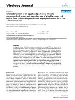
Figure 3a shows how as-grown CNTs stick together to
form microbundles as a result of evaporation following
exposition in a liquid environment. This phenomenon is
completely avoided by performing an SiO
2
coating. The
SiO
2
thin film, in fact, improves CNT mechanical features
against the capillary force of water droplets during the
drying process, thus preserving vertical alignment
(Fig. 3b). High magnification (50 kX) FIB imaging reveals
a non-uniform coating, having on the tips a higher thick-
ness than at the walls (about 40 ± 2 nm at CNT tip and
CNT base, 6 ± 1 nm at the wall).
Alginate Film Properties
In order to define a thickness of the film polymer compa-
rable to the height of CNTs, different alginate solutions at
several concentrations were tested producing films on Si-
clean surface. Subsequently, via FIB analysis, the film
thicknesses for the different conditions were measured, and
finally the alginate concentration corresponding to a film
thickness of approximately 5 lm was chosen.
The typical temporal trend of the protein release from
the alginate thin films is reported in Fig. 4. The protein
amount is given as percentage of the initial amount
entrapped into the film (200 lg/cm
3
of film). The trend is
well fitted (R
2
= 97.65%) with a bi-exponential curve as
already reported for alginate fibers [18] and microspheres
[19] and described by the following expression:
C
2
ðtÞ¼
C
10
1 þ
V
2
V
1
Áð1 Àe
ÀhÁSÁð
1
V
1
þ
1
V
2
ÞÁt
Þþ
S ÁC
s0
V
2
Áð1 Àe
À2Ák
S
Át
Þ
ð1Þ
Fig. 2 Schematic illustration of the proposed CNT-based system
670 Nanoscale Res Lett (2009) 4:668–673
123
where C
2
is the protein in the bulk, C
10
is the concentration
inside the gel, S and V
1
are, respectively, the surface and
the volume of film, V
2
is the volume of the bulk, h is the
massive exchange coefficient, C
s0
is the protein concen-
tration on the surface of the film and finally k
s
is the
desorption rate constant.
Substituting known values and by fitting the experi-
mental data with the mathematical model of Eq. 1, the h
value resulted 10
-9
m/s, in agreement with data given in
the literature for alginate microsphere [20].
Induction of Cell Differentiation
In vitro tests were performed in order to demonstrate that
proteins entrapped in CNT array are successfully released
in cell medium and fully retain their biological activity.
Figure 5 shows clearly differentiated PC12 cells after
incubation on the CNT array coated with the releasing film,
as described in section ‘‘Cell Culture and In Vitro Testing’’
The microscope analysis was carried out up to three days of
incubation, and, specifically, after 8 (Fig. 5a), 24 (Fig. 5b),
48 (Fig. 5c) and finally after 72 h (Fig. 5d). Number of
differentiated cells incremented during the time: at the
third day of culture, the PC12 cells generate a neural
network that is a demonstration that the NGF is completely
released from the film and still maintains its bioactivity.
Figure 6a and b show, respectively, the percentage of
differentiated cells and the neurite length at the different
time points. Figure 6a shows that already after 8 h, not a
negligible number of cells (about 10%) are differentiated.
After 24 h, there is a spread of the number of differentiated
cells, being about 85% of the total cells. In the second day,
the number increased up to 90% and, in the third day, about
96% of the cells have well-developed neurites. Figure 6b
reports the trend of neurite length in the time: already after
24 h, the mean length of the neurite is 33.1 ± 17.9 lm and
after 72 h, the length increases up to 27.7 ± 15.9 lm.
These data do not significantly differ (P [ 0.1, Student’s
t-test) from control tests performed with ‘‘free’’ NGF
(80 ng/mL in the culture medium) where, after three days of
incubation, almost 95% of the cells were differentiated with
an average neurite length of about 30 lm (data not shown).
Conclusions
In this article, the authors demonstrated that a thin poly-
meric film-based drug delivery system can be combined to
a CNT array and efficiently exploited for biomedical
applications.
The system proposed in this study was developed by
deposing a thin film of SiO
2
onto a CNT array in order to
prevent the CNT sticking in a liquid environment. A thin
film of alginate containing NGF was thereafter deposited
on the CNT array. The polymer fills the array for few
microns (*5 lm) allowing the CNTs to expose their tips
to the microenvironment. We showed that PC12 cells––
cultured on ad hoc substrate and positioned on the array––
differentiated thanks to the protein released from the
polymer embedded in the interface.
The results achieved indicate that polymer technology
could be efficiently embedded in CNT array [21] acting as
Fig. 3 Bare (a), and SiO
2
-
coated (b) CNT array (samples
dipped in water and dried in air
before the imaging)
0
20
40
60
80
100
120
0 50 100 150 200 250 300 350 400 450 500
time (h)
% of release
Experimental data
Model fitting - R2 = 97.65%
Fig. 4 Alginate release profile: experimental data and model fitting
(n = 3)
Nanoscale Res Lett (2009) 4:668–673 671
123
drug delivery system at cellular level. The implication of
this study opens several perspectives in particular in the
field of neurointerfaces, combining several functions into a
single platform [22, 23]. The nanostructured architecture of
CNTs presents features that could mimic the biological
complexity of the nervous system, making them suitable
for clinical applications. Electrical properties could enable
neural stimulation and signal recording at cellular level or
offer an exciting test-bench to study the cellular behaviour
at the neuronal interface. Finally, CNT-based interfaces, as
demonstrated, could be used for controlled drug delivery:
any bioactive factor could be released in a spatially and
temporally controlled manner.
The proposed approach represents an interesting solu-
tion for building an innovative neuronal interface that
could provide record of activity and/or stimulation of the
nervous tissue as well as delivery of therapeutic agents at
cellular level.
Although neuronal interfaces have reached clinical
utility, reducing the size of the bioelectrical interface in
order to minimize damage to neural tissue and maximize
selectivity is still most problematic. Moreover, the efficacy
of any clinical applications is ultimately determined by the
quality of the neuron–electrode interface. Recently, new
insights are emerging about the interactions between brain
cells and carbon nanotubes, which could eventually lead to
the development of nanoengineered neural devices [24].
Very interestingly, reports show that nanotubes can sustain
and promote neuronal electrical activity in networks of
cultured cells, by favouring electrical shortcuts between the
proximal and distal compartments of the neuron [25]. The
strategy of the proposed study has the possibility to couple
one interface with enhanced electrical functionality with a
Fig. 5 Differentiated PC12
after 8 (a), 24 (b), 48 (c) and
72 h (d)
0
25
50
75
3210
day
neurite length (µm)
0
25
50
75
100
0123
day
(a)
(b)
% of differentiated cells
Fig. 6 Percentage of differentiated cells (a) and neurite length (b)
versus time (n = 3)
672 Nanoscale Res Lett (2009) 4:668–673
123
system for the release of neurotrophic factors. It is well
proven, in fact, that biomolecular therapy is a well-estab-
lished methodology for stimulation of nerve regeneration
[26]. We have demonstrated the potential of polymeric,
neurotrophin-eluting hydrogels to be incorporated into
existing neural prosthesis designs, to improve the condi-
tions of surrounding cells and, eventually, of the tissue-
electrode interface in case of in vivo applications. In future,
enabling bionanotechnology should open new perspectives
in the design of the NI, allowing the integration of multi-
sites for specific and simultaneous tasks with high spatial
resolution [27].
Acknowledgements The reserach study described in this article
was partially supported by the NINIVE (Non Invasive Nanotrans-
ducer for In Vivo gene thErapy, STRP 033378) project, co-financed
by the 6FP of the European Commission, and by the IIT (Italian
Institute of Technology) Network. Authors gratefully thank Mr. Carlo
Filippeschi for his kind help by allowing the use of the FIB micro-
scope for this study.
References
1. D. Maysinger, A. Morinville, Drug delivery to the nervous sys-
tem. Trend. Biotechnol. 15, 410–418 (1997). doi:10.1016/S0167-
7799(97)01095-0
2. M. Danckwerts, A. Fassihi, Implantable controlled release drug
delivery systems: a review. Drug Dev. Ind. Pharm. 17, 1465–
1502 (1991). doi:10.3109/03639049109026629
3. M. Ferrari, Nanovector therapeutics. Curr. Opin. Chem. Biol. 9,
343–346 (2005). doi:10.1016/j.cbpa.2005.06.001
4. D. Tasis, N. Tagmatarchis, A. Bianco, M. Prato, Chemistry of
carbon nanotubes. Chem. Rev. 106, 1105–1136 (2006). doi:
10.1021/cr050569o
5. T.D. Nguyen-Vu, H. Chen, A.M. Cassell, R.J. Andrews, M.
Meyyappan, J. Li, Vertically aligned carbon nanofiber architec-
ture as a multifunctional 3-D neural electrical interface. IEEE
Trans. Biomed. Eng. 54, 1121–1128 (2007). doi:10.1109/TBME.
2007.891169
6. T.D. Nguyen-Vu, H. Chen, A.M. Cassell, R. Andrews, M.
Meyyappan, J. Li, Vertically aligned carbon nanofiber arrays: an
advance toward electrical-neural interfaces. Small 2, 89–94
(2006). doi:10.1002/smll.200500175
7. T. Gabay, M. Ben-David, I. Kalifa, R. Sorkin, Z.R. Abrams, E.
Ben-Jacob, Y. Hanein, Electro-chemical and biological properties
of carbon nanotube-based multi-electrode arrays. Nanotechnol-
ogy 18, 1–6 (2007). doi:10.1088/0957-4484/18/3/035201
8. C. Chretien, J.C. Chaumeil, Release of a macromolecular drug
from alginate-impregnated particles. Int. J. Pharm. 304, 18–28
(2005). doi:10.1016/j.ijpharm.2005.06.030
9. A. Mikkelsen, A. Eigsaeter, Density distribution of calcium
induces alginate gels: a numerical study. Biopolymers 36, 17–41
(1995). doi:10.1002/bip.360360104
10. W.R. Gombotz, S.F. Wee, Protein release from alginate matrices.
Adv. Drug Deliv. Rev. 31, 267–285 (1998). doi:10.1016/S0169-
409X(97)00124-5
11. G. Ciofani, M.G. Cascone, L.P. Serino, L. Lazzeri, Urease loaded
alginate microspheres for blood purification. J. Microencapsul.
25, 569–576 (2008). doi:10.1080/02652040802081227
12. G. Murtas, M. Capuani, Dentini, C. Manetti, G. Masci, M.
Massimi, A. Miccheli, V. Crescenzi, Alginate beads as immobi-
lization matrix for hepatocytes perfused in a bioreactor. J.
Biomater. Sci. Polym. Ed. 16, 829–846 (2005). doi:10.1163/156
8562054255718
13. Kimberly, L. Douglas, C.A. Piccirillo, M. Tabrizian, Effects of
alginate inclusion on the vector properties of chitosan-based
nanoparticles. J. Control Release 115, 354–361 (2006). doi:
10.1016/j.jconrel.2006.08.021
14. J.O. Winter, S.F. Cogan, J.F. Rizzo III, Neurotrophin-eluting
hydrogel coatings for neural stimulating electrodes J. Biomed.
Mater. Res. B Appl. Biomater. 81B, 551–563 (2007). doi:
10.1002/jbm.b.30696
15. N.E. Simpson, C.L. Stabler, C.P. Simpson, A. Sambanis, I.
Constantinidis, The role of the CaCl
2
-guluronic acid interaction
on alginate encapsulated betaTC3 cells. Biomaterials 25, 2603–
2610 (2004)
16. C.M. Stoscheck, Quantitation of protein. Methods Enzymol. 182,
50–69 (1990). doi:10.1016/0076-6879(90)82008-P
17. L.A. Greene, S.E. Farinelli, M.E. Cunningham, D.S. Park, in
Culture and experimental use of the PC12 rat pheochromocytoma
cell line, ed. by F. Banker, K. Gosling. Culturing Nerve Cells
(1998), p. 2
18. G. Ciofani, V. Raffa, T. Pizzorusso, A. Dario, P. Dario, Char-
acterization of an alginate based drug delivery system for
neurological applications. Med. Eng. Phys. 30, 848–855 (2008).
doi:10.1016/j.medengphy.2007.10.003
19. G. Ciofani, V. Raffa, Y. Obata, A. Menciassi, P. Dario,
S. Takeoka, Magnetic driven alginate nanoparticles for targeted
drug delivery. Curr. Nanosci. 4, 212–218 (2008). doi:10.2174/
157341308784340886
20. A. Laca, L.A. Garcia, F. Argueso, M. Diaz, Protein diffusion in
alginate beads monitored by confocal microscopy, The applica-
tion of wavelets for data reconstruction and analysis. J. Ind.
Microbiol. Biotechnol. 23, 155–165 (1999). doi:10.1038/sj.jim.
2900703
21. B.J. Hinds, N. Chopra, T. Rantell, R. Andrews, V. Gavalas, L.G.
Bachas, Aligned multiwalled carbon nanotube membranes. Sci-
ence 303, 62–65 (2004). doi:10.1126/science.1092048
22. X. Navarro, S. Calvet, C.A. Rodriguez, C. Blau, M. Buti, E.
Valderrama, J.U. Meyer, T. Stieglitz, Stimulation and recording
from regenerated peripheral nerves through polyimide sieve
electrodes. J. Peripher. Nerv. Syst. 3, 91–101 (1998)
23. X. Navarro, T. Lago, S. Micera, T. Stieglitz, P. Dario, A critical
review of interfaces with the peripheral nervous system for the
control of neuroprostheses and hybrid bionic systems. J. Peripher.
Nerv. Syst. 10, 229–258 (2005). doi:10.1111/j.1085-9489.2005.
10303.x
24. G.A. Silva, Nanomedicine: shorting neurons with nanotubes. Nat.
Nanotechnol. 4, 82–83 (2009). doi:10.1038/nnano.2008.424
25. G. Cellot, E. Cilia, S. Cipollone, V. Rancic, A. Sucapane, S.
Giordani, L. Gambazzi, H. Markram, M. Grandolfo, D. Scaini, F.
Gelain, L. Casalis, M. Prato, M. Giugliano, L. Ballerini, Carbon
nanotubes might improve neuronal performance by favouring
electrical shortcuts. Nat. Nanotechnol. 4, 126–133 (2009). doi:
10.1038/nnano.2008.374
26. G. Terenghi, Peripheral nerve regeneration and neurotrophic
factors. J. Anat. 194, 1–14 (1999). doi:10.1046/j.1469-7580.1999.
19410001.x
27. J.J. Pancrazio, Neural interfaces at the nano scale. Nanomedicine
3, 823–830 (2008). doi:10.2217/17435889.3.6.823
Nanoscale Res Lett (2009) 4:668–673 673
123