Báo cáo hóa học: " Facile Fabrication of Ultrafine Copper Nanoparticles in Organic Solvent" potx
Bạn đang xem bản rút gọn của tài liệu. Xem và tải ngay bản đầy đủ của tài liệu tại đây (323.57 KB, 4 trang )
NANO EXPRESS
Facile Fabrication of Ultrafine Copper Nanoparticles
in Organic Solvent
Han-Xuan Zhang Æ Uwe Siegert Æ Ran Liu Æ
Wen-Bin Cai
Received: 20 January 2009 / Accepted: 24 March 2009 / Published online: 10 April 2009
Ó to the authors 2009
Abstract A facile chemical reduction method has been
developed to fabricate ultrafine copper nanoparticles whose
sizes can be controlled down to ca. 1 nm by using poly
(N-vinylpyrrolidone) (PVP) as the stabilizer and sodium
borohyrdride as the reducing agent in an alkaline ethylene
glycol (EG) solvent. Transmission electron microscopy
(TEM) results and UV–vis absorption spectra demonstrated
that the as-prepared particles were well monodispersed,
mostly composed of pure metallic Cu nanocrystals and
extremely stable over extended period of simply sealed
storage.
Keywords Copper nanoparticles Á Ultrafine
nanoparticles Á Chemical reduction Á Polyol method
Introduction
Nanoparticles of the coinage metals have raised great
attention due to their fascinating optical, electronic, and
catalytic properties [1–3]. In the last two decades, a sub-
stantial body of research has been directed toward the
synthesis and application of Au and Ag nanoparticles.
Brilliant achievements have been made toward the suc-
cessful control of the nanoparticle sizes and shapes [4],
and the broad applications in optical waveguides [1],
catalysis [2], surface-enhanced Raman scattering (SERS)
[3], and surface-enhanced IR absorption spectroscopy
(SEIRAS) [5, 6]. Copper is a highly conductive, much
cheaper, and industrially widely used material, possessing
a valence shell electron structure similar to the other two
coinage metals. Furthermore, it is unique with the chem-
ical reactivity capable of serving as precursors for the
fabrication of conductive structures by ink-jet printing [7]
or forming CuInSe
2
or CuIn
x
Ga
1-x
Se
2
semiconducting
nanomaterials for photodetectors and photovoltaics [8].
Nevertheless, over the years, fabrication of Cu nanopar-
ticles has received less attention as compared to that of Au
and Ag ones [7, 9–17], and is still open for more intensive
investigations.
Several methods have been developed for the prepara-
tion of copper nanoparticles, including thermal reduction
[9], sonochemical reduction [13], metal vapor synthesis
[10], chemical reduction [7, 11, 14, 15], vacuum vapor
deposition [12], radiation methods [16], and microemulsion
techniques [17]. Among all these methods as mentioned,
chemical reduction in aqueous or organic solvents exhibits
the greatest feasibility to be extended to further applica-
tions in terms of its simplicity and low cost. However, Cu
nanoparticles so far obtained via the chemical reduction
tactics are mostly located in the range of 10–50 nm, and
H X. Zhang Á W B. Cai (&)
Shanghai Key Laboratory of Molecular Catalysis and Innovative
Materials and Department of Chemistry, Fudan University,
Shanghai 200433, China
e-mail:
H X. Zhang
e-mail:
U. Siegert
Tech Univ Chemnitz, Fak Nat Wissensch, Inst Chem,
Lehrstuhl Anorgan Chem, 09111 Chemnitz, Germany
e-mail:
R. Liu
The State Key Lab of ASIC & System and Department of
Microelectronic, Fudan University, Shanghai 200433, China
e-mail:
123
Nanoscale Res Lett (2009) 4:705–708
DOI 10.1007/s11671-009-9301-2
well dispersed Cu nanoparticles with a mean diameter of
5.1 nm could only be synthesized in a CTAB solution [14].
As is well known, the chemical and physical properties
including catalytic activities and melting points of the
metal nanoparticles are significantly influenced by the
particle size [7]. Along this line, synthesis of copper
nanoparticles with smaller sizes based on simple chemical
reduction is highly demanded.
Here, we present a facile fabrication of ultrafine and
monodispersed copper nanoparticles in an organic solvent
with average diameters down to 1.4 ± 0.6 nm. The whole
synthesis was just taken at room temperature under nitro-
gen atmosphere. Poly(N-vinylpyrrolidone) (PVP) was used
as the stabilizer and sodium borohyrdride as the main
reducing agent in the alkaline solvent. Ethylene glycol
(EG) was chosen as the solvent for better preventing the
oxidation and aggregation of the nanoparticles. The char-
acterization of the Cu colloids was accomplished by using
transmission electron microscopy (TEM) as well as
UV–vis spectroscopy.
Experimental Section
Preparation of Copper Nanoparticles
Synthesis of ultrafine copper nanoparticles in organic sol-
vent was typically processed as follows. A certain amount
of poly(N-vinylpyrrolidone) (PVP, MW = 55,000), acting
as the capping molecule, was dissolved in ethylene glycol
(EG) in a flask. Afterward, at room temperature, copper(II)
sulfate (1.5 ml of a 0.1 M solution in EG) was added under
strong magnetic stirring followed by adjusting the solution
pH up to 11 with dropwise addition of 1 M NaOH EG
solution. After stirring for an additional 10 min, 4 ml of
0.5 M NaBH
4
EG solution was quickly added into the
flask. In the first few minutes, the deep blue solution
gradually became colorless, and then it turned burgundy,
suggestive of the formation of a copper colloid. All
procedures were carried out with nitrogen gas bubbling to
prevent the reoxidation of reduced copper.
Characterization
The ultrafine copper nanoparticles were characterized
using transmission electron microscopy (TEM) (JEOL
JEM-2010) with an accelerating voltage of 100 kV. TEM
samples were prepared by placing a drop of a dilute dis-
persion of Cu nanoparticles on the surface of a 400-mesh
copper grid backed with Formvar and were dried in a
vacuum chamber for 20 min. The UV–vis absorbance was
measured on a PerkinElmer LAMBDA 40 spectrometer.
Results and Discussion
During synthesis, the dropwise addition of NaOH-EG
solution to the pale blue CuSO
4
-EG bulk solution resulted
initially in the formation of a white blue precipitate
attributable likely to Cu(OH)
2
. Then, this precipitate
gradually dissolved and turned to a deep blue clear solution
with further addition of NaOH to reach pH 11, as clearly
demonstrated by the significant blueshift of ca. 214 nm in
absorption peak of the relevant solutions (see curves a and
b in the left panel of Fig. 1). In considering that hydroxide
ions and ethylene glycol may coordinate with copper ions,
we assume that an intermediate Cu(II)-hydroxyl-EG com-
plex may form at this stage before it is reduced by NaBH
4
.
Addition of NaBH
4
-EG solution first turned the deep blue
solution to a nearly colorless one (see curve c in the left
panel of Fig. 1), and eventually to a burgundy one (see
curves in the right panel of Fig. 1). The above colorless
solution can be explained if we assume that Cu(I) species is
formed at this stage, since the d–d transitions would be
forbidden due to a full electronic structure in the 3d orbital
of Cu(I) species. The final burgundy solution can be
assigned to the Cu colloid (vide infra). Along this line, we
may conclude that the reduction proceeds through
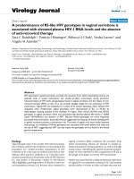
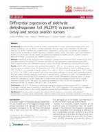
Fig. 1 Left panel: UV–vis
absorption spectra of (a) CuSO
4
EG solution, (b) pH 11solution,
and (c) transitional colorless
solution. Right panel: UV–vis
absorption spectra of copper
colloids A, B, and C, which are
synthesized under different
molecular ratios of PVP and
CuSO
4
, viz., (a) 2:1, (b) 10:1,
and (c) 20:1, respectively
706 Nanoscale Res Lett (2009) 4:705–708
123
Cu(II) ? Cu(I) ? Cu(0). The flow chart of color changes
during the synthesis depicted in Chart 1, for a better visual
understanding.
The right panel of Fig. 1 shows the UV–vis absorption
spectra of three colloidal solutions synthesized under
otherwise the same conditions except differences in
molecular ratios of PVP (counted as the repeat union) and
CuSO
4
, viz. 2:1, 10:1, and 20:1 (designated as colloids A,
B, and C). A band centered at ca. 560 nm for each solution
can be seen, characteristic of surface plasmon absorption of
copper colloids. In addition, no enhanced background
absorption around 800 nm can be observed implicating that
the colloidal particles are nominally reduced copper in
nature without being oxidized to copper oxide on surface
[17–19]. The as-prepared Cu colloids exhibit a blueshift of
ca. 10 nm in absorption peak as compared to the previously
reported Cu colloid [14], suggestive of much smaller par-
ticle sizes [20]. The peak position in curve a is redshifted
by ca. 2 nm with respect to curves b and c, while the
corresponding peak intensity decreases apparently from
colloid A to C, revealing that the particle size decreases
with increasing molecular ratio of PVP and CuSO
4
as
predicted by the Mie’s theory [19]. Notably, no precipita-
tion was observed and the UV–vis absorption profile
remained virtually unchanged even after 2 months storage
in a simply sealed container, suggestive of long-term sta-
bility of the as-prepared Cu colloids.
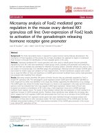
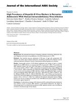
Figure 2 are the TEM images for Cu nanoparticles from
colloids A, B, and C together with the histograms of par-
ticle size distributions, indicating that Cu nanoparticles are
rather monodispersed in nearly sphere shape. Careful sta-
tistical examination of the nanoparticles revealed that
average sizes of 3.1 ± 0.5 nm, 2.6 ± 0.6 nm, and
1.4 ± 0.6 nm were for colloids A, B, and C, respectively,
in agreement with the UV–vis results. As PVP molecules
strongly adsorb on as-prepared metal nanoparticles, they
effectively prevent the aggregation in reducing metal ions
[21–23]. Consequently, at a higher molecular ratio of PVP
and CuSO
4
, more PVP molecules are adsorbed on Cu
nanoparticle surfaces, keeping them from the excessive
growth, and leading to the formation of smaller
nanoparticles.
Chart 1 The flow chart
showing the synthesis procedure
and related color variations
Fig. 2 TEM images and corresponding histograms of copper colloid
(a)A,(b) B, and (c) C, respectively
Nanoscale Res Lett (2009) 4:705–708 707
123
It should be pointed out that in a traditional polyol
process to obtain metal nanoparticles, EG acts not only as
an organic solvent but also as a reducing agent [24]. The
use of EG to reduce Cu(II) species at a relative high tem-
perature would produce Cu nanoparticles [30 nm due to
its inherently mild reducing ability [7]. At room tempera-
ture, EG is unlikely involved in the reduction of Cu(II)
species, and the introduction of a much stronger reducing
agent is believed to be a crucial factor for achieving much
smaller particle sizes, given that the nucleation and growth
processes are greatly dependent on the power of reducing
agent [15]. Briefly, weaker reducing agents benefit further
growth of the existing nuclei leading to larger particles.
With stronger reducing agent, the larger nucleation prob-
ability would be expected, in favor of the formation of
more ultrafine nanoparticles.
Along this line, the use of stronger reducing agent
NaBH
4
played an important role as well in the controlled
synthesis of ultrafine copper nanoparticles in this study.
The reduction ability of NaBH
4
is related to its concen-
tration and the solution pH. For a given concentration, the
solution pH should be properly regulated to attain a satis-
factory size control. We found that for 0.5 M NaBH
4
,
pH 11 was optimized for the fabrication of ultrafine copper
nanoparticles. At too low pH, rapid release of strong
hydrogen bubbles produces large amount of Cu nuclei,
which tend to aggregate into large agglomerates; at too
high pH, newly reduced Cu atoms prefer to deposit onto the
nuclei already formed, eventually causing larger Cu
nanoparticles.
Conclusions
In summary, nominally reduced Cu nanoparticles was syn-
thesized by a modified polyol method, leading to ultrafine
nanoparticles ranging from 1.4 ± 0.6 nm to 3.1 ± 0.5 nm
in average with narrow size distribution, uniform shape, and
great stability. The size of the nanoparticles decreases with
increasing the ratio of PVP and Cu(II) concentrations in the
precursor solution by using NaBH
4
as the reducing agent.
The organic solution EG, stabilizer PVP, and the pH value
showed a co-effect to ensure the formation of the desired
particles, and the reduction process may go through
Cu(II) ? Cu(I) ? Cu(0).
Acknowledgment This work is supported by the NSFC (Nos.
20673027 and 20833005) and Sino-German IRTG program.
References
1. S.J. Oldenburg, R.D. Averitt, S.L. Westcott, N.J. Halas, Chem.
Phys. Lett. 288, 243 (1998). doi:10.1016/S0009-2614(98)00277-2
2. A. Henglein, J. Phys. Chem. B 104, 6683 (2000). doi:10.1021/
jp000746j
3. A.M. Michaels, J. Jiang, L. Brus, J. Phys. Chem. B 104, 11965
(2000). doi:10.1021/jp0025476
4. Y.G. Sun, Y.N. Xia, Science 298, 2176 (2002). doi:10.1126/
science.1077229
5. S.J. Huo, Q.X. Li, Y.G. Yan, Y. Chen, W.B. Cai, Q.J. Xu, M.
Osawa, J. Phys. Chem. B 109, 15985 (2005). doi:10.1021/
jp052585v
6. S.J. Huo, X.K. Xue, Q.X. Li, S.F. Xu, W.B. Cai, J. Phys. Chem. B
110, 25721 (2006). doi:10.1021/jp064036a
7. S. Jeong, K. Woo, D. Kim, S. Lim, J.S. Kim, H. Shin, Y. Xia, J.
Moon, Adv. Funct. Mater. 18, 679 (2008). doi:10.1002/
adfm.200700902
8. J. Tang, S. Hinds, S.O. Kelley, E.H. Sargent, Chem. Mater. 20,
6906 (2008). doi:10.1021/cm801655w
9. N.A. Dhas, C.P. Raj, A. Gedanken, Chem. Mater. 10, 1446
(1998). doi:10.1021/cm9708269
10. G. Vitulli, M. Bernini, S. Bertozzi, E. Pitzalis, P. Salvadori, S.
Coluccia, G. Martra, Chem. Mater. 14, 1183 (2002). doi:10.1021/
cm011199x
11. H.H. Huang, F.Q. Yan, Y.M. Kek, C.H. Chew, G.Q. Xu, W. Ji,
P.S. Oh, S.H. Tang, Langmuir 13, 172 (1997). doi:10.1021/
la9605495
12. Z.W. Liu, Y. Bando, Adv. Mater. 15, 303 (2003). doi:
10.1002/adma.200390073
13. R.V. Kumar, Y. Mastai, Y. Diamant, A. Gedanken, J. Mater.
Chem. 11, 1209 (2001). doi:10.1039/b005769j
14. S.H. Wu, D.H. Chen, J. Colloid. Interface Sci. 273, 165 (2004).
doi:10.1016/j.jcis.2004.01.071
15. B.K. Park, S. Jeong, D. Kim, J. Moon, S. Lim, J.S. Kim, J.
Colloid. Interface Sci. 311, 417 (2007). doi:10.1016/j.jcis.
2007.03.039
16. I.G. Casella, T.R.I. Cataldi, A. Guerrieri, E. Desimoni, Anal.
Chim. Acta 335, 217 (1996). doi:10.1016/S0003-2670
(96)00351-0
17. I. Lisiecki, M.P. Pileni, J. Am. Chem. Soc. 115, 3887 (1993). doi:
10.1021/ja00063a006
18. A. Yanase, H. Komiyama, Surf. Sci. 248, 11 (1991). doi:
10.1016/0039-6028(91)90056-X
19. I. Lisiecki, F. Billoudet, M.P. Pileni, J. Phys. Chem. 100, 4160
(1996). doi:10.1021/jp9523837
20. N. Shirtcliffe, U. Nickel, S. Schneider, J. Colloid. Interface Sci.
211, 122 (1999). doi:10.1006/jcis.1998.5980
21. P. Jiang, S.Y. Li, S.S. Xie, Y. Gao, L. Song, Chem. Eur. J. 10,
4817 (2004). doi:10.1002/chem.200400318
22. H.S. Shin, H.J. Yang, S.B. Kim, M.S. Lee, J. Colloid. Interface
Sci. 274, 89 (2004). doi:10.1016/j.jcis.2004.02.084
23. Z.T. Zhang, B. Zhao, L.M. Hu, J. Solid State Chem. 121, 105
(1996). doi:10.1006/jssc.1996.0015
24. C. Bock, C. Paquet, M. Couillard, G.A. Botton, B.R. MacDou-
gall, J. Am. Chem. Soc. 126, 8028 (2004). doi:10.1021/
ja0495819
708 Nanoscale Res Lett (2009) 4:705–708
123