Báo cáo hóa học: " Studies on Preparation of Photosensitizer Loaded Magnetic Silica Nanoparticles and Their Anti-Tumor Effects for Targeting Photodynamic Therap" ppt
Bạn đang xem bản rút gọn của tài liệu. Xem và tải ngay bản đầy đủ của tài liệu tại đây (512.92 KB, 9 trang )
NANO EXPRESS
Studies on Preparation of Photosensitizer Loaded Magnetic Silica
Nanoparticles and Their Anti-Tumor Effects for Targeting
Photodynamic Therapy
Zhi-Long Chen Æ Yun Sun Æ Peng Huang Æ
Xiao-Xia Yang Æ Xing-Ping Zhou
Received: 13 November 2008 / Accepted: 8 January 2009 / Published online: 31 January 2009
Ó to the authors 2009
Abstract As a fast developing alternative of traditional
therapeutics, photodynamic therapy (PDT) is an effective,
noninvasive, nontoxic therapeutics for cancer, senile mac-
ular degeneration, and so on. But the efficacy of PDT was
compromised by insufficient selectivity and low solubility.
In this study, novel multifunctional silica-based magnetic
nanoparticles (SMNPs) were strategically designed and
prepared as targeting drug delivery system to achieve
higher specificity and better solubility. 2,7,12,18-Tetra-
methyl-3,8-di-(1-propoxyethyl)-13,17-bis-(3-hydroxypropyl)
porphyrin, shorted as PHPP, was used as photosensitizer,
which was first synthesized by our lab with good PDT
effects. Magnetite nanoparticles (Fe
3
O
4
) and PHPP were
incorporated into silica nanoparticles by microemulsion
and sol–gel methods. The prepared nanoparticles were
characterized by transmission electron microscopy, X-ray
diffraction, Fourier transform infrared spectroscopy and
fluorescence spectroscopy. The nanoparticles were
approximately spherical with 20–30 nm diameter. Intense
fluorescence of PHPP was monitored in the cytoplasm of
SW480 cells. The nanoparticles possessed good biocom-
patibility and could generate singlet oxygen to cause
remarkable photodynamic anti-tumor effects. These sug-
gested that PHPP-SMNPs had great potential as effective
drug delivery system in targeting photodynamic therapy,
diagnostic magnetic resonance imaging and magnetic
hyperthermia therapy.
Keywords Targeting photodynamic therapy Á
Photosensitizer Á Silica Á Magnetic nanoparticles Á
Tumor
Introduction
Photodynamic therapy (PDT) is an effective, noninvasive
and nontoxic therapeutics for cancer, senile macular
degeneration, actinic keratosis, port-wine stains, rheuma-
toid arthritis, and so on [1, 2]. After bio-distribution,
photosensitizer (PS) administered systemically or topically
is activated by light of appropriate wavelength and dosage.
The activated PS transfers its excited-state energy to
nearby oxygen molecular to generate reactive oxygen
species, such as singlet oxygen (
1
O
2
) or peroxides induc-
ing oxidative damage to target tissue and blood vessels
that feed them [1–4]. Due to minimal invasion and non-
toxicity, PDT provides patients, weak or failed in
traditional therapy, opportunities to be treated painlessly
and repeatedly.
However, the PDT efficacy is compromised by insuffi-
cient selectivity and low solubility. Although several
methods including drug delivery systems were investigated
[3–9], developing a PS delivery system for higher selec-
tivity and less dark toxicity is still a challenge [5, 6].
Magnetic drug delivery system is a promising drug
delivery system, which can be steered to the target tissue
simply by an external magnetic field [10, 11]. Silica
nanoparticles, easily prepared with desired size, shape and
porosity, are water-soluble, stable and biocompatible.
More importantly, silica nanoparticles are permeable to
Z L. Chen (&) Á Y. Sun Á P. Huang Á X X. Yang Á
X P. Zhou (&)
Department of Pharmaceutical Science and Technology,
College of Chemistry and Biology, Donghua University,
Shanghai 201620, China
e-mail:
X P. Zhou
e-mail:
123
Nanoscale Res Lett (2009) 4:400–408
DOI 10.1007/s11671-009-9254-5
small molecular such as singlet oxygen [4, 5], which is the
key effector of PDT. Therefore, photosensitizer loaded
silica nanoparticles are different from conventional
delivery systems which need releasing of the loaded drug
[9].
Previous investigations of fluorescent-magnetic nano-
particles mainly focused on the MRI imaging and
fluorescence imaging for diagnosis; however, there are
few studies on the multifunctional magnetic targeting
drug delivery system for diagnosis and therapy [12, 13].
In the earliest study of magnetic targeting, a magnetic
fluid was developed to which epirubicin was chemically
bound to enable those agents to be directed within an
organism by high-energy magnetic fields. In vitro and in
vivo study of the epirubicin-magnetic fluid indicated
biosafety and complete tumor response [10, 11], demon-
strating the potential of magnetic targeting. Recently, the
investigation of PS encapsulated magnetic silica nano-
particles (SMNPs) showed efficient cellular uptake [14]
and obvious generation of singlet oxygen in vitro [15, 16],
which indicated the potential of SMNPs as targeting drug
delivery system.
Herein multifunctional PS encapsulated magnetic silica
nanoparticles were strategically designed and synthesized,
the silica shell of which can provide a porous environ-
ment for oxygen diffusion. 2,7,12,18-Tetramethyl-3,8-di-
(1-propoxyethyl)-13,17-bis-(3-hydroxypropyl) porphyrin,
shorted as PHPP, was used as photosensitizer, which was
first synthesized by our lab with good PDT effects [17, 18].
The SMNPs were characterized by transmission electron
microscopy, X-ray diffraction, Fourier transform infrared
spectroscopy and fluorescence spectroscopy. The genera-
tion of singlet oxygen was monitored by RNO bleaching
assay, and the photodynamic efficacy of the SMNPs to
SW480 colon carcinoma cells was detected by MTT assay
(Scheme 1).
Experimental Section
Materials
Ferrous(II) sulfate heptahydrate (FeSO
4
Á 7H
2
O, 99%),
ferric chloride hexahydrate (FeCl
3
Á 6H
2
O, 99%), anhy-
drous ethanol (99.7%), ammonium hydroxide (25.2–
28.0%), 1-butanol (99.8%), dimethyl sulfoxide (DMSO,
99.8%), tetrahydrofuran (THF, 99.9%), hydrochloric acid
(36%) and oleic acid (99%) were purchased from Sinop-
harm Co. (China). Surfactant aerosol OT (AOT, 98%),
tetraethylorthosilicate (TEOS, 99.99%), (3-mercaptopro-
pyl) trimethoxysilane (MPS, 95%), N,N-dimethyl-4-
nitrosoaniline (RNO, 99%), imidazole (C99%), trypsinase
(0.25%), and 3-(4,5-dimethylthiazol-2-yl)-2,5-diphenyltet-
razolium bromide (MTT) were obtained from Aldrich. The
PS PHPP was synthesized by our lab with purity C98%.
SW480 cell was available in the cell store of Chinese
Academy of Science. Other materials for cell culture,
unless mentioned otherwise, were purchased from GIBCO.
All the above-mentioned chemicals were used without any
further purification.
Preparation of Fe
3
O
4
Nanoparticles
In total, 2.51 g (9 mmol) FeCl
3
Á 6H
2
O and 1.25 g
(4.5 mmol) FeSO
4
Á 7H
2
O were dissolved in 20 mL water.
The solution was vigorously stirred, followed by adding
10 mL 1.5 mol L
-1
NH
3
Á H
2
O. The color of the solution
was changed into black and the black solid produced was
precipitated to the bottom. The Fe
3
O
4
nanoparticles were
obtained after the precipitants were washed for five times
with 20 mL distilled water and 20 mL ethanol alternatively
to remove unreacted chemicals.
Preparation of Silica-Based Fe
3
O
4
Nanocarriers
In total, 1 g Fe
3
O
4
nanoparticles and 10 g oleic acid were
mixed with 10 mL ethanol. The suspension was refluxed
for 30 min. Fe
3
O
4
/OA nanoparticles were obtained after
the excess oleic acid was scoured off with ethanol by the
magnetic decantation.
Micelles were prepared by dissolving 0.90 g AOT and
1600 lL 1-butanol in 40 mL doubly distilled water by
vigorous magnetic stirring. A solution of 60 lL PHPP
(15 mmol L
-1
) in 1-butanol and 0.003 g Fe
3
O
4
/OA
nanoparticles were added to above micellar system. After
30 min stirring, a new micellar system containing PHPP-
Fe
3
O
4
/OA was formed. A total of 200 lL TEOS and
1.2 mL aqueous ammonia were added to the PHPP-Fe
3
O
4
/
OA system prior to 1 h stirring. Then, 10 lL MPS was
added, followed by continued 20 h stirring. The resultant
Scheme 1 Chemical structure of PHPP
Nanoscale Res Lett (2009) 4:400–408 401
123
was treated by magnetic separation and washed with eth-
anol until no PS could be detected in the supernatant by
UV–Vis spectroscopy. All the above-mentioned experi-
ments were conducted at room temperature. The products
were dried at 60 °C for 3 h in vacuum oven.
Characterization
The X-ray diffraction pattern of silica-based magnetic
nanocarriers powders was obtained using D/max-2550PC
(Geigerflex, Rigaku, Japan) with monochromated CuKa
radiation operated at 40 kV and 100 mA. Transmission
electron microscopy (TEM) was employed to determine
the morphology and size of the aqueous dispersion of
nanocarriers, using a HITACHIH-800 electron microscope,
operating at an accelerating voltage of 200 kV. UV–Vis
absorption spectra were recorded using a Jasco V-530
spectrophotometer, in a quartz cuvette with 1 cm path
length. Fluorescence spectra were recorded on a HIT-
ACHIH FL-4500 spectrofluorimeter.
Encapsulation Efficiency Measurements
The UV–Vis measurements of the PHPP-SMNPs were
carried out contrasted to other six groups: (a) PHPP; (b)
Fe
3
O
4
? PHPP; (c) PHPP ? HCl; (d) Fe
3
O
4
? HCl; (e)
Fe
3
O
4
? PHPP ? HCl; (f) PHPP ? |SMNPs ? HCl. The
amount of the mixed solvent was 0.1 mL the concentrated
HCl and 2.9 mL ethanol. The absorbance at 409 nm was
used to validate the PS presence and estimate the PS
encapsulation efficiency. Each measurement was repeated
three times.
The standard curve was established in the drug
concentration range from 7.65 9 10
-7
mol L
-1
to
1.02 9 10
-5
mol L
-1
. Different concentrations of PHPP
(7.65 9 10
-7
, 2.55 9 10
-6
, 5.10 9 10
-6
, 7.65 9 10
-6
,
1.02 9 10
-5
mol L
-1
) were mixed with 0.1 mL concen-
trated HCl and 2.9 mL ethanol. The samples were
measured at 409 nm wavelength. Each experiment was
repeated three times.
Detection of Singlet Oxygen
The PHPP-SMNPs in phosphate buffer (pH = 7.4) were
irradiated in the presence of imidazole (10 mmol L
-1
) and
RNO (50 mmol L
-1
). The RNO bleaching by
1
O
2
was
followed spectrophotometrically with observing the
decrease in the 440 nm absorption peak of RNO as a
function of irradiation time. The reaction mixture in a 1 cm
spectrometric cuvette, placed at a distance of 12 cm, was
continuously irradiated using 632.8 nm laser.
In Vitro Studies with Tumor Cells
Preparation of PHPP-SMNPs Solution
PHPP-SMNPs was diluted to 100 lmol L
-1
with 0.5%
carboxymethylcellulose sodium. The solution was then
diluted with RPMI-1640 medium (supplemented with
100 U mL
-1
penicillin, 10 U mL
-1
streptomycin and 10%
calf serum) using a dilution factor of 5 to varied concen-
trations: 0, 0.03, 0.13, 0.64, 3.20, 16.00, and 80 lmol L
-1
.
Biosafety Assessment
SW480 carcinoma cells (3 9 10
3
cells per well) were
seeded in 96-well plates and incubated overnight at 37 °C
in a humidified 5% CO
2
atmosphere. After being rinsed
with PBS (pH 7.4), the cells were incubated with 100 lL
varied concentration of PHPP-SMNPs prepared above for
24 h at 37 °C in the dark under the same conditions. Rinsed
with PBS, the cells were incubated another 48 h. Cell
viability was determined by the colorimetric 3-(4,5-
dimethylthiazol-2-yl)-2,5-diphenyltetrazolium bromide
(MTT) assay. Cells were rinsed with PBS and then incu-
bated with culture medium containing 0.5 mg mL
-1
MTT
reagent for 3 h. The medium was then removed and the
formazan crystals formed were dissolved in 100 lL
DMSO. The absorbance at 492 nm for each well was
recorded by a microplate reader.
Photodynamic Activity Assay
Two plates were set up as dark control and experimental
group for the MTT assay and these plates were seeded,
exposed identically to the plates prepared for the biosafety
assessment. The cells of experimental group were then
rinsed again with PBS and immersed in 100 lL of fresh
culture medium before being illuminated using a 488 nm
argon-ion laser with energy density of 4.35 J/cm
2
from the
underside of the culture plate. After 10 min illumination,
cells were incubated 48 h in a 5% CO
2
, 95% air humidified
incubator at 37 °C. Dark control group keep identical to
experimental group except illumination. Photodynamic
activity assay was also determined by MTT assay as
mentioned above.
Statistical Analysis
Cell viability was calculated using the following formula:
average A value of experimental group/average A value of
control group 9 100%. Results were expressed as
means ± SD. Comparisons between two groups were
made by unpaired two-tailed Student’s t-test using SPSS
402 Nanoscale Res Lett (2009) 4:400–408
123
15.0 software. P-value of less than 0.05 was taken to
indicate statistical significance.
Results and Discussion
Preparation of Fe
3
O
4
Nanoparticles
Fe
3
O
4
nanoparticles, prepared by co-precipitation method
[19, 20], were 10 ± 2 nm in diameter by measuring 200
randomly selected particles in enlarged TEM images,
which agreed well with the value calculated from Scherrer
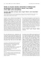
Equation. Figure 1 shows the spherical morphology
(Fig. 1a) and the characteristic peaks (Fig. 1b), which was
compatible with the values of standard pattern. In addition
to the dispersed and well-separated features, the formed
Fe
3
O
4
nanoparticles also exhibited some degree of aggre-
gated morphology. The particles show an attraction to
magnetic field, demonstrating the magnetic responsibility.
The Fe
3
O
4
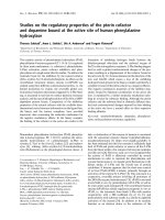
nanoparticles were coated with oleic acid
according to the procedure described by Reimers [21, 22].
By adding oleic acid at melting point, the Fe
3
O
4
nano-
particles were hydrophobized as illustrated in Fig. 2. The
precipitate could be readily redispersed in the solvents such
as cyclohexane, chloroform, or 1-butanol.
Preparation of PHPP-SMNPs
Considering the size of the emulsion droplet is directly
related to the final nanoparticle size, the formation of the
emulsion is the key aspect. Emulsions can be classified in
macroemulsions, microemulsions and miniemulsions (or
nanoemulsions) The o/w microemulsion method is easy to
scale up, it does not need high shear stress, and it is
transparent and thermodynamically stable, with droplets
mean sizes from 20 to 50 nm [23, 24]. So it is widely used
for entrapment of hydrophobic compounds. Here, the o/w
microemulsion method was combined with sol–gel method
which was the classical approach to synthesize SiO
2
nanoparticles [25, 26]. The PHPP-SMNPs were prepared in
the nonpolar core of AOT/1-butanol/water micelles, as
shown schematically in a pictorial representation (Fig. 3).
PHPP-absorbed Fe
3
O
4
/OA had priority to disperse in
1-butanol droplets redounding to form the nanocarriers.
Characterization of PHPP-SMNPs
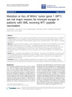
TEM image (Fig. 4a) of PHPP-SMNPs showed that the
PHPP-SMNPs were approximately spherical, sized in the
range of 20–30 nm, but some agglomeration could be
observed. Dynamic light scattering measurements were
performed to study the behavior of a suspension containing
PHPP-SMNPs. The hydrodynamic diameter of the nano-
carriers was about 126 nm and the polydispersion was
about 0.116, which verified the aggregates in the
Fig. 1 TEM image (a) and
XRD pattern (b)ofFe
3
O
4
nanoparticles
Fig. 2 The Fe
3
O
4
nanoparticles hydrophobized by coating of oleic
acid, dispersed in different system
Nanoscale Res Lett (2009) 4:400–408 403
123
suspension of particles. These aggregates were not stable
and could be easily redispersed by simply shake or
sonication.
Figure 4b illustrates the XRD pattern of PHPP-SMNPs.
The (111) peak is derived from the amorphous mesoporous
silica spheres and the characteristic (311), and (440) peaks
are typical of a cubic structure. The result showed that the
crystallinity has not changed after encapsulation.
Figure 5 shows the typical FT-IR spectra of (a) PHPP,
(b) SMNPs, and (c) PHPP-SMNPs. The bands C–H at
740 cm
-1
(Fig. 5a) and C–O/C–N at 1079 cm
-1
(Fig. 5c)
indicated the encapsulation of the drug in the SMNPs.
According to (b) and (c), it was found that the Si–O
vibration absorption peak of the 1044 cm
-1
shifted to
1079 cm
-1
, which might be contributed to the integration
of Si–O vibration absorption and C–O/C–N vibration
absorption peak. The above facts suggested that PHPP was
successfully wrapped in the SMNPs.
The photoluminescence peaks of PHPP were at 625 and
690 nm, as shown in Fig. 6. Figure 7 represents the fluo-
rescence emission spectra of mother liquor and aqueous
dispersion of the PHPP-SMNPs at same concentration. The
emission signal from the PHPP in the PHPP-SMNPs was
almost 20% of the all solution. Fluorescence intensity
decreased significantly from (a) to (b), illustrating that
PHPP was not completely encapsulated in the SMNPs.
Fig. 3 Scheme depicting the
synthesis and purification of
(PHPP-Fe
3
O
4
/OA)/SiO
2
Fig. 4 TEM image (a) and
XRD pattern (b) of PHPP-
SMNPs
Fig. 5 FT-IR spectra of (a) PHPP, (b) (Fe
3
O
4
/OA)/SiO
2
, and (c)
(PHPP-Fe
3
O
4
/OA)/SiO
2
Fig. 6 Fluorescence emission spectra of PHPP. The excitation
wavelength is 420 nm
404 Nanoscale Res Lett (2009) 4:400–408
123
Figure 8 shows a concentration-dependent increase in
photoluminescence signal, demonstrating the encapsulation
of the PHPP again.
Encapsulation Efficiency Measurements
The amount of drug entrapped within PHPP-SMNPs was
determined by dissolving PHPP-SMNPs into hydrochloric
acid to destroy the Fe
3
O
4
cores of PHPP-SMNPs for the
release of PHPP. After ethanol addition, the absorbance of
PHPP was detected and was performed by ultraviolet
spectrophotometer at 409 nm.
Figure 9a shows the UV spectra of different substances
in the 200–700 nm wavelength range, using ethanol as the
solvent. The spectrum of PHPP (a) showed the special
absorption peaks of PHPP at 399 nm. The increased
absorption of PHPP and SMNPs mixture was caused by the
absorption of SMNPs. The reaction of PHPP with HCl
caused a red shift of the special absorption peak of PHPP
from 399 to 409 nm (c). Spectrum (d) indicated absorp-
tions of SMNPs and HCl at 240, 314, and 364 nm,
demonstrating that the 240, 314, and 364 nm peak of the
spectrum (e) attributed to SMNPs and HCl. Therefore, the
409 nm was the characteristic absorption peaks of PHPP
with or without SMNPs.
The PHPP-SMNPs treated with HCl were detected by
UV–Vis spectrophotometer and the spectrums are dis-
played in Fig. 10.
The standard curve had a good linear relation
(r = 0.99905) within the range of 7.65 9 10
-7
to
1.02 9 10
-5
mol L
-1
, described by the following typical
equation: Y = 0.04447 ? 0.02571x (see Fig. 11). PHPP
encapsulation efficiency was 20.8%, estimated by the
typical equation.
Fig. 7 Fluorescence emission spectra of (a) mother liquor and (b)
PHPP-SMNPs
Fig. 8 Fluorescence emission spectra of (a) 0.0 mg, (b) 0.2 mg, and
(c) 0.6 mg PHPP-SMNPs. The excitation wavelength is 420 nm
Fig. 9 UV spectra of (a) PHPP, (b) PHPP mixed with SMNPs, (c)
PHPP dissolved in HCl and ethanol, (d) SMNPs dissolved in HCl and
ethanol, and (e) SMNPs and PHPP dissolved in HCl and ethanol
Fig. 10 UV spectra of the PHPP-SMNPs treated with HCl and
ethanol
Nanoscale Res Lett (2009) 4:400–408 405
123
Detection of Singlet Oxygen
The N,N-dimethyl-4-nitrosoaniline (RNO) was used as an
indicator for photo-induced singlet oxygen with imidazole
as a chemical trap for singlet oxygen [27–29]. The prin-
ciple of this method is shown in the following formula:
1
O
2
þ imidazole ! imidazole À
1
O
2
ÂÃ
imidazole À
1
O
2
ÂÃ
þ RNO ! RNO
2
þ Products
The bleaching of RNO by
1
O
2
was followed spectropho-
tometrically with observing the decrease in the 440 nm
absorption peak of RNO as a function of irradiation time.
A decrease in the 440 nm absorption peak of RNO was
caused by the Fe
3
O
4
nanoparticles without irradiation. So a
24 h aging of the PHPP-SMNPs and RNO system was
necessary prior to detecting the
1
O
2
productivity, until the
absorption of RNO at 440 nm did not decline. The system
was irradiated. The continued decrease at 440 nm absorp-
tion peak was caused by the significant generation of
1
O
2
released from the PHPP-SMNPs (Fig. 12), indicating the
potential for efficient PDT.
In Vitro Studies with Tumor Cells: Cellular Uptake,
Biosafety Assessment and Photodynamic Activity
Assay
Intracellular Uptake
As an essential tool in material science and biology, fluo-
rescence microscopy demonstrates the ability to monitor
the precise location of intracellular fluorescence materials
excited by light of specific wavelengths, as well as their
associated diffusion coefficients, transport characteristics,
and interactions with other biomolecules. To test the
intracellular uptake of PHPP-SMNPs, fluorescence imag-
ing was performed on human SW480 colon carcinoma
cells after incubation with 50 lmol L
-1
PHPP-SMNPs for
4 h in cell culture incubator. As shown in Fig. 13, PHPP-
SMNPs were taken up by SW480 cells and showed sig-
nificant intracellular fluorescence (extra nuclear) compared
to unincubated cell. As the PDT effects depend on the
uptake of PS by tumor cells, the intense fluorescence of
intracellular PHPP-SMNPs, related to the PS concentra-
tion, predicted an available obvious PDT effects.
Subcellular distribution of PHPP-SMNPs in the cytoplasm
primarily demonstrated slight effects on DNA.
Biosafety Assessment
Biosafety assessment was essential to evaluate the potential
application of silica nanoparticle in clinics. Here, MTT
assay was performed to detect the dark toxicity of PHPP-
SMNPs. No obvious dark toxicity of PHPP-SMNPs on
SW480 carcinoma cells was detected within 0.03–
80 lmol L
-1
concentration range in comparison with
control (Fig. 14a). In addition, negligible cell death and
physiological state changes of SW480 cells treated with
highest dosage of PHPP-SMNPs were observed in
Fig. 14b. It could be predicted that the PHPP-SMNPs had
minimal, if any, impact on cellular functions, which indi-
cated the low dark toxicity and good biocompatibility.
In Vitro Photodynamic Efficacy
Likewise, the MTT assay was performed to examine the
phototoxicity of PHPP-SMNPs to SW480 colon carcinoma
cell lines, which indicated the PDT efficacy in vitro [30].
Fig. 11 The standard curve of PHPP dissolved in HCl and ethanol,
measured at 409 nm. Typical equation: Y = 0.04447 ? 0.02571x
(where x is the concentration and Y is the absorbance)
Fig. 12 Photosensitized RNO bleaching measured at 440 nm as a
function of irradiation time
406 Nanoscale Res Lett (2009) 4:400–408
123
Cell viability was normalized to control cells (no drug and
unirradiated) in Fig. 14. The combination of 24 h exposure
of tumor cells to PHPP-SMNPs and 4.35 J/cm
2
irradiation
induced a drug concentration-dependent cytotoxicity to
SW480 tumor cells, which was significantly different from
unirradiated control in statistics, as shown in Fig. 15. With
a 10 min light exposure, 80 lmol L
-1
PHPP-SMNPs in the
safety range measured as above caused approximately 40%
cell viability lost, demonstrating obvious photodynamic
activity. The group treated with the drug without light
exposure showed that the drug alone had no effects on
tumor cells which coincided with the result of biosafety
assay. In this case, the cell viability at the maximum of
concentration (80 lmol L
-1
), slightly lower than the con-
trol, was caused by the natural light during the execution of
experiments.
Fig. 13 Intracellular uptake of
PHPP-SMNPs. Cells alone
(a, b), and cells incubated with
50 lmol L
-1
PHPP-SMNPs for
4h(c, d)
Fig. 14 Dark cytotoxicity of
PHPP-SMNPs. SW480 cells
were incubated with
0–80 lmol L
-1
PHPP-SMNPs
for 24 h at 37 °C in the dark.
Cell toxicity was determined by
MTT assay. Data represent
mean ± SD (n = 3)
Nanoscale Res Lett (2009) 4:400–408 407
123
Conclusions
Novel multifunctional silica-based magnetic nanoparticles
containing photosensitizer PHPP were prepared. The
PHPP-SMNPs were approximately spherical and 20–
30 nm in diameter, achieving 20.8% encapsulation effi-
ciency of PHPP. They showed no obvious toxicity without
irradiation, but significant generation of singlet oxygen and
remarkable photodynamic efficacy after irradiation. The
PHPP-SMNPs were primarily distributed in the cytoplasm.
It can be concluded that the silica-based magnetic
nanoparticles are of great value as effective drug delivery
system in targeting photodynamic therapy. The potential of
the magnetic core for magnetic resonance imaging and
magnetic hyperthermia therapy could also be expected.
Acknowledgments This work was supported by National Natural
Science Foundation of China (grant nos. 30070862, 30271534),
Shanghai Municipal Foundation (grant nos. 05ZR14002, 06PJ14001,
064319020).
References
1. J. Levy, M. Obochi, Photochem. Photobiol. 64, 737 (1996). doi:
10.1111/j.1751-1097.1996.tb01828.x
2. Y. Konan, R. Gurny, E. Alle
´
mann, J. Photochem. Photobiol. B
66, 89 (2002)
3. T. Dougherty, Photochem. Photobiol. 45, 879 (1987). doi:
10.1111/j.1751-1097.1987.tb07898.x
4. I. Roy, T. Ohulchanskyy, H. Pudavar, E. Bergey, A. Oseroff, J.
Morgan, T. Dougherty, P. Prasad, J. Am. Chem. Soc. 125, 7860
(2003). doi:10.1021/ja0343095
5. S. Wang, R. Gao, F. Zhou, M. Selke, J. Mater. Chem. 14, 487
(2004). doi:10.1039/b311429e
6. R. De Gao, H. Xu, M. Philbert, R. Kopelman, Nano Lett. 6, 2383
(2006). doi:10.1021/nl0617179
7. C. van Nostrum, Adv. Drug Deliv. Rev. 56, 9 (2004). doi:
10.1016/j.addr.2003.07.013
8. A. Derycke, P. de Witte, Adv. Drug Deliv. Rev. 56, 17 (2004).
doi:10.1016/j.addr.2003.07.014
9. J. Snyder, E. Skovsen, J. Lambert, P. Ogilby, J. Am. Chem. Soc.
127, 14558 (2005). doi:10.1021/ja055342p
10. T. Jain, I. Roy, T. De, A. Maitra, J. Am. Chem. Soc. 120, 11092
(1998). doi:10.1021/ja973849x
11. T.K. Jain, M.K. Reddy, M.A. Morales, D.L. Leslie-Pelecky,
V. Labhasetwar, Mol. Pharm. 5, 316 (2008). doi:10.1021/mp700
1285
12. A. Quarta, R. Di Corato, L. Manna, A. Ragusa, T. Pellegrino,
IEEE Trans. Nanobiosci. 6, 298 (2007). doi:10.1109/TNB.2007.
908989
13. S. Corr, Y. Rakovich, Y. Gun’ko, Nanoscale Res. Lett. 3,87
(2008). doi:10.1007/s11671-008-9122-8
14. C.W. Lai, Y.H. Wang, C.H. Lai, M.J. Yang, C.Y. Chen, P.T.
Chou, C.S. Chan, Y. Chi, Y.C. Chen, J.K. Hsiao, Small 4, 218
(2008). doi:10.1002/smll.200700283
15. L.O. Cinteza, T.Y. Ohulchanskyy, Y. Sahoo, E.J. Bergey,
R.K. Pandey, P.N. Prasad, Mol. Pharm. 3, 415 (2006). doi:
10.1021/mp060015p
16. D. Tada, L. Vono, E. Duarte, R. Itri, P. Kiyohara, M. Baptista,
L. Rossi, Langmuir 23, 8194 (2007). doi:10.1021/la700883y
17. Z L. Chen, W Q. Wan, J R. Chen, F. Zhao, D Y. Xu, Het-
erocycles 48, 1739 (1998)
18. Z L. Chen, W Q. Wang, K. Fan, Y. Zhou, W. Liu, D. Xu, Chin.
J. Med. Chem. 8, 1 (1998)
19. X. Zhou, S. Ni, X. Wang, F. Wu, Curr. Nanosci. 3, 259 (2007).
doi:10.2174/157341307781422942
20. D. Kim, S. Lee, K. Kim, M. Lee, Y. Lee, Curr. Appl. Phys. 6, 242
(2006)
21. S. Khalafalla, G. Reimers, U.S. Patent No 3, 1974
22. L. Ramirez, K. Landfester, Macromol. Chem. Phys. 204,22
(2003). doi:10.1002/macp.200290052
23. R. Alex, R. Bodmeier, J. Microencapsul. 7, 347 (1990). doi:
10.3109/02652049009021845
24. B. Paul, S. Moulik, Curr. Sci. 80, 990 (2001)
25. W. Stober, A. Fink, E. Bohn, J. Colloid Interface Sci. 26,62
(1968). doi:10.1016/0021-9797(68)90272-5
26. J. Kim, J.E. Lee, J. Lee, J. Yu, B. Kim, K. An, Y. Hwang,
C. Shin, J. Park, J. Am. Chem. Soc. 128, 688 (2006). doi:
10.1021/ja0565875
27. I. Kraljic, S. Mohsni, Photochem. Photobiol. 28, 577 (1978). doi:
10.1111/j.1751-1097.1978.tb06972.x
28. J. Inbaraj, M. Vinodu, R. Gandhidasan, R. Murugesan, M. Pad-
manabhan, J. Appl. Polym. Sci. 89, 3925 (2003). doi:
10.1002/app.12610
29. Y. Choi, R. Weissleder, C. Tung, Cancer Res. 66, 7225 (2006).
doi:10.1158/0008-5472.CAN-06-0448
30. T. Mosmann, J. Immunol. Methods 65, 55 (1983). doi:10.1016/
0022-1759(83)90303-4
Fig. 15 In vitro photodynamic activity of PHPP-SMNPs. SW480
cells were incubated with 0–80 lmol L
-1
PHPP-SMNPs for 24 h at
37 °C in the dark prior to irradiation for 10 min (4.35 J/cm
2
) with
488-nm argon-ion laser (4.35 J/cm
2
) from the underside of the culture
plate. Cell viability was determined by MTT assay. Data represent
mean ± SD (n = 3). Comparisons between two groups were made by
unpaired two tailed Student’s t-test using SPSS 15.0 software
408 Nanoscale Res Lett (2009) 4:400–408
123