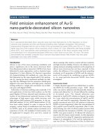
Báo cáo hóa học: " Field Emission and Radial Distribution Function Studies of Fractal-like Amorphous Carbon Nanotips" potx
Bạn đang xem bản rút gọn của tài liệu. Xem và tải ngay bản đầy đủ của tài liệu tại đây (622.03 KB, 6 trang )
NANO EXPRESS
Field Emission and Radial Distribution Function Studies
of Fractal-like Amorphous Carbon Nanotips
F. Sola
´
Æ A. Biaggi-Labiosa Æ L. F. Fonseca Æ
O. Resto Æ M. Lebro
´
n-Colo
´
n Æ M. A. Meador
Received: 30 September 2008 / Accepted: 28 January 2009 / Published online: 13 February 2009
Ó to the authors 2009
Abstract The short-range order of individual fractal-like
amorphous carbon nanotips was investigated by means of
energy-filtered electron diffraction in a transmission elec-
tron microscope (TEM). The nanostructures were grown in
porous silicon substrates in situ within the TEM by the
electron beam-induced deposition method. The structure
factor S(k) and the reduced radial distribution function G(r)
were calculated. From these calculations a bond angle of
124° was obtained which suggests a distorted graphitic
structure. Field emission was obtained from individual
nanostructures using two micromanipulators with sub-
nanometer positioning resolution. A theoretical three-stage
model that accounts for the geometry of the nanostructures
provides a value for the field enhancement factor close to
the one obtained experimentally from the Fowler-Nord-
heim law.
Keywords Carbon nanotips Á Graphite-like a-C Á
EELS Á EFED Á Field emission
Introduction
Nanotips made of carbon can have many different
applications such as scanning microscope probes [1] and
field emission (FE) sources [2]. For instance, recently
carbon nanofibers were used as the electron source in
order to test and build an FE display device prototype
where a new nanocrystalline silicon—polymer film—was
used as the phosphor material [3]. Carbon nanotubes,
from single to film dispersed cases, have been exten-
sively studied due to their tensile strength, electrical
properties, chemical inertness, and high aspect ratio
[4–10]. However, an advantage that amorphous carbon
(a-C) nanotips have over carbon nanotubes is that when
a-C nanotips are synthesized using the electron beam-
induced deposition method with a transmission electron
microscope (TEM-EBID) their growth process can be
followed in real-time and the nanostructures can be
grown at preferred positions by controlling the electron
beam [11]. Recently, FE studies were done on an indi-
vidual one-dimensional a-C nanotip grown by the TEM-
EBID method, where a field enhancement factor of the
order of 10 was found [12]. In this study, a field
enhancement factor of the order of 10
3
was obtained for
fractal-like a-C nanotips consisting of several branches,
each branch similar in shape to the previously mentioned
one-dimensional nanotips. A theoretical three-stage
model that accounts for those findings is presented and
discussed. Multistage models have been used for other
types of nanostructures such as carbon nanotubes and
tungsten oxide nanowires [7, 13–15]. In addition, we also
present information of the short-range order of our
nanostructures, using the radial distribution function
(RDF) obtained by electron diffraction patterns. With this
information average nearest-neighbor distances and their
bond angle were obtained. The results are consistent with
distorted graphitic-like structure that can account for the
moderate conductivity of the tips observed in the FE
results.
F. Sola
´
Á A. Biaggi-Labiosa Á L. F. Fonseca (&) Á O. Resto
Department of Physics, Institute for Functional Nanomaterials,
University of Puerto Rico, Rio Piedras, P.O. Box 23343,
San Juan, PR 00931, USA
e-mail:
M. Lebro
´
n-Colo
´
n Á M. A. Meador
Polymeric Materials Branch, Materials and Structures Division,
National Aeronautics and Space Administration Glenn Research
Center, 21000 Brookpark Road, Cleveland, OH 44135, USA
123
Nanoscale Res Lett (2009) 4:431–436
DOI 10.1007/s11671-009-9270-5
Experimental
The nanostructures were grown in situ in a TEM by the
TEM-EBID method [11] using porous silicon (PSi) sub-
strates. Full details about the PSi preparation procedure can
be found elsewhere [16]. Pieces of the PSi films of the
order of 2 mm
2
were attached to copper TEM grids. TEM-
EBID experiments were made in a JEOL JEM-100S TEM
at 100 kV. Electron energy loss spectroscopy (EELS)
measurements were made in a Carl Zeiss LEO-922 TEM
equipped with an Omega filter. Energy-filtered electron
diffraction studies were done in a Phillips CM-200 TEM at
200 kV using a Gatan imaging filter. The energy selecting
window used to filter the electron diffraction pattern
was 10 eV and was centered at the zero loss peak of the
electron energy loss spectrum. The inverse space was
calibrated using a polycrystalline aluminum standard
sample.
FE studies were done in an FEI Strata 235 Dual Beam
FIB (DB-FIB) with a vacuum better than 10
-5
Torr. Two
electrochemically etched tungsten tips were connected to
two Kleindiek micromanipulators, each having a posi-
tioning resolution of 0.25 nm. One of the tungsten tips is
landed on a platinum contact which ends at the base of the
nanostructure and the other is positioned close to the outer
branches of the nanostructure to collect the FE current. The
platinum contact was carefully deposited by SEM-EBID
method using only the electron source of the DB-FIB. FE
experiments were controlled with a LabView program and
the beam was blanked before collecting the FE currents.
Inspection of the samples after the FE measurements did
not show additional growth of nanotrees.
Results and Discussion
A TEM image of fractal-like a-C nanotips is shown in
Fig. 1. A qualitative growth mechanism for this type of
nanostructure has been reported previously [17]. In a few
words, the poor vacuum conditions in the JEOL JEM-100S
TEM working without the liquid nitrogen trap brings
hydrocarbon contamination inside the chamber which
generally comes from the diffusion pump. Due to the
incident electron beam, the hydrocarbon molecules are
ionized. The high resistivity of the PSi samples allows the
charging of the irradiated area, which becomes positively
charged as secondary electrons leave the sample into
the vacuum. Since there is no charge outside the sample,
the Laplace equation is fulfilled. With these conditions, the
hydrocarbons are attracted to the surface of the sample and
preferentially deposited on hot spots with the highest local
electric fields. Branching takes place during growth due to
the preferential deposition near the tip regions. Finally, the
deposited hydrocarbons are then transformed into a-C due
to the continuous electron irradiation. With this technique,
we recently developed methods to synthesize nanopalm-
like silica/carbon heterostructures [16] and arrays of frac-
tal-like a-C nanotips [11].
RDF Studies
Previous to the FE experiments we made energy-filtered
convergent-beam electron diffraction (EFCBED) studies
on the nanostructures to clarify the bonding and atomic
order properties of the tips on which the FE properties will
depend. Figure 2a is a low loss electron energy loss spec-
trum of fractal-like a-C nanotips. The first peak at 0 eV is
the zero loss peak associated to the elastically scattered
electrons. The second peak is located at 21.4 eV and it is
related to the Plasmon energy (E
p
) associated to the col-
lective resonant oscillations of the valence electrons [18].
The Plasmon energy can be used to estimate the density of
the nanostructures according to Eq. 1
p
0
¼
1
4
M
C
N
A
m Ã
e
0
"h
2
e
2
E
2
p
; ð1Þ
where m* = 0.87m, m is the electron rest mass and the
other factors have their usual meaning [19]. Taking the
molar mass of carbon (M
C
) as 12 g/mol, the density is
found to be 1.44 g/cm
3
, which is consistent with the den-
sity of graphite-like a-C [20]. The C–K energy-loss near-
edge structure (ELNES) spectrum of the nanotips (Fig. 2b)
exhibits two peaks. The first peak corresponds to a
1s ? p* transition and the second peak to a 1s ? r*
Fig. 1 TEM image of a fractal-like a-C nanotips obtained by TEM-
EBID method
432 Nanoscale Res Lett (2009) 4:431–436
123
transition. These two transitions are consistent with the
ELNES spectrum of a-C [21]. Based on this type of
spectrum we previously found a sp
2
bonding percentage
around 80% for our nanostructures and when these results
were compared with visible Raman studies, they were
classified as graphite-like a-C with low hydrogen content
[11].
Figure 3a shows the EFCBED pattern of fractal-like a-C
nanotips. In order to avoid any damage to the CCD camera,
the central spot of the EFCBED was positioned on the right
corner. Cockayne and McKenzie [22] presented a pio-
neering study on calculating nearest-neighbor distances
using EFED patterns. However, several improvements
have been made to this study [23, 24]. Here we use the
procedure presented in ref. [24] by splicing together two
electron diffraction patterns with v
2
= 0.0038. First, the
static structure factor was calculated. In Fig. 3b, we present
the static structure factor S(k) plot obtained using Eq. 2
SðkÞ¼
IðkÞ
Nf
2
ðkÞ
; ð2Þ
where I(k) is the azimuthally average intensity, f(k) is the
atomic scattering factor, N a fitting parameter, and
k = 2 sin h/k is the magnitude of the scattering vector.
Secondly, the short-range order of individual fractal-like
a-C nanotips can be investigated using the RDF defined as
the probability to find an atom at a given distance from a
particular atom. RDF is characterized by peaks from each
shell of neighbors. The reduced RDF G(r) is calculated by
applying an inverse Fourier sine transformation to Eq. 3,
UðkÞ¼kSðkÞÀ1½¼
Z
1
0
GðrÞsinðkrÞdr; ð3Þ
where U(k) is the reduced intensity function,
G(r) = 4pr[p(r) - p
0
] and p(r) is the average density of
neighbors a distance r from a particular atom. Figure 3c
shows the reduced RDF G(r) plot. At this point it is worth
mentioning that the hydrogen contribution is excluded in p
0
because hydrogen is a weak scatterer and, hence, its
contribution to the scattering intensity can be assumed to
be negligible [25]. Taking first and second nearest-
neighbor distances as the peak maxima of peaks 1 and 2
in the G(r) plot, respectively, the values for r
1
and r
2
are:
r
1
= 1.49 A
˚
and r
2
= 2.65 A
˚
. Furthermore, the average
bond angle can be acquired using Eq. 4 [25]
h ¼ 2 sin
À1
r
2
2r
1
: ð4Þ
From this expression, a bond angle of 124° was
obtained. This bond angle differs from a sp
2
trigonally
bonded carbon, which has a bond angle of 120°.This
difference suggests that our nanostructures have a distorted
graphitic structure which is consistent with our previous
Raman and EELS results. From these findings it is
expected that the a-C fractal nanotips will present good FE
properties [26].
FE Studies
A SEM image of our FE set-up is shown in Fig. 4a, and the
inset is the TEM image representative of the type of fractal
nanotips tested. A typical FE current (I) curve is presented in
Fig. 4b. For these measurements the distance (d) between
the collection tip and the outer tips of the nanostructure was
252 nm. The turn on field was around 24 V/lm and is
defined here as the field required for extracting a current of
10 nA, which is much lower than the 88 V/lm turn on field
found in ref. [12] for a single nanotip. The fact that several
branches are contributing to the total current can justify this
difference. The inset of Fig. 4b is the corresponding Fowler-
-10 0 10 20 30 40
Counts
0
20
40
60
80
100
120
140
(a)
Energy Loss (eV)
280 290 300 310 320
Counts
120
130
140
150
160
170
180
(b)
π*
σ*
Fig. 2 a Low loss EELS showing the Plasmon energy at 21.4 eV.
b C–K ELNES of a nanostructure typical of highly sp
2
amorphous
carbon
Nanoscale Res Lett (2009) 4:431–436 433
123
Nordheim (FN) plot (Ln(I/V
2
) versus 1/V) which shows a
fairly linear relation at low electric field intensities. At high
electric fields the slope increases, which can be an indication
of heating [2]. At the intermediate values of the electric
field, the curve slightly deviates from the straight line. This
deviation has been observed previously and explained in
terms of the presence of adsorbates that enhances the tun-
neling probability [27]. In general, the curve suggests that
the FE from the nanostructures follows the FN law expres-
sed as
I ¼ Aa
E
2
/
exp Àb
/
3=2
E
!
; ð5Þ
where A is the emitting area (pr
2
), E is the applied field,
/ the work function of the material, and a and b are
constants equal to 1.54 9 10
-6
AeVV
-2
and 6.83 9
10
3
eV
-3/2
V lm
-1
, respectively [4]. Due to the tip-like
geometry of our field emitters one can approximate E to
bV/d, where b is the field enhancement factor that accounts
for the geometry of the emitter. With this definition and
rearranging Eq. 5, we can estimate b from the slope of the
following relation
Ln
Id
2
AV
2
¼
Àb/
3=2
d
b
1
V
þ Ln
ab
2
/
ð6Þ
Taking a work function of a graphitic structure of 5 eV
[26], and the radius of the emitting area as r * 0.5 lm, the
slope of Eq. 6 gives a b equal to 889. In trying to find a
possible explanation of the previous enhancement factor,
the multistage model [14] is used. We interpret the previ-
ous result of the field enhancement as a product of three
stages. The schematic representation of our three-stage
model is presented in Fig. 4c in which each stage repre-
sents an effective branch of the fractal-like a-C nano-
structure. Each stage is defined with a particular length (l
i
)
and radius (r
i
), where stage 1 has the biggest length and
radius. In a multistage model the total enhancement factor
can be written as a product of individual stages [7, 13–15].
With that interpretation b is written as
b ¼ b
s
Y
3
i¼1
b
i
; ð7Þ
where
b
i
¼
l
i
l
i
þ d
1 þ
d
r
i
and b
s
¼ 1 À exp Àa
s
L
hi
: ð8Þ
The expression for b
i
was derived by Huang et al. [14].
The factor b
s
is a factor that accounts for the screening
effect of adjacent tips [28–30], where s is interpreted here
as the average distance between adjacent tips, L is the sum
of the lengths of three stages, and a is a fitting parameter
[30]. Taking the common fitting parameter a equal to
2.3172 [28, 29], and measuring average values for l
i
as
491 nm (i = 1), 146 nm (i = 2) and 74 nm (i = 3),
average values of r
i
as 13.4, 5.6, and 2.1 nm and with s
r (Å)
012345
G(r)
-60
-40
-20
0
20
40
60
k (Å
-1
)
0123456
S(k)
0
5
10
15
20
(a)
(b)
(c)
1
2
Fig. 3 a EFCBED pattern showing diffuse rings typical of an
amorphous structure. The central spot is at the right corner. b Plot of
the structure factor S(k) with k
max
= 6A
˚
-1
. c Corresponding reduced
RDF plot of (b), where first and second nearest-neighbors distance
peaks are marked 1 and 2, respectively
434 Nanoscale Res Lett (2009) 4:431–436
123
equal to 54.7 nm, we obtain (from Eqs. 7 and 8) a field
enhancement factor, b, equal to 981. This result agrees well
with the value found experimentally of 889.
Conclusions
In summary, the reduced RDF analysis on the fractal-like
a-C nanotips grown by the TEM-EBID method shows first
and second bond lengths at 1.49 and 2.65 A
˚
. Those nearest-
neighbors distances defined an average bond angle of 124°
which deviates from a trigonally sp
2
bonded carbon (120°)
indicating a distorted graphite-like structure, which
explains the moderate conductivity of the tips observed in
the FE results. The electron FE measurements of the
individual fractal-like a-C nanorods showed a turn on field
of 24 V/lm. A three-stage model that accounts for the
geometry of the nanostructures described well the value of
the enhancement factor obtained experimentally from the
FN law, and hence suggests a possible explanation of our
observations. Hence, our observations show that these
nanostructures are promising to be used in FE applications.
In particular, the growth mechanism of the nanotrees’ tips
follows the path of maximum local field at the tips thus
forming a nanostructure where the relationship between the
density of tips and the local electric field intensity at the
tips is optimized. It is worth to mention that for our cal-
culations of the emission current densities we have used as
the active emitting area the cross section of the total
nanotree such that the values for the current density per
branch’s tip are lowest bound values.
Acknowledgments This study was supported by the following
grants numbers and projects: NASA NNX08BA48A, NASA Space
Grant NNG05GG78H, NSF 0701525, Fundamental Aeronautics
Program and Subsonic Fixed Wing Project. The authors acknowledge
the National Center for Electron Microscopy, Lawrence Berkeley
Lab, which is supported by the U.S. Department of Energy under
Contract #DE-AC02-05CH11231, and in particular to Dr. A. Minor
for helping us with the FE experimental setup. FÁS. kindly acknowl-
edges Dr. D. J. H. Cockayne from Oxford University and D. Hull
from NASA GRC for providing helpful information related to the
electron diffraction analysis.
References
1. I.C. Chen, L.H. Chen, O. Christine, A. Quist, R. Lal, S. Jin,
Nanotechnology 17, 4322 (2006)
2. K.S. Yeong, B.C. Boothroyd, J.T.L. Thong, Nanotechnology 17,
3655 (2006)
1
2
3
Platinum
contact
(a)
Voltage (V)
2 4 6 8 10 12 14 16
Current (
µΑ)
Α
0.0
0.2
0.4
0.6
0.8
1.0
1.2
I/V (V
-1
)
0.060.080.100.120.140.160.180.200.220.240.26
Ln (I / V
2
)
-10
-9
-8
-7
-6
-5
-4
(c)(b)
Fig. 4 a SEM image of our FE
set-up. The inset is the TEM
image representative of the type
of fractal nanorods tested; the
scale bar is 200 nm. b FE
current curve obtained at a
distance of 252 nm and the inset
is the linear FN plot. c
Schematic representation of the
three-stage model. The solid
line corresponds to the first
stage and so on
Nanoscale Res Lett (2009) 4:431–436 435
123
3. A. Biaggi-Labiosa, F. Sola
´
, O. Resto, L.F. Fonseca, A. Gonza
´
lez-
Berrı
´
os, J. De Jesu
´
s, G. Morell, Nanotechnology 19, 225202
(2008)
4. M. Passacantando, F. Bussolotti, S. Santucci, A. Di Bartolomeo,
F. Giubileo, L. Iemmo, A.M. Cucolo, Nanotechnology 19,
395701 (2008)
5. X.J. Li, W.F. Jiang, Nanotechnology 18, 065203 (2007)
6. Z. Xu, X.D. Bai, E.G. Wang, Z.L. Wang, Appl. Phys. Lett. 87,
163106 (2005)
7. D.L. Niemann, B.P. Ribaya, N. Gunther, M. Rahman, J. Leung,
V. Nguyen, Nanotechnology 18, 485702 (2007)
8. R.C. Smith, D.C. Cox, S.R.P. Silva, Appl. Phys. Lett. 87, 103112
(2005)
9. S.H. Jo, Y. Tu, Z.P. Huang, D.L. Carnahan, D.Z. Wang, Z.F.
Reng, Appl. Phys. Lett. 82, 3520 (2003)
10. J.M. Bonard, K.A. Dean, B.F. Coll, C. Klinke, Phys. Rev. Lett.
89, 1976021 (2002). doi:10.1103/PhysRevLett.89.197602
11. F. Sola
´
, O. Resto, A. Biaggi-Labiosa, L.F. Fonseca, Micron 40,
80 (2009)
12. C.H. Jin, J.Y. Wang, Q. Chen, L.M. Peng, J. Phys. Chem. B 110,
5423 (2006). doi:10.1021/jp057240r
13. R. Seelaboyina, S. Boddepalli, K. Noh, M. Jeon, W. Choi,
Nanotechnology 19, 065605 (2008)
14. J.Y. Huang, K. Kempa, S.H. Jo, S. Chen, Z.F. Ren, Appl. Phys.
Lett. 87, 053110 (2005)
15. R. Seelaboyina, J. Huang, J. Park, D.H. Kang, W.B. Choi,
Nanotechnology 17, 4840 (2006)
16. F. Sola
´
, O. Resto, A. Biaggi-Labiosa, L.F. Fonseca, Nanotech-
nology 18, 405308 (2007)
17. F. Banhart, Phys. Rev. E 52, 5156 (1995)
18. R.F. Egerton, Electron Energy Loss Spectroscopy in the Electron
Microscope (Plenum Press, New York, 1996), Chapter 3
19. A.C. Ferrari, A. Libassi, B.K. Tanner, V. Stolojan, J. Yuan, L.M.
Brown, S.E. Rodil, B. Kleinsorge, J. Robertson, Phys. Rev. B 62,
11089 (2000). doi:10.1103/PhysRevB.62.11089
20. S.R.P. Silva, Properties of Amorphous Carbon (Inspec, London,
UK, 2003), Chapter 1
21. K. Uppireddi, O. Resto, B.R. Weiner, G. Morell, Nanoscale Res.
Lett. 3, 65 (2008)
22. D.J.H. Cockayne, D.R. McKenzie, Acta Crystallogr. A 44, 870
(1988)
23. W. McBride, D.J.H. Cockayne, J. Non-Cryst. Solids 318, 233
(2003). doi:10.1016/S0022-3093(02)01908-7
24. T.C. Petersen, W. McBride, D.G. McCulloch, I.K. Snook, I.
Yarovsky, Ultramicroscopy 103, 275 (2005)
25. R.D. Evans, J. Bentley, K.L. More, G.L. Doll, J.T. Glass, J. Appl.
Phys. 96, 273 (2004). doi:10.1063/1.1760232
26. A.C. Ferrari, B.S. Satyanarayana, J. Robertson, W.I. Milne, E.
Barborini, P. Piseri, P. Milani, Europhys. Lett. 46, 245 (1999)
27. K.S. Yeong, J.T.L. Thong, Appl. Surf. Sci. 233, 20 (2004).
doi:10.1016/j.apsusc.2004.03.222
28. J. Xiao, X. Zhang, G. Zhang, Nanotechnology 19, 295706 (2008)
29. Y.W. Zhu, T. Yu, F.C. Cheong, X.J. Xu, C.T. Lim, V.B.C. Tan,
J.T.L. Thong, C.H. Sow, Nanotechnology 16, 88 (2005)
30. X. Qian, H. Liu, Y. Guo, Y. Song, Y. Li, Nanoscale Res. Lett. 3,
303 (2008)
436 Nanoscale Res Lett (2009) 4:431–436
123