Acute Ischemic Stroke Part 6 pot
Bạn đang xem bản rút gọn của tài liệu. Xem và tải ngay bản đầy đủ của tài liệu tại đây (2.73 MB, 18 trang )
4
Dysphagia and Respiratory
Infections in Acute Ischemic Stroke
Claire Langdon
Sir Charles Gairdner Hospital & Curtin University of Technology,
Australia
1. Introduction
Eating and swallowing are activities that are normally performed without conscious
thought. This complex behaviour – involving 5 pairs of cranial nerves and 26 pairs of
muscles – can be interrupted by a stroke, leading to dysphagia. Dysphagia is associated with
aspiration (where material passes into the respiratory tract) and aspiration carries a risk of
pneumonia seven times greater than that of the normal population. Around 15% - 20% of all
stroke patients will develop respiratory tract infections during the acute phase of their
stroke. Pneumonia is one of the leading causes of mortality in the acute stroke patient.
Respiratory infections add to hospital length of stay and are associated with significant
increases in the cost of patient care, as well as being associated with poorer outcomes for the
patient. This chapter will outline the association of dysphagia and other risk factors in the
development of respiratory infections in acute ischemic stroke patients.
2. High early incidence of dysphagia, its causes, presentation and implications
Swallowing is a vital motor activity that serves alimentary purposes and protects the upper
airway (Jean, 2001). Dysphagia (difficulty eating and swallowing) is very common after an
ischemic stroke, affecting between 13% and 94% of all ischemic stroke patients, with the
incidence depending on the size and location of the lesion (Barer, 1989; DePippo, Holas,
Reding, Mandel & Lesser, 1994; Daniels, 2000; Ding & Logemann, 2000; Aydogdu, Ertekin,
Tarlaci, Turman & Klyliogly, 2001; Marik, 2001). Dysphagia can lead to malnutrition,
dehydration, aspiration pneumonia and increased length of hospital stay.
In a recent review of the costs to the US health system, dysphagia was found to be
associated with significantly increased costs due to increased length of stay and infections.
The median hospitalization days for patients with dysphagia was 4.04 compared with 2.40
days for those patients without dysphagia. Mortality was substantially increased in patients
with dysphagia associated with rehabilitation, intervertebral disk disorders, and heart
diseases (Altmann, Yu & Schaefer, 2010). A cohort study of 330 stroke survivors found that
those who developed infections during their hospital admission had a median length of stay
of 26 days, significantly longer than the median length of stay of 11 days of those who did
not require antibiotic treatment (Langdon, Lee and Binns, 2010).
Best practice management of dysphagia in acute stroke encompasses the need to consider
the patient holistically. The impact of various eating difficulties on nutritional status has not
Acute Ischemic Stroke
80
received great attention in research (Westergren, 2006). Impaired arm movement, lip closure
and swallowing have all been found to be significant predictors of decreased energy intake
over 24-hours in patients with stroke (McLaren & Dickerson 2000).
Dysphagia that occurs due to a stroke is associated with an increased risk of material being
aspirated during swallowing. This is due to the disruption to motor and sensory input and
to protective reflexes (Holas, DePippo & Reding, 1994; Aviv, Martin, Sacco, Zager,
Diamond, Keen & Blitzer, 1996; Daniels, Brailey, Priestly, Herrington, Weisberg & Foundas,
1998; Addington, Stephens, Gilliland, Rodriguez, 1999; Nakajoh, Nakagawa, Sekizawa,
Matsui, Arai & Sasaki, 2000). ‘Aspiration’ occurs when matter (food, fluid, saliva or
secretions), enters the airway and passes below the level of the true vocal folds. Pneumonia
is up to seven times more likely to occur in patients who are known to aspirate (DePippo,
Holas & Redding, 1994; Kidd, Lawson, Nesbitt & MacMahon, 1995; Smithard, O’Neill,
England, Park, Wyatt, Martin & Morris, 1997). Dysphagia has been found to be associated
with an increased risk of chest infections, dehydration and death (Gordon, Hewer & Wade,
1987). In stroke patients, pneumonia has been associated with an increased cost to the health
system of US$14,836 per patient (Katzan, Dawson, Thomas, Votruba & Cebul, 2007) and is
strongly associated with poorer outcomes (Smithard, O’Neill, Park, Morris, 1996; Wang,
Lim, Levi, Heller & Fischer, 2001; Wang, Lim, Heller, Fisher & Levi, 2003).
2.1 Infection in acute stroke
Infection in acute stroke has been, and remains, a significant problem, with pneumonia and
urinary tract infections occurring the most frequently. In the GAIN study, a multicentre,
multinational study of 1455 patients with strokes, 142 died during the first week following
hospital admission. Thirty four (23.9%) of these died from pneumonia (Aslanyan, Weir,
Diener, Kaste & Lees, 2004).
A study of 124 stroke patients who were admitted to Neurological Intensive Care Units in
Cologne reported an incidence of pneumonia of 21%, occurring an average 1.8 days (±1.9
days), post stroke, although these figures did not distinguish between patients who
required ventilator support for respiration and those who did not (Hilker, Poetter,
Findeisen, Sobesky, Jacobs, Neveling & Heiss, 2003). Ventilator-associated pneumonia is
common in patients who require mechanical respiratory support and is discussed in
greater detail later in the chapter.
A Scandanavian study of 1,156 patients reported 19.4% developed infections within 3 days
of hospital admission, with 49% of the infections diagnosed males and 27% of infections in
females being respiratory tract infections. Early infection added 9.3 days on average to the
patient’s hospital admission (Kammersgaard, Jorgensen, Reith, Nakayama, Houth, Weber,
Pederse, & Olsen, 2001).
Using videofluoroscopy to examine swallowing, Kidd et. al., (1995) found 25 (42%) of a
cohort of 60 stroke patients were aspirating at 72 hours post stroke. In the first 14 days after
their stroke, 19 patients developed lower respiratory tract infections. Of these 19 patients, 17
(89%) were known to be aspirating. Of the 25 aspirators, 22 had returned to normal
swallowing function when followed up at 3 months post stroke (Kidd, Lawson, Nesbitt &
MacMahon, 1995).
Mann, Hankey and Cameron followed 128 acute stroke patients for six months and reported
an incidence of dysphagia in 82 patients (64%) when examined using videofluoroscopy. In
the six months of follow up, 26 patients (20%) experienced a chest infection (Mann, Hankey
& Cameron, 1999). A limitation of this study was the exclusion of stroke patients who were
Dysphagia and Respiratory Infections in Acute Ischemic Stroke
81
unable to tolerate the videofluoroscopy procedure, which excludes those patients with
impaired conscious state. Often these patients with poor conscious level are the ones who
are at greatest risk of aspiration due to the impairment in their ability to protect their
airway.
A study of 88 patients admitted to hospital with ischemic strokes found infection occurred
in 25 of 80 survivors during the first month post stroke. Respiratory infection was
significantly more likely to occur in patients with dysphagia (Langdon, Lee & Binns, 2007).
In a cohort of 330 acute ischemic stroke patients followed up for the first month after their
stroke, there were 51 respiratory infections, with dysphagia again a significant predictor of
patients who developed infections (Langdon, Lee & Binns, 2009).
2.2 Normal swallowing
Swallowing is something that is often taken for granted, yet is a complex and tightly
controlled event that coordinates breathing and deglutition, with an average of 2,400
swallows occurring daily. The frequency of swallowing changes depending on the activity.
It occurs on the average at 6 swallows an hour during sleep to 10 per hour during normal
activity to around 300 per hour while eating (Miller, 1982). The body produces 1500ml -
2000ml saliva daily (Witt, 2005), which is swallowed without conscious thought.
Successful swallowing is an extremely complex and dynamic process, involving 5 pairs of
cranial nerves and coordination of some 26 pairs of striated muscles (Dodds, Stewart &
Logemann, 1990; Bass & Morrell, 1992; Matsuo & Palmer, 2008) involved in the act of
moving food or fluid from the oral cavity to the stomach. There is an extremely elaborate
reflex mechanism that provides a close functional relationship between the pharynx
(throat), larynx and oesophagus during swallowing, belching and reflux to help prevent
aspiration or food/fluids going ‘the wrong way’ (Shaker, 2006). Studies have provided
evidence that the process of swallowing is governed by specialised neural networks in a
finely-tuned partnership with respiration and speech (Zald & Pardoe, 1999). Neural
control of swallowing is multidimensional. The brainstem contains the swallowing central
pattern generator – the first level of control. The second level of control incorporates the
sub cortical structures; basal ganglia, hypothalamus, amygdala and midbrain. The third
level of control is represented by suprabulbar cortical swallowing centres (Mistry &
Hamdy, 2008).
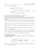
Normal swallowing is generally arbitrarily divided into four stages for convenience of
description, however, the normal swallow is a complex, fast, continuous sequence of
coordinated muscle movements and there is some overlap between the phases. An
efficient swallow involves an anticipatory phase, an oral phase, a pharyngeal phase and
an oesophageal phase. These are illustrated in Figure 1 and described in greater detail
below. The oral phase of swallowing is initiated voluntarily while the pharyngeal and
oesophageal phases occur via intraphase reflexes (Lang, 2009). Control of the phases
represent a coordination among the brain stem and central cortical pathways (Miller,
2008).
2.2.1 Oral preparatory phase (voluntary control)
The oral preparatory stage of swallow incorporates prior knowledge of feeding and
swallowing, environmental, visual and olfactory cues. Once food enters the oral cavity, the
lips seal, the tongue accepts and cradles the food and it is tasted. Information about the food
is transmitted from the taste buds to the cortex and brainstem. The preparatory stage allows
Acute Ischemic Stroke
82
prior knowledge to impact on eating and swallowing: for example, if a stroke patient has
had recent choking events due to dysphagia, they may be reluctant to accept a particular
texture or type of food.
2.2.2 Oral phase (voluntary and reflexive control)
Food is chewed and mixed with saliva using movement of the jaw, tightly coordinated with
movements of the tongue, cheeks, soft palate, and hyoid bone (Matsuo & Palmer, 2008).
When the prepared food has been formed into a ‘bolus’ suitable for swallowing, it is centred
on the tongue and propelled backwards by the tongue to the pharynx. Difficulties with the
oral stage of the swallow may occur because of muscle weakness or nerve dysfunction.
These will often lead to an extension in the time taken for oral transit and/or retention of
material in the oral cavity as residue, or may cause premature spillage of the bolus into the
pharynx (Dodds, Stewart & Logemann, 1990; Matsuo & Palmer, 2008) which can impact on
airway protection.
Disruption to the oral phase can occur due to poor dentition or poorly fitting dentrues.
Absent molar teeth can significantly impair bolus preparation, as these teeth grind food into
smaller, more digestible particles. Gum disease and teeth in poor condition may cause pain
when eating, which is associated with decreased intake of food. A weak tongue or jaw will
also contribute to impairment of the oral phase of the swallow.
2.2.3 Pharyngeal phase (reflexive control)
The pharynx consists of the nasopharynx (superior and anterior to the soft palate) and
oropharynx (from the nasopharynx to the larynx). It serves two purposes, acting as a
conduit for air to and from the lungs and also moving food and liquids from the mouth to
the esophagus (Miller, 2002). The pharyngeal stage of the swallow is an important and
complex activity that coordinates: (1) food or liquids passing through the pharynx and
upper esophageal sphincter (UES) to the osophagus and (2) airway protection – isolating the
larynx and trachea from the pharynx during swallowing to prevent the bolus from entering
the airway (Matsuo & Palmer, 2008). The pharynx is made up of the pharyngeal constrictor
muscles (superior, middle and inferior) that overlap to form a sheath that extends from the
base of skull to the esophagus. The pharyngeal swallow also involves muscles of the soft
palate, the tongue, pharynx, larynx and hyoid bone. The pharyngeal swallowing muscles
are innervated by the trigeminal (V), facial (VII) glossopharyngeal (IX), vagal (X), spinal
accessory (XI) and hypoglossal (XII) nerves (Miller, 2002).
Breathing and swallowing are tightly coordinated by the brainstem. During swallowing,
there is a brief cessation of breathing known as ‘deglutition apnea‘. Studies of normal
subjects have found that there is a small exhalation prior to the swallow being initiated,
followed by the swallow, and finally a larger exhalation once the bolus has entered the
esophagus (Martin, Logemann, Shaker & Dodds, 1994). Swallowing normally finishes with
an exhalation of air. This serves to assist clearance of any material that may have entered the
laryngeal vestibule during the swallow. This normal breathing/swallowing rhythm has
been shown to break down in stroke patients (Leslie, Drinnan, Ford & Wilson, 2002), while
the apnea associated with swallowing increases with age (Leslie, Drinnan, Ford & Wilson,
2005).
Stroke can impact on the timing of the pharyngeal swallow, or weaken the muscles of one
side of the pharynx, resulting in a weak or incoordinated swallow, often associated with
material being aspirated before, during or after the swallow is initiated.
Dysphagia and Respiratory Infections in Acute Ischemic Stroke
83
Fig. 1. Stages of Normal Swallowing:
1. Food is chewed and mixed with saliva. This is shaped into a bolus by the tongue, and
centred on the tongue prior to initiation of the swallow. The soft palate elevates to form a
seal with the nasopharyx.
2. The tongue tip is pressed against the alveolar ridge, then the tongue base drops and the
bolus is pushed into the pharynx. The vocal folds adduct and breathing is ceased momentarily.
3. The epiglottis deflects downward and the bolus enters the esophagus due to (a) tonic
relaxation of the upper esophageal sphincter (b) hyolaryngeal traction opening the sphincter
(c) pharyngeal squeeze.
4. The bolus is cleared into the esophagus by pharyngeal muscles exerting a stripping action.
2.2.4 Oesophageal phase (reflexive control)
After the bolus enters the oesophagus through the UES, peristalsis moves it down to the
stomach and through the lower esophageal sphincter to the stomach. The peristaltic wave
consists of an initial wave of relaxation to accommodate the bolus followed by a wave of
contraction that propels it onward (Matsuo & Palmer, 2008). This phase of the swallow is
considered the least complex, although it can still be subject to impairment leading to
decreased safety and poor oral intake.
1.
2.
3.
4.
Acute Ischemic Stroke
84
2.3 Causes of dysphagia in ischemic stroke
Stroke is a brain injury, and may impact on swallowing, either by damage to cranial
nerves or nuclei, or by interrupting the interconnecting neural networks that regulate
normal deglutition. A stroke patient may be drowsy in the acute phase, which impacts on
their ability to remain conscious long enough to eat or drink sufficient amounts to ensure
that their nutrition and hydration needs are being met. Poor conscious level may mean
that the stroke patient’s ability to protect his or her airway is compromised. Difficulty in
maintaining sitting balance and hemiparesis affecting the dominant hand may contribute
to the person’s difficulties in self-feeding. Loss of facial tone may cause dentures to
become ill-fitting, making chewing difficult for the patient. Loss of facial tone may also
mean that the patient has difficulty containing the bolus in the oral cavity, forming a seal
with the lips to drink easily, or prevent saliva spilling from the mouth (drooling). Loss of
sensation from damage to the trigeminal nerve may mean that the patient is unaware that
they are drooling, or that food may be pocketed in a flaccid cheek following mealtimes.
The impact of different types of stroke on feeding and swallowing is more fully discussed
below.
2.3.1 Cortical lesions
Swallowing has been shown using fMRI studies to involve the precentral and postcentral
gyri, the anterior cingulated gyrus and the insula (Miller, 2008). By using Transcranial
Magnetic Stimulation, Hamdy and his colleagues showed that swallowing is bilaterally but
asymmetrically represented in the cortical hemispheres, and that this representation is
unrelated to handedness. If a stroke involves the dominant swallowing centre, dysphagia is
highly likely, though this has been seen to resolve if the non-affected side subsumes the
functions of the dominant centre (Hamdy, Aziz, Rothwell, Singh, Barlow, Hughes, Tallis &
Thompson, 1996).
A cortical stroke may interfere with motor planning of the swallow. Large lesions may
involve association or projection tracts of the brain, penetrating into the internal capsule.
Significant dysphagia is commonly associated with a TACI stroke (Langdon, 2007; Sundar,
Pahuja, Dwivedi, Yeolekar, 2008; Langdon, 2010). Stroke that occurs in the right hemisphere
has been shown to impact on the pharyngeal phase of the swallow, with impairment to
initiation and duration and increased frequency of penetration and aspiration seen
(Robbins, Levine, Maser, Rosenbek & Kampster, 1993), while strokes affecting the left
hemisphere result in impairement in pharyngeal transit and longer oral transit (Miller,
2008).
For many patients with unilateral cortical strokes that affect the dominant swallowing
centre, dysphagia is transient, with a large percentage recovering their ability to eat and
swallow very quickly. Around 50% of patients admitted with strokes demonstrate
dysphagia (Gordon, Hewer & Wade, 1987; Smithard, O’Neill, England, Park, Wyatt, Martin
& Morris, 1997; Broadley, Croser, Cottrell, Creevy, Teo, Yiu, Pathi, Taylor & Thompson,
2003). This incidence tends to resolve by the end of the 5-7 days post stroke once acute
edema and ‘cerebral shock‘ start to resolve (Broadley et. al., 2003). Patients who have
bilateral cortical strokes tend to have more severe and prolonged dysphagia; possibly due to
the impairment affecting both hemispheres and precluding the subsumption of swallowing
function by an unimpaired swallowing representation.
A cortical stroke patient who presents with dysphagia may demonstrate some or all of the
following:
Dysphagia and Respiratory Infections in Acute Ischemic Stroke
85
- Facial droop
- Difficulty controlling saliva/secretions
- Slurred speech (dysarthria)
- Dysphasia/aphasia
- Weak or impaired cough
- Dysphonia
- Impaired conscious level
Any of these, or a combination of these, can impact adversely on the voluntary and reflexive
aspects of the normal swallow and should be investigated and treated. This assessment is
usually performed by a Speech and Language Pathologist. Formal dysphagia screening
programmes for acute stroke patients are associated with a significant decrease in the risk of
pneumonia and should be offered to all stroke patients (Hinchey, Shepherd, Furey, Smith,
Wang & Tonn, 2005). There has been consideration in the past that patients who present
with an impaired gag reflex are at high risk of swallowing problems or aspiration. The gag
reflex should not be considered to be predictive of dysphagia: published evidence clearly
shows that there is little or no relationship between the gag reflex and the ability to swallow
safely (Smithard & Spriggs, 2003).
2.3.2 Brainstem lesions
In the brainstem, there are motor nuclei that are responsible for acting as swallowing
Central Pattern Generators (CPG). The main motor nuclei involved in swallowing are the
hypoglossal motor nucleus and the nucleus ambiguus. These contain motoneurons, which
innervate the intrinsic and extrinsic muscles of the tongue, such as the genioglossus,
geniohyoid, styloglossus and hyoglossus, and the pharynx, larynx, and esophagus (Jean,
2001). Swallowing neurons are located in two main brain stem areas:
1. In the dorsal medulla (within the nucleus tractus solitarius) (NTS) and the adjacent
reticular formation, where they form the dorsal swallowing group (DSG)
2. In the ventrolateral medulla, just superior to the nucleus ambiguus, where they form
the ventral swallowing group (VSG)
Anatomically, swallowing neurons are located in the same sites as neurons that belong to
CPGs involved in respiration and cardiovascular regulation (Jean, 2001). Both breathing and
swallowing share some interneurons. This may help to explain the close relationship
between breathing and swallowing. This close relationship may be affected by an ischemic
stroke in the brainstem, predisposing the person to aspiration and dysphagia.
Brainstem strokes may account for up to 15% of all strokes (Kruger, Teasell, Salter &
Hellings 2007). They have been associated with severe and persisting dysphagia, although
this is by no means seen in every patient who has a stroke involving the brainstem. A
patient who has experienced a brainstem stroke usually presents quite differently to one
who has had a cortical stroke. Commonly seen dysfunction includes hemi- or quadriplegia,
ataxia, dysphagia, dysarthria, gaze abnormalities and visual disturbances. In contrast to
hemispheric lesions, new onset of cortical deficits, such as aphasia and cognitive
impairments, are absent. Brainstem stroke patients may demonstrate some other
characteristic clinical features, including
- Dysarthria
- Vertigo, nystagmus, nausea and vomiting, due to involvement of the vestibular
system
- Visual field loss or visuospatial deficits if there is occipital lobe involvement
Acute Ischemic Stroke
86
Common findings observed in patients with vertebrobasilar stroke include an abnormal
level of consciousness, as well as hemiparesis or quadriparesis. Pupillary abnormalities and
oculomotor signs are common, and bulbar manifestations, such as facial weakness,
dysphonia, dysarthria, and dysphagia, occur in more than 40% of patients (Kaye &
Brandstarter, 2009).
Patients with brainstem lesions tend to demonstrate the most prolonged course of recovery
or poorest outcomes. In an early study of dysphagia which reported on four patients with
strokes involving the brainstem, two patients subsequently died, while the other two had
resolution of their swallowing impairment by 25 days post onset and were discharged home
(Gordon, Hewer & Wade, 1987). In a case series over a six year period, Chua & Kong (1996)
reported 21 of 53 patients (40%) demonstrated dysphagia on admission, but did make some
progress during their stay at a rehabilitaton facility.
3. The time course of dysphagia
While dysphagia incidence is very high in stroke patients who are admitted to hospital,
there tends to be a sharp decrease in prevalence over a short period, usually in the first few
days to a week.
3.1 Studies of dysphagia in acute stroke patients
In an early study of dysphagia in stroke, (Gordon, Hewer & Wade, 1987) reported on a
cohort of 91 stroke patients. Forty one patients in their study (45%) had swallowing
problems on admission to hospital. This study used a water swallow test; bedside
swallowing examinations have been found to be less sensitive than instrumental evaluation
in determining the true incidence of dysphagia and aspiration, although instrumental
evaluation tends to select only those patients with conscious level good enough to tolerate
testing. The reported duration of dysphagia in survivors was
8 days or less 15
9 - 14 days 3
14 – 40 days 3
Kidd examined a cohort of 60 acute stroke patients using videofluoroscopy and found 25
(42%) were aspirating at 72 hours post stroke, with resolution of aspiration in 22 of 25
patients by 3 months post stroke (Kidd, Lawson, Nesbitt & McMahon, 1995). Another study
which followed 121 acute stroke patients found 61 (51%) had dysphagia on admission to
hospital. When reviewed at 7 days post stroke, dysphagia had resolved in a significant
number of patients, with only 28 still demonstrating swallowing impairment. By six-months
post stroke, this had decreased further and dysphagia persisted in only 6 of the original 61
patients with swallowing impairment on admission (Smithard, O’Neill, England, Park,
Wyatt, Martin & Morris, 1997).
Table 1 below clearly shows the decreasing prevalence of dysphagia during the first week
post stroke, with a reduction from 51% to 27%, although it should be noted that this
reported prevalence included cases of dysphagia that began during the first week post
stroke, presumably from an extension of the original stroke, or a new event.
Dysphagia and Respiratory Infections in Acute Ischemic Stroke
87
Day
N
Dysphagic (%) New Persistent
0 121 61 (51) - -
1 113 33 (39) 4 29
2 111 35 (32) 6 28
3 105 43 (39) 6 35
7 110 28 (27) 1 27
28 105 18 (17) 5 13
180 73 8 (11) 2 6
Table 1. Number of dysphagic patients at different time points in the study by Smithard et.
al., (1997).
Using a combination of videofluoroscopy and bedside swallowing examination, Mann,
Hankey & Cameron (2000) reported that aspiration was present in 49% of a cohort of 128
acute stroke patients. By six months post stroke, 97 of 112 survivors had returned to their
pre-stroke diet (Mann, Hankey & Cameron, 1999).
Daniels, Ballo, Mahoney and Foundas (2000) reported on a cohort of 56 ischemic stroke
survivors and noted that, although initially 38 (68%) presented with moderate to severe
dysphagia, at the time of their discharge from hospital 52 of 54 survivors consumed a
regular diet, one required food to be diced and one remained on enteral feeding.
Steinhagen, Grossman, Benecke and Walter (2008) reported on a cohort of 60 patients with
ischemic strokes. This cohort demonstrated an incidence of pneumonia of 39 patients:
median time to infection was 3 days (1-16 days).
In a large cohort study of 369 stroke patients, 125 of 330 survivors (38%) demonstrated some
degree of dysphagia on bedside examination at 7 days post stroke. Between 48 hours post
stroke and seven days post stroke, dysphagia prevalence in acute ischemic stroke patients
decreased from 153 (46.4%) to 125 (37.9%). Even among the patients with the most seriously
impaired swallowing function, those who were ‘Nil by Mouth’, there was a significant
amount of swallow improvement, with the initial 63 patients who were ‘Nil by Mouth’
decreasing to 41 by 7 days post stroke (Langdon, Lee & Binns, 2010).
As the studies above show, dysphagia is extremely common following a stroke, but tends to
resolve quite quickly in the majority of patients. It has been recommended that
percutaneous endoscopic gastrostomy (PEG) tubes be considered for those patients with
severe dysphagia that persists beyond 7 to 10 days post stroke if the treating medical team
feels the patient is likely to survive (Broadley et. al., 2003). Previous studies have
demonstrated a relatively consistent mortality rate of around 15% of all acute stroke patients
(Gordon, Hewer & Wade, 1987; Aslanyan et. al., 2004; Mann, Hankey & Cameron, 1999;
Langdon, Lee & Binns, 2010).
4. How good management of dysphagia prevents respiratory infections
In the early acute phase of a stroke, quality management of dysphagia is focused upon
ensuring that patients’ nutrition and hydration requirements are appropriately met, and that
they receive medications. This needs to be done in such a way that aspiration is prevented
or minimised. Stroke unit care using a multidisciplinary team with expertise in management
of this population has been shown to be responsible for improved outcomes for patients.
There have been few randomized control trials of dysphagia management: this is because
having a control group where treatment or management of dysphagia is withheld is
Acute Ischemic Stroke
88
unethical. In a review of the literature, Doggett, Tappe, Mitchell, Chapell, Coates and
Turkelson (2001) reported an estimated incidence of 43% - 54% of stroke patients with
dysphagia leading to aspiration, with approximately 37% of these patients going on to
develop pneumonia. Acute-care stroke patients with dysphagia were estimated to
experience malnutrition at a rate of 48%. The authors also reported that introduction of a
dysphagia management program dramatically reduces pneumonia rates: one study reported
frequency decreased from 6.7% (95% confidence interval 3.1% - 14%) to 4.1% (95%CI 1.8% -
9.3%) in the first year, and reduced to zero (95%CI 0% - 3%) in the second year.
4.1 Assessment of dysphagia
Optimal stroke care involves early dysphagia assessment: this often takes the form of an
initial screening by medical or nursing personnel, with patients who fail the screen
undergoing more thorough clinical assessment from a Speech-Language Pathologist, with
the option of patients undergoing instrumental assessment. It has been argued that a reliable
bedside assessment is useful in identifying patients at risk of nutritional compromise,
aspiration and poorer outcomes (Smithard, et. al., 1996). The current gold standard of
dysphagia assessment is the videofluoroscopic swallow study (also known as the Modified
Barium Swallow Study), where patients are assessed using a moving x-ray, which is
recorded to allow detailed analysis. Another alternative is a fibreoptic endoscopic
evaluation of swallowing (FEES). Both assessments have been shown to have excellent
reliability in detecting and assessing dysphagia.
The choice of instrumental examination should be made based upon the information that
the clinician seeks to obtain from the test. Videofluoroscopy is the superior study for
obtaining information about the oral phase of the swallow, and to quantify aspiration. It also
provides clear visualisation of the opening of the upper oesophageal sphincter, and is very
useful if a cricopharyngeal dysfunction or Zenker’s pharyngoesophageal diverticulum is
suspected. Negative aspects of videofluoroscopy include
• the need for patients to sit upright during the examination, which makes it a difficult
procedure for patients with impaired conscious level or poor sitting balance
• the exposure to radiation, albeit a small amount of less than 0.2 mSv per procedure
• the taste and density of barium that is added to the materials to be swallowed changes
the properties of the food or liquid
• the captured image is a two-dimensional representation of a three-dimensional act
The FEES examination allows excellent visualisation of the structures of the pharynx and of
the larynx. It can be carried out in patients with poorer conscious state, and is comparatively
portable compared to the videofluoroscopy equipment, allowing a clinician to conduct the
examination in the ICU or at bedside. FEES allows the clinician to determine whether there
are pooled secretions in the pharynx, and whether these are being aspirated. Due to the
nature of the equipment, during the actual moment of swallowing, the view is obscured due
to the action of pharyngeal squeeze against the fibreoptic camera. This phenomenon is
called ‘whiteout’. Judgements regarding the oral phase of the swallow are not possible, and
it is not possible to quantify the amount of material that is aspirated. FEES does allow for a
much longer examination, as there is no radiation exposure associated with the procedure,
so it is a much better instrument to identify the effects of fatigue over the course of a meal.
Once a diagnosis of dysphagia has been made, there is liaison between members of the
multidisciplinary team in how best to manage the patient’s swallowing impairment. This
involves communication between
Dysphagia and Respiratory Infections in Acute Ischemic Stroke
89
- Nursing
- Speech language Pathology
- Pharmacist
- Dietitian
- Medical
- Occupational therapist
- Physiotherapist
- Family members
- Social workers
The roles of each of these are briefly discussed below.
4.1.1 Nursing
Nursing are responsible for the minute-to-minute management of the acute stroke patient.
They are the eyes and ears of the ward. Nurses will be able to assist the patient with feeding
and with drinking, if assistance is required. This is a skill, as careful assistance helps in
preventing aspiration. Langmore et. al., (1998) showed that being dependent for feeding was
associated with an increased risk of developing aspiration pneumonia of nearly 20 times
greater than those subjects who were able to feed themselves.
Other important tasks that are the responsibility of the stroke patient’s nurse are monitoring
the performance during the meal. In acute stroke, fatigue is often a problem for the patient,
and their ability to protect their airway may decline as they get increasingly tired. The
patient may have demonstrated excellent control and airway protection during their
swallowing assessment with the Speech Language Pathologist: this may change rapidly. A
proactive nurse who is aware of the variability of the patient’s swallow may decide that a
reassessment is needed and act to ensure that an aspiration event is avoided. The nurse may
assist the patient with finishing a meal if they are becoming too fatigued to feed themselves,
or she may monitor the patient’s ability to maintain sitting balance throughout the duration
of the meal, and re-position them into optimal sitting position if this is required.
Nurses are responsible for one of the most important factors that contribute to the stroke
patient’s wellbeing – oral hygiene and care. A patient’s nutritional status and immune
function are closely linked. Poor oral health is associated with poor nutrition in the elderly
(Mojon, Budtz-Jorgensen & Rapin, 1999). The association between poor oral hygiene and
development of pneumonia is discussed in greater detail later in this chapter.
4.1.2 Speech language pathologist
The Speech Language Pathologist is well placed to assess, manage and treat the stroke
patient’s dysphagia. They may also determine the presence of communication difficulties
and assess and treat these.
Communication impairment is common in acute stroke. This may be as a result of the stroke
affecting the language areas of the brain, with the person presenting with aphasia. The
person may demonstrate impairment to the motor planning of speech, with verbal
dyspraxia resulting. Cranial nerve lesions may cause dysarthria, with the patient presenting
with difficulty with execution of the smooth, controlled muscular movements required for
precise articulation. These communication difficulties may further impact on an acute stroke
patient’s eating and drinking: for example, an aphasic patient who is unable to read the
hospital menu may require special assistance in completing their meal requests.
Acute Ischemic Stroke
90
It is a recommendation that stroke patients be screened for dysphagia upon admission to
hospital, due to the very high initial incidence of dysphagia. If dysphagia is suspected, a
more comprehensive examination by the Speech Language Pathologist should be requested.
As well as assessing the patient’s swallowing impairment, the Speech Language Pathologist
may begin to treat the dysphagia. For some acute stroke patients, a fairly normal diet can be
followed utilising strategies that overcome the impairments caused by the stroke. For
example, for some patients a postural modification of tucking the chin slightly when
swallowing helps to compensate for a slight delay in the triggering of the swallow.
Studies have shown that early intervention for dysphagia in acute stroke is efficacious. In a
randomized control trial of 306 acute stroke patients assigned to ‘usual care’, ‘low level
intervention’ or ‘high level intervention’, Carnaby, Hankey and Pizzi (2006) showed a
significantly greater proportion of dysphagic patients who received the high level
intervention had returned to their pre-morbid level of swallowing function by six months
post stroke.
4.1.3 Pharmacist
A thoughtful review of a stroke patient’s medications by a Clinical Pharmacist will be of
benefit, particularly for the dysphagic patient. If the Speech Language Pathologist has
determined that the patient has difficulty swallowing medications, the Pharmacist may be
able to suggest alternative methods of delivering pharmacology. This may include
transdermal, liquid form, intravenous or intra muscular or medications may be
administered via suppository.
Another reason that the Pharmacist is an important member of the multidisciplinary team is to
review medications to determine which may be ceased, or if a medication interaction is likely.
Potential for an adverse drug reaction occurring has been estimated at 6% if two medications
are taken, rising to 50% for five different medications and 100% if eight or more different
medications are taken (Larsen & Martin, 1999). Polypharmacy is a problem in elderly patients.
Langmore, et.al. (1998) studied a cohort of 189 elderly male subjects and demonstrated that
those who were taking 6 or more different medications per day were more likely to develop
aspiration pneumonia. One study in 1998 found that a typical 65-year old patient will be
taking two to three medications daily, while those that are in high-level residential care take an
average of eight medications each day (Barczi, Sullivan & Robbins, 2000).
In a cohort study of 330 acute stroke survivors, 109 patients were found to be taking 8 or
more different medications per day, with the majority of these 109 patients aged between 65
and 84 years (n=75). Langdon, (2007) noted that of the patients taking 6 or more different
medications daily, 30 (27.5%) were later diagnosed with a respiratory infection. Chi-square
test indicated a significant association between taking 6 or more medications daily and
developing respiratory infections, χ
2
(1)=18.143, p<.0001, with an associated Odds Ratio of
2.08 (95% CI 1.54, 2.79).
Number of Medications per Day
Pneumonia? 0-2 3-5 6-8 9 or more Totals
Yes 13 8 16 14
51
No 96 104 58 21
279
Total 109 112 74 35 330
Table 2. Association between multiple medications and subsequent development of
respiratory infections in acute stroke patients.
Dysphagia and Respiratory Infections in Acute Ischemic Stroke
91
4.1.4 Dietitian
Dehydration and malnutrition are common in stroke patients, due to dysphagia, immobility
and communication difficulties. These are worst in the first week after a stroke and are
associated with poorer outcomes (NSF Clinical Guidelines, 2010). The dietitian’s role in
managing the acute stroke patient is to ensure that their nutrition and hydration are being
optimally maintained. This may take the form of an enteral feeding regime, an oral feeding
regime that takes into account special dietary needs (diabetic, renal, low sodium etc.) or a
transitional feeding regime, where the patient is moving from enteral to full oral feeding.
Speech Language Pathologists and Dietitians work closely to achieve optimal outcomes for
the acute stroke patient. Patients who are re-commencing oral nutrition will require a
transitional feeding regime to ensure that they continue to receive adequate hydration and
calories: this may mean partial oral diet; with the Dietitian re-adjusting enteral feeding
regimes based on how much is consumed orally.
4.1.5 Medical
The medical team’s role includes liaising with the patient’s GP or the rehabilitation facility
to ensure seamless transition of care. In an acute stroke unit, the medical team is led by a
Consultant Neurologist, who supervises the management of patients with support from
more junior medical colleagues.
The medical team is responsible for the overall coordination of the acute stroke patient’s
care. They will monitor the person’s recovery, and ensure that they lead and coordinate the
multidisciplinary team (SIGN, 2002). Their day to day responsibilities include ordering tests
and medications and reviewing these to ensure that the patient’s physiological homeostasis
is maintained.
4.1.6 Occupational therapist
The role of the Occupational Therapist in managing dysphagia in acute stroke patients is to
assist with the promotion of self-feeding. Adaptive equipment may be needed to maximise
functional independence. This includes angled cutlery, built up plates and equipment that
will assist the patient overcome hemiplegia in preparing meals. Occupational Therapists
will also work to provide equipment that optimises the patient’s positioning, such as
customising wheelchairs to compensate for poor trunk control.
4.1.7 Physiotherapist
The Physiotherapist will work towards the patient regaining movement on the hemiplegic
side, and will also contribute to respiratory health. If a patient has a weak cough or has
developed a chest infection, the Physiotherapist will assist with clearance of purulent
secretions.
Improving movement and control allows stroke patients to sit upright and eat or drink
independently and also to perform their own oral care. Being dependent for feeding and/or
oral care are factors that have been shown to contribute to patients developing aspiration
pneumonias (Langmore, et. al., 1998).
Physiotherapists will assist the patient to regain mobility; early mobilisation is a feature of
stroke unit care which is associated with benefits to the patient (SIGN, 2002). In a cohort of
330 stroke survivors, severely impaired mobility on admission to hospital was strongly
associated with subsequent development of infections, with an Odds Ratio of 2.56 (95% CI
2.01, 3.34) (Langdon, Lee and Binns, 2010).
Acute Ischemic Stroke
92
4.1.8 Family members
In an acute ischemic stroke, stress and anxiety are frequently experienced by family
members. Stroke patients and their families and carers should be offered information about
stroke, its treatment and rehabilitation (SIGN, 2002). Good quality information provided to
stroke survivors and family members on an ongoing basis has been shown to be beneficial
in minimising stress and depression caused by the complex adjustments that need to be
made by the patient and their family (NSF Clinical Guidelines, 2010).
4.1.9 Social worker
Stroke may cause additional anxiety and depression among patients and their families and
carers as it may mean unexpected absence from work, with the financial implications
associated with this. There may be a complicated array of government departments to
negotiate. In addition, the stroke patient may require high-level care due to ongoing
disability. Social workers will bring their expertise to assist patients and their families in
accessing benefits and services. They also bring expertise in counselling, and may be able to
assist with families and patients’ adjustment to the changes that a stroke has necessitated.
5. Additional risk factors for respiratory infections in acute ischemic stroke
patients
There may be other factors that the multidisciplinary team need to consider in order to
provide the acute stroke patient with optimal care. These are factors other than dysphagia
that may contribute to patients’ developing respiratory infections during the acute stroke
period.
They include
- Ventilators
- Tracheostomy
- Oral Hygiene
- Nasogastric tubes
- Gastroesophageal Reflux
These risk factors are briefly discussed below.
5.1 Ventilators
Patients who require mechanical assistance to breathe are at an increased risk of respiratory
complications. Ventilator-assisted pneumonia (VAP) is a common occurrence in Intensive
Care Units (ICU), with specific organisms (Pseudomonas aeruginosa and Acinectobacter
baumannii) associated with the highest incidence. VAP is associated with the duration of
mechanical ventilation. Other factors associated with an increased risk of VAP developing
include reintubation, tracheostomy, nasogastric tube, enteral feeding, supine positioning
and use of gastric pH-altering agents (Cook & Kollef, 1998).
5.2 Tracheostomy
A tracheostomy tube may be needed for patients who are slow to wean from ventilators.
Generally, a cuffed tube is inserted, as this helps prevent aspiration of saliva and secretions.
However, it has been demonstrated that an inflated tracheostomy cuff is more likely to lead
to patients aspirating (Davis, Bears, Barone, Corvo & Tucker, 2002). Tracheostomy is
Dysphagia and Respiratory Infections in Acute Ischemic Stroke
93
associated with a high incidence of silent aspiration (aspiration without clinical signs), and it
has been argued that all patients with tracheostomy should undergo instrumental
examination of their swallowing function (Matthews & Coyle, 2010).
5.3 Oral hygiene
The mouth is colonised by over 400 different species of bacteria (Brook, 2003). Normal saliva
has a concentration o f 10
8
organisms per millilitre, which can increase to 10
11
per millilitre
in a person with gingivitis (Mojon, 2002). Langmore et. al., (1998) demonstrated that poor
oral hygiene and being dependent for oral care are both associated with a significantly
increased risk of patients developing aspiration pneumonia. One small study looked at
residents of chronic care and compared them with community-dwellers. Respiratory
pathogens were present in dental plaque for 25% of the care facility residents, with 4 of
these 7 residents classified as ‘positively colonised’, compared to none of the people living
independently (Russell, Boylan, Kaslick, Scannapieco, Katz, 1999).
Elderly patients and hemiplegic patients may require assistance for oral care, which may
mean that this is sub-optimal. Dentures of 50 elderly patients who received help from carers
to maintain their dentures were analyzed. Aerobic bacteria were isolated from all 50
patients, with 23 of 50 demonstrating dental plaque that was colonised by potential
respiratory pathogens (Sumi, Sunakawa, Michiwaki, Sakagami, 2002).
Acute stroke patients with dysphagia are at risk of aspirating their saliva. If this saliva is
colonised by bacteria, these bacteria will be aspirated into the lungs, where they may
overwhelm respiratory defences. The patient may then proceed to develop a respiratory
infection. A study of 95 patients with severe aspiration pneumonia took bronchial secretion
samples and compared this to the pathogens found in dental plaque. They found that the
bacteria in the respiratory samples and those found in plaque were the same (El-Solh,
Pietrantoni, Okada, Bhat, Zambon, Aquilina, and Berbery, 2004).
Stringent oral care and strategies to minimize reflux in the acute stroke population has the
potential to reduce respiratory infections (Langdon, Lee & Binns, 2009).
5.4 Nasogastric tubes
Colonisation by bacteria has been reported to have a high incidence in patients fed by
nasogastric tubes. In one study of 103 elderly patients living in residential care facilities,
those patients being fed using nasogastric tubes had a much greater incidence of
colonisation with gram-negative bacteria, with 34 (64%) of tube-fed patients compared to 4
(8%) of orally fed patients showing colonisation. The pathogen most frequently recovered
from the nasogastric tubes was Pseudomonas aeruginosa (Leibovitz, Dan, Zinger, Carmeli,
Habot & Segal, 2003).
One prospective cohort study investigated 100 acute stroke patients tube fed due to dysphagia:
44% were diagnosed with pneumonia in the 2nd or 3rd day after onset. The authors concluded
nasogastric tubes offered limited protection against aspiration pneumonia in dysphagic stroke
patients (Dziewas, Ritter, Schilling, Konrad, Oelenberg, et. al., 2004).
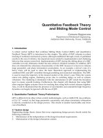
Langdon, Lee and Binns (2009) studied acute ischemic stroke patients and found the
incidence of overall infections (respiratory, urinary tract and other infections) in stroke
patients who were tube fed was 69%, with 51 infections occurring in 74 patients. Thirty of
these infections were respiratory. They reported a significant effect of time-to-event, with
the majority of respiratory infections diagnosed on days 3 and 4 after stroke, and 39/51
Acute Ischemic Stroke
94
(76%) of all infections diagnosed in tube-fed survivors occurring by day 7 after stroke. The
relationship between tube feeding and subsequent development of respiratory infections is
shown in Figure 2.
Fig. 2. Relationship Between Tube Feeding and Respiratory Infections in the First Month
Following Stroke.
It has been hypothesized that nasogastric feeding tubes actually predispose patients to
aspiration by
1. Loss of anatomical integrity of the upper and lower oesophageal sphincters
2. Increase in frequency of transient lower oesophageal sphincter relaxations
3. Desensitization of the pharyngoglottal adduction reflex (Gomes, Pisani, Macedo &
Campos, 2003).
Ischemic Stroke
Patients n = 369
Survivors
n=330
Deaths
n=39
Tube Fed
n = 74
Fed orally
n = 256
No Infection
n = 23
Infection
n = 51
No Infection
n = 192
Infection
n = 64
Other
n = 21 (41%)
Respiratory
n = 30 (59%)
Other
n = 43 (67%)
Respiratory
n = 21 (33%)
Dysphagia and Respiratory Infections in Acute Ischemic Stroke
95
In addition, presence of a feeding tube may lead to formation of biofilms which become
colonized by bacteria, found to have an incidence of 60% of day one and 100% on
subsequent days (Leibovitz, Baumoehl, Steinberg & Segal, 2005).
Data from the FOOD trial was inconclusive on efficacy of nasogastric feeding in early stroke,
however it has been suggested that enteral feeding need not begin in the first few days after a
stroke (Donnan & Dewey, 2005). Langdon, Lee and Binns suggested that it may be appropriate
to hold off from instituting enteral feeding in the first 3-4 days post stroke, as this appears to be
a period when patients are especially susceptible to developing respiratory infections.
5.5 Gastroesophageal reflux
Patients who have gastroesophageal reflux (GOR) and also a poor conscious state are
considered at very high risk of aspiration, particularly if they have a large volume of gastric
content (DeLegge, 2002). Langdon, Lee and Binns (2010) found that subjects with a prior
history of GOR had a significantly increased risk of developing respiratory infections in
acute stroke, with 19 of 55 patients with pre-existing GOR developing respiratory infections
OR 1.89 (95% CI 1.23, 2.9), p=.01.
It may be that acute stroke affects gastric motility due to the neurological insult,
predisposing patients to an increased risk of reflux. Oesophageal manometry studies
performed in five patients with dysphagic stroke and PEG tubes found lower oesophageal
sphincter dysfunction occurred in 80% with significant gastroesophageal reflux seen in two
patients. One of these two patients recovered swallow function and resumed oral feeding,
while the other developed three bouts of aspiration pneumonia and died four months after
PEG insertion (Elphick, Elphic, Smith, DaCosta & Riley 2006). An earlier study examined 35
acute stroke patients with severe impairment requiring tube feeding and found lower
oesophageal sphincter dysfunction in 24 patients; upper oesophageal sphincter function was
lower than normal in 30 patients (Lucas, Yu, Vlahos & Ledgerwood (1999).
Positioning in stroke patients can be an important prophylactic measure for GOR events.
Elphick et. al., (2006) found reflux events occurred significantly more frequently when
patients were in either right lateral or supine position. Supine positioning is considered a
risk factor for development of reflux- related and ventilator-assisted pneumonia (Cook &
Kollef, 1998; Metheny, Chang, Ye, Edwards, Defer et. al., 2002).
6. A critical period of susceptibility to infection in acute ischemic stroke
Previous studies have shown that infection commonly occurs in the acute period
immediately following a stroke. Gordon, Hewer & Wade (1987) reported that all eleven
chest infections developed during the first seven days post stroke in their cohort of 91
dysphagic stroke patients. Mann, Hankey and Cameron (1999) reported 26/112 patients
developed chest infections during the six-month post stroke period, with 10/26 diagnosed
during the first week post-onset.
In the Copenhagen study, 24.5% of female subjects and 13.6% of males developed early
infections (Kammersgaard, Jorgensen, Reith, Nakayama, Houth, Weber, Pederse & Olsen,
2001). The GAIN trial noted that the majority of infections occurred in the first week of the
acute stroke period (Aslanyan, 2004). In an Australian cohort of 330 ischemic stroke
survivors, 115 infections were treated in the first month post stroke. Sixty (52%) of these
infections were diagnosed by the third day post admission. During the first seven days post
stroke, 51 infections in females, and 45 infections in males were diagnosed (Langdon, Lee &
Acute Ischemic Stroke
96
Binns, 2010). These represent 83% of all the infections treated in the first month, and
demonstrate a strong argument that the first week post stroke is associated with a greater
period of susceptibility to infections.
Recent studies in animal models have suggested that stroke induces a severe depression of
immune function, and predisposes patients to infections. This phenomenon, stroke induced
immunodepression (SIDS) leads to lymphocytopneia and functional deactivation of
monocytes and T-helper cells (Dirnafl, Klehmet, Braun, Harms, Neisel, et. al., 2007). This has
been shown to be strongly related to the infarct volume and stroke severity (Hug, Dalpke,
Wieczorek, Giese, Lorenz et. al., 2009).
In animal studies, stroke increases susceptibility to aspiration-induced pneumonia. In a
murine model, control animals required at least a 3 order of magnitude increase in the
numbers of bacteria required to induce pneumonia compared to mice with induced MCA
stroke (Prass, Meisel, Hoflich, Braun, Halle, et.al., 2003).
There is evidence that overactivation of the sympathetic nervous system occurs in the first
two days post stroke in humans. Within 3 days of the initial insult, up to 61% of patients will
become febrile, with the most common cause being infection. Of these infections,
pneumonia is most commonly reported (Mergenthaler, Dirnagl & Meiser, 2004).
In a cohort of 330 stroke survivors, 81 cases of nosocomial infection were diagnosed in the
first month post stroke. Of these 81 infections, 56 (69%) were diagnosed in the first week
post stroke; 28 infections in males and 28 in female subjects. Female subjects demonstrated a
definite peak of infections occurring on Day 3 post stroke, while males were diagnosed on
Day 3 and Day 4. Once the first 7 days post stroke were past, infection rates declined
significantly for both genders (Langdon, Lee & Binns, 2010). Prass and colleagues (2006)
have suggested that a stroke causes a shift from harmless aspiration to severe potentially life
threatening infection.
7. Conclusions
Respiratory infections and urinary tract infections are the most common infections in acute
stroke patients. Studies indicate that patients are particularly vulnerable to infection in the
very acute period of their stroke: the first few days following an infarct. During this period
it may be appropriate for patients to be ‘Nil by Mouth’ if they are at high risk of aspiration.
Hydration can be maintained using either intravenous hydration or hypodermoclysis (sub
cutaneous fluids), while medications can be administered transdermally, intravenously or
by suppository.
Multidisciplinary care that concentrates on minimizing risk factors for infection is
essential. These include minimising aspiration risk with good dysphagia management,
positioning and chest care, early mobilisation, stringent oral hygiene and encouragement
of self-feeding.
While dysphagia is very common in the acute stroke period, for the majority of patients it
is a transitory phenomenon and they can look forward to resuming normal eating and
drinking within a short period of time. For those patients with more severe and persistent
dysphagia, good management will help prevent respiratory complications. Early referral
to a Speech Language Pathologist with expertise in dysphagia assessment and
rehabilitation will ensure that patients have their best possible chance of return to eating
and drinking normally – one of life’s great pleasures as well as an act essential to
maintaining life.