Proteomic Applications in Biology Part 7 doc
Bạn đang xem bản rút gọn của tài liệu. Xem và tải ngay bản đầy đủ của tài liệu tại đây (2.76 MB, 17 trang )
Proteomics as a Tool for the Characterization of Microbial Isolates and Complex Communities
91
bacteria involved in acid mine drainage formation. PLoS Biology, Vol. 6, No. 7, pp.
e177.
Smedley, D., Haider, S., Ballester, B., Holland, R., London, D., Thorisson, G., & Kasprzyk, A.
(2009). BioMart biological queries made easy. BMC Genomics, Vol. 10, pp. 22.
Taylor, C. F., Paton, N. W., Lilley, K. S., Binz, P A., Julian, R. K., Jones, A. R., Zhu, W.,
Apweiler, R., Aebersold, R., Deutsch, E. W., Dunn, M. J., Heck, A. J. R., Leitner, A.,
Macht, M., Mann, M., Martens, L., Neubert, T. A., Patterson, S. D., Ping, P.,
Seymour, S. L., Souda, P., Tsugita, A., Vandekerckhove, J., Vondriska, T. M.,
Whitelegge, J. P., Wilkins, M. R., Xenarios, I., Yates, J. R., & Hermjakob, H. (2007).
The minimum information about a proteomics experiment (MIAPE). Nature
Biotechnology, Vol. 25, No. 8, pp. 887-893.
Taylor, E. B., & Williams, M. A. (2010). Microbial protein in soil: influence of extraction
method and C amendment on extraction and recovery. Microbial Ecology, Vol. 59,
No. 2, pp. 390-399.
Vallenet, D., Labarre, L., Rouy, Z., Barbe, V., Bocs, S., Cruveiller, S., Lajus, A., Pascal, G.,
Scarpelli, C., & Médigue, C. (2006). MaGe: a microbial genome annotation system
supported by synteny results. Nucleic Acids Research, Vol. 34, No. 1, pp. 53-65.
Vizcaíno, J. A., Côté, R., Reisinger, F., M. Foster, J., Mueller, M., Rameseder, J., Hermjakob,
H., & Martens, L. (2009). A guide to the Proteomics Identifications Database
proteomics data repository. Proteomics, Vol. 9, No. 18, pp. 4276-4283.
Weiss, S., Carapito, C., Cleiss, J., Koechler, S., Turlin, E., Coppee, J Y., Heymann, M., Kugler,
V., Stauffert, M., Cruveiller, S., Médigue, C., Van Dorsselaer, A., Bertin, P. N., &
Arsène-Ploetze, F. (2009). Enhanced structural and functional genome elucidation
of the arsenite-oxidizing strain Herminiimonas arsenicoxydans by proteomics data.
Biochimie, Vol. 91, No. 2, pp. 192-203.
Werner, J. J., Ptak, A. C., Rahm, B. G., Zhang, S., & Richardson, R. E. (2009). Absolute
quantification of Dehalococcoides proteins: enzyme bioindicators of chlorinated
ethene dehalorespiration. Environmental Microbiology, Vol. 11, No. 10, pp. 2687-
2697.
Wilkins, M. J., Verberkmoes, N. C., Williams, K. H., Callister, S. J., Mouser, P. J., Elifantz, H.,
N’guessan, A. L., Thomas, B. C., Nicora, C. D., Shah, M. B., Abraham, P., Lipton, M.
S., Lovley, D. R., Hettich, R. L., Long, P. E., & Banfield, J. F. (2009). Proteogenomic
monitoring of Geobacter physiology during stimulated uranium bioremediation.
Applied and Environmental Microbiology, Vol. 75, No. 20, pp. 6591-6599.
Wilmes, P., & Bond, P. L. (2004). The application of two-dimensional polyacrylamide gel
electrophoresis and downstream analyses to a mixed community of prokaryotic
microorganisms. Environmental Microbiology, Vol. 6, No. 9, pp. 911-920.
Yan, J. X., Devenish, A. T., Wait, R., Stone, T., Lewis, S., & Fowler, S. (2002). Fluorescence
two-dimensional difference gel electrophoresis and mass spectrometry based
proteomic analysis of Escherichia coli. Proteomics, Vol. 2, No. 12, pp. 1682-1698.
Yilmaz, P., Kottmann, R., Field, D., Knight, R., Cole, J. R., et al. (2011). Minimum
information about a marker gene sequence (MIMARKS) and minimum
information about any (x) sequence (MIxS) specifications. Nature Biotechnology,
Vol. 29, No. 5, pp. 415-420.
Proteomic Applications in Biology
92
Zanzoni, A., Carbajo, D., Diella, F., Gherardini, P. F., Tramontano, A., Helmer-Citterich, M.,
& Via, A. (2011). Phospho3D 2.0: an enhanced database of three-dimensional
structures of phosphorylation sites. Nucleic Acids Research, Vol. 39, No. Database
issue, pp. D268-271.
5
Life in the Cold: Proteomics of the Antarctic
Bacterium Pseudoalteromonas haloplanktis
Florence Piette, Caroline Struvay,
Amandine Godin, Alexandre Cipolla and Georges Feller
Laboratory of Biochemistry, Center for Protein Engineering, University of Liège
Belgium
1. Introduction
It is frequently overlooked that the majority (>80%) of the Earth’s biosphere is cold and
permanently exposed to temperatures below 5 °C (Rodrigues & Tiedje, 2008). Such low
mean temperatures mainly arise from the fact that ~70% of the Earth’s surface is covered by
oceans that have a constant temperature of 4°C below 1000 m depth, irrespective of the
latitude. The polar regions account for another 15%, to which the glacier and alpine regions
must be added, as well as the permafrost representing more than 20% of terrestrial soils. All
these low temperature biotopes have been successfully colonized by cold-adapted
microorganisms, termed psychrophiles (Margesin et al., 2008). These organisms do not
merely endure such low and extremely inhospitable conditions but are irreversibly adapted
to these environments as most psychrophiles are unable to grow at mild (or mesophilic)
temperatures. Extreme psychrophiles have been traditionally sampled from Antarctic and
Arctic sites, assuming that low temperatures persisting over a geological time-scale have
promoted deep and efficient adaptations to freezing conditions. In addition to ice caps and
sea ice, polar regions also possess unusual microbiotopes such as porous rocks in Antarctic
dry valleys hosting microbial communities surviving at -60 °C (Cary et al., 2010), the liquid
brine veins between sea ice crystals harboring metabolically-active microorganisms at -20 °C
(Deming, 2002) or permafrost cryopegs, i.e. salty water pockets that have remained liquid at
-10 °C for about 100 000 years (Gilichinsky et al., 2005). Psychrophiles and their biomolecules
also possess an interesting biotechnological potential, which has already found several
applications (Margesin & Feller, 2010).
Cold exerts severe physicochemical constraints on living organisms including increased
water viscosity, decreased molecular diffusion rates, reduced biochemical reaction rates,
perturbation of weak interactions driving molecular recognition and interaction,
strengthening of hydrogen bonds that, for instance, stabilize inhibitory nucleic acid
structures, increased solubility of gases and stability of toxic metabolites as well as reduced
fluidity of cellular membranes (D'Amico et al., 2006; Gerday & Glansdorff, 2007; Margesin et
al., 2008; Rodrigues & Tiedje, 2008). Previous biochemical studies have revealed various
adaptations at the molecular level such as the synthesis of cold-active enzymes by
psychrophiles or the incorporation of membrane lipids promoting homeoviscosity in cold
conditions. It was shown that the high level of specific activity at low temperatures of cold-
adapted enzymes is a key adaptation to compensate for the exponential decrease in
Proteomic Applications in Biology
94
chemical reaction rates as the temperature is reduced. Such high biocatalytic activity arises
from the disappearance of various non-covalent stabilizing interactions, resulting in an
improved flexibility of the enzyme conformation (Feller & Gerday, 2003; Siddiqui &
Cavicchioli, 2006; Feller, 2010). Whereas membrane structures are rigidified in cold
conditions, an adequate fluidity is required to preserve the integrity of their physiological
functions. This homeoviscosity is achieved by steric hindrances introduced into the lipid
bilayer via incorporation of cis-unsaturated and branched-chain lipids, a decrease in average
chain length, and an increase both in methyl branching and in the ratio of anteiso- to iso-
branching (Russell, 2007).
More recently, several genomes from psychrophilic bacteria have been sequenced (Danchin,
2007; Casanueva et al., 2010) but only a few of them have been analyzed with respect to cold
adaptation (Saunders et al., 2003; Rabus et al., 2004; Medigue et al., 2005; Methe et al., 2005;
Riley et al., 2008; Rodrigues et al., 2008; Allen et al., 2009; Ayala-del-Rio et al., 2010). However,
the lack of common features shared by all these psychrophilic genomes has suggested that
cold adaptation superimposes on pre-existing cellular organization and, accordingly, that
the adaptive strategies may differ between the various microorganisms (Bowman, 2008;
Piette et al., 2010).
The Gram-negative bacterium Pseudoalteromonas haloplanktis is a typical representative of γ-
proteobacteria found in cold marine environments and, in fact, strain TAC125 has been
isolated from sea water sampled along the Antarctic ice-shell (Terre Adélie). Such strains
thrive permanently in sea water at about -2 °C to +4 °C but are also anticipated to endure
long term frozen conditions when entrapped in the winter ice pack. The genome of P.
haloplanktis TAC125 has been fully sequenced and has undergone expert annotation
(Medigue et al., 2005). This work has allowed a proteomic study of its cold-acclimation
proteins (CAPs)
1
, i.e. proteins that are continuously overexpressed at a high level during
growth at low temperatures (Piette et al., 2010). This has demonstrated that protein synthesis
and protein folding are the main up-regulated functions, suggesting that both cellular
processes are limiting factors for bacterial development in cold environments. Furthermore,
a proteomic survey of cold-repressed proteins at 4 °C has revealed a strong repression of
most heat shock proteins (Piette et al., 2011). This chapter describes the various proteomic
features analyzed in the context of adaptation to life at low temperature.
2. Temperature dependence of growth
The ability of P. haloplanktis to grow at low temperatures is illustrated in Fig. 1. This
psychrophilic Antarctic bacterium maintains a doubling time of ~4 h at 4 °C in a marine
broth, with an extrapolated generation time of 5 h 15 at 0 °C (Fig. 1a). This can be compared
with the behavior of a mesophilic bacterium such as E. coli, which displays a doubling time
of ~8h at 15 °C and which fails to grow below ~8 °C (Strocchi et al., 2006). When the culture
temperature is raised up to 20 °C, the generation time moderately decreases (e.g. 1 h 40 at 18
°C) with a concomitant increase in the biomass produced at the stationary phase (Fig. 1b). At
temperatures higher than 20 °C, the doubling time of P. haloplanktis slightly increases again
with, however, a drastic reduction in cell density at the stationary phase (Fig. 1b), indicating
a heat-induced stress on the cell. P. haloplanktis TAC125 fails to grow above 29 °C, thereby
1
The abbreviations used are: CAPs, cold acclimation proteins; CRPs, cold repressed proteins; TF, trigger
factor; ROS, reactive oxygen species
Life in the Cold: Proteomics of the Antarctic Bacterium Pseudoalteromonas haloplanktis
95
Fig. 1. (a) Temperature dependence of the generation time of Pseudoalteromonas haloplanktis
TAC125 grown in a marine broth (solid line and circles). A typical curve for E. coli RR1 in LB
broth is shown for comparison (dashed) (b) Growth curves of P. haloplanktis at 4°C (○), 18°C
(●) and 26°C (■). Reprinted with permission from Piette et al., 2011. © 2011 American Society
for Microbiology.
defining its upper cardinal temperature. According to this growth behavior, the
temperatures of 4 °C and 18 °C were selected for the differential comparison of the
proteomes, as 18 °C does not induce an excessive stress as far as growth rate and biomass
are concerned.
The fast growth rate of the Antarctic bacterium is primarily achieved by a low temperature
dependence of the generation times when compared with a mesophilic bacterium, i.e. the
generation time of P. haloplanktis is moderately increased when the culture temperature is
decreased (Fig. 1a). It should be stressed that enzymes from cold-adapted organisms are
characterized by both a high specific activity at low temperatures and a low temperature
dependence of their activity (formally, a weak activation enthalpy), i.e. reaction rates of
psychrophilic enzymes are less reduced by a decrease in temperature as compared with
mesophilic enzymes (D'Amico et al., 2003; Feller & Gerday, 2003). Accordingly, the growth
characteristics of the Antarctic bacterium (Fig. 1a) appear to be governed by the properties
of its enzymatic machinery: high enzyme-catalyzed reaction rates maintain metabolic fluxes
Proteomic Applications in Biology
96
and cellular functions at low temperatures, whereas the weak temperature dependence of
enzyme activity counteracts the effect of cold temperatures on biochemical reaction rates.
3. Cold-induced versus cold-repressed proteins
The proteomes expressed by the Antarctic bacterium at 4 °C and 18 °C during the
logarithmic phase of growth have been compared by two-dimensional differential in-gel
electrophoresis (2D-DIGE), enabling the co-migration in equal amounts of cell extracts
obtained from both conditions (labeled by distinct CyDye fluorophores) in triplicate gels
(Fig. 2).
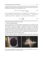
Fig. 2. Comparison of intracellular soluble proteins from P. haloplanktis grown at 4°C (red-
labeled) and 18°C (green-labeled) on 2D-DIGE gels analyzed by fluorescence. From left to
right, non-linear gradient from pH 3 to pH 10. From top to bottom, mass scale from ~150 to
~15 kDa. The intense red fluorescence of the trigger factor (TF) spot correlates with its up-
regulation at 4°C, whereas the intense green fluorescence of the DnaK spot correlates with
its down-regulation Adapted with permission from Piette et al., 2010. © 2010 Wiley.
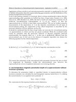
In a typical single 2D-gel (Fig. 3), 142 protein spots are more abundant at 4 °C. As protein
extracts were prepared from cells growing exponentially at this temperature, all up-
regulated proteins at 4°C are regarded as CAPs. Furthermore, 309 protein spots are less
Life in the Cold: Proteomics of the Antarctic Bacterium Pseudoalteromonas haloplanktis
97
Fig. 3. Differential analyses of soluble cellular proteins from Pseudoalteromonas haloplanktis
grown at 4°C (left panels) and 18°C (right panels) on 2D-DIGE gels analyzed by
fluorescence. (a) 142 protein spots that are more intense at 4°C are indicated. (b) 309 protein
spots that are less intense at 4°C are indicated. Reprinted with permission from Piette et al.,
2011. © 2011 American Society for Microbiology.
intense at 4 °C as compared with 18 °C. This unexpected large number of cold-repressed
proteins (CRPs) already indicates that numerous cellular functions are down-regulated
during growth at low temperature.
The induction factors for CAPs and the repression factors for CRPs, given by the spot
volume ratio between 4 °C and 18 °C are illustrated in Fig. 4. This distribution shows that
most CAPs and CRPs have a five-time higher or lower relative abundance at 4 °C. However,
about 20% of these differentially expressed proteins display up- or down-regulation factors
higher than 5, revealing that some key cellular functions are strongly regulated. Amongst all
these differentially expressed proteins, 40 CAPs and 83 CRPs were retained, which satisfied
both statistical biological variation analysis and mass spectrometry identification scores, as
Proteomic Applications in Biology
98
Fig. 4. Distribution of the relative abundance of cold-repressed proteins (dashed, negative
values) and of cold acclimation proteins (positive values) in the proteome of P. haloplanktis
grown at 4°C and 18°C. Reprinted with permission from Piette et al., 2011. © 2011 American
Society for Microbiology.
detailed in the original publications (Piette et al., 2010; Piette et al., 2011). Accordingly, the
identified proteins should be analyzed as markers of a pathway or of a general function,
rather than for their specific function as they represent 27% of the differentially expressed
proteins at 4°C.
4. Cold shock and heat shock proteins
One of the most remarkable features of the differentially expressed proteome of P.
haloplanktis is the strong up-regulation at 4°C of proteins that are regarded as cold shock
proteins in mesophilic bacteria, as well as the down-regulation to nearly undetectable levels
of proteins classified as heat shock proteins (Fig. 5).
Cold shock proteins that have been identified as CAPs in P. haloplanktis include Pnp (+4x),
TypA (+5x) and the trigger factor TF (+38x) that are involved in distinct functions
(degradosome, membrane integrity and protein folding, respectively). Sustained synthesis
of various cold shock protein-homologues has been also reported in other cold-adapted
bacteria (Bakermans et al., 2007; Kawamoto et al., 2007; Bergholz et al., 2009). There are
therefore striking similarities between the cold shock response in mesophiles and cold
adaptation in psychrophiles. From an evolutionary point of view, it can be proposed that
one of the adaptive mechanisms to growth in the cold was to regulate the cold shock
response, shifting from a transient expression of cold shock proteins to a continuous
synthesis of at least some of them. Interestingly, nearly all proteins displaying the highest
repression factors at 4°C are heat shock proteins (Rosen & Ron, 2002) including the main
chaperones DnaK (-13x) and GroEL (-3.4x), the accessory chaperones such as Hsp90 (-28x),
the small heat shock proteins IbpA (-24x) and IbpB (-18x), as well as LysS (-17x).
Life in the Cold: Proteomics of the Antarctic Bacterium Pseudoalteromonas haloplanktis
99
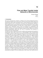
Fig. 5. Comparative analysis of spots containing the trigger factor TF (a cold shock protein)
and DnaK (a heat shock protein) from P. haloplanktis grown at 4°C (left panels) and 18°C
(right panels). Spot views on 2D-gels (circled) and three-dimensional images. Adapted with
permission from Piette et al., 2010; Piette et al., 2011. © 2010 Wiley and © 2011 American
Society for Microbiology.
Proteomic Applications in Biology
100
In mesophilic bacteria such as E. coli, cold shock and heat shock proteins are transiently
expressed in response to temperature downshift and upshift, respectively. By contrast, the
Antarctic bacterium continuously over-expresses some cold shock proteins (Piette et al.,
2010) whereas most heat shock proteins are continuously repressed at 4 °C. It is obvious that
regulation of the expression of these proteins involved in thermal stress is a primary
adaptation to bacterial growth at low temperatures that remains to be properly explained.
5. Protein folding at low temperature rescued by the trigger factor
In bacteria, the three main chaperones are the trigger factor TF, a cold shock protein that
stabilize nascent polypeptides on ribosomes and initiate ATP-independent folding, DnaK
that mediates co- or post-transcriptional folding and the GroEL/ES chaperonin that acts
downstream in folding assistance (Hartl & Hayer-Hartl, 2009). Both latter chaperones are
also well-known heat shock proteins. The trigger factor TF (+38x up-regulated at 4°C) is the
first molecular chaperone interacting with virtually all newly synthesized polypeptides on
the ribosome. It delays premature chain compaction and maintains the elongating
polypeptide in a non-aggregated state until sufficient structural information for productive
folding is available and subsequently promotes protein folding (Merz et al., 2008; Hartl &
Hayer-Hartl, 2009; Martinez-Hackert & Hendrickson, 2009). Furthermore, TF also contains a
domain catalyzing the cis-trans isomerization of peptide bonds involving a proline residue
(Kramer et al., 2004). This cis-trans isomerization is a well-known rate-limiting step in
protein folding (Baldwin, 2008). On the other hand the major heat shock proteins were
identified as strongly cold-repressed proteins in the proteome of P. haloplanktis (or, in other
words, they are up-regulated at 18°C). The overexpression of bacterial heat shock proteins at
elevated temperatures is well recognized as being indicative of a heat-induced cellular stress
(Rosen & Ron, 2002; Goodchild et al., 2005). Although this is obviously relevant for the
Antarctic bacterium grown at 18 °C, the implications for the psychrophilic strain appear to
be more complex. Indeed, these heat shock proteins are chaperones assisting co- or post-
translational protein folding (Hartl & Hayer-Hartl, 2009). Furthermore, it has been
demonstrated that GroEL from P. haloplanktis is not cold-adapted, it is inefficient at low
temperatures as its activity is reduced to the same extent than that of its E. coli homologue
(Tosco et al., 2003). Accordingly, under this imbalanced synthesis of folding assistants,
protein folding at low temperature is apparently compromised in the Antarctic bacterium.
Considering the down-regulation of heat shock chaperones and the inefficiency of GroEL
from P. haloplanktis at low temperature, as well as the essential function of TF in the
initiation of proper protein folding, it can be proposed that TF rescues the chaperone
function at low temperatures, therefore explaining its unusual overexpression level. It
follows that TF becomes the primary chaperone of the Antarctic bacterium for growth in the
cold. Although the psychrophilic bacterium maintains a minimal set of chaperones, this is
obviously sufficient to allow bacterial development at low temperature.
6. Possible origins of heat shock protein repression at low temperature
The strong overexpression of TF at low temperature can be understood according to its
above mentioned essential function. By contrast, the reasons for the concomitant repression
of heat shock chaperones in the Antarctic bacterium remain hypothetical. At least four
possible origins, not mutually exclusive, can be mentioned. i) Low temperature slows down
Life in the Cold: Proteomics of the Antarctic Bacterium Pseudoalteromonas haloplanktis
101
the folding reaction and is well known to reduce the probability of misfolding and
aggregation (King et al., 1996), therefore possibly reducing the need for heat shock
chaperones that act downstream from TF. ii) In E. coli, it has been shown that synthesis of
heat shock proteins is repressed during growth at low temperatures, but also that these heat
shock proteins are harmful to cells at 4 °C, as their induced expression reduces cell viability
at this temperature (Kandror & Goldberg, 1997). Accordingly, the observed cold-repression
of heat shock proteins would be beneficial to the psychrophilic bacterium. iii) To our
knowledge, the reasons for this harmful effect of heat shock proteins have not been
investigated. A possible explanation could be found in the second function of these
chaperones. Indeed, besides their role in folding assistance, many heat shock proteins bind
partly folded polypeptides and promote their fast degradation by proteases Lon and Clp. In
addition, TF enhances the binding affinity of some heat shock proteins for these partly
folded polypeptides (Kandror et al., 1995; Kandror et al., 1997). In the case of the Antarctic
bacterium, heat shock chaperones would then have an increased ability to bind slowly
folding polypeptides at low temperatures and to promote their unwanted degradation: this
would account for the cold repression of heat shock chaperones by P. haloplanktis. iv) The
observation that GroEL from the Antarctic bacterium is non cold-adapted (Tosco et al., 2003)
suggests that this chaperonin is well suited to function during sudden temperature increases
of the environment. Indeed, microorganisms subjected to seasonal or local temperature
variations (e.g. melting sea ice, polar surface soils, etc.) would advantageously maintain a
heat shock response involving non cold-adapted chaperones remaining active at transiently
high temperatures.
7. Structural properties of the psychrophilic trigger factor
According to its essential function in P. haloplanktis, the psychrophilic TF has been analyzed
into more details. Its amino acid sequence (47,534 Da) displays 61% identity (85% similarity)
on 434 residues with its homologue from E. coli. Its sequence is also close to that of some
known TF from psychrophilic bacteria. The pronounced sequence similarity and predicted
secondary structure conservation with E. coli TF suggest that the psychrophilic chaperone
should also folds into an extended “crouching dragon” conformation (Ferbitz et al., 2004)
comprising three domains (Fig. 6). The N-terminal domain mediates ribosome attachment
via an exposed loop, the PPIase activity domain located at the opposite end of the molecule
(Kramer et al., 2004) and the C-terminal domain forming the body of the protein and bearing
the central module of chaperone activity (Merz et al., 2006).
In order to analyze the psychrophilic TF, its gene has been cloned and overexpressed in E.
coli and the recombinant protein has been purified to homogeneity (Piette et al., 2010). Its
thermal stability was investigated by differential scanning calorimetry. Fig. 7 shows that TF
from the Antarctic bacterium is a marginally stable protein, exhibiting a melting point T
m
at
33°C. It follows that at a typical mesophilic temperature of 37°C, almost all the protein
population is already in the unfolded state. In addition, the calorimetric enthalpy is also
very weak (
Δ
H
cal
= 82.5 kcal mol
-1
, the sum of all enthalpic contributions to protein stability
disrupted during unfolding and calculated from the area under the transition). By
comparison, a T
m
of 54°C and a calorimetric enthalpy of 178 kcal mol
-1
have been reported
for the E. coli trigger factor analyzed by DSC (Fan et al., 2008). Despite its modular structure,
P. haloplanktis TF unfolds according to a perfect 2-state transition (Fig. 7), i.e. without
significantly populated intermediates between the native and the unfolded states. This
Proteomic Applications in Biology
102
Fig. 6. Domain organization in the trigger factor structure (based on E. coli trigger factor:
PDB 1W26). The N-terminal domain mediating ribosome attachment (aa 1-144) is in red, the
PPiase domain (aa145-247) is in yellow and the C-terminal domain bearing the central
module of the chaperone activity (aa 248-432) is in green. As a result of the strong
conservation of primary and predicted secondary structures in P. haloplanktis TF, a model of
its structure built by homology modeling is undistinguishable from the E. coli crystal
structure. Reprinted with permission from Piette et al., 2010. © 2010 Wiley.
indicates that the psychrophilic TF is uniformly unstable and unfolds cooperatively. To the
best of our knowledge, P. haloplanktis TF is the least stable protein reported so far. This
strongly suggests that the essential chaperone function requires considerable flexibility and
dynamics to compensate for the reduction of molecular motions at freezing temperatures.
The E. coli trigger factor has been reported to undergo in vitro a concentration-dependent
dynamic equilibrium between the monomeric and the dimeric forms. Static light scattering
experiments performed in batch mode provided a mean particle mass of 51 kDa and 106
kDa for P. haloplanktis and E. coli TF, respectively. This is in agreement with a monomeric
psychrophilic TF and a dimeric E. coli TF. However, in dynamic light scattering the particle
polydispersity (size distribution) of P. haloplanktis TF was twice that of E. coli TF, suggesting
that the psychrophilic TF may possibly perform transient intermolecular interactions. These
observations are in line with a report showing that the trigger factor from the psychrophile
Psychrobacter frigidicola is a monomeric chaperone (Robin et al., 2009). The interpretation of
these differences in oligomerization state remains to be properly explained but suggest
noticeable differences between psychrophilic and mesophilic bacteria for the TF function in
Life in the Cold: Proteomics of the Antarctic Bacterium Pseudoalteromonas haloplanktis
103
Fig. 7. Microcalorimetric analysis of the trigger factor from P. haloplanktis. The melting point
T
m
corresponds to the top of the transition at 33°C. The calorimetric enthalpy
Δ
H
cal
corresponds to the area under the transition. The red dashed line corresponds to the fit of
the DSC data to a two-state unfolding transition. Baseline-subtracted data have been
normalized for protein concentration (2.6 mg/ml in 30 mM Mops, 250 mM NaCl, pH 7.6.).
Adapted with permission from Piette et al., 2010. © 2010 Wiley.
the cytoplasmic fraction, when not bound to the ribosome. Finally, in a typical refolding
assay monitoring chaperone activity, it has been found that P. haloplanktis TF is inactive at
20°C and recovers partial activity at 15°C. It was also shown that this TF requires near-zero
temperatures (Fig. 8) to efficiently bind an unfolded protein (Piette et al., 2010). This
illustrates a remarkable cold adaptation of the chaperone function in the psychrophilic TF.
8. Protein synthesis and folding are limiting factors in the cold
Thirty percent of the identified CAPs are directly related to protein synthesis and cover all
essential steps, from transcription (including RNA polymerase RpoB) to translation and
folding (TF, PpiD). Amongst these CAPs, for instance, genes pnp and rpsA encode
components of the degradosome that regulates transcript lifetimes. The Rho termination
factor is a RNA/DNA helicase that can contribute to relieve nucleic acid secondary
structures strengthened in cold conditions. Interestingly, mutations in the ribosomal
protein L6 (RplF) have been reported to cause loss of E. coli cells viability at 0°C (Bosl &
Bock, 1981) and it is also a CAP in P. haloplanktis. Methionyl-tRNA synthetase MetG
displays one of the highest up-regulation ratio (+7.6x): this can be tentatively related to
the requirement of an increased pool of initiation tRNA to promote protein synthesis.
Two putative proteases were also identified as CAPs and can potentially participate to
proteolysis of misfolded proteins.
Proteomic Applications in Biology
104
Fig. 8. Detection of the chaperone activity of the psychrophilic TF. Aggregation at 15°C of a
chemically unfolded protein (glyceraldehyde-3-phosphate dehydrogenase) is monitored by
absorbance at 600 nm after incubation in melting ice. In the absence of TF (black trace),
aggregation occurs as soon as the temperature is raised. By contrast, TF suppresses
aggregation for 10-15 min (blue trace). Adapted with permission from Piette et al., 2010. ©
2010 Wiley
In the last step of protein synthesis, the folding catalyst TF acts on proteins synthesized by
the ribosome and also catalyses peptidyl-prolyl cis-trans isomerisation (PPiase) while PpiD
(another PPiase) is involved in the folding of outer membrane proteins. Peptidyl-prolyl cis-
trans isomerisation appears therefore as a limiting factor for a wide range of proteins in P.
haloplanktis. Furthermore, some previous studies on cold-adapted microorganisms have
reported either PPiases (Goodchild et al., 2004b; Suzuki et al., 2004) or the trigger factor (Qiu
et al., 2006; Kawamoto et al., 2007) as potential CAPs. It seems therefore that the constraints
imposed by protein folding in the cold are common traits in several psychrophilic
microorganisms.
Altogether, these observations strongly suggest that low temperatures impair protein
synthesis and folding, resulting in up-regulation at 4°C of the associated cellular processes.
9. Metabolism depression at low temperatures
Nearly half of down-regulated proteins at 4°C are related to superclasses of function
involved in the bacterial general metabolism. This includes the degradation or biosynthesis
of compounds and the production of energy. Most of these proteins belong to the oxidative
metabolism, in particular to glycolysis, the pentose phosphate pathway, Krebs cycle and
electron chain transporters. Accordingly, the Antarctic bacterium depresses its general
metabolism when grown at low temperature. This is in agreement with the reduced biomass
produced at 4 °C as compared with cultures run at 18 °C (Fig. 1b). As mentioned in the
previous section, protein synthesis and folding are limiting factors for the growth of P.
Life in the Cold: Proteomics of the Antarctic Bacterium Pseudoalteromonas haloplanktis
105
haloplanktis at cold temperatures (Piette et al., 2010). However, the proteomic data indicates
that when these limitations are alleviated at 18 °C, the bacterium proliferates by activation of
its general metabolism and therefore divides actively and produces more biomass. The high
number of identified ribosomal proteins and of elongation factors involved in translation
indicates that protein synthesis is no longer limiting but is also stimulated at 18 °C.
10. Down-regulation of iron metabolism at low temperatures
Iron uptake and iron-related proteins are clearly down-regulated at 4 °C in P. haloplanktis.
The uptake of this essential element in an aquatic environment is mediated by several iron
transport systems. Two systems were found to be down-regulated at 4 °C: the ABC
transporter (FbpA) and a TonB-dependent receptor. The first is involved in the uptake of the
weakly soluble ferric ion (Fe
3+
) directly from the environment and the second is required for
the transport of heme complexes and ferric siderophores through the membrane (Clarke et
al., 2001). The reduced needs for iron by P. haloplanktis at 4 °C can be partly explained by the
down-regulation of the Krebs cycle and respiratory chain (and their iron-containing
complexes such as SdhB), by the repression of HmgA, which requires Fe
2+
to degrade cyclic
amino-acids or by the strong down-regulation of catalase (which is made up of four heme
groups). Hemes are tetrapyrroles that have porphobilinogen as a precursor: this is in
agreement with the down-regulation of both GltX (glutamyl-tRNA synthetase) and HemB
(5-aminolevulinate dehydratase), which are responsible for porphobilinogen synthesis.
Various metallic ions are essential to the cell metabolism and therefore the fact that
proteomic data only points to cold repression of iron-related proteins is puzzling. Iron in a
redox-active form (Fe
2+
) is potentially deleterious, as it is able to induce oxidative cell
damage by the Fenton reaction, for instance (Valko et al., 2005). It can be tentatively
proposed that, as a result of the improved stability of ROS (reactive oxygen species) at low
temperatures, the down-regulation of iron-related proteins could contribute to an avoidance
of such detrimental iron-based reactions. In this respect, it should be mentioned that the
genome of P. haloplanktis entirely lacks the ubiquitous ROS-producing molybdopterin
metabolism (Medigue et al., 2005). This suggests that the Antarctic bacterium tends to avoid
ROS production involving metallic ions.
11. Oxidative stress-related proteins
The pattern of oxidative stress-related proteins in P. haloplanktis is complex because some
have been identified as CAPs, while others were found to be CRPs. For instance, glutathione
synthetase is the second main up-regulated protein at 4°C (+13.2x) and superoxide
dismutase (+1.6x) was also detected as a CAP. This is a clear indication of a cellular response
to an oxidative stress arising from increased dioxygen solubility and ROS stability. On the
other hand, the second group of proteins that displays the highest repression factors at 4 °C
is represented by the oxidative stress-related proteins catalase (-6.5x), glutathione reductase
(-8.1x) and peroxiredoxin (-15.7x). At first sight, this may be regarded as a conflicting result
because conclusive evidences have indicated that psychrophiles are exposed to a permanent
oxidative stress at low temperatures, which originates from improved dioxygen solubility
and increased ROS stability (Rabus et al., 2004; Medigue et al., 2005; Methe et al., 2005;
Duchaud et al., 2007; Bakermans et al., 2007; Ayub et al., 2009; Piette et al., 2010). In order to
reconcile these apparent contradictions, it should be recalled that the general aerobic
Proteomic Applications in Biology
106
metabolism of the Antarctic bacterium is stimulated at 18 °C, also resulting in ROS
production. Accordingly, the identified oxidative stress-related proteins would be better
regarded as being induced at 18 °C, rather than repressed at 4 °C.
The up-regulation of catalase and peroxiredoxin at 18°C shows that the bacterium needs to
be protected against ROS like H
2
O
2
as both enzymes catalyze its decomposition into O
2
and
H
2
O. Under oxidative stress, the NADPH supply for reduced glutathione regeneration is
also dependent on glucose-6-phosphate dehydrogenase (Zwf) in the first step of the pentose
phosphate pathway, and indeed Zwf is positively regulated at 18 °C. Glutathione reductase
(Gor) plays a central role in the reoxidation of NADPH from the pentose phosphate
pathway, allowing formation of reduced glutathione, an important cellular antioxidant. The
up-regulated DNA-binding DPS protein (DpsB) plays a major role in the protection of
bacterial DNA from damage by ROS and is induced under stress conditions. Some DPS
proteins are also able to bind iron and are involved in its storage and in the protection of the
cell (Haikarainen & Papageorgiou, 2010).
There is obviously a finely-tuned balance between the cellular mechanisms protecting
against oxidative stresses generated by low temperatures (resulting from ROS stability and
oxygen solubility) and by high temperatures (resulting from stimulated metabolic activity).
The number of identified proteins does not allow a detailed description of this balance but
the strong involvement of glutathione synthetase, glutathione reductase and the
Microor
g
anism Source Technique Ref.
Bacillus
p
sychrosaccharolyticus
Soil, marshes 2D-PAGE 1
M
ethanococcoides burtonii
Ace Lake, Antarctica LC/LC-MS/MS 2
M
ethanococcoides burtonii
Ace Lake, Antarctica 2D-PAGE 3
M
ethanococcoides burtonii
Ace Lake, Antarctica ICAT LC MS 4
M
ethanococcoides burtonii
Ace Lake, Antarctica LC MS 5
M
ethanococcoides burtonii
Ace Lake, Antarctica SDS-PAGE LC-MS/MS 6
Exiguobacterium sibiricum
Siberian permafrost 2D LC MS 7
Shewanella livingstonensis
Antarctic seawater 2D-PAGE 8
Psychrobacter cryohalolentis
Siberian permafrost 2D-PAGE 9
Psychrobacter articus
Siberian permafrost 2-D HPLC & MS 10
M
oritella viscosa
Atlantic salmo
n
2D-PAGE 11
Psychrobacter articus
Siberian permafrost 2D-PAGE 12
Sphingopyxis alaskensis
Alaska seawater GeLC-MS/MS 13
M
ethanococcoides burtonii
Ace Lake, Antarctica LC/LC-MS/MS 14
Lactococcus piscium strain
seafood products 2D-PAGE 15
Pseudoalteromonas haloplanktis
Antarctic seawater 2D-DIGE 16
M
ethanococcoides burtonii
Ace Lake, Antarctica 8-plex iTRAQ 17
Acidithiobacillus ferrooxidans
Mine draina
g
e, Canada 2D-PAGE 18
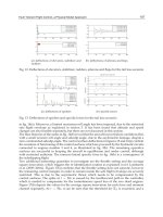
Table 1. Proteomic studies performed on psychrophilic microorganisms. References: 1, Seo et
al., 2004; 2, Goodchild et al., 2004a; 3, Goodchild et al., 2004b; 4, Goodchild et al., 2005; 5,
Saunders et al., 2005; 6, Saunders et al., 2006; 7, Qiu et al., 2006; 8, Kawamoto et al., 2007; 9,
Bakermans et al., 2007; 10, Zheng et al., 2007; 11, Tunsjo et al., 2007; 12, Bergholz et al., 2009;
13, Ting et al., 2010; 14, Williams et al., 2010; 15, Garnier et al., 2010; 16, Piette et al., 2010;
Piette et al., 2011; 17, Williams et al., 2011; 18, Mykytczuk et al., 2011.
Life in the Cold: Proteomics of the Antarctic Bacterium Pseudoalteromonas haloplanktis
107
identification of enzymes belonging to the pentose phosphate pathway suggest that
regulation of the cytoplasmic redox buffering capacity via glutathione is a key component.
12. Other proteomic studies
A selection of recent proteomic studies on psychrophilic and cold-adapted microorganisms
is listed in Table 1. It is worth mentioning that the CAPs and CRPs identified in these
studies do not constitute a conserved set of proteins in terms of identification and
expression level. Nevertheless, a survey of these data shows that the main upregulated
functions for growth at low temperatures are protein synthesis (transcription, translation),
RNA and protein folding, membrane integrity and transport, antioxidant activities and
regulation of specific metabolic pathways. Such heterogeneous upregulation of CAPs
supports the view that cold-adaptation mechanisms are constrained by the species-specific
cellular structure and organization, resulting in distinct adaptive strategies. This hypothesis
is based on a previous observation made by Bowman (2008). In a review of genome data
from psychrophiles, he concluded that the lack of common features shared by these
genomes suggests that cold adaptation superimposes on pre-existing cellular organization
and, accordingly, that the adaptive strategies may differ between the various
microorganisms.
13. Conclusions
The capacity of psychrophilic bacteria to thrive successfully in permanently cold
environments obviously requires a vast array of adaptations. At least two prerequisites to
this environmental adaptation can be cited: i) from a functional standpoint, the synthesis of
cold-active enzymes is required to support the bacterial metabolism and its energy
production (Feller & Gerday, 2003; Siddiqui & Cavicchioli, 2006; Feller, 2010), and ii) from a
structural standpoint, the synthesis of cold-adapted lipids is required to maintain the cell
membrane integrity, fluidity and functions (Russell, 2007). It should be noted that the first
adaptation is genetically encoded in the protein sequence (and results from a long term
adaptation), whereas the second adaptation involves regulation of pre-existing biosynthetic
pathways. However, neither of these basic adaptations is sufficient because low temperature
induces physicochemical constraints that are unavoidable but that can be attenuated by
cellular mechanisms. For instance, low temperature reduces molecular diffusion rates and
also increases water and cytoplasmic viscosity. It can be proposed that both
physicochemical constraints are responsible for the rate limiting steps of P. haloplanktis
growth in the cold, namely protein synthesis and folding, as deduced from proteomic
experiments. Indeed, bacterial protein synthesis is one of the most complex cellular
processes and requires diffusion and docking of numerous partners with ribosomes
(mRNA, tRNA, initiation factors, elongation factors, GTP…). Assuming a high, cold-active
ribosomal efficiency (although this has not been demonstrated to date), its synthetic activity
would be nevertheless restricted by diffusion and availability of the required partners. The
rate of protein folding is also limited by low temperatures. This is an entropically-driven
process governed by the chemical nature of the polypeptide chain and of water molecules.
Furthermore, the main protein chaperones are not catalysts per se but rather they assist in
protein folding and prevent or relieve misfolding. The above mentioned physicochemical
constraints exert their effects on all psychrophiles and it can be anticipated that protein