Báo cáo hóa học: " Nanoparticles for Applications in Cellular Imaging" pot
Bạn đang xem bản rút gọn của tài liệu. Xem và tải ngay bản đầy đủ của tài liệu tại đây (297.78 KB, 12 trang )
NANO REVIEW
Nanoparticles for Applications in Cellular Imaging
K. Ted Thurn Æ Eric M. B. Brown Æ Aiguo Wu Æ Stefan Vogt Æ
Barry Lai Æ Jo
¨
rg Maser Æ Tatjana Paunesku Æ Gayle E. Woloschak
Received: 28 May 2007 / Accepted: 18 July 2007 / Published online: 15 August 2007
Ó to the authors 2007
Abstract In the following review we discuss several
types of nanoparticles (such as TiO
2
, quantum dots, and
gold nanoparticles) and their impact on the ability to image
biological components in fixed cells. The review also dis-
cusses factors influencing nanoparticle imaging and uptake
in live cells in vitro. Due to their unique size-dependent
properties nanoparticles offer numerous advantages over
traditional dyes and proteins. For example, the photosta-
bility, narrow emission peak, and ability to rationally
modify both the size and surface chemistry of Quantum
Dots allow for simultaneous analyses of multiple targets
within the same cell. On the other hand, the surface
characteristics of nanometer sized TiO
2
allow efficient
conjugation to nucleic acids which enables their retention
in specific subcellular compartments. We discuss cellular
uptake mechanisms for the internalization of nanoparticles
and studies showing the influence of nanoparticle size and
charge and the cell type targeted on nanoparticle uptake.
The predominant nanoparticle uptake mechanisms include
clathrin-dependent mechanisms, macropinocytosis, and
phagocytosis.
Keywords Nanoparticle Á Cellular uptake Á
Quantum dots Á Titanium dioxide
Introduction
Implementation of nanoparticle use in cell biology has
been one of the most exciting developments in this field in
the past 5 years. The number of articles describing the use
of nanoparticles is increasing so rapidly that this review
will be limited only to applications of nanoparticles on
whole cells, fixed or alive, and not on the numerous strictly
in vitro or in vivo uses. The focus on cells in this review is
based on the fact that understanding of the interactions
between nanoparticles and cells is the first step toward
mechanistic understanding of the relationship between
organisms and nanomaterials. Therefore, cellular studies
provide a preliminary step for nanoparticle use in in vivo
therapeutic or imaging purposes. Herein we are particularly
interested in nanoparticles applied to cells, and used for
imaging of subcellular components. Although cytotoxicity
and the effects of cell loading by nanoparticles are of little
consequence in fixed cells, nanoparticle biocompatibility
and cellular uptake mechanisms are particularly relevant to
live cell studies. Studies of the effects of nanoparticles on
K. T. Thurn Á E. M. B. Brown Á A. Wu Á T. Paunesku Á
G. E. Woloschak (&)
Department of Radiation Oncology, Northwestern University,
Robert E. Lurie Cancer Center, Feinberg School of Medicine,
303 E. Chicago Ave. Ward Building Room 13-007, Chicago, IL
60611, USA
e-mail:
S. Vogt Á B. Lai
X-Ray Science Division, Advanced Photon Source, Argonne
National Laboratory, Argonne, IL 60439, USA
J. Maser
Center for Nanoscale Materials, Advanced Photon Source,
Argonne National Laboratory, Argonne, IL 60439, USA
T. Paunesku Á G. E. Woloschak
Department of Radiology, Northwestern University, Robert E.
Lurie Cancer Center, Feinberg School of Medicine, 303 E.
Chicago Ave. Ward Building Room 13-007, Chicago, IL 60611,
USA
G. E. Woloschak
Department of Cell and Molecular Biology, Northwestern
University, Robert E. Lurie Cancer Center, Feinberg School of
Medicine, 303 E. Chicago Ave. Ward Building Room 13-007,
Chicago, IL 60611, USA
123
Nanoscale Res Lett (2007) 2:430–441
DOI 10.1007/s11671-007-9081-5
cellular proliferation and viability have shown that in most
cases toxicity/biocompatibility of nanoparticles depends on
their concentration [1–9]. Depending on the type of cell
treated, the size, and the surface charge of the nanoparticle
conjugate (nanoconjugate), different cellular uptake
mechanisms are used by cells—most often clathrin-
dependent mechanisms, macropinocytosis, and phagocy-
tosis [10–17].
Surface modifications are often used to increase the
functionality of nanoconjugates. In work with cells, surface
modifiers serve to (i) increase cellular uptake of nanocon-
jugates, (ii) increase the specificity of cellular uptake, and
(iii) increase the efficiency of intracellular targeting or
retention of nanoconjugates. These nanoparticle modifiers/
conjugants include various antibodies and peptides which
improve cell type and subcellular compartment targeting,
while nucleic acids (and their mimics) have been demon-
strated to modify subcellular retention of nanoconjugates.
This review focuses on optically fluorescent semicon-
ductor quantum dots and noble metal nanoparticles with
size- and shape-dependent optical properties. In addition,
particular attention is given to a different type of semi-
conductor material—TiO
2
which is easily functionalized
by both optically fluorescent agents and molecules for
subcellular targeting. For detection of nanoparticles in cells
some of the most powerful techniques are still optical
microscopy and electron microscopy. However, comple-
mentary newly emerging imaging approaches such as four
photon microscopy [18], near-infrared surface enhanced
Raman scattering [19, 20], X-ray fluorescence micro- and
nano-probe imaging [21–25], and coherent X-ray diffrac-
tion imaging [26–28] will significantly improve imaging
work with nanoparticles in cells. Some of the future
developments with these techniques are expected to allow
for 3D imaging with resolution as good as 5 nm
3
voxel
(coherent X-ray diffraction imaging), permitting imaging
of whole frozen cells with the nanoparticles distributed at
specific destinations in the cellular interior.
Nanoparticle Chemistry
Nanoparticles are mesostructures with some unique prop-
erties compared to bulk materials on one hand and atomic
or molecular structures on the other. Compared to the bulk
materials with constant physical and chemical properties
regardless of their sizes (until it reaches the nano-regime),
the nanoparticles have size-dependent properties: for
example, quantum confinement in different semiconductor
nanoparticles, an absorbance of surface plasmon resonance
in metal (particularly noble metals) nanoparticles, super-
paramagnetism in magnetic nanoparticles etc. Three main
types of nanoparticles used for cellular imaging described
in this review are: polymer/biomacromolecule nanoparti-
cles, semiconductor nanoparticles, and metal nanoparticles.
Polymer/biomacromolecule nanoparticles are made of
biocompatible nanomaterials. Often, they are used in
combination with other types of materials to improve their
biocompatibility or functionality. On their own they are
also used for the applications in cellular imaging. Nano-
particles of this group discussed in this review are:
• polymer nanoparticles such as Poly(
D,L-lactic-co-gly-
colic acid) (PLGA) nanoparticles [29–32], polystyrene
[11, 33], polyethylene glycol (PEG) covered or PEGy-
lated nanoparticles [15, 34], poly(ethylene glycol)-
block-poly(aspartic acid) (PEG-PAA)-coated calcium
phosphate [35, 36], poly-vinyl-chloride (PVC) [37];
• lipids and lipoproteins [17, 38];
• proteins condensed nanoparticles made with albumin
and oligonucleotides [39, 40];
• nanoparticles containing DNA in addition to inorganic
molecules or non-nucleic acid polymers: polyethylene
glycol/DNA nanoparticles [41], poly(methyl methacry-
late)/poly(ethyleneimine)–nanoparticle/pDNA com-
plexes [41, 42], poly-
L-Lysine-DNA complexes [15, 43];
• various fluorescent polymer nanoparticles [14, 44];
Semiconductor nanoparticles mentioned in this review
include quantum dots [18, 45–59], and other semiconductor
metal oxides: SiO
2
,ZnO,Al
2
O
3
, CrO, SnO
2
and TiO
2
[10,
12, 14, 33, 37, 60–69].
The elemental components of quantum dots are from
groups II–VI, III–V, or IV–IV in the periodic table. They
are considered inorganic salts or metal oxides. The size of
quantum dots is usually less than 10 nm which is smaller
than a bulk excitation Bohr radius. The scale of quantum
dots results in their unique photoelectron emission. After
excitation of a quantum dot, electrons in the valence band
of the quantum dot hop to its conductive band. When the
excited electrons with higher energy move back to the
valence band, photons are emitted and provide a fluores-
cent signal. Due to this, quantum dots have advantages
over ‘‘classical’’ organic fluorescent dyes including out-
standing photostability and narrow emission peaks; at the
same time, quantum dots collectively cover a wide range of
fluorescence emission wavelengths from blue to infrared
light depending on their physical size, shape, and chemical
components. This is very useful for photoluminescent
labels and simultaneous multiple targets.
Different from quantum dots, TiO
2
is a wide-gap
semiconductor nanoparticle with photocatalytic ability.
Upon excitation, TiO
2
nanoparticles can trap multiple
electrons, producing at the same time positively charged
holes in the conjugated molecules (if present) or leading to
formation of reactive oxygen species in the nanoparticle
vicinity by removal of electrons from the molecules of
Nanoscale Res Lett (2007) 2:430–441 431
123
water in contact with the TiO
2
surface [70]. These electro-
positive holes can lead to oxidation of nearby biomolecules
which may be useful for therapeutic purposes. The surface
chemistry of TiO
2
nanoparticles smaller than 20 nm relies
on formation of ‘‘corner defects’’ on the surface of the
nanoparticle which are very reactive with bidentate ligands
[71, 72]. Therefore, any molecule that can be synthesized
or modified to include, for example, dopamine can be
easily attached to the surface of TiO
2
nanoparticles. This
approach was used for attachment of DNA oligonucleo-
tides enabling subcellularly specific retention of TiO
2
-
DNA oligonucleotide nanoconjugates [24, 66, 67].
Of the metal nanoparticles discussed in this review, the
greatest emphasis will be given to gold nanoparticles [14,
19, 54, 69, 73–82] and then silver, cobalt and nickel nano-
particles [37]. Compared to other types of nanoparticles,
metal nanoparticles, particularly the noble metal nanoparti-
cles, easily form various stable nanostructures, are non-toxic
and able to bind different targeting molecules. In particular,
gold nanoparticles are easily modified with alkanethiols
forming a chemical bond between gold and sulfur, while
silver nanoparticles react with amino-compounds due to the
formation of silver–nitrogen bond. This surface chemistry
provides diverse ways for functionalizing through conjuga-
tion of nucleic acids (DNA, RNA, and synthetic nucleic
acids such as locked nucleic acids [LNAs], peptide nucleic
acids [PNAs] etc.), (poly)peptides or cellular ligands; e.g.
thiocitic acid–polyethylene glycol–folate gold conjugates
developed for targeting of cells with folate receptors [74].
The noble metal nanoparticles such as gold and silver have
strong, size-dependent and shape-dependent optical prop-
erties with an absorbance of surface plasmon resonance.
Thus different colors of the nanoparticles can be prepared
from the same bulk metal by making nanoparticles of dif-
ferent sizes or shapes.
In summary, many nanoparticles have unique properties
when compared to bulk materials of the same chemical
composition. These unique chemical properties can be
exploited for use in a variety of different applications
including cellular imaging and delivery. The major types of
nanoparticles that have been used for cellular imaging
include polymer/biomacromolecular nanoparticles, semi-
conductor nanoparticles, and metal nanoparticles. Each of
these types of nanoparticles has different properties that
permit binding of proteins and nucleic acids that can be
used for cellular and intracellular targeting.
Nanoparticles for Imaging in Fixed Cells
New developments in nanoparticle technology in recent
years have offered numerous improvements to the study of
fixed cells. Compared to traditional fluorescent dyes and
proteins, modified quantum dots and gold nanoparticles
possess alternative properties that enhance their imaging
capabilities in cells that are fixed before imaging. Addi-
tionally, these nanoparticles enable multi-functional
analyses of single samples using different forms of detec-
tion. Disadvantages of using nanoparticles are relatively
minor and are increasingly being circumvented as tech-
nologies improve. For example, when conjugated to an
antibody and used as a fluorescent tag, a quantum dot may
be transformed into a non-fluorescent state upon initial
illumination (termed blinking). This blinking may result in
a false negative fluorescence reading during shorter periods
of illumination. Li-Shishido et al. have demonstrated that
this problem may be avoided by increasing the length of
time that quantum dots are illuminated prior to recording of
fluorescence intensity [52]. In that study, the majority of
single dots were in the non-fluorescent state at the begin-
ning of the illumination period, however, a 2- to 3-fold
increase in fluorescence was observed after 10 min of
illumination. Blinking (as well as bleaching) of the quan-
tum dots was further suppressed in this study adding b-
mercaptoethanol and glutathione to the sample [52]. Future
nanoparticles will likely have an increased number and
diversity of properties which will widen the scope of their
use as cellular biomarkers [49].
Nanoparticle Based Biosensors have Enhanced Imaging
Capabilities
Quantum dot nanocrystals functionalized by biomolecules
are excellent fluorescent biosensors [55], proven to be
active in many of the main cellular regions. Wu et al.
(2003) used quantum dots to image cell surface markers
(Her2), cytoplasmic proteins (actin and microtubules), and
nuclear antigens [59]. Quantum dots can be designed to
interact with a biological sample through electrostatic or
hydrogen bonding [83] and are modifiable to suit their
target. When coated with trimethoxysilylpropyl urea and
acetate groups, quantum dots have shown the ability to
bind to the nuclear membrane [45]. It is also possible for
quantum dot nanocrystals to interact through ligand
receptor interaction. CdSe–CdS core-shell nanocrystals
with biotin covalently linked to the nanocrystal surface,
have served as the secondary antibody, binding F-actin
filaments in 3T3 mouse fibroblasts that had previously been
labeled with phalloidin–biotin and streptavidin [45].
Relative to traditional fluorescent dyes and fluorescent
proteins, the smaller size and increased photostability of
quantum dots allow for prolonged and enhanced visuali-
zation of cellular detail. Wang et al. (2004) used quantum
dots with maximum emission wavelength 605 nm (QD605)
to detect the ovarian carcinoma marker CA125 in fixed
432 Nanoscale Res Lett (2007) 2:430–441
123
cells. Antibody-conjugated quantum dots have demon-
strated brighter and more specific signals as well as
superior photostability compared to traditional organic
FITC dyes. In one study, continuous illumination by an
Argon laser 100 mW at 488 nm caused FITC signals to
become undetectable after 24 min, while quantum dot-
based probes maintained a bright signal after an hour [58].
The photostability of quantum dots also enables repeated
imaging [50], which is valuable for higher resolution three-
dimensional confocal imaging of fixed cells.
The broad excitation, narrow emission peak wavelengths
of individual quantum dots, and availability of a wide range
of quantum dots with different emission peaks allow for
simultaneous imaging of multiple targets with multiple
quantum nanoparticles. Taking advantage of spectral
properties of quantum dots, flow cytometry has been used to
resolve as many as 17 different fluorescent emissions,
providing insight into complex phenotypic variations of
numerous antigen-specific T-cell populations that would
have previously eluded study [46]. There is an ever-
increasing demand for multi-level analyses on single cell
samples, where quantum dots and nanogold nanoparticles
may be able to satisfy that need. Mittag et al. (2006) have
demonstrated that a hyperchromatic cytometry approach,
using quantum dots, allows for quantification and analysis
of numerous areas of interest in a single cell [53]. Using a
laser scanning cytometer and quantum dots, they were able
to stain a sample with eight or more fluorochromes simul-
taneously through iterative restaining. The ability to
relocate immobilized cells on a microscope slide enables
extraction of many layers of information from a single cell
after numerous rounds of treatments. The only factors that
can limit the information gained by this multi-faceted
approach are steric hindrance, the number of available
antibodies [53], and resolution of optical microscopy
(200 nm). Since quantum dots have broad excitation
wavelengths and narrow emission wavelengths which can
be varied through manipulation of nanoparticle size, they
are well-suited for concurrent tracking. Using such multi-
plex assays and CdSe–ZnS core-shell quantum dots, Kriete
et al. were able to quantify rapid changes in epidermal
growth factor receptor internalization over time [84].
Additionally, quantum dots provide flexibility in that
their particle size and surface chemistry can be varied to
manipulate their chemical, optical, and electronic properties
[85]. Quantum dots can even be used in molecular sensing
as the optical properties of ZnS-capped CdSe quantum dots
are affected by changes in pH and the presence of divalent
cations [47]. This characteristic may even be used to
monitor and optimize conditions during staining.
The relatively small size of quantum dots and gold
nanoparticles provides an alternative manner to study fine
cellular detail. Immunochemically functional quantum dots
have been used for high magnification, three-dimensional
erythrocyte reconstruction [57]. The quantum dot nano-
crystals used in this study consisted of a \ 10 nm CdSe
semiconductor core surrounded by an inorganic ZnS shell.
Conjugation of a monoclonal antibody to this quantum dot
allowed for the detection of raft-like distribution of band 3
proteins in the erythrocyte membrane. Small differences in
mtHSP70 and HSP60 between cancer and normal cells
were visualized with the aid of quantum dots for simulta-
neous imaging [51].
Nanoparticles Used for Multi-modal Analyses
Nanoparticles enable multiple new approaches for imaging
cellular samples. Since quantum dots are capable of narrow-
spectrum emission when excited with light and readily
absorb electrobeams, they can serve as imaging agents for
both light microscopy and transmission electron micros-
copy [56]. Quantum dots have been used to label numerous
endogenous proteins in fixed cells, permitting the visuali-
zation of these proteins by both light confocal and electron
microscopy [50]. Quantum dots and immunogold nano-
particles have been used simultaneously, facilitating high
resolution study of the potential interactions of multiple
proteins. Streptavidin-conjugated Quantum Dot 605 and
immunogold were used to detect primary rabbit anti-NH2-
terminal CBP and mouse monoclonal anti-PML antibody
5E10, respectively [56]. Tang et al. (2007) have reported
the ability of 60 nm colloidal gold nanoparticles to provide
high spatial resolution data within individual fixed or live
osteosarcoma cells using near-infrared surface-enhanced
Raman scattering (SERS) [19, 20]. Fahrni’s group used gold
nanoparticles to do both optical imaging and X-ray fluo-
rescence microscopy on the same cells [23, 25]. To
surmount the limitations of fluorescent microscopy and
conventional multi-photon microscopy, Medda et al. (2006)
have used quantum dots in conjunction with four photon
microscopy to visualize the three dimensional co-localiza-
tion of microtubule and mitochondrial networks with great
detail [18]. This demonstrates the proof of principle of the
manner in which quantum dots can be combined with cur-
rently ‘‘less common’’ imaging techniques to provide high
resolution of detail that was not possible before.
In summary, innovations in nanoparticle technology
over the last several years have provided many benefits to
the imaging of fixed cells. The unique physical properties
of nanoparticles make them highly photostable, convey a
narrow emission spectra, and enable reiterative, high res-
olution imaging of samples using multiple forms of
detection. Continued developments in the field of nano-
technology are likely to further enhance the benefits
obtained from using nanoparticles for fixed cell imaging.
Nanoscale Res Lett (2007) 2:430–441 433
123
Nanoparticle Imaging in Live Cells
The use of nanoparticles in live cell imaging is already
showing great promise. The ability to both rationally
modify the surface chemistry of nanoparticles and conju-
gate them to biologically relevant molecules allows for an
enhanced means to overcome some of the current limita-
tions in live cell imaging, namely the rapid and efficient
uptake of reagents. Moreover, nanoconjugates can be
designed in such a way to take advantage of the physio-
logical/molecular processes ongoing in cells in order to
image subcellular compartments or illuminate certain
aspects of cellular processes. A greater understanding of
the effects nanoparticles have on living cells and the
mechanisms the cell uses to take them up will have a direct
impact on the ability to image with nanoparticles. This
section describes some of the pertinent factors to consider
when imaging live cells such as nanoparticle concentration,
charge, size, and surface modifications.
Interactions of Nanoparticles and Living Cells
The growing selection and understanding of nanoparticles
is opening new doors for cellular and medical imaging.
They are also providing new insight into approaches such
as antisense research [39, 79], gene therapy [15, 36, 42],
and drug delivery [30, 86]. Despite this revolutionary
potential, admittedly relatively little is known about the
effects that nanoparticles have on living cells which could
greatly impact live cell studies. There is currently a valid
debate as to the possible deleterious effects that nano-
technology, if unchecked, will have on the environment
and those exposed to it [87, 88]. Several reviews on
nanoparticle toxicity are available [1–6, 8, 9], and in this
article we will not delve much into biocompatibility of
nanoparticles.
TiO
2
nanoparticles might be one of the best studied
nanoparticles over the past several decades due to their
potential uses in disinfection of polluted water and air (as
reviewed in [89]). Although there are conflicting reports as
to the extent of cytotoxicity that TiO
2
nanoparticles exert
on living cells [9, 37, 62, 89–91], most studies to date show
there is little effect on cell viability, even at high concen-
trations [90, 92]. A study looking into the pathways
activated by exposure to TiO
2
nanoparticles, show that
macrophage-like brain microglia BV2 cells have an
increase in intracellular reactive oxygen species (ROS) due
to oxidative burst and abnormal mitochondrial function
[64]. A proteomics approach by Cha et al. showed that
there are 20 proteins in bronchial epithelial BEAS-2B cells
whose expression changed at least 2-fold upon exposure to
TiO
2
particles (0.29 lm) [60]. One of these proteins is
macrophage migratory inhibitory factor (MIF) which has
been shown to sustain a pro-inflammatory response by
inhibiting p53 [93]. Another study comparing the effects of
5 different nanoparticles (TiO
2
, Co, Ni, Poly-Vinyl Chlo-
ride, and SiO
2
) on human dermal microvascular endothelial
cells (HDMEC) showed that only Co and SiO
2
nanoparti-
cles had a significant effect on proliferation, viability, and
pro-inflammatory potential [37]. TiO
2
caused a minor, but
detectable, increase in pro-inflammatory interleukin 8 (IL-
8) release as detected by ELISA. The effects were slight
compared to the response induced by Co and SiO
2
[37]. A
separate study comparing the effects of several metal oxide
nanoparticles (TiO
2
, ZnO, Fe
3
O
4
,Al
2
O
3
, CrO
3
) on mouse
neuroblastoma Neuro-2A cells found that only ZnO
nanoparticles were extremely toxic [62]. TiO
2
and Fe
3
O
4
nanoparticles had a slight effect on mitochondrial function
at concentrations of 100 lg/ml, but cells treated with TiO
2
or Al
2
O
3
nanoparticles at the same concentration induced
apoptosis in only 2% of cells. At high concentrations of
200 lg/ml, however, there was a noticeable effect on lac-
tate dehydrogenase leakage, an indicator of cytotoxicity
[62].
Quantum dots represent another prominent type of
nanoparticle used in imaging whose cytotoxicity remains
uncertain. Quantum dots commonly consist of a cadmium-
selenide or cadmium-telluride core (CdSe or CdTe)
enclosed within a zinc–sulfur shell [2, 94]. The cadmium-
based core is toxic to cells, but by coating it in a ZnS shell
the core is sufficiently separated from the cell [95]tobe
non-toxic under functionally useful concentrations. Pro-
tection is also provided by coating the quantum dots with
peptides, polyethylene glycol (PEG) or other biocompati-
ble polymers [96–98]. A gene array experiment showed
that human skin fibroblast (HSF-42) cells treated with PEG
coated CdSe quantum dot had only 50 genes out of nearly
22,000 examined ($0.2%) whose expression was altered
significantly at concentrations of 8 or 80 nM [99]. Sur-
prisingly, these did not include immune or inflammatory-
related genes. The protection of PEG was further verified in
human epidermal keratinocytes where carboxylic acid and
PEG-amine coated quantum dots induced the release of
pro-inflammatory cytokines IL-1b, IL-6, and IL-8, but PEG
coated CdSe quantum dots did not [98]. Mercaptopropionic
acid coated CdTe quantum dots, on the other hand, at
10 lg/ml were shown to cause a significant increase in
intracellular reactive oxygen species (ROS) levels in MCF-
7 cells and induced caspase-independent cell death [97].
Choi et al. showed that neuroblastoma SH-SY5Y cells
treated with cysteamine-capped and N-acetylcysteine con-
jugated CdTe quantum dots have an increase in surface Fas
expression, which is a known downstream target of ROS
[100]. TEM showed the presence of autophagosomes in
human mesenchymal stem cells treated with 5 nM of
434 Nanoscale Res Lett (2007) 2:430–441
123
Q525, but not with larger Q605 having identical chemical
composition [7]. This was confirmed with fluorescent
confocal microscopy which showed elevated levels of LC3
expression in cells treated with Q525, but not Q605 [7].
Taken together, these studies clearly show some of the
potential drawbacks of using nanoparticles for live cell
experiments. By taking into consideration the type, size,
surface chemistry, and the concentration of the nanoparti-
cle being used to treat the cells, minimal cytotoxic effects
can be achieved.
Cellular Uptake Mechanisms of Nanoparticles In vitro
Advancing the use of nanoparticles in cellular imaging and
as potential drug delivery devices can only occur with a
fundamental understanding of the cellular mechanisms
involved in their uptake. Nanoparticle internalization in
most cells occurs primarily through an active endocytic or
phagocytic mechanism that is temperature and energy
dependent (Table 1). For many cells, the key mechanisms
of nanoparticle uptake include clathrin-mediated endocy-
tosis, caveolin-dependent endocytosis, macropinocytosis,
phagocytosis, and/or new uncharacterized mechanisms
[10–15]. Clathrin-mediated endocytosis is the predominant
mechanisms involved in non-macrophage cell nanoparticle
uptake (reviewed elsewhere [17]); it results in the accu-
mulation of extracellular macromolecules into clathrin
coated vesicles which fuse to early endosomal vesicles
eventually becoming degradative lysosomes. The function
of many nanoparticles requires escape from the endosomes.
Endosomal escape of fluorescent Poly(
D,L-lactic-co-gly-
colic acid) nanoparticles was observed as the decreasing
pH in maturing endosomes was believed to change the
surface characteristics of the nanoparticles from anionic to
cationic [101]. This reversal of surface charge was believed
to cause association of the nanoparticles with the mem-
brane of late endosomes, resulting in their rapid escape into
the cytoplasm [101]. Bypassing endosomes can also occur
by directly conjugating targeting molecules to the surface
of the nanoparticle such as protein transduction domains
[73].
Clathrin-mediated endocytosis has been involved, at
some level, in the uptake of a majority of the nanoparticles
in non-macrophage cells. In osteosarcoma MNNG/HOS
cells, fluorescently labeled FITC-layered double hydroxide
nanoparticles were observed to co-localize with several
proteins significant for clathrin-mediated endocytosis, but
not with caveolin-1 [102]. Immunofluorescent confocal
microscopy revealed that the FITC-LDH nanoparticle
distribution matched that of fluorescent anti-clathrin,
anti-eps15, and anti-dynamin antibodies [102]. This was
further validated by treatment of the cells with the
clathrin-mediated endocytosis inhibitor, chlorpromazine
[102]. In human cervical epithelial carcinoma (HeLa)
and primary human umbilical vein endothelial cells (HU-
VEC), 43 nm carboxyl-modified fluorescent polystyrene
nanoparticles were also found to enter the cell via
clathrin-dependent endocytosis [13]. Treatment with
chlorpromazine inhibited uptake by as much as 43%, while
caveolin-dependent uptake inhibitors (filipin and genistein)
had no effect [13]. This was confirmed by the co-
localization of the nanoparticles with the lysosomal stain
Lysotracker (Molecular Probes). In A549 lung cancer cells,
hyperosmotic sucrose was used to suppress coated pit
function, resulting in decreased silica coated magnetic
nanoparticle uptake [16]. Transmission Electron Micros-
copy (TEM) analysis also showed the presence of the
magnetic nanoparticles localized within endosomes, all
pointing to the fact that clathrin-mediated endocytosis is
responsible for uptake [16].
The considerable absence of evidence implicating
caveolin-dependent endocytosis in nanoparticle uptake
in vitro may be due to particle size. A thorough study by
Rejman et al. showed fluorescent latex microspheres were
taken up primarily by clathrin-mediated endocytosis at
sizes ranging from 50 to 200 nm, while particles 500 nm
and above were taken up in a caveolin-dependent fashion
by murine melanoma B16-F10 cells [103]. The lack of
absolute specificity for some of the inhibitors used in many
of the experiments might also contribute to some confusion
discerning the exact mechanism involved in nanoparticle
uptake [104]. Finally, the discrepancy in caveolin expres-
sion among different cell lines may also affect results.
When NIH/3T3 cells were transformed by oncogene
expression, caveolin expression was dramatically
decreased at both the mRNA and protein levels [105].
Macropinocytosis is expected to be responsible for
uptake of pegylated poly-lysine (C
1
K
30
-polyethylene gly-
col)-compacted DNA nanoparticles in Cos-7 cells [15].
Rhodamine labeled DNA was complexed with C
1
K
30
-
polyethylene glycol and only slightly co-localized with
early endosomal antigen-1 (EEA1). The distribution of the
nanoparticle-DNA complex did not overlap with that of
receptor-mediated endocytosis (a subset of clathrin-
mediated endocytosis) marker transferrin or with late
endosomal-marker lysobisphosphatidic acid (LBPA) [15].
Additionally, treatment with chlorpromazine or filipin had
no effect on the amount of C
1
K
30
-DNA uptake. When cells
were incubated with amiloride, an inhibitor of macropin-
ocytosis [106], intracellular fluorescent rhodamine was
significantly reduced [15].
In primary rabbit conjunctival epithelial cells (RCEC)
uptake of fluorescent Poly(
D,L-lactic-co-glycolic acid)
nanoparticles was inhibited upon potassium depletion
(clathrin-mediated endocytosis inhibitor) but not by filipin
Nanoscale Res Lett (2007) 2:430–441 435
123
Table 1 Variables affecting nanoparticle uptake and subcellular localization
Nanoparticle Cell type Localization Uptake mechanism References
1. NP size
50 nm silica magnetic NP A549 lung cancer Endosomal CME Kim et al. [12]
24 and 43 nm Polystyrene(PST) HeLa 24 nm = perinuclear, 43 nm = lysosome 24 nm = CME independent,
43 nm = CME
Lai et al. [13]
100 nm PLGA Primary RHEC Membrane bound, intracellular Clathrin?, Caveolin independent Qaddoumi et al. [32]
78 nm–1 lm Microspheres RBC Cytoplasm Passive uptake? Rothen-Rutishauser et al. [14]
40 nm–4.5 lm Microshperes Dendritic Cytoplasm and membrane bound No experimental evidence Foged et al. [11]
2. NP charge
PEG-PLA NP(+) and (À) charge HeLa Both types perinuclear (+) NP = CME/macropinocytosis,
(À) NP = CME/caveolin
independent
Harush-Frenkel et al. [30]
100 nm MSN (uncoated, weak, moderate,
and strong (+) charge)
hMSC and 3T3-L1 No experimental evidence hMSC: uncoated, weak, mod.
(+) = CME, strong (+)
unknown. 3T3-L1 = All CME
Chung et al. [10]
3. Cell type
78 nm–1 lm PST Microsphere Macrophage vs. RBC Intracellular, not membrane bound Macrophage:
1 lm = phagocytosis, .078–
0.2 lm = actin-independent;
RBC: all actin-independent
Geiser et al. [33]
MSN strongly (+) hMSC vs. 3T3-L1 No experimental evidence hMSC = CME-independent, 3T3-
L1 = CME
Chung et al. [10]
4. Surface modifications
Folic acid-LDL NP KB Cells (FR+) Cytoplasm, not in nucleus Receptor mediated endocytosis Zheng et al. [38]
PVA and vitamin E TPGS coated PLGA
NPs
Caco-2 Cytoplasm and nucleus No experimental evidence Win and Feng [108]
Trastuzumab—HSA NP BT-474 and SK-BR-3 No experimental evidence Receptor mediated endocytosis Steinhauser et al. [40]
Tat peptide conjugated Gold NP hTERT-BJ1 fibroblast Nucleus No experimental evidence de la Fuente and Berry [73]
NP = nanoparticle, PEG = poly(ethylene glycol), CME = clathrin-mediated endocytosis, PLGA = Poly(
D,L-lactic-co-glycolic acid), (+) = positively charged, (À) = negatively charged,
MSN = mesoporous silica nanoparticle, hMSC = human mesenchymal stem cell, RBC = red blood cell, FR+ = folate receptor positive, LDL = low density lipoprotein, PVA = polyvinyl
alcohol, TPGS = d-alpha-tocopheryl polyethylene glycol 1000 succinate, HSA = human serum albumin
436 Nanoscale Res Lett (2007) 2:430–441
123
and nystatin (caveolin inhibitors) [32]. However, when
clathrin was specifically knocked-down using antisense
oligonucleotides targeting the rabbit clathrin HC gene,
there was no effect on Poly(
D,L-lactic-co-glycolic acid)
nanoparticle uptake. Fluorescent transferrin internalization
was decreased upon treatment with the antisense oligonu-
cleotides, suggesting clathrin-mediated endocytosis was
specifically targeted [32]. The authors concluded that
uptake was clathrin- and caveolin-independent, and they
hypothesized that it may occur via macropinocytosis or
adsorptive endocytosis.
The study of nanoparticles has also brought to light and
helped characterize some potentially new uptake mecha-
nisms. A study by Chung et al. found that in human
mesenchymal stem cells (hMSC) uptake of strongly posi-
tive mesoporous silica nanoparticles was not affected by
any of the inhibitors used targeting clathrin-mediated
endocytosis, caveolin-dependent endocytosis, actin poly-
merization, or microtubule polymerization [10]. Similarly,
a study looking at ultrafine particles (78 nm–1 lm)
observed that non-phagocytic red blood cells were able to
internalize particles in the presence of cytochalasin D,
which inhibits actin polymerization [33]. The authors
concluded that internalization must occur through
‘‘adhesive interaction’’ or diffusion.
Variables Affecting In vitro Uptake of Nanoparticles in
Living Cells
The ability to rationally design nanoparticles allows for the
manipulation of their size, surface chemistry, and charge,
invariably affecting their mechanism of uptake. Since the
mode of internalization has a direct consequence on the
subcellular localization and stability of the nanoparticle, it
is imperative to consider these factors in live cell studies.
The key variables elucidated thus far, appearing to be the
most critical for the efficiency and mechanism of nano-
particle uptake include the size of the nanoparticle, the
charge of the nanoparticle surface (ignoring targeting
molecule conjugations), and the cell type being used [10,
22, 29, 107] (Fig. 1 and Table 1). Several studies have
shown that by simply altering one of these three variables
the type and efficiency of uptake can be considerably
changed.
The relevance of size was dramatically exemplified by
looking at the variation in internalization of polystyrene
nanoparticles whose only difference was geometric size.
Lai et al. compared the mechanism of uptake and subcel-
lular localization of 24 and 43 nm nanoparticles in HeLa
cells [13]. Although uptake of both nanoparticles was
temperature-dependent and caveolin-independent, the lar-
ger nanoparticles appeared to enter the cell through a
clathrin/degradative pathway while the smaller nanoparti-
cles did not [13]. In fact, the smaller 24 nm particles
appeared in a perinuclear localization that did not signifi-
cantly overlap with early endosome markers or
Lysotracker. Therefore, it appears that the polystyrene
nanoparticles entered the cell via an entirely different
mechanism based purely on size, with the smaller nano-
particles able to avoid endosomal/lysosomal entrapment
[13]. Dendritic cells, treated with fluorescent polystyrene
nano- and micro-particles (40 nm–15 lm) of similar
charge, showed a preferential uptake of smaller rather than
larger particles [11]. In fact nanoparticles ranging from 40
to 500 nm had increased cell association compared to
particles ranging from 1 to 4.5 lm as determined by flow
cytometry [11]. The authors hypothesized that the 40–
100 nm particles were taken up by macropinocytosis while
larger particles up to 15 lm were taken up by phagocytosis
[11]. In human colon adenocarcinoma Caco-2 cells,
however, it was noted that the smaller polystyrene nano-
particles did not necessarily have the highest uptake
efficiency [108]. In fact, 50 nm nanoparticles appeared to
be taken up about half as well as 100 nm particles after 1 h
of treatment [108]. Also, PEG coated quantum dots of
different sizes (Q565 and Q655) but with similar charges
did not co-localize within human epidermal keratinocyte
cells, further demonstrating the effect of nanoparticle size
on subcellular localization [98].
The effect of surface charge also has a profound effect
on internalization capability. This is partly due to the fact
that the cell membrane is negatively charged and will have
a higher affinity for positively charged molecules. In
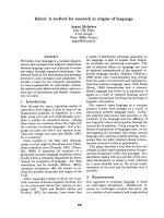
Fig. 1 Factors affecting nanoparticle uptake. (A) Generally, smaller
nanoparticles are internalized more efficiently than larger ones with
similar surface characteristics. (B) Due to the negative charge of the
cellular membrane, positively charged particles are preferentially
taken up by living cells. (C) Cell-specific targeting by conjugating
ligands for surface receptors to nanoparticles. (D) Rapid uptake and
endosome bypassing can be achieved by conjugating protein trans-
duction domains to the surface of the nanoparticle. (E) Conjugation of
ODN was found to aid in specific subcellular localization based on the
presence of complimentary cellular DNA. (F) Endosome escape has
been reported to occur for nanoparticles whose surface is positively
charged inside the low pH of late endosomes. Small nanoparticles
have been reported to bypass degradation pathways better than larger
particles of same chemical composition (see text for details)
Nanoscale Res Lett (2007) 2:430–441 437
123
dendritic cells, coating large 1 lm fluorescent polystyrene
particles with positively charged poly-
L-lysine increased
uptake almost 10-fold compared to uncoated and nega-
tively charged tetanus toxoid coated particles [11]. In
smaller 100 nm nanoparticles, although uptake of poly-
L-
lysine coated nanoparticles was significantly higher than
that of uncoated nanoparticles, there was no difference
compared to uptake of the negatively charged nanoparticles
[11]. In a separate study, confocal microscopy revealed a
higher fluorescence intensity for cells treated with fluo-
rescent positively charged polyethylene glycol-
D,L-
polylactide nanoparticles compared to negatively charged
nanoparticles in HeLa cells [30]. Flow cytometry analysis
also showed the rate of uptake was significantly higher for
the positively charged nanoparticles than for their negative
counterparts. In order to determine if the mechanism was
also affected by changing the nanoparticle surface charge,
cells were also infected with adenoviruses expressing
dominant negative alleles of proteins involved in endocy-
tosis [30]. From this the authors deduced that the inferior
rate of uptake of negative nanoparticles occurs through a
clathrin- and caveolin-independent mechanism while the
more rapid uptake of positively charged nanoparticles
occurs through a clathrin-dependent mechanism [30].
Interestingly, when a dominant negative form of Dynamin I
was expressed (inhibiting both clathrin-mediated endocy-
tosis and caveolin-dependent endocytosis) there was a
significant increase in cellular fluorescence of cells treated
with positively charged fluorescent nanoparticles [30]. This
suggests that if the predominant mechanisms involving
clathrin and caveolin are interrupted, a more efficient
compensatory mechanism takes over. The authors specu-
lated that this compensatory mechanism may in fact be
macropinocytosis, although further study is required [30].
Different cell types obviously have unique efficiencies
of nanoparticle uptake and respond differently to various
kinds of nanoparticles. For example, when treating hMSC
and 3T3-L1 cells with mesoporous silica nanoparticles of
different surface charges, it was observed that uptake dif-
fered between the cell types [10]. Regardless of the extent
of charge, 3T3-L1 cells took up the nanoparticles via
clathrin-mediated endocytosis, but uptake of strong posi-
tively charged nanoparticles in hMSC cells occurs by an
alternate and undefined mechanism [10].
Effect of Surface Modifications on In vitro
Nanoparticle Uptake
In an attempt to target nanoparticles to specific cell types,
to increase uptake efficiency, and to bypass intracellular
obstacles (e.g. endosomes) there is an increasing amount of
work being done to conjugate targeting molecules to the
surface of nanoparticles. Among the most effective and
interesting conjugants are protein transduction domains
[73, 109]. These are short amphipathic peptide sequences
that translocate across cell membranes in a rapid manner
[110–112]. Although there is much debate as to the
mechanism they use to cross the cell membrane, there is
little doubt that it occurs rapidly and efficiently. When the
protein transduction domain of HIV-Tat (GRKKRRQRRR)
was conjugated to 2.8 nm gold nanoparticles, TEM showed
that it translocated across the cell membrane and was
localized within the nucleus of human fibroblast cells [73].
Gold nanoparticles lacking the Tat peptide, on the other
hand, were found surrounding the mitochondria or in
cytoplasmic vacuoles [73]. In another study, when 20 nm
gold nanoparticles were conjugated to the HIV-Tat peptide,
the nanoparticle-peptide nanoconjugate was found to be
localized mainly in the cytoplasm and not in the nucleus
[80], once again substantiating the fact that nanoparticle
size is one of the key factors affecting the uptake. In HeLa
cells, when iron oxide CLIO nanoparticles were labeled
with Cy3.5 and conjugated to a fluorescent Tat peptide-
FITC conjugate, there was a rapid and sustained internal-
ization of the nanoparticles as determined by flow
cytometry [109]. Fluorescent confocal microscopy
revealed that at 24 h post-treatment there was extensive
co-localization of FITC and Cy3.5 in the nucleus and
cytoplasm of cells treated with Tat peptide-FITC-Cy3.5-
CLIO nanoparticles [109]. The nuclear localization was
lost by 72 h.
An alternative approach is to specifically target tumor
cells that overexpress surface receptors such as Her2/Neu or
folate receptor. Therefore, by conjugating the ligands of
these receptors to nanoparticles it is possible to achieve cell-
specific internalization for potential drug delivery or imag-
ing. Human Serum Albumin nanoparticles complexed with
Trastuzumab (antibody against Her2) showed specific uptake
of nanoparticles only in Her2 overexpressing cell lines [40].
Likewise, conjugation of nanoparticles to folate has been
successful in targeting folate receptor overexpressing pros-
tate and nasopharyngeal cancer cells [38, 74, 113].
While peptides direct nanoparticle uptake, conjugation
of nucleic acids have a marked effect on nanoparticle
subcellular retention [66, 67, 79]. Our own laboratory has
shown the effects of conjugating oligonucleotides to the
surface of TiO
2
nanoparticles that target different organ-
elles in living cells. X-ray-fluorescence microscopy
(reviewed in [24, 114]) and TEM have shown that by
altering the oligonucleotide sequence bound to the nano-
particle the subcellular localization of the TiO
2
-DNA
nanoconjugate can change based on the location of avail-
able cellular complimentary DNA [66, 67]. For example,
when breast cancer MCF-7/WS8 cells were treated with
TiO
2
nanoconjugates complimentary to genomic DNA
438 Nanoscale Res Lett (2007) 2:430–441
123
encoding 18S rRNA (of which 200–300 copies reside in the
nucleolus [115]), nanoconjugates were detected by X-ray-
fluorescence microscopy and TEM within the nucleus. On
the other hand, when the oligonucleotide sequence bound
to the TiO
2
nanoparticle was complimentary to mitochon-
drial DNA, there was a more disperse Ti signal found
throughout the cytoplasm as detected by X-ray fluores-
cence microscopy. Also, TEM showed the presence of
electron dense nanoparticles in the mitochondria [67].
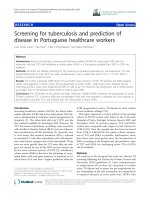
Furthermore, results in Fig. 2 show the combination of X-
ray-fluorescence microscopy and fluorescent confocal
microscopy for imaging the same cell treated with a TiO
2
-
DNA nanoconjugate whose nucleic acid component is
labeled with tetramethylrhodamine (TAMRA). Clearly
there is both a titanium signal and fluorescent TAMRA
signal in the nucleus as well as in the perinuclear region.
This strongly suggests that TiO
2
-DNA nanoconjugates are
stable in cells.
In conclusion, imaging of live cells is important in
assessing biological function, and nanotechnology offers
many new approaches for such studies. In most cases,
imaging of live cells is dependent upon the development of
nanomaterials that can penetrate the cell membrane and not
cause the subsequent death of the cell. Many groups are
exploring the use of bionanoconjugates that can be
designed to probe functional biological pathways in living
cells, and the identification of pathways important in
nanomaterial uptake into cells will facilitate this work.
Conclusions
In this review we focused on nanoparticles that have been
used for cellular imaging; either in live or fixed cells. We
chose this as a focus area because whole cell imaging and
manipulation by nanoparticles are at this time gathering
momentum. The cell is the best starting point for devel-
opment of new therapeutics and new cellular molecular
biology techniques, because the whole cell as a biological
entity has always been the first target en route to mecha-
nistic understanding of both intracellular and whole
organism pathways and processes. New types of nanopar-
ticles are developed daily and we can anticipate that new
uses for them in the field of cell imaging and manipulation
will be discovered with great rapidity as well. Most of the
cellular manipulation with nanoparticles is, at this moment,
devoted to improvements of new therapies and imaging
tools. It is our opinion, however, that development of
nanoparticles as tools for basic science may revolutionize
cellular and molecular biology techniques as much as
understanding and utilization of enzymatic reactions did,
leading to creation of molecular biology we know today.
Fig. 2 Combining X-ray
fluorescence microscopy and
fluorescent confocal microscopy
for the imaging of intracellular
nanoconjugates. MCF-7 cells
were transfected with TiO
2
-
DNA nanoconjugates
complimentary to genomic
DNA encoding r18S rRNA. The
DNA was fluorescently labeled
with TAMRA. After treatment,
cells were washed, fixed, and
stained with Hoechst dye. Then
they were analyzed by
fluorescent confocal microscopy
for the localization of TAMRA.
Next, the same cells were
dehydrated in 100% ethanol and
analyzed at the 2-ID-D
Beamline at the Advanced
Photon Source at Argonne
National Laboratories for the
presence of titanium. Black bar
scale represents 10 lm for XFM
(top left and middle), and the
white bar 10 lm for fluorescent
confocal microscopy (top right,
bottom row)
Nanoscale Res Lett (2007) 2:430–441 439
123
Acknowledgements The authors would like to extend their grati-
tude to Benjamin Haley and David Paunesku for their input and
advice. Work was supported by NIH Grants: CA107467, EB002100,
P50 CA89018, U54CA119341. Use of the Advanced Photon Source
was supported by the U.S. Department of Energy Basic Energy Sci-
ences; under contract number DE-AC02-06CH11357.
References
1. A. Gojova, B. Guo, R.S. Kota, J.C. Rutledge, I.M. Kennedy, A.I.
Barakat, Environ. Health Perspect. 115, 3 (2007)
2. R. Hardman, Environ. Health Perspect. 114, 2 (2006)
3. S. Lanone, J. Boczkowski, Curr. Mol. Med. 6, 6 (2006)
4. C. Medina, M.J. Santos-Martinez, A. Radomski, O.I. Corrigan,
M.W. Radomski, Br. J. Pharmacol. 150, 5 (2007)
5. A. Nel, T. Xia, L. Madler, N. Li, Science 311, 5761 (2006)
6. B.G. Priestly, A.J. Harford, M.R. Sim, Med. J. Aust. 186,4
(2007)
7. O. Seleverstov, O. Zabirnyk, M. Zscharnack, L. Bulavina, M.
Nowicki, J.M. Heinrich, M. Yezhelyev, F. Emmrich, R. O’Re-
gan, A. Bader, Nano Lett 6, 12 (2006)
8. J.S. Tsuji, A.D. Maynard, P.C. Howard, J.T. James, C.W. Lam,
D.B. Warheit, A.B. Santamaria, Toxicol. Sci. 89, 1 (2006)
9. D.B. Warheit, R.A. Hoke, C. Finlay, E.M. Donner, K.L. Reed,
C.M. Sayes, Toxicol. Lett. 171(3), 99 (2007)
10. T.H. Chung, S.H. Wu, M. Yao, C.W. Lu, Y.S. Lin, Y. Hung,
C.Y. Mou, Y.C. Chen, D.M. Huang, Biomaterials 28(19), 2959
(2007)
11. C. Foged, B. Brodin, S. Frokjaer, A. Sundblad, Int. J. Pharm.
298, 2 (2005)
12. J.S. Kim, T.J. Yoon, K.N. Yu, M.S. Noh, M. Woo, B.G. Kim,
K.H. Lee, B.H. Sohn, S.B. Park, J.K. Lee, M.H. Cho, J. Vet. Sci.
7, 4 (2006)
13. S.K. Lai, K. Hida, S.T. Man, C. Chen, C. Machamer, T.A.
Schroer, J. Hanes, Biomaterials 28, 18 (2007)
14. B.M. Rothen-Rutishauser, S. Schurch, B. Haenni, N. Kapp, P.
Gehr, Environ. Sci. Technol. 40, 14 (2006)
15. M. Walsh, M. Tangney, M.J. O’Neill, J.O. Larkin, D.M. Soden,
S.L. McKenna, R. Darcy, G.C. O’Sullivan, C.M. O’Driscoll,
Mol. Pharm. 3, 6 (2006)
16. M. Huang, Z. Ma, E. Khor, L.Y. Lim, Pharm. Res. 19, 10 (2002)
17. S.A. Mousavi, L. Malerod, T. Berg, R. Kjeken, Biochem. J.
377(Pt 1), 1 (2004)
18. R. Medda, S. Jakobs, S.W. Hell, J. Bewersdorf, J. Struct. Biol.
156, 3 (2006)
19. H.W. Tang, X.B. Yang, J. Kirkham, D.A. Smith, Anal. Chem.
79(10), 3646 (2007)
20. P.H. Yang, X. Sun, J.F. Chiu, H. Sun, Q.Y. He, Bioconjug.
Chem. 16, 3 (2005)
21. B. Lai, J. Maser, T. Paunesku, G.E. Woloschak, Int. J. Radiat.
Biol. 78, 8 (2002)
22. B. Lai, J. Maser, S. Vogt, T. Paunesku, G.E. Woloschak, Int. J.
Radiat. Biol. 80, 6 (2004)
23. R. McRae, B. Lai, S. Vogt, C.J. Fahrni, J. Struct. Biol. 155,1
(2006)
24. T. Paunesku, S. Vogt, J. Maser, B. Lai, G. Woloschak, J. Cell.
Biochem. 99, 6 (2006)
25. L. Yang, R. McRae, M.M. Henary, R. Patel, B. Lai, S. Vogt, C.J.
Fahrni, Proc. Natl. Acad. Sci. USA 102, 32 (2005)
26. M.A. Pfeifer, G.J. Williams, I.A. Vartanyants, R. Harder, I.K.
Robinson, Nature 442, 7098 (2006)
27. I.K. Robinson, I.A. Vartanyants, G.J. Williams, M.A. Pfeifer,
J.A. Pitney, Phys. Rev. Lett. 87, 19 (2001)
28. J. Miao, P. Charalambous, J. Kirz, D. Sayre, Nature 400, 6742
(1999)
29. J. Davda, V. Labhasetwar, Int. J. Pharm. 233(1–2), 51 (2002)
30. O. Harush-Frenkel, N. Debotton, S. Benita, Y. Altschuler,
Biochem. Biophys. Res. Commun. 353, 1 (2007)
31. S.H. Kim, J.H. Jeong, K.W. Chun, T.G. Park, Langmuir 21,19
(2005)
32. M.G. Qaddoumi, H.J. Gukasyan, J. Davda, V. Labhasetwar, K.J.
Kim, V.H. Lee, Mol. Vis. 9, 559 (2003)
33. M. Geiser, B. Rothen-Rutishauser, N. Kapp, S. Schurch, W.
Kreyling, H. Schulz, M. Semmler, V. Im Hof, J. Heyder, P.
Gehr, Environ. Health Perspect. 113, 11 (2005)
34. G.R. Reddy, M.S. Bhojani, P. McConville, J. Moody, B.A.
Moffat, D.E. Hall, G. Kim, Y.E. Koo, M.J. Woolliscroft, J.V.
Sugai, T.D. Johnson, M.A. Philbert, R. Kopelman, A. Rehem-
tulla, B.D. Ross, Clin. Cancer Res. 12, 22 (2006)
35. Y. Kakizawa, S. Furukawa, K. Kataoka, J. Control Release 97,2
(2004)
36. V. Sokolova, A. Kovtun, R. Heumann, M. Epple, J. Biol. Inorg.
Chem. 12, 2 (2007)
37. K. Peters, R.E. Unger, C.J. Kirkpatrick, A.M. Gatti, E. Monari,
J. Mater. Sci. Mater. Med. 15, 4 (2004)
38. G. Zheng, J. Chen, H. Li, J.D. Glickson, Proc. Natl. Acad. Sci.
USA 102, 49 (2005)
39. A. Arnedo, J.M. Irache, M. Merodio, M.S. Espuelas Millan, J.
Control Release 94, 1 (2004)
40. I. Steinhauser, B. Spankuch, K. Strebhardt, K. Langer, Bioma-
terials. 27, 28 (2006)
41. I. Brigger, C. Dubernet, P. Couvreur, Adv. Drug Deliv. Rev. 54,
5 (2002)
42. M. Feng, D. Lee, P. Li, Int. J. Pharm. 311(1–2), 209 (2006)
43. B. Lucas, K. Remaut, N.N. Sanders, K. Braeckmans, S.C. De
Smedt, J. Demeester, Biochemistry (Mosc.) 44, 29 (2005)
44. A. Vogt, B. Combadiere, S. Hadam, K.M. Stieler, J. Lademann,
H. Schaefer, B. Autran, W. Sterry, U. Blume-Peytavi, J. Invest.
Dermatol. 126, 6 (2006)
45. M. Bruchez Jr., M. Moronne, P. Gin, S. Weiss, A.P. Alivisatos,
Science 281, 5385 (1998)
46. P.K. Chattopadhyay, D.A. Price, T.F. Harper, M.R. Betts, J. Yu,
E. Gostick, S.P. Perfetto, P. Goepfert, R.A. Koup, S.C. De Rosa,
M.P. Bruchez, M. Roederer, Nat. Med. 12, 8 (2006)
47. X. Gao, W.C. Chan, S. Nie, J. Biomed. Opt. 7, 4 (2002)
48. X. Gao, Y. Cui, R.M. Levenson, L.W. Chung, S. Nie, Nat.
Biotechnol. 22, 8 (2004)
49. B.N. Giepmans, S.R. Adams, M.H. Ellisman, R.Y. Tsien, Sci-
ence 312, 5771 (2006)
50. B.N. Giepmans, T.J. Deerinck, B.L. Smarr, Y.Z. Jones, M.H.
Ellisman, Nat. Methods 2, 10 (2005)
51. Z. Kaul, T. Yaguchi, S.C. Kaul, R. Wadhwa, Ann. N.Y. Acad.
Sci. 1067, 469 (2006)
52. S. Li-Shishido, T.M. Watanabe, H. Tada, H. Higuchi, N. Ohu-
chi, Biochem. Biophys. Res. Commun. 351, 1 (2006)
53. A. Mittag, D. Lenz, A.O.H. Gerstner, A. Tarnok, Cytom Part A
69A, 7 (2006)
54. T. Prow, J.N. Smith, R. Grebe, J.H. Salazar, N. Wang, N. Kotov,
G. Lutty, J. Leary, Mol. Vis. 12, 606 (2006)
55. K. Sapsford, T. Pons, I. Medintz, H. Mattoussi, Sensors 6, 925
(2006)
56. R. Nisman, G. Dellaire, Y. Ren, R. Li, D.P. Bazett-Jones, J.
Histochem. Cytochem. 52, 1 (2004)
57. F. Tokumasu, J. Dvorak, J. Microsc. 211(Pt 3), 256 (2003)
58. H.Z. Wang, H.Y. Wang, R.Q. Liang, K.C. Ruan, Acta Biochim.
Biophys. Sin. (Shanghai) 36, 10 (2004)
59. X. Wu, H. Liu, J. Liu, K.N. Haley, J.A. Treadway, J.P. Larson,
N. Ge, F. Peale, M.P. Bruchez, Nat. Biotechnol. 21, 1 (2003)
440 Nanoscale Res Lett (2007) 2:430–441
123
60. M.H. Cha, T. Rhim, K.H. Kim, A.S. Jang, Y.K. Paik, C.S. Park,
Mol. Cell. Proteomics 6, 1 (2007)
61. J.K. Herr, J.E. Smith, C.D. Medley, D. Shangguan, W. Tan,
Anal. Chem. 78, 9 (2006)
62. H.A. Jeng, J. Swanson, J. Environ. Sci. Health A Tox. Hazard.
Subst. Environ. Eng. 41, 12 (2006)
63. J. Liu, Z. Saponijc, N.M. Dimitrijevic, S. Luo, D. Preuss, T.
Rajh, Proc. SPIE 6096, 48 (2006)
64. T.C. Long, N. Saleh, R.D. Tilton, G.V. Lowry, B. Veronesi,
Environ. Sci. Technol. 40, 14 (2006)
65. S.J. Mechery, X.J.J. Zhao, L. Wang, L.R. Hilliard, A. Munteanu,
W.H. Tan, Chem-Asian J. 1, 3 (2006)
66. T. Paunesku, T. Rajh, G. Wiederrecht, J. Maser, S. Vogt, N.
Stojicevic, M. Protic, B. Lai, J. Oryhon, M. Thurnauer, G.
Woloschak, Nat. Mater. 2, 5 (2003)
67. T. Paunesku, S. Vogt, B. Lai, J. Maser, N. Stojicevic, K.T.
Thurn, C. Osipo, H. Liu, D. Legnini, Z. Wang, C. Lee, G.E.
Woloschak, Nano Lett. 7, 3 (2007)
68. R. Peters, Traffic 6, 5 (2005)
69. H.D. Zhang, P.S. Williams, M. Zborowski, J.J. Chalmers,
Biotechnol. Bioeng. 95, 5 (2006)
70. A. Fujishima, K. Honda, Nature 238, 5358 (1972)
71. A. Michelmore, W. Gong, P. Jenkins, J. Ralston, Phys. Chem.
Chem. Phys. 2, 2985 (2000)
72. T. Rajh, L.X. Chen, K. Lukas, T. Liu, M. Thurnauer, D.M.
Tiede, J. Phys. Chem. B 106, 41 (2002)
73. J.M. de la Fuente, C.C. Berry, Bioconjug. Chem. 16, 5 (2005)
74. V. Dixit, J. Van den Bossche, D.M. Sherman, D.H. Thompson,
R.P. Andres, Bioconjug. Chem. 17, 3 (2006)
75. A.R. Herdt, S.M. Drawz, Y. Kang, T.A. Taton, Colloids Surf. B
Biointerf. 51, 2 (2006)
76. D.B. Kirpotin, D.C. Drummond, Y. Shao, M.R. Shalaby, K.
Hong, U.B. Nielsen, J.D. Marks, C.C. Benz, J.W. Park, Cancer
Res. 66, 13 (2006)
77. S.Y. Park, J.S. Lee, D. Georganopoulou, C.A. Mirkin, G.C.
Schatz, J. Phys. Chem. B Condens. Matter Mater. Surf. Inter-
faces Biophys. 110, 25 (2006)
78. W.J. Qin, L.Y. Yung, Biomacromolecules 7, 11 (2006)
79. N.L. Rosi, D.A. Giljohann, C.S. Thaxton, A.K. Lytton-Jean,
M.S. Han, C.A. Mirkin, Science 312, 5776 (2006)
80. A.G. Tkachenko, H. Xie, D. Coleman, W. Glomm, J. Ryan, M.F.
Anderson, S. Franzen, D.L. Feldheim, J. Am. Chem. Soc. 125,
16 (2003)
81. A.G. Tkachenko, H. Xie, Y. Liu, D. Coleman, J. Ryan, W.R.
Glomm, M.K. Shipton, S. Franzen, D.L. Feldheim, Bioconjug.
Chem. 15, 3 (2004)
82. V. Salnikov, Y.O. Lukyanenko, C.A. Frederick, W.J. Lederer,
V. Lukyanenko, Biophys. J. 92, 3 (2007)
83. M. Wilchek, E.A. Bayer, Trends Biochem. Sci. 14, 10 (1989)
84. A. Kriete, E. Papazoglou, B. Edrissi, H. Pais, K. Pourrezaei, J.
Microsc. 222(Pt 1), 22 (2006)
85. J.K. Jaiswal, H. Mattoussi, J.M. Mauro, S.M. Simon, Nat. Bio-
technol. 21
, 1 (2003)
86. M. Ferrari, Nat. Rev. Cancer 5, 3 (2005)
87. V.E. Kagan, H. Bayir, A.A. Shvedova, Nanomedicine 1,4
(2005)
88. G. Oberdorster, E. Oberdorster, J. Oberdorster, Environ. Health
Perspect. 113, 7 (2005)
89. D.M. Blake, P.C. Maness, Z. Huang, E.J. Wolfrum, J. Huang,
W.A. Jacoby, Separ. Purif. Methods. 28, 1 (1999)
90. A.P. Zhang, Y.P. Sun, World J. Gastroenterol. 10, 21 (2004)
91. D.H. Garabrant, L.J. Fine, C. Oliver, L. Bernstein, J.M. Peters,
Scand. J. Work. Environ. Health 13, 1 (1987)
92. J.W. Seo, H. Chung, M.Y. Kim, J. Lee, I.H. Choi, J. Cheon,
Small 3, 5 (2007)
93. R.A. Mitchell, H. Liao, J. Chesney, G. Fingerle-Rowson, J.
Baugh, J. David, R. Bucala, Proc. Natl. Acad. Sci. USA 99,1
(2002)
94. I.L. Medintz, H.T. Uyeda, E.R. Goldman, H. Mattoussi, Nat.
Mater. 4, 6 (2005)
95. A.M. Derfus, W.C. Chan, S.N. Bhatia, Nano Lett. 4, 1 (2004)
96. A. Hoshino, K. Fujioka, T. Oku, M. Suga, Y.F. Sasaki, T. Ohta,
M. Yasuhara, K. Suzuki, K. Yamamoto, Nano Lett. 4, 11 (2004)
97. J. Lovric, S.J. Cho, F.M. Winnik, D. Maysinger, Chem. Biol. 12,
11 (2005)
98. J.P. Ryman-Rasmussen, J.E. Riviere, N.A. Monteiro-Riviere,
J. Invest. Dermatol. 127, 1 (2007)
99. T. Zhang, J.L. Stilwell, D. Gerion, L. Ding, O. Elboudwarej,
P.A. Cooke, J.W. Gray, A.P. Alivisatos, F.F. Chen, Nano Lett. 6,
4 (2006)
100. A.O. Choi, S.J. Cho, J. Desbarats, J. Lovric, D. Maysinger,
J. Nanobiotechnol. 5, 1 (2007)
101. J. Panyam, W.Z. Zhou, S. Prabha, S.K. Sahoo, V. Labhasetwar,
FASEB J. 16, 10 (2002)
102. J.M. Oh, S.J. Choi, S.T. Kim, J.H. Choy, Bioconjug. Chem. 17,
6 (2006)
103. J. Rejman, V. Oberle, I.S. Zuhorn, D. Hoekstra, Biochem.
J. 377(Pt 1), 159 (2004)
104. I.A. Khalil, K. Kogure, H. Akita, H. Harashima, Pharmacol.
Rev. 58, 1 (2006)
105. A.J. Koleske, D. Baltimore, M.P. Lisanti, Proc. Natl. Acad. Sci.
USA 92, 5 (1995)
106. L.J. Hewlett, A.R. Prescott, C. Watts, J. Cell Biol. 124, 5 (1994)
107. M.D. Chavanpatil, A. Khdair, J. Panyam, J. Nanosci. Nano-
technol. 6(9–10), 2651 (2006)
108. K.Y. Win, S.S. Feng, Biomaterials 26, 15 (2005)
109. A.M. Koch, F. Reynolds, M.F. Kircher, H.P. Merkle, R.
Weissleder, L. Josephson, Bioconjug. Chem. 14, 6 (2003)
110. B. Gupta, T.S. Levchenko, V.P. Torchilin, Adv. Drug Deliv.
Rev. 57, 4 (2005)
111. E. Vives, P. Brodin, B. Lebleu, J. Biol. Chem. 272, 25 (1997)
112. M. Zorko, U. Langel, Adv. Drug Deliv. Rev. 57, 4 (2005)
113. Y. Hattori, Y. Maitani, Cancer Gene Ther. 12, 10 (2005)
114. C.J. Fahrni, Curr. Opin. Chem. Biol. 11, 2 (2007)
115. W. Makalowski, Acta Biochim. Pol. 48, 3 (2001)
Nanoscale Res Lett (2007) 2:430–441 441
123