Báo cáo hóa học: " In Vitro Structural and Functional Evaluation of Gold Nanoparticles Conjugated Antibiotics" pdf
Bạn đang xem bản rút gọn của tài liệu. Xem và tải ngay bản đầy đủ của tài liệu tại đây (527.1 KB, 9 trang )
NANO EXPRESS
In Vitro Structural and Functional Evaluation of Gold
Nanoparticles Conjugated Antibiotics
Biswarup Saha Æ Jaydeep Bhattacharya Æ Ananda Mukherjee Æ
Anup Kumar Ghosh Æ Chitta Ranjan Santra Æ Anjan K. Dasgupta Æ
Parimal Karmakar
Received: 26 June 2007 / Accepted: 30 October 2007 / Published online: 17 November 2007
Ó to the authors 2007
Abstract Bactericidal efficacy of gold nanoparticles
conjugated with ampicillin, streptomycin and kanamycin
were evaluated. Gold nanoparticles (Gnps) were conju-
gated with the antibiotics during the synthesis of
nanoparticles utilizing the combined reducing property of
antibiotics and sodium borohydride. The conjugation of
nanoparticles was confirmed by dynamic light scattering
(DLS) and electron microscopic (EM) studies. Such Gnps
conjugated antibiotics showed greater bactericidal activity
in standard agar well diffusion assay. The minimal inhib-
itory concentration (MIC) values of all the three antibiotics
along with their Gnps conjugated forms were determined in
three bacterial strains, Escherichia coli DH5a, Micrococ-
cus luteus and Staphylococcus aureus. Among them,
streptomycin and kanamycin showed significant reduction
in MIC values in their Gnps conjugated form whereas;
Gnps conjugated ampicillin showed slight decrement in the
MIC value compared to its free form. On the other hand, all
of them showed more heat stability in their Gnps conju-
gated forms. Thus, our findings indicated that Gnps
conjugated antibiotics are more efficient and might have
significant therapeutic implications.
Keywords Gold nanoparticles Á Antibiotics Á
Dynamic light scattering Á
Transmission electron microscope Á
Scanning electron microscope Á
Minimal inhibitory concentration Á Agar well diffusion
Introduction
Nanotechnology is a rapidly developing field of new
therapeutic and diagnostic concept in all areas of medicine
[1–3]. Due to their unique characteristics, nanoparticles are
considered to have wide applications in detection of bio-
molecules, drug delivery and release. Of them, Gnps have
already been used to deliver protein-based drugs, and are of
particular utility because the particles can carry multiple
active groups [4–6]. The chemical, optical and electronic
properties of Gnps made them well suited for applications
in biosensing and therapeutic delivery. Gnps based bio-
sensors [7, 8], drug delivery [9–11] was demonstrated to be
more sensitive and effective.
Moreover, nanoparticles were shown to take up by
phagocytic cells and held promises as carrier for the
treatment of intracellular infections with several antibiotics
[12]. It was reported that Gnps as drug carriers allow
increased drug concentration at infected sites as well as
reduce toxicity of the drug [13]. Thus, Gnps as carrier for
Biswarup Saha and Jaydeep Bhattacharya authors contributed
equally.
B. Saha Á A. Mukherjee Á P. Karmakar
Department of Life Science and Biotechnology, Jadavpur
University, Kolkata 700 032, WB, India
J. Bhattacharya
Department of Microbiology, Vijoygarh Jyotish Roy College,
University of Calcutta, Jadavpur, Kolkata 700 032, WB, India
A. K. Ghosh
Department of Instrumentation Science, Jadavpur University,
Kolkata 700 032, WB, India
C. R. Santra
Department of Chemistry, Netaji Nagar Day College,
NSC Bose Road, Regent Estate, Kolkata 700 092, WB, India
A. K. Dasgupta
Department of Biochemistry, University of Calcutta, 35
Ballygunge Circular Road, Kolkata 700 019, WB, India
123
Nanoscale Res Lett (2007) 2:614–622
DOI 10.1007/s11671-007-9104-2
the antibacterial drug ciprofloxacin and subsequent release
of the drug over an extended period of time was observed
[14]. This is essential for ideal antibiotic therapy. Nano
carriers were also found to be more effective for the drugs
like gentamycin [15], tuberculosis drugs [16, 17], ampi-
cillin [18–20], anticancer drugs [21, 22], anti fungal drug
amphotericin B [23] etc.
For successful application of nano-antibiotic conjuga-
tion, apart from better delivery, their activities should be
evaluated properly because the amount of antibiotics often
given for therapy is much more higher than the dose
required for killing the pathogens. This in turn could pro-
duces toxic effect, which was demonstrated in several
reports too [24, 25]. For a successful antibiotic therapy, the
dose should be reduced to avoid their side effects at the
same time the stability should be increased to make them
more economic. With the advancement of nanotechnology,
functionalized nanoparticles have been used to conjugate
different drugs. Among the different nanoparticles, Gnps
were found to be less toxic and hence widely used for this
purpose. In most of the cases, the conjugation was done by
functionalized gold particles, where amino acids, glutathi-
one, polyethylene glycol etc were used as functionalizing
agents [26]. But to avoid the possible effects of these agents
on biological system, we have conjugated antibiotics
directly without any functionalizing agents at the time of the
Gnps synthesis [27].
While there were many reports about the delivery of
different drugs in nanoparticles conjugated form, little or
no efforts were made, so far, to determine the efficiency,
stability of antibiotics conjugated with Gnps in vitro. In
this study, we compared the efficiency and stability of
Gnps conjugated antibiotics with respect to their free forms
in vitro. We found that the MIC of Gnps conjugated
ampicillin, streptomycin and kanamycin on Escherichia
coli DH5a (Microbial type culture collection (MTCC)
No.1652, India), Micrococcus luteus (MTCC No. 106) and
Staphylococcus aureus (MTCC No. 96) were reduced when
compared to their respective unconjugated free forms.
Moreover, the activity of all Gnps conjugated antibiotics
showed higher stability compared to their corresponding
free forms. Thus our results suggest that antibiotics con-
jugated with Gnps might be used in therapy for their
greater efficiency and stability.
Experimental Procedures
Preparation of Bare Gold Nanoparticles
Gold nanoparticles (Gnps) were prepared by the reduction
of chloroauric acid (H[AuCl
4
]) by sodium borohydride.
The normal reduction process was performed according to
the standard protocol [27]. The size of Gnps obtained by
this process was 14 nm.
Preparation of Conjugated Gold Nanoparticles using
Antibiotics as Template
The combined reducing property of sodium borohydride
and antibiotics were used to reduce H[AuCl
4
]. The seeding
of Gnps was done in presence of the antibiotics (Ampi-
cillin, Streptomycin and Kanamycin, Fig. 1) individually
and thus Gnps conjugated antibiotics were formed [27].
Dynamic Light Scattering (DLS)
The Nano-ZS (Malvern) instrument (5 mW HeNe laser
k = 632 nm) was used for this purpose. The sample was
taken in a DTS0112—low volume disposable sizing cuv-
ette of 1.5 ml volume (path length 1 cm). The operating
procedure was programmed (using the DTS software sup-
plied with the instrument) such that there were average of
25 runs, each run being averaged for 15 s, with an equili-
bration time of 3 min at 25°C. A particular hydrodynamic
diameter (d
h
) was evaluated several times and the result
was presented in terms of distribution of d
h
[28].
Transmission Electron Microscopy
All the three Gnps conjugated antibiotics along with the
free Gnps were prepared after drying on carbon coated
copper grid and observed under a transmission electron
microscope (FEI, Model: STWIN) with an accelerating
potential of 200 KV and analyzed with TECNAI G
2
software.
Scanning Electron Microscopy
Gnps conjugated antibiotics along with bare Gnps were
lyophilized on glass slides and then coated with gold. The
samples were then observed under a scanning electron
microscope (JEOL JSM 5200).
MIC Study of Free and Gnps Conjugated Antibiotics
MIC of ampicillin, streptomycin and kanamycin along with
their respective Gnps conjugated forms against E. coli
DH5a, M. luteus and S. aureus in Luria-Bertani (LB) broth
were determined by standard method [29]. Each tube
contained 5 ml of LB medium inoculated with 10
6
bacteria
Nanoscale Res Lett (2007) 2:614–622 615
123
per ml. Decreasing concentrations of each antibiotic and
their corresponding Gnps conjugated form were added to
the respective tubes. After 16 h. the turbidity of each tube
was measured at 600 nm using a spectrophotometer.
Bactericidal Activity Measurement
This assay was conducted by standard agar well diffusion
method. The E. coli DH5a, M. luteus and S. aureus strains
were grown on LB Broth at 37°C overnight upto a turbidity
of 0.5 Mac Farland standard (10
8
CFU per ml) [30]. About
100 ll of this suspension was used to inoculate 90 mm
diameter petridish filled with 35 ml of LB agar. Wells
(diameter
2
= 0.563 cm
2
) were punched in the agar plates
and filled with 100 ll of either antibiotics or their respec-
tive Gnps conjugated forms. The concentrations of both the
forms of antibiotics were at their respective MIC values,
generally used in common laboratory purpose (50 lg/ml
for ampicillin, 10 lg/ml for streptomycin and 50 lg/ml for
kanamycin) [31]. Plates were incubated at 37°C for over-
night. Antibacterial activities were evaluated by measuring
the area of zone of inhibition (diameter
2
). We used auto-
claved water and only Gnps as negative control.
Results
Production of Gnps on reduction with citrate or borohy-
dride generally resulted in a size less than 20 nm but the
molar ratio of reductant to H[AuCl
4
] was the key factor for
the synthesis of Gnps below 20 nm (d
h
). We used the ratio
of reductant and H[AuCl
4
] in such a way that the synthe-
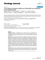
sized Gnps produced a size of 13.54 nm (Fig. 2a) when
measured by photon correlation spectroscopy. The plasmon
resonances of the Gnps varied with the diameter of the
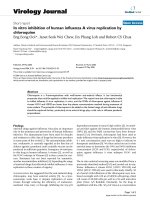
reduced particles. The plasmon resonance was obtained at
526 nm and the produced Gnps were of red wine colour
(Fig. 3). Thus, larger particles appeared more bluish in
colour while smaller particles showed red colour [27].
Conjugated with Antibiotics
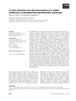
The antibiotics (Fig. 1) were conjugated with Gnps by
reducing H[AuCl
4
] with the combined reducing effect of
both antibiotics and sodium borohydride. The antibiotics
themselves were able to reduce H[AuCl
4
] to synthesize the
Gnps but the reducing power was much less. It took around
4 h for ampicillin and 24 h for streptomycin and kana-
mycin to reduce H[AuCl
4
] to form Gnps conjugated
nanoparticles (data not shown). Also, in case of ampicillin,
the particles produced in this way formed larger aggregates
and precipitated out from the solution quickly whereas
streptomycin and kanamycin reduced H[AuCl
4
] very
poorly. But the Gnps produced by using the combined
reducing property of both sodium borohydride and the
antibiotics showed much higher stability. The produced
Gnps conjugated antibiotics appeared more bluish (Fig. 3).
So, it was obvious that the size of the particles would be
larger and that was reflected in the intensity distribution of
the size of the Gnps (Fig. 2b). The intensity distribution
was obtained due to the Rayleigh scattering (i.e., propor-
tional to R
6
, where R is the radius of particle). We found
that there were distributions of large and small particles but
the number distribution showed (*R) that there were
major numbers of particle, which have the hydrodynamic
radius less than 10 nm (Fig. 2a). The colour showed bluish
because of the presence of some larger particles too.
As DLS study showed size distribution of Gnps conju-
gated particles, we then wanted to visualize and validate
the size of the particles directly. For this, we did electron
microscopic study of the free Gnps and Gnps conjugated
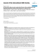
antibiotics. In the transmission electron microscopy
(TEM), we observed that the Gnps conjugated with the
antibiotics produce larger particles. The conjugation with
antibiotics resulted an irregular but consistence change in
the particles association for all the three antibiotics tested
(Fig. 4b, c, d). But only Gnps showed very regular spher-
ical shaped particles with much smaller size (Fig. 4a). We
further used scanning electron microscope (SEM) to
determine the conjugation of Gnps with antibiotics.
Fig. 1 Chemical structure of
antibiotics. (a) Ampicillin, (b)
Streptomycin, (c) Kanamycin
616 Nanoscale Res Lett (2007) 2:614–622
123
Distinct structures were found for all the three antibiotics
conjugated with Gnps. Gnps conjugated ampicillin showed
cubic structure (Fig. 5b), Gnps conjugated streptomycin
showed rectangular rod shaped structure (Fig. 5c) and
Gnps conjugated kanamycin showed extended star like
structures (Fig. 5d). This observation clearly demonstrated
the conjugation of antibiotics with Gnps. These structure
formations were absent when pre-synthesized Gnps and
antibiotics were mixed separately (data not shown).
Gnps conjugated ampicillin, streptomycin and kanamy-
cin along with their corresponding free antibiotics were
then tested on bacterial strains E. coli DH5a, M. luteus and
S. aureus by comparing corresponding zone of inhibition
(diameter
2
). In Fig. 6, the zone of inhibition by agar well
diffusion assay for E. coli DH5a was shown to increase at a
particular concentration for Gnps conjugated antibiotics
compared to their respective unconjugated forms. The
concentrations of all the three antibiotics taken in the above
experiments were the standard concentrations used in the
laboratory (50 lg/ml for ampicillin, 10 lg/ml for strepto-
mycin and 50 lg/ml for kanamycin) [31]. Similar results
were obtained for M. luteus and S. aureus too (pictures not
shown). We also tested a wide range of concentrations for
all the antibiotics and observed that the Gnps conjugated
antibiotics were more efficient than their respective free
forms (data not shown). In the Fig. 7, the percentage
increment in the zone of inhibition for Gnps conjugated
antibiotics were compared to their respective free forms at
the concentrations mentioned above. In Fig. 7a, the incre-
ment in the zone of inhibition (diameter
2
) of Gnps
conjugated ampicillin with respect to the free ampicillin
was shown for all the three bacterial strains we had tested.
Similar data for streptomycin and kanamycin were plotted
also in Fig. 7b and c, respectively. The percentage incre-
ments in the zone of inhibition (diameter
2
) for the Gnps
conjugated antibiotics compared to their respective free
forms were summarized in Table 1. As seen in the
Table 1, kanamycin in Gnps conjugated form was more
effective than its free form in the case of E. coli DH5a
and S. aureus, whereas streptomycin was more effective
in its Gnps conjugated form in the case of M. luteus.
Fig. 2 Measurement of the
hydrodynamic diameter of bare
and Gnps conjugated antibiotics
by dynamic light scattering
experiment. (a) represents the
number distribution of the
hydrodynamic diameter of bare
Gnps and Gnps conjugated
antibiotics. (b) represents the
intensity distribution of the
hydrodynamic diameter of the
free Gnps and the antibiotics
conjugated Gnps
WAVE LENGTH (in nm)
nillicipmA
an
a
Kcymin
ertStpicymon
0.4
0.5
0.6
0.7
0.8
1.6
1.5
1.4
1.3
1.2
1.1
1
0.9
0.8
450 500 550 600
450 500 550 600
Wave Length (in nm)
Absor bance
ABSORBANCE
Fig. 3 The spectroscopic measurement of plasmon resonance of the
antibiotics conjugated with Gnps. The figure in the inset represents
the plasmon resonance of bare Gnps
Nanoscale Res Lett (2007) 2:614–622 617
123
On the other hand, Gnps conjugated ampicillin showed
uniform increment in the zone of inhibition compared to
its free form in the case of all the three bacterial strains
tested. S. aureus strain was resistant to streptomycin, so
neither the free antibiotic nor the Gnps conjugated anti-
biotic produced any inhibition to their growth. Further, in
one of the control experiments we determined the zone of
inhibition with the mixture of previously synthesized
Gnps and antibiotics. In that case, the zone of inhibition
did not increase compare to the free antibiotics. Also, by
adding only sodium borohydride to the antibiotics, we
could not see significant increase in the activity of anti-
biotics (only 3–6%).
We next determined the minimal inhibitory concentra-
tion (MIC) of each antibiotic compared to their Gnps
conjugated form in each bacterial strain. MIC for each of
the Gnps conjugated antibiotic reduced significantly
(Table 2) compared to their respective free forms. For
Gnps conjugated ampicillin, the MIC value was 45 lg/ml
compared to 50 lg/ml for free ampicillin (10% decrement),
for streptomycin the corresponding values were 7 and
14 lg/ml (50% decrement) and for kanamycin the values
were 12 and 30 lg/ml (60% decrement) in E. coli DH5a.
For other strains, the values of MIC were also reduced for
all antibiotics conjugated with Gnps compared to their
respective free forms (Table 2).
We then wanted to determine the stability of the Gnps
conjugated antibiotics compared to the free antibiotics.
Both forms of all the three antibiotics were given heat
shock by incubating them at different temperature for
10 min and then their antibacterial activity was measured
by agar well diffusion method. It was observed that Gnps
conjugated antibiotics were more stable than corresponding
free antibiotics (Table 3). The antibacterial activity of free
ampicillin did not decrease much with the elevation of
temperature while the Gnps conjugated ampicillin showed
more activity at higher temperature. On the other hand, for
free streptomycin and kanamycin, the antibacterial activi-
ties were reduced significantly but the antibacterial activity
of Gnps conjugated streptomycin and kanamycin decrease
slightly with the increment in temperature. One step fur-
ther, we then measured the rate of functional degradation
of the antibiotics (both free and Gnps conjugated forms) by
storing them at room temperature. Both the forms of
antibiotics were stored at room temperature (25–28°C) and
used to evaluate the zone of inhibition by agar well
Fig. 4 Transmission electron
micrographs of free Gnps and
antibiotics conjugated Gnps. (a)
Bare Gnps, (b) Ampicillin
conjugated Gnps, (c)
Streptomycin conjugated Gnps,
(d) Kanamycin conjugated
Gnps
618 Nanoscale Res Lett (2007) 2:614–622
123
diffusion method. All the antibiotics in their respective
Gnps conjugated form had more antibacterial activity
compared to the corresponding free antibiotics, except
Gnps conjugated ampicillin (Table 3). This is true for all
the three bacterial strains tested (data not shown).
Discussions
Our results for the first time demonstrated that the in vitro
bactericidal activity of Gnps conjugated ampicillin,
streptomycin and kanamycin were more efficient compared
to their respective free forms. We had also developed a
simple technique for the conjugation of antibiotics with
Gnps during its synthesis step. Usually, such conjugation
needs functionalization process. But we avoided the
interference of such functionalizing agent in determining
the bactericidal activity of the antibiotics. Using the com-
bined reducing property of antibiotics and borohydride,
antibiotics were conjugated with Gnps. The interaction
between antibiotics and Gnps is likely to be mediated by
Fig. 5 Scanning electron
micrographs of free Gnps and
antibiotics conjugated Gnps. (a)
Bare Gnps, (b) Ampicillin
conjugated Gnps, (c)
Streptomycin conjugated Gnps,
(d) Kanamycin conjugated
Gnps
Fig. 6 Comparison of antibacterial activity of antibiotics conjugated
Gnps along with respective free antibiotics in E. coli DH5a by agar
well diffusion method. (a) Ampicillin (50 lg/ml), (b) Streptomycin
(10 lg/ml), (c) Kanamycin (50 lg/ml). The well 1, 2, and 3
represents free antibiotics; Gnps conjugated antibiotics and bare
Gnps respectively in each plate
Nanoscale Res Lett (2007) 2:614–622 619
123
the adsorption of the antibiotic molecules on the nanopar-
ticle surfaces. The average particles size after conjugation
were shown to decrease (Fig. 2a). This was possibly again
due to the combined reducing property of both antibiotics
and borohydride in situ. However, the plasmon resonance
study (Fig. 3) showed a red shift, indicating the presence of
larger particles (Fig. 2b), though they were less in number
(Fig. 2a). In case of Gnps conjugated ampicillin, the
Table 1 Represents the zone of inhibition (in terms of diameter square) for free antibiotics and antibiotics conjugated with Gnps in three
bacterial strains
Name of the bacterial strain Name of antibiotics Inhibitory zone in sq. diameter (cm
2
) % Change in inhibitory sq. diameter
Free antibiotics Gnps-conjugated antibiotics
E. coli DH5a (Gram -Ve) Ampicillin 3.085 ± 0.146 3.569 ± 0.160 +15.688
Streptomycin 2.189 ± 0.057 2.453 ± 0.102 +12.060
Kanamycin 3.371 ± 0.164 4.545 ± 0.223 +34.826
M. luteus (Gram +Ve) Ampicillin 8.740 ± 0.201 10.493 ± 0.354 +20.057
Streptomycin 0.818 ± 0.091 1.712 ± 0.241 +109.291
Kanamycin 2.507 ± 0.118 2.960 ± 0.149 +18.069
S. aureus (Gram +Ve) Ampicillin 14.839 ± 0.321 16.659 ± 0.678 +12.265
Kanamycin 1.588 ± 0.098 2.132 ± 0.150 +34.257
The concentrations of free as well as Gnps conjugated antibiotics are 50 lg/ml for ampicillin, 10 lg/ml for streptomycin and 50 lg/ml for
kanamycin. The data is the average of three experiments ± SD. Percentage change in each case is calculated and mentioned above
Table 2 Represents minimal inhibitory concentrations (MIC) for free antibiotics along with their respective Gnps conjugated form in three
bacterial strains
Name of the bacterial strain Name of antibiotics Minimal inhibitory concentration (lg/ml) for 10
6
bacteria/ml % Change in MIC
Free antibiotics Gnps-conjugated antibiotics
E. coli DH5a (Gram -Ve) Ampicillin 50.0 ± 0.50 45.0 ± 1.50 -10.00
Streptomycin 14.0 ± 2.00 7.0 ± 1.00 -50.00
Kanamycin 30.0 ± 2.50 12.0 ± 1.00 -60.00
M. luteus (Gram +Ve) Ampicillin 0.52 ± 0.02 0.45 ± 0.03 -13.46
Streptomycin 22.0 ± 2.00 17.0 ± 1.00 -22.73
Kanamycin 32.5 ± 0.50 23.0 ± 1.50 -29.23
S. aureus (Gram +Ve) Ampicillin 0.45 ± 0.03 0.37 ± 0.01 -17.78
Kanamycin 9.0 ± 0.50 5.8 ± 0.20 -35.56
The data is the average of three experiments ± SD. Percentage change in each case is calculated and mentioned above
Fig. 7 Comparative study of different antibiotics along with their
respective Gnps conjugated forms in three bacterial strains. (a)
Ampicillin (50 lg/ml), (b) Streptomycin (10 lg/ml), (c) Kanamycin
(50 lg/ml). The data is the average of three experiments ± SD. The
first column in each pair represents free form of antibiotics and the
second column represents its respective Gnps conjugated form
620 Nanoscale Res Lett (2007) 2:614–622
123
plasmon resonance showed a flatten plateau in the plasmon
region due to the presence of such poly dispersed particles.
The dynamic light scattering study (Fig. 2b) and TEM
study (Fig. 4b) also supported the above statement. In one
step further, we directly showed evidences by scanning
electron microscopic studies that, all the three antibiotics
formed some specific three-dimensional structures when
conjugated with Gnps. Also, to prove the conjugation of
antibiotics with Gnps, we found that after spinning down
the Gnps conjugated antibiotics, the functional activity of
the precipitate (pellet-suspension) was about 60–80% and
that of the supernatant was about 20–40%. Thus, majority
of the antibiotic molecules were associated with Gnps.
Using standard agar well diffusion assay, we compared
the bactericidal activity of Gnps conjugated antibiotics
with their respective free forms. The relative bactericidal
activity of Gnps conjugated ampicillin was less effective
than Gnps conjugated streptomycin and kanamycin (Fig. 7
and Table 1). Consequently, for E. coli DH5a strain, the
MIC values of Gnps conjugated ampicillin decreased 10%,
while the percentage decrement for Gnps conjugated
streptomycin and kanamycin were 50% and 60%, respec-
tively. Such differential activity might be due to the
differences in the mode of action of the antibiotics.
Ampicillin inhibits the cell wall biosynthesis by inhibiting
the cross-linking reaction mediated by transpeptidase,
while both streptomycin and kanamycin bind with ribo-
some and block translation process during protein synthesis
[32]. The binding affinity of Gnps conjugated antibiotics
with the said enzyme or even ribosome might be the key
factor for this differential response. Although, in the con-
trol experiments, only Gnps did not show any bactericidal
activity (Fig. 6) so the antibiotics conjugated with Gnps
might have a higher binding affinity to their respective
targets. On the other hand, the Gnps conjugated antibiotics
might have greater chance to penetrate bacterial cell
membrane compared to their respective free forms. In the
control experiments, we also mixed pre-synthesized Gnps
and antibiotics externally to determine the bactericidal
activity. None of these antibiotics mixed with Gnps showed
significant increment in bactericidal activity compared to
the respective free antibiotics (data not shown). Thus, only
Gnps did not promote the penetration of the antibiotics into
the bacterial cells. So, Gnps conjugated antibiotics might
have some other mechanisms that could enhance the effi-
cacy of the antibiotics. On the other hand, presence of
sodium borohydride during the Gnps synthesis step might
alter the function of antibiotics, but when we mixed only
sodium borohydride with antibiotics, the functional activity
of antibiotics did not increase much (only 3–6%). Thus the
reduction process in our reaction condition does not change
the antibiotic structure abruptly. The conjugation between
the antibiotics and Gnps is probably based on the adsorp-
tion phenomenon mediated by intermolecular forces. Thus
having the larger surface area of these adsorbed antibiotics
in Gnps conjugated form, their bactericidal activity might
increases compared to their respective free forms. How-
ever, the exact mechanisms of action of Gnps conjugated
antibiotics are highly speculative and needs further study.
The Gnps conjugated antibiotics were seen to be more
stable than their respective free forms. Stability of the most
antibiotics is temperature and parenteral solutions depen-
dent [33]. We introduced stresses by heat shock and by
prolong storage at room temperature (25–28°C). In both the
Table 3 Represents the bactericidal activity in E. coli DH5a by agar well diffusion assay for free antibiotics and their respective Gnps
conjugated form after different temperature and time stresses
Agents Zone of inhibition for E. coli DH5a strain in sq. cm
Ampicillin Streptomycin Kanamycin
Free Gnps
conjugated
% Change Free Gnps
conjugated
% Change Free Gnps
conjugated
% Change
Incubated
for 10 min at
26 °C 3.085 3.569 +15.688 2.189 2.453 +12.060 3.371 4.545 +34.826
50 °C 2.806 6.141 +118.85 1.378 1.622 +17.707 2.063 3.663 +77.557
75 °C 2.198 6.635 +201.87 1.116 1.411 +26.434 1.834 3.389 +84.787
90 °C 2.107 6.707 +218.32 0.053 1.324 +2398.1 1.491 3.263 +118.85
Storage at room
temp. (25–28°C)
for
0 day 3.085 3.569 +15.688 2.189 2.453 +12.060 3.371 4.545 +34.826
3 days 2.929 3.142 +7.272 2.126 2.361 +11.054 2.283 4.199 +83.925
7 days 2.646 2.823 +6.689 1.486 2.049 +37.887 1.562 4.024 +157.62
14 days 2.561 2.593 +1.250 1.055 1.483 +40.569 1.501 3.879 +158.43
21 days 2.540 1.941 -23.583 0.913 1.345 +47.317 1.338 3.645 +172.42
28 days 1.965 1.209 -38.473 0.547 0.945 +72.761 1.239 3.459 +179.18
The concentrations of free as well as Gnps conjugated antibiotics are 50 lg/ml for ampicillin, 10 lg/ml for streptomycin and 50 lg/ml for
kanamycin
Nanoscale Res Lett (2007) 2:614–622 621
123
cases Gnps conjugated antibiotics were observed to be
more stable compared to their respective free forms except
Gnps conjugated ampicillin during its temporal study. This
was perhaps due to the close association between Gnps and
antibiotics, the bond energy of antibiotic molecules were
increased which in turn stabilized them. Whatever the
mechanisms of such stability of Gnps conjugated antibi-
otics be, we showed further that at elevated temperature the
Gnps conjugated forms were even more active for ampi-
cillin. This was perhaps due to the delocalization of the
electron in the carbonyl group of the b-lactam ring in
ampicillin at elevated temperature. Elevation in tempera-
ture might induce breakage in the b-lactam ring of the free
ampicillin, whereas Gnps conjugation might stabilize the
ring and thereby allowing the delocalization of electron.
Hence, in case of free ampicillin we found a decrease in the
activity, whereas Gnps conjugated form showed more
activity than the activity at its lower temperature. In this
regard, one of the important findings was the deactivation
of streptomycin at 90°C, whereas its Gnps conjugated form
retained its activity at the same condition. Here also, the
Gnps conjugation might stabilize the structure of the
streptomycin molecules.
We found that the activity of Gnps conjugated ampi-
cillin decreased compared to its free form after two weeks
(Table 3). Actually, we observed that the Gnps conjugated
ampicillin (Table 3) was precipitated out from the solution.
This might be the reason for its decreased efficiency
compared to its free form.
It was reported that antibiotic solutions used for longer
than 7 days should be stored at 4°C, those stored at 24°C
should be discarded after 7 days [34]. Our data also sup-
ported this observation. Moreover, we provided evidences
that Gnps conjugated antibiotics were more stable and
might withstand more harsh storage conditions, which
raised a hope to use Gnps conjugated antibiotics with
greater efficiency in the remote area, where proper storage
condition is unavailable.
Acknowledgements We thank Dr. Joydeep Mukherjee, School of
Environmental Sciences, Jadavpur University, Kolkata, for the gift of
bacterial strains, Micrococcus luteus and Staphylococcus aureus. We
thank Mr. Pallab Dasgupta, Department of Instrumentation Science,
Jadavpur University, Kolkata for helping us to avail the SEM facility.
We also acknowledge Dr. Pulak Ray and Mr. Tapan Kumar Ray of
TEM Section, Saha Institute of Nuclear Physics, Kolkata, for pro-
viding the TEM facilities. This work was partially supported by
CSIR, India (financial grant No. 37(1231)/02/EMR-II).
References
1. G.A. Silva, Surg. Neurol. 61, 216 (2004)
2. D.A. Groneberg, M. Giersig, T. Welte, U. Pison, Current. Drug.
Targets. 7, 643 (2006)
3. E. Katz, I. Willner, Integrated Nanoparticle–Biomolecule Hybrid
Systems: Synthesis, Properties, and Applications (Wiley-VCH
Verlag GmbH & Co., KgaA Weinheim, 2004)
4. M. Hu, J. Chen, Z.Y. Li, L. Au, G.V. Hartland, X. Li, M. Mar-
quez, Y. Xia, Chem. Soc. Rev. 35, 1084 (2006)
5. P.K. Jain, K.S. Lee, I.H. El-Sayed, M.A. El-Sayed, J. Phys.
Chem. B, Condens. Matter. Mater. Surf. Int. Biophys. 110, 7238
(2006)
6. M. Chen, Y. Cai, Z. Yan, D.W. Goodman, J. Am. Chem. Soc.
128, 6341 (2006)
7. M.T. Castaneda, A. Merkoci, M. Pumera, S. Alegret, Biosens.
Bioelectron. 22, 1961 (2007)
8. P.V. Baptista, M. Koziol-Montewka, J. Paluch-Oles, G. Doria,
R. Franco, Clin. Chem. 52, 1433 (2006)
9. A.G. Cuenca, H. Jiang, S.N. Hochwald, M. Delano, W.G. Cance,
S.R. Grobmyer, Cancer 107, 459 (2006)
10. G.F. Paciotti, L. Myer, D. Weinreich, D. Goia, N. Pavel, R.E.
McLaughlin, L. Tamarkin, Drug. Deliv. 11, 169 (2004)
11. X. Huang, P.K. Jain, I.H. El-Sayed, M.A. El-Sayed, Photochem.
Photobiol. 82, 412 (2006)
12. J. Kreuter, Infection. 19, S224 (1991)
13. H. Pinto-Alphandary, A. Andremont, P. Couvreur, Int. J. Anti-
microb. Agents. 13, 155 (2000)
14. R.T. Tom, V. Suryanarayanan, P.G. Reddy, S. Baskaran,
T. Pradeep, Langmuir. 20, 1909 (2004)
15. C. Lecaroz, C. Gamazo, M.J. Blanco-Prieto, J. Nanosci. Nano-
technol. 6, 3296 (2006)
16. R. Pandey, A. Sharma, A. Zahoor, S. Sharma, G.K. Khuller,
B. Prasad, J. Antimicrob. Chemother. 52, 981 (2003)
17. R. Pandey, S. Sharma, G.K. Khuller, Drug. Deliv. 13, 287 (2006)
18. E. Fattal, M. Youssef, P. Couvreur, A. Andremont, Antimicrob.
Agents. Chemother. 33, 1540 (1989)
19. E. Fattal, J. Rojas, L. Roblot-Treupel, A. Andremont, P. Couvreur,
J. Microencapsul. 8, 29 (1991)
20. H. Pinto-Alphandary, O. Balland, M. Laurent, A. Andremont,
F. Puisieux, P. Couvreur, Pharm. Res. 11, 38 (1994)
21. S.M. Moghimi, Anticancer. Agents. Med. Chem. 6, 553 (2006)
22. S. Maesaki, Curr. Pharm. Des. 8, 433 (2002)
23. R.B. Umamaheshwari, S. Ramteke, N.K. Jain, AAPS. Pharm. Sci.
Tech. 5, e32 (2004)
24. R.J. Geller, R.L. Chevalier, D.A. Spyker, Toxicol. Clin. Toxicol.
24, 175 (1986)
25. S.F. Schellie, T. Groshong, Mol. Med. 96, 209 (1999)
26. D. Shenoy, W. Fu, J. Li, C. Crasto, G. Jones, C. DiMarzio,
S. Sridhar, M. Amiji, Int. J. Nanomedicine. 1, 51 (2006)
27. J. Bhattacharya, S. Jasrapuria, T. Sarkar, R. GhoshMoulick, A.K.
Dasgupta, Nanomedicine 3
, 14 (2007)
28. J. Bhattacharya, R. GhoshMoulick, U. Choudhuri, P. Chakra-
barty, P.K. Bhattacharya, P. Lahiri, B. Chakraborti, A.K.
Dasgupta, Analytica. Chimica. Acta. 522, 207 (2004)
29. G. Lancini, F. Parenti, Antibiotics: An Integrated View (Springer-
Verlag, New York, 1982)
30. C. Perez, M. Pauli, P. Bazerque, Acta. Biol. Med. Exper. 15, 113
(1990)
31. J. Sambrook, E.F. Fritsch, T. Maniatis, Molecular Cloning: A
Laboratory Manual (Cold Spring Harbor Laboratory Press,
New York, 1989)
32. G. Lancini, F. Parenti, Antibiotics: An Integrated View (Springer-
Verlag, New York, 1982)
33. G. Richard, M.D. Wyatt, A. Gary, M.D. Okamoto, D. Ralph,
M.D. Feigin, Pediatrics 49, 22 (1972)
34. M.K. Arici, Z. Sumer, C. Guler, O. Elibol, G. Saygi, S. Cetin-
kaya, Aust. N Z. J. Ophthalmol. 27, 426 (1999)
622 Nanoscale Res Lett (2007) 2:614–622
123