Báo cáo hóa học: " Research Article Genome-Wide Analysis of Intergenic Regions of Mycobacterium tuberculosis H37Rv Using Affymetrix GeneChips" docx
Bạn đang xem bản rút gọn của tài liệu. Xem và tải ngay bản đầy đủ của tài liệu tại đây (493.13 KB, 7 trang )
Hindawi Publishing Corporation
EURASIP Journal on Bioinformatics and Systems Biology
Volume 2007, Article ID 23054, 7 pages
doi:10.1155/2007/23054
Research Article
Genome-Wide Analysis of Intergenic Regions
of Mycobacterium tuberculosis H37Rv Using
Affymetrix GeneChips
Li M. Fu
1
and Thomas M. Shinnick
2
1
Pacific Tuberculosis and Cancer Research Organization, 8 Corporate Park, Suite 300, Irvine, CA 92606, USA
2
Centers for Disease Control and Prevention, Atlanta, GA 30333, USA
Received 24 April 2007; Accepted 14 August 2007
Recommended by Z. Jane Wang
Sequencing the complete genome of Mycobacterium tuberculosis H37Rv is a major milestone in the genome project and it sheds
new light in our fight with tuberculosis. The genome contains around 4000 genes (protein-coding sequences) in the original
genome annotation. A subsequent reannotation of the genome has added 80 more genes. However, we have found that the inter-
genic regions can exhibit expression signals, as evidenced by microarray hybridization. It is then reasonable to suspect that there
are unidentified genes in these regions. We conducted a genome-wide analysis using the Affymetrix GeneChip to explore genes
contained in the intergenic sequences of the M. tuberculosis H37Rv genome. A working criterion for potential protein-coding
genes was based on bioinformatics, consisting of the gene structure, protein coding potential, and presence of ortholog evidence.
The bioinformatics criteria in conjunction with transcriptional evidence revealed potential genes with a specific function, such
as a DNA-binding protein i n the CopG family and a nickle binding GTPase, as well as hypothetical proteins that had not been
reported in the H37Rv genome. This study further demonstrated that microarray-based transcriptional evidence would facilitate
genome-wide gene finding, and is also the first report concerning intergenic expression in M. tuberculosis genome.
Copyright © 2007 L. M. Fu and T. M. Shinnick. This is an open access article distributed under the Creative Commons Attribution
License, which permits unrestricted use, distribution, and reproduction in any medium, provided the original work is properly
cited.
1. INTRODUCTION
Unraveling the complete genome sequence of Mycobacterium
tuberculosis H37Rv [1] has led to a better understanding of
the biology and pathogenicity of the organism. This is a ma-
jor advance in combating tuberculosis (TB), a deadly infec-
tious disease caused by M. tuberculosis. With this accomplish-
ment, new molecular targets for diagnostics and therapeutics
can be invented at a fast pace by searching the genome.
To utilize the information embedded in a genome, the
genome must be annotated thoroughly. In essence, genome
annotation is to identify the locations of genes and all of
the coding regions in a genome, and determine their pro-
tein products as well as functions. As hundreds of bacterial
genome sequences are publicly available and the number will
soon reach the milestone of 1000, the need for automated,
large-scale, high-throughput genome annotation is rapidly
increasing [2–4]. A recent study indicates that many genomes
couldbeeitherover-annotated(toomanygenes)orunder-
annotated (too few genes), and a large percentage of genes
may have been assigned a wrong start codon [5]. Even if
the original genome annotation looks accurate and complete
upon submission, it needs to be updated on a regular basis
in accordance with new experimental evidence and knowl-
edge that is evolving over time. However, reannotation of the
whole genome is not very fruitful, as most of the genes have
been identified in the first annotation. For example, the re-
annotation of the H37Rv genome resulted in about 2% of
new protein-coding sequences (CDS) added to the genome.
Some intergenic sequences in M. tuberculosis genome
exhibit expression signals, as detected by the Affymetrix
GeneChip. The same observations have been made for other
bacteria, such as Bacillus subtilis [6], and also in the eu-
karyotic system [7]. At present, it is not clear whether or
how intergenic expression represents gene activity. Here,
we conducted a genome-wide analysis using the Affymetrix
GeneChip to explore genes contained in the intergenic se-
quences of the M. tuberculosis H37Rv genome. Potential
protein-coding genes were determined based on the bioin-
formatics criteria [8, 9] consisting of the gene structure,
2 EURASIP Journal on Bioinformatics and Systems Biology
protein coding potential, and presence of ortholog evidence.
We present the first report concerning intergenic expression
in M. tuberculosis genome and show that microarray-based
transcriptional evidence would facilitate genome-wide gene
finding.
2. MATERIALS AND METHODS
2.1. Bacterial culture of M. tuberculosis
M. tuberculosis strain H37Rv was obtained from the culture
collection of the Mycobacteriology Laboratory Branch, Cen-
ters for Disease Control and Prevention at Atlanta, GA, USA.
A portion of a recently frozen stock was inoculated into 5 ml
of complete Middlebrook 7H9 broth (7H9) supplemented
with 10% albumin-dextrose-catalase v/v (Difco Laborato-
ries, Detroit, Mich, USA) and 0.05% Tween 80 v/v (Sigma, St.
Louis, Mo, USA) and incubated at 37
◦
C for 5 days. Then the
culture was transferred into 50 ml of 7H9 media and incu-
bated at 37
◦
C with 50 rpm shaking until the OD600 reached
0.35. The cells were harvested by centrifugation for RNA
preparation.
2.2. RNA isolation
Bacterial lysis and RNA isolation were performed following
the procedure of [10] at the CDC lab. (Atlanta). Briefly, cul-
tures were mixed with an equal volume of RNALater
TM
(Am-
bion, Austin, Tex) and the bacteria harvested by centrifuga-
tion (1 minute, 25 000 g , 8
◦
C) and transferred to Fast Prep
tubes ( Bio 101, Vista, Calif) containing Trizol (Life Tech-
nologies, Gaithersburg, Md). Mycobacteria were mechani-
cally disrupted in a Fast Prep apparatus (Bio 101). The aque-
ous phase was recovered, treated with Cleanascite (CPG, Lin-
coln Park, NJ), and extracted with chloroform-isoamyl al-
cohol (24 : 1 v/v). Nucleic acids were ethanol precipitated.
DNAase I (Ambion) treatment to digest contaminating DNA
was performed in the presence of Prime RNase inhibitor
(5
−3
, Boulder, Colo). The RNA sample was precipitated
and washed in ethanol, and redissolved to make a final con-
centration of 1 mg/ml. The purity of RNA was estimated by
the ratio of the readings at 260 nm and 280 nm (A260/A280)
in the UV. 20 ul RNA samples were sent to the UCI DNA
core and further checked through a quality and quantity test
based on electrophoresis before microarray hybridization.
2.3. Microarray hybridization
In this study, we used the antisense Affymetrix M. tuberculo-
sis genome array (GeneChip). The probe selection was based
on the genome sequence of M. tuberculosis H37Rv [1]. Each
annotated open reading frame (ORF) or intergenic region
(IG) was interrogated with oligonucleotide probe pairs. An
IG refers to the region between two consecutive ORFs. The
GeneChip represented all 3924 ORFs and 740 intergenic re-
gions of H37Rv. The selection of these IGs in the original
design was based on the sequence length. Twenty 25-mer
probes were selected within each ORF or IG. These probes
are called PM (perfect-match) probes. The sequence of each
PM probe is perturbed with a single substitution at the mid-
dle base. They are called MM (mismatch) probes. A PM
probe and its respective MM probe constitute a probe pair.
The MM probe serves as a negative control for the PM probe
in hybridization.
Microarray hybridization followed the Affymetrix pro-
tocol. In brief, the assay utilized reverse transcriptase and
random hexamer primers to produce DNA complementary
to the RNA. The cDNA products were then fragmented by
DNAase and labeled with terminal transferase and biotiny-
lated GeneChip DNA Labeling Reagent at the 3
terminal.
Each RNA sample underwent hybridization with one
gene array to produce the expression data of al l genes on the
array. We performed eleven independent bacterial cultures
and RNA extractions at different times, and collected eleven
sets of microarray data for this study. A global normalization
scheme is applied so that each array’s median value is ad-
justed to a predefine value (500). The scale factor for achiev-
ing this transformed median value for an array is uniformly
applied to all the probe set values on a specific array to result
in the determined signal value for all the probe sets on the
array. In this manner, corresponding probe sets can now be
directly compared across arrays.
2.4. Bioinformatic analysis
2.4.1. Gene expression analysis
The gene expression data were analyzed by the program
GCOS (GeneChip Operating Software) version 1.4. In the
program, the Detection algorithm determines whether a
measured transcript is detected (P Call) or not detected (A
Call) on a single array according to the detection P-value that
is computed by applying the one-sided Wilcoxon’s signed
rank test to test the discrimination scores (R) against a pre-
defined adjustable threshold τ. The discrimination score cal-
culated for each probe pair is a function of the PM intensity
(PMI) and the MM intensity (MMI), as given by
R
=
PMI − MMI
PMI + MMI
. (1)
The parameter τ controls the sensitivity and specificity of the
analysis, and was set to a typical value of 0.015, and the detec-
tion p-value cutoffs, α
1
and α
2
, set to their typical values, 0.04
and 0.06, respectively, according to the Affymetrix system.
2.4.2. Gene prediction
Protein-coding region identification and gene prediction
were performed by the programs, GeneMark and Gene-
Mark.hmm [8, 9]( re-
spectively. The prokaryotic version and the M. tuberculosis
H37Rvgenomewereselected.Bothprogramsuseinhomo-
geneous Markov chain models for coding DNA and homo-
geneous Markov chain models for noncoding DNA. Gen-
eMark adopts Bayesian formalism, while GeneMark.hmm
uses a hidden Markov model (HMM).
L. M. Fu and T. M. Shinnick 3
2.4.3. Protein domain search
The Pfam program version 20.0 [11](tl
.edu) was employed to conduct protein domain search af-
ter the input DNA sequence was translated into a protein
sequence in six possible frames. The search mode was set
to “global and local alignments merged,” and the cut-off E-
value set to 0.001, which is more stringent than the default
value of 1.0. Pfam maintains a comprehensive collection of
multiple sequence alignments and hidden Markov models
for 8296 common protein families based on the Swissprot
48.9 and SP-TrEMBL 31.9 protein sequence databases.
2.4.4. Homology search
The BLASTx program [12](.
gov/BLAST) was used to identify high-scoring homologous
sequences. The program first translated the input DNA
sequence into a protein sequence in six possible frames, and
then matched it against the nonredundant protein sequence
database (nr) in the GenBank and calculated the statistical
significance of the matches. The default cut-off E-value was
10.0butwesetitto1.0
× 10
−10
. Potential protein-coding
genes are defined based on the bioinformatics criteria con-
sisting of the gene structure, protein coding potential, and
presence of ortholog evidence. Orthologs refer to homologs
in different strains of M. tuberculosis. A typical prokaryotic
gene has the following structure: the promoter, transcription
initiation, the 5
untranslated region, translation initiation,
the coding region, translation stop, the 3
untranslated
region, transcription stop.
3. RESULTS
We conducted a genome-wide expression analysis on inter-
genic regions using the Affymetrix GeneChip. Each inter-
genic sequence is subject to gene prediction and coding po-
tential analysis based on bioinformatics. Each candidate gene
is validated by sequence comparison with orthologs among
other Mycobacterium tuberculosis strains.
To analyze the transcriptional activit y of intergenic re-
gions, we collected a set of eleven independent RNA samples
from M. tuberculosis. Each RNA sample contained the infor-
mation of genome-wide expression of genes, including those
residing in the intergenic regions that have yet to be revealed.
The Affymetrix GeneChip was used since it contained en-
coded intergenic sequences whereas other types of microar-
ray like the cDNA array did not.
3.1. Identification of potential genes
in intergenic regions
In our analysis, an intergenic region is assumed to transcribe
if there exist transcripts that can bind to the probes encod-
ing that intergenic sequence. The presence or absence of a
given tr a nscript is determined in accordance with the detec-
tion algorithm of the Affymetrix system. A gene or intergenic
region was determined to express (transcriptionally active)
only if the derived mRNA was present (P-call) in more than
90% of the collected RNA samples with a detection P-value <
.001. The active-transcription status assigned to an intergenic
sequence signifies the possible presence of a gene within that
sequence. However, if a piece of DNA transcribes into a regu-
latory RNA instead of mRNA, it should not be considered as
a protein-coding sequence. Furthermore, it is not clear how
much cross-hybridization can occur between genic and inter-
genic sequences. To minimize false positives for gene identi-
fication, the functional criterion based on expression activity
should be strengthened by structural analysis.
Gene structure and coding potential are the two mu-
tually supportive elements in the sequence-based approach
to gene prediction. The GeneMark algorithm was ap-
plied to an intergenic sequence for checking whether
it contained a probable coding region, and the Gene-
Mark.hmm algorithm for predicting a gene within the se-
quence. The criteria based on the predefined transcriptional
evidence, coding potential, and gene prediction yielded
65 candidate genes in the intergenic regions of M. tb.
H37Rv; their locations in the genome are provided at
( />H37Rv IG.html).
3.2. Protein domain search
The intergenic sequences that satisfied the criteria based on
transcr iption and predicted gene/coding potential were ex-
amined for possessing any domain of known function. Pfam
search on the protein sequences of candidate genes showed
that twelve of them had a known domain (Tables 1, 2). In
these cases, a domain was found within the predicted gene,
but there were a few exceptions (i.e., IG398 and IG1140)
where a domain was found within the intergenic sequence
but outside the predicted gene. The function of a gene may
be deducible from its associated domain but cannot be con-
firmed until there is sufficientevidencefromhomologyor
biochemistry.
3.3. Gene function prediction
Identification of orthologs is a reliable means for predict-
ing the function of an unknown gene sequence. BLAST, a
bioinformatics program for inferring functional and evolu-
tionary relationships between sequences, was employed to
retrieve from sequence databases all proteins that produce
statistically significant alignment with a given intergenic se-
quence under study. The sequences thus obtained are homol-
ogous to the query sequence. The highest-scoring homolo-
gous sequences with
≥ 98% identity consistently turned out
to be those belonging to the same strain (H37Rv) or different
strains of Mycobacterium tuberculosis (e.g., CDC1551, F11,
and C) in this analysis.
A homologous sequence found in different strains of the
same species often represents an ortholog that shares sim-
ilar function, whereas a homologous sequence in the same
organism could be a paralog that tends to have different
function. Paralogs were not found. In fact, given an inter-
genic sequence, when the BLAST program returned a ho-
mologous sequence pertaining to the H37Rv strain, it was
actually the same protein-coding sequence contained in the
4 EURASIP Journal on Bioinformatics and Systems Biology
Table 1: Intergenic sequences in the genome of Mycobacterium tuberculosis H37Rv. This list includes intergenic sequences that exhibit gene
expression and contain a predicted gene as well as a known domain. The starting and ending positions refer to those in the genome. The
strand refers to the coding strand or the strand associated with a higher expression signal. “Exp” is the mean level of the gene expression.
IG Start End Exp Gene-Start Gene-End Strand
IG1061 1485277 1485859 3900 1485311 1485766 −
IG499 731675 731927 2230 731710 731877 +
IG617 882417 882757 1072 882522 882755 +
IG1741 2486986 2487612 698 2486992 2487414 +
IG2500 3571209 3571598 624 3571332 3571586 +
IG2053 2958344 2958905 521 2958346 2958867 +
IG1179 1678903 1679319 502 1678940 1679170 +
IG2522 3600696 3601011 371 3600697 3601009 +
IG1567 2234648 2234988 413 2234650 2234889
−
IG2229 3167800 3168579 237 3168209 3168424 +
Table 2: Each intergenic sequence shown is characterized by its flanking genes or ORFs and the functional domain identified in the translated
protein sequence. Most of IGs with a functional domain contain a gene in the reannotated H37Rv genome.
IG Lt Flank Rt Flank Domain Reannotated H37Rv Gene
IG1061 Rv1322 Rv1323 Glyoxalase Rv1322A
∗
IG499 Rv0634c Rv0635 Ribosomal L33 Rv0634B
IG617 Rv0787 Rv0788 PurS Rv0787A
IG398 Rv0500 Rv0501 DUF1713 Rv0500A
∗
IG1741 Rv2219 Rv2220 RDD Rv2219A
IG2500 Rv3198c Rv3199c Glutaredoxin Rv3198A
IG2053 Rv2631 Rv2632c UPF0027 Rv2631
∗
IG1179 Rv1489c Rv1490 MM CoA mutase Rv1489A
∗
IG1140 Rv1438 Rv1439c TetR NNone
IG2522 Rv3224 Rv3225c YbaK Rv3224B
∗
IG1567 Rv1991c Rv1992c RHH 1None
IG2229 Rv2856 Rv2857c cobW None
∗
Hypothetical protein.
intergenic sequence, as evident from the fact that they both
occupied the same location in the H37Rv genome. This
situation arose because the intergenic sequence was taken
from the original version of the H37Rv genome while the
homologous sequence was based on the later revised ver-
sion stored in the database. The significance of this find-
ing is twofold. First, a noncoding sequence could be up-
graded to one containing a coding region as a result of
more research. Secondly, our method based on bioinformat-
ics and transcriptional evidence has correctly predicted these
changes in a more time-economical way. The changes re-
fer to IG1061
→ (containing) Rv1322A, IG499 → Rv0634B,
IG617
→ Rv0787A, IG1741 → Rv2219A, IG2500 → Rv3198A,
IG2053
→ Rv2631, IG1179 → Rv1489A, IG2522 → Rv3224B,
IG1291
→ Rv1638A, IG398 → Rv0500A, IG2870 → Rv3678A,
IG188
→ Rv0236A, IG2498 → Rv3196A, IG2591 → Rv3312A,
IG595
→ Rv0755A, IG1814 → Rv2309A, IG1030 → Rv1290A,
and IG2141
→ Rv2737A. Here each intergenic region con-
tained an independent gene/CDS with the only exception
that part of IG2053 was incorporated in its left-flanking CDS.
The presence of a gene structure in an IG and its lack of func-
tional correlation with its adjacent genes suggest that it is not
a run-away segment from adjacent genes.
Potential protein-coding genes in our analysis refer to
those satisfying the bioinformatics criteria defined earlier. A
probable function can be assigned to a candidate gene if it is
homologous to another gene of know function, but the strat-
egy of inferring the function of an uncharacterized sequence
from its orthologs had limited value in analyzing intergenic
data in the present study mainly because most of the found
orthologs were hypothetical proteins with unknown func-
tion. A candidate gene that contained a known functional
domain was not assigned a specific function unless it had an
ortholog of known function. Without a specific function as-
signed, we would term a CDS a hypothetical protein rather
than a gene.
The bioinformatics criteria in conjunction with tran-
scriptional evidence revealed potential protein-coding genes
with a specific function implied by orthologs in 6 inter-
genic sequences: IG499, IG617, IG1741, IG2500, IG1567, and
IG2229, among which 4 genes had been reported in the M.
tuberculosis H37Rv genome (Ta ble 2). A hypothetical protein
L. M. Fu and T. M. Shinnick 5
Table 3: The locations of new hypothetical proteins found in the genome of Mycobacterium tuberculosis H37Rv. Each IG listed contains a
predicted gene (not shown), whose locations in the genome are given at />H37Rv IG.html.
IG Start End Exp Strand Orthologs in M. tube rculosis
IG914 1271907 1272420 3130 − MT1178
IG1753 2510255 2510595 1294
− MT2297
IG2456 3502934 3503389 942 + MT3222
IG1680 2398405 2398717 912 + MtubF
01002217, MtubC 01001975
IG2210 3136331 3136616 893
− MT2896
IG985 1371476 1371774 880
− MT1266
IG454 665382 665848 782
− MT0600
IG1989 2869236 2869724 651
− MT2625, MtubF 01002636, MtubC 01002404
IG3016 4319638 4320700 538
− MT3957
IG23 31820 32056 520
− MT0031
IG789 1113582 1114290 505 + MT1025.2, MtubF
01001043, MtubC 01000775
IG1093 1539210 1539509 502 + MT1413, MtubF
01001433, MtubC 01001168
IG1670 2387971 2388613 493 + MtubF
01002203, MtubC 01001961
IG1140 1616348 1616958 492
− MtubF 01001501, MtubC 01001241
IG1359 1961787 1962225 409
− MT1777, MtubF 01001795, MtubC 01001544
IG2681 3848802 3849289 407
− MtubF 01003537, MtubC 01003989
IG717 1016684 1017214 401
− MT0937
IG1685 2402509 2402974 391
− MT2201
IG525 767319 767681 384 + MT0697
IG1652 2364780 2365462 375
− MT2165
IG1812 2581134 2581761 359
− MT2367.1
IG1546 2205272 2205579 293
− MT2013
IG53 68361 68617 266 + MT0069, MtubF
01000066, MtubC 01003319
IG713 1014123 1014678 254 + MT0932, MtubF
01000953, MtubC 01000683
IG758 1073272 1073542 249 + MT0987, MtubF
01001005, MtubC 01000736
IG2313 3317459 3318326 232 + MT3041.1
IG1087 1530924 1531345 217
− MT1404, MtubF 01001425, MtubC 01001160
IG54 71558 71818 186 + MtubF
01000069, MtubC 01003322
IG2849 4092876 4093628 185 + MT3755
IG2360 3378241 3378707 154 + MT3103, MtubF
01003110
IG2492 3558343 3559366 151
− MT3282
IG1498 2141868 2142518 119
− MT1945
IG2618 3755030 3755947 115 + MT3456.1
IG331 503123 503493 106 + MT0431, MtubF
01000431, MtubC 01000146
IG1849 2632074 2632920 102 + MT2418
IG1560 2225831 2226241 97
− MT2035
IG2363 3380680 3381371 92
− MT3106.1
IG841 1178391 1179393 78 + MT1086, MtubF
01001104, MtubC 01000837
was found in 52 intergenic sequences and 14 among them
had been reported in the H37Rv genome. Taken together,
there were two genes with a specific function and 38 hy-
pothetical proteins (Table 3) that had not been reported in
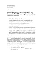
the H37Rv genome. The two genes mentioned are a DNA-
binding protein in the CopG family and a nickle binding GT-
Pase, located in IG1567 and IG2229, respectively (Figure 1).
Importantly, 4.3% of intergenic regions exhibiting transcrip-
tional evidence contained a gene in the reannotated H37Rv
genome, compared with 1.0% of intergenic regions in the
absence transcriptional evidence. The four-fold increase in
likelihood in the results suggests that microarray-based tran-
scriptional evidence would facilitate genome-wide gene find-
ing.
4. DISCUSSION
The computational part of the gene prediction problem is
dealt with by two classes of algorithms. One is based on se-
quence similarity while the other based on gene structure and
signal is known as ab initio prediction. The first class of algo-
rithms, exemplified by BLAST [12], finds sequences (DNA,
6 EURASIP Journal on Bioinformatics and Systems Biology
protein, or ESTs) in the database that match the given se-
quence, whereas the second class of algorithm, notably hid-
den Markov model [8, 9, 13], builds a model of gene struc-
ture from empirical data. They both have their ow n limi-
tations. For instance, the sequence-based approach cannot
handle the case of having no homology, and the model-
based approach the case of inadequate training data. The
method devised in this study would offer a more reliable
gene-prediction mechanism by combining sequence align-
ment, transcr iptional evidence, and homology. In particular,
the transcriptional activity of a piece of DNA is direct ev-
idence that it is funct ioning. As the whole H37Rv genome
sequence has been intensively searched for genes, transcrip-
tional analysis of intergenic regions could only provide more
insight into hidden genes. The integrated method suggested
by this study makes sense from our data showing that tran-
scriptional evidence can support finding potential protein-
coding genes in the intergenic regions. Thus the idea of com-
bining the evidence from the sequence- and function-based
analyses lends itself to not just gene characterization but also
gene prediction. Notice, however, genes that are silent in the
standard in vitro growth condition are not subject to exam-
ination in this study, but the same method can be used gen-
erally for gene finding in other genomes and conditions.
We studied the intergenic regions of M. tuberculosis
H37Rv because of our observation that some of the inter-
genic regions exhibit expression signals. This observation has
little to do with our traditional understanding about pro-
moter and cis-regulatory elements since the former is in-
volved in binding of RNA polymerase and the latter in bind-
ing transcriptional factors but the DNA-protein binding pro-
cess dose not require transcription in the intergenic region.
Relevant to this discourse is the fact that there are a num-
ber of regulatory, noncoding RNAs assuming a distinct role
from mRNA, rRNA, and tRNA. Many such RNAs have been
identified and characterized both in prokaryotes and eukary-
otes and their main function is posttranscriptional regula-
tion of gene expression and RNA-directed DNA methylation
[14, 15]. A noncoding RNA has neither a long open read-
ing frame nor a gene structure. The DNA sequence that en-
codes a noncoding RNA may be viewed as a gene if its reg-
ulatory function can be defined. An isolated expression ele-
ment unaccompanied by a gene structure may hint at non-
coding or regulatory RNA. We confirmed that the poten-
tial protein-coding genes found in this study did not match
any RNA family published in the RNA-families database
(www.sanger.ac.uk/Software/Rfam).
New genes continue to be discovered over time, but the
accumulated discovery will approach to saturation if the true
number of genes is a constant, albeit unknown. Advanced
genome annotation technology enables the identification of
most, if not all, protein-coding sequences in the genome
as soon as it is sequenced. Thus, it is reasonable that the
number of new protein-coding sequences due to reannota-
tion is merely 2% of that in the original submission of M.
tuberculosis genome [16]. Through homology and pattern-
based search, most protein-coding sequences with a pre-
dicted function have been reported. It is encouraging that
we have still been able to find a small number of those in
(1) [Location]: Between Rv1991c and Rv1992c
[Product]: DNA-binding protein, CopG family
[Nucleotide Sequence]: atcgtccatggtttctagcacgcggtatgc-
gttggccacggcgagggcctccgcttcgtcggtgccatggatgctctctagag-
ccctgtcgatctggcccgtgagcaattgggcgtccagctcgtgcaggtagcg-
ctgcgcagccttcgtgaagaactcggaccgactcatgccgagctcactcgca-
cgccgcgatacccgatcgaacgtctcatccggcagagaaatagctgtcttcat
[Protein Sequence]: mktaislpdetfdrvsrraselgmsrsefftka-
aqrylheldaqlltgqidralesihgtdeaealavanayrvletmdd
(2) [Location]: Between Rv2856 and Rv2857c
[Product]: Nickle binding GTPase involved in regula-
tion of expression urease and hydrogenase
[Nucleotide Sequence]: atggtctcctcggtcaccgagggcaagga-
caagccgctgatgtacccggcgacgttccgctcgagggatgtagtgctgctc-
gacaagatcgacttggtgccctttctggacgccgacgtggacgcgtatatcgc-
gcatgtccgcgaggtcaacgcagccgcgacgatcctgccgaccagcacgcg-
caccggagccggcatggggtcctggtcatga
[Protein Sequence]: mvssvtegkdkplmypatfrsrdvvlldkid-
lvpfldadvdayiahvrevnaaatilptstrtgagmgsws
Figure 1: New genes with a predicted function found in the genome
of Mycobacterium tuberculosis H37Rv.
this study. The current knowledge concerning M. tuberculosis
genes is derived from intensive research in the field involv-
ing biological experiments, such as gene deletion and com-
plementation, and bioinfor matics analysis. The gap between
the existing knowledge about M. tuberculosis genes in the
genome and our findings in this study can be ascribed to the
lack of timely update of genome-annotation with the latest
research results in bioinformatics and genomics rather than
the inconsistency in stringency of computational parameters
used. The integrity and advancement of the knowledge base
in genomics would hinge upon the maintenance of complete
and accurate information about the whole genome, espe-
cially for model organisms, such as M. tuberculosis H37Rv.
A critical element in this research is the Affymetrix
oligonucleotide GeneChip, which allowed us to detect the
gene expression of the intergenic regions in M. tube rculo-
sis H37Rv. The Affymetrix system can compute the absolute
signal intensity of mRNA hybridized on the array in a sin-
gle condition as well as the signal ratio between two con-
ditions. The built-in statistical algorithm arrives at the so-
called detection P-value that determines the presence or ab-
sence of any given mRNA. In contrast, the cDNA microarray,
another major platform, generally does not indicate whether
and to what extent a gene expresses in each condition. While
there exist a couple of other types of oligonucleotide mi-
croarray, only the Affymetrix array implements the probes
for interrogating intergenic sequences in the H37Rv genome.
As an additional strength, the Affymetrix array is designed
to minimize cross-hybridization by using unique oligonu-
cleotide probes and the pair of PM (perfect-match) and MM
(mismatch) probes. The cross-hybridization of related or
overlapping gene sequences often contributes to false pos-
itive signals, especially in the case when long cDNA se-
quences are used as probes. A study demonstrated that the
Affymetrix GeneChip produced more reliable results in de-
tecting changes in gene expression than cDNA microarrays
L. M. Fu and T. M. Shinnick 7
[17]. Thus, the choice of the Affymetrix GeneChip for this
study is well justified. To validate genome-wide microarray
data, a basic means is to demonstrate a high correlation be-
tween the data of duplicate experiments [18]. In the present
study, the correlation between any pair of the gene expres-
sion data derived from independent RNA samples is > .9. In
addition, PCR analysis has been performed to verify that the
AffymetrixGenechipsystemworkedproperlyinourprior
work [19, 20].
5. CONCLUSION
Current computational programs for gene prediction have
no guarantee to identify all genes in a sequenced genome be-
cause the knowledge about gene structure has yet to be per-
fected. Genome reannotation using the same kind of heur is-
tics offers limited help unless its predictive power has been
improved. Reannotation based on new experimental evi-
dence that trickles in at its own pace is probably slow.
We conducted a genome-wide analysis using the
Affymetrix GeneChip to explore genes contained in the in-
tergenic sequences of the M. tuberculosis H37Rv genome. Po-
tential protein-coding genes were determined according to
the bioinformatics criteria constituted by the gene structure,
protein coding potential, and the presence of ortholog evi-
dence. The bioinformatics criteria in conjunction with tran-
scriptional evidence have led to the discovery of genes with
a specific function, such as a DNA-binding protein in the
CopG family and a nickle binding GTPase, as well as hypo-
thetical proteins that have not been reported in the M. tu-
berculosis H37Rv genome. This work has demonstrated that
microarray-based transcriptional evidence would help gene
finding on the genomic scale.
ACKNOWLEDGMENTS
This work is supported by National Institutes of Health un-
der the Grant HL-080311 and the Centers of Disease Con-
trol and Prevention. The authors would like to thank CDC
for the use of the facilities and UCI for providing service for
microarray hybridization. They also thank Thomas R. Gin-
geras at Affy metrix, Inc. for designing Mycobacterium tuber-
culosis GeneChip. Bacterial culture and RNA isolation were
performed by Pramod Aryal.
REFERENCES
[1] S. T. Cole, R. Brosch, J. Parkhill, et al., “Deciphering the biol-
ogy of Mycobacterium tuberculosis from the complete genome
sequence,” Nature, vol. 393, no. 6685, pp. 537–544, 1998.
[2] R. Overbeek, T. Begley, R. M. Butler, et al., “The subsystems
approach to genome annotation and its use in the project
to annotate 1000 genomes,” Nucleic Acids Research, vol. 33,
no. 17, pp. 5691–5702, 2005.
[3] G. H. Van Domselaar, P. Stothard, S. Shrivastava, et al.,
“BASys: a web server for automated bacterial genome anno-
tation,” Nucleic Acids Research, vol. 33, Web Server issue, pp.
W455–W459, 2005.
[4] P. Stothard and D. S. Wishart, “Automated bacterial genome
analysis and annotation,” Current Opinion in Microbiology,
vol. 9, no. 5, pp. 505–510, 2006.
[5] P. Nielsen and A. Krogh, “Large-scale prokaryotic gene predic-
tion and comparison to genome annotation,” Bioinformatics,
vol. 21, no. 24, pp. 4322–4329, 2005.
[6] J M. Lee, S. Zhang, S. Saha, S. Santa Anna, C. Jiang, and J.
Perkins, “RNA expression analysis using an antisense Bacillus
subtilis genome array,” Journal of Bacter i ology, vol. 183, no. 24,
pp. 7371–7380, 2001.
[7] D. Zheng, Z. Zhang, P. M. Harrison, J. Karro, N. Carriero, and
M. Gerstein, “Integrated pseudogene annotation for human
chromosome 22: evidence for transcription,” Journal of Molec-
ular Biology, vol. 349, no. 1, pp. 27–45, 2005.
[8] A. V. Lukashin and M. Borodovsky, “GeneMark.hmm: new so-
lutions for gene finding,” Nucleic Acids Research, vol. 26, no. 4,
pp. 1107–1115, 1998.
[9] J. Besemer and M. Borodovsky, “GeneMark: web software for
gene finding in prokaryotes, eukaryotes and viruses,” Nucleic
Acids Research, vol. 33, Web Server issue, pp. W451–W454,
2005.
[10] M. A. Fisher, B. B. Plikaytis, and T. M. Shinnick, “Microarray
analysis of the Mycobacterium tuberculosis transcriptional re-
sponse to the acidic conditions found in phagosomes,” Journal
of Bacteriology, vol. 184, no. 14, pp. 4025–4032, 2002.
[11] R. D. Finn, J. Mistry, B. Schuster-B
¨
ockler, et al., “Pfam:
clans, web tools and services,” Nucleic Acids Research, vol. 34,
Database issue, pp. D247–D251, 2006.
[12] S. F. Altschul, W. Gish, W. Miller, E. W. Myers, and D. J. Lip-
man, “Basic local alignment search tool,” Journal of Molecular
Biology, vol. 215, no. 3, pp. 403–410, 1990.
[13] C. Burge and S. Karlin, “Prediction of complete gene struc-
tures in human genomic DNA,” Journal of Molecular Biology,
vol. 268, no. 1, pp. 78–94, 1997.
[14] V. A. Erdmann, M. Z. Barciszewska, A. Hochberg, N. de Groot,
and J. Barciszewski, “Regulatory RNAs,” Cellular and Molecu-
lar Life Sciences, vol. 58, no. 7, pp. 960–977, 2001.
[15] A. S. Pickford and C. Cogoni, “RNA-mediated gene silencing,”
Cellular and Molecular Life Sciences, vol. 60, no. 5, pp. 871–882,
2003.
[16]J C.Camus,M.J.Pryor,C.M
´
edigue, and S. T. Cole, “Re-
annotation of the genome sequence of Mycobacterium tuber-
culosis H37Rv,” Microbiology, vol. 148, no. 10, pp. 2967–2973,
2002.
[17] J. Li, M. Pankratz, and J. A. Johnson, “Differential gene expres-
sion patterns revealed by oligonucleotide versus long cDNA
arrays,” Tox icolog ical Sciences, vol. 69, no. 2, pp. 383–390, 2002.
[18]J.L.DeRisi,V.R.Iyer,andP.O.Brown,“Exploringthe
metabolic and genetic control of gene expression on a genomic
scale,” Science, vol. 278, no. 5338, pp. 680–686, 1997.
[19] L. M. Fu, “Exploring drug action on Mycobacterium tubercu-
losis using affymetrix oligonucleotide genechips,” Tuberculosis,
vol. 86, no. 2, pp. 134–143, 2006.
[20] L. M. Fu and T. M. Shinnick, “Genome-wide exploration of
the drug action of capreomycin on Mycobacterium tuberculosis
using Affymetrix oligonucleotide GeneChips,” Journal of Infec-
tion, vol. 54, no. 3, pp. 277–284, 2007.