Structural and vibrational investigations and molecular docking studies of a vinca alkoloid vinorelbine
Bạn đang xem bản rút gọn của tài liệu. Xem và tải ngay bản đầy đủ của tài liệu tại đây (3.96 MB, 21 trang )
Journal of Biomolecular Structure and Dynamics
ISSN: (Print) (Online) Journal homepage: www.tandfonline.com/journals/tbsd20
Structural and vibrational investigations and
molecular docking studies of a vinca alkoloid,
vinorelbine
Sefa Celik, Sevim Akyuz & Aysen E. Ozel
To cite this article: Sefa Celik, Sevim Akyuz & Aysen E. Ozel (2023) Structural
and vibrational investigations and molecular docking studies of a vinca alkoloid,
vinorelbine, Journal of Biomolecular Structure and Dynamics, 41:19, 9666-9685, DOI:
10.1080/07391102.2022.2145369
To link to this article: />
View supplementary material
Published online: 11 Nov 2022.
Submit your article to this journal
Article views: 353
View related articles
View Crossmark data
Citing articles: 6 View citing articles
Full Terms & Conditions of access and use can be found at
/>
JOURNAL OF BIOMOLECULAR STRUCTURE AND DYNAMICS
2023, VOL. 41, NO. 19, 9666–9685
/>
Structural and vibrational investigations and molecular docking studies
of a vinca alkoloid, vinorelbine
Sefa Celika, Sevim Akyuzb and Aysen E. Ozela
a
Physics Department, Science Faculty, Istanbul University, Istanbul, Turkey; bPhysics Department, Science and Letters Faculty, Istanbul Kultur
University, Istanbul, Turkey
Communicated by Ramaswamy H. Sarma
ABSTRACT
ARTICLE HISTORY
Vinorelbine, a vinca alkaloid, is an antimitotic drug that inhibits polymerisation process of tubulins to
microtubules, and is widely used in cancer chemotherapy. Due to the importance of the structureactivity relationship, in this work the conformational preferences of the vinorelbine molecule were
surched by PM3 method. The obtained lowest energy conformer was then optimized at DFT/B3LYP/631G(d,p) level of theory and the structural characteristics were determined. Frontier orbital (HOMO,
LUMO) and molecular electrostatic potential (MEP) analyses were performed for the optimized struc
ture. The experimental FT-IR, Raman and UV-VIS spectral data of vinorelbine along with the theoretical
DFT/B3LYP/6-31G(d,p) calculations were investigated in detail. The vibrational wavenumbers were
assigned based on the calculated potential energy distribution (PED) of the vibrational modes. To
shed light into the anticancer property of vinorelbine as microtubule destabilizer, the most favourable
binding mode and the interaction details between vinorelbine and tubulin were revealed by molecular
docking studies of vinorelbine into the a,b-tubulin (PDB IDs: 4O2B; 1SA0; 7CNN) and binding free ener
gies were calculated by the combination of Molecular Mechanics/Generalized Born Surface Area
(MMGBSA) and Molecular Mechanics/Poisson-Boltzmann Surface Area (MM-PBSA) methods fMM/
PB(GB)SAg. The calculated vinorelbine-7CNN binding free energy, using by MM/PB(GB)SA approach,
was found to be the best (-50.39 kcal/mol), and followed by vinorelbine-4O2B (-28.5 kcal/mol) and
vinorelbine-1SA0 (-17.59 kcal/mol) systems. Moreover, the interaction of vinorelbine with the cyto
chrome P450 enzymes (CYP), which are known to help in the metabolism of many drugs in the body,
was investigated by docking studies against CYP2D6 and CYP3A4 targets.
Received 1 August 2022
Accepted 3 November 2022
1. Introduction
Vinorelbine is a vinca alkaloid that is produced semi-synthet
ically and suppresses cellular growth by binding to tubulin,
like other vinca alkaloids, although it varies from them in
terms of its spectrum of antitumor action (Ngan et al., 2000).
It was shown by in vitro experiments that vinorelbine affects
on microtubule dynamic instability and treadmilling and
reported that its major effects were a slowing of the micro
tubule growth rate, an increase in growth duration, and a
reduction in shortening duration (Ngan et al., 2000).
Vinorelbine has been used to treat non-small cell lung
cancer (NSCLC), as well as breast, ovarian, and cervical can
cers (Altinoz et al., 2018; McQueen, 2010). It is also a vinca
plant-based antimicrotubule antineoplastic agent that has
been studied in both first and second-line NSCLC treatments.
In the United States, the FDA (U.S. Food and Drug
Administration) has authorized vinorelbine for NSCLC treat
ment (Laurent & Shapiro, 2006). The antitumor activity of
vinorelbine has been demonstrated in several preclinical
studies against various cancer cell lines and by xenograft
tumor models (Waldman & Terzic, 2008). Vinorelbine has
Vinorelbine; density
functional theory; molecular
docking; FT-IR and Raman
become an important anticancer agent in adjuvant chemo
theraphy of of patients with NSCLC (stages 1B-III) and the
palliative chemotheraphy in the NSCLC (stages IIIB and IV)
offen in combination with cisplatin (Aronson, 2015). It also
plays an important role in the treatment of advanced and
metastatic breast cancer (Aronson, 2015; Gregory & Smith,
2000; Stravodimou et al., 2014). In Europe, vinorelbine has
been approved for the treatment of NSCLC, breast cancer,
and in some countries, prostate cancer (Debernardis
et al., 2009).
The human cytochrome P450 isoenzymes involved in in
the liver metabolism of vinorelbine was investigated and
determined that CYP3A4 was the main enzyme involved in
the hepatic metabolism of vinorelbine (Beulz-Riche et al.,
2005). Chi et al. investigated the structural mechanism for
the binding of a vinca alcoloid, vinblastine, with tubulin by
molecular docking studies (Chi et al., 2015).
Literature survey reveals that for vinorelbine no molecular
structure analysis and vibrational assignments have been
reported so far. This study aimed to enlighten the molecular
structure, electronic properties, and anticancer action mecha
nisms of vinorelbine. To reveal the molecular structure,
CONTACT Sefa Celik
Physics Department, Science Faculty, Istanbul University, Istanbul, Turkey.
Supplemental data for this article can be accessed online at />� 2022 Informa UK Limited, trading as Taylor & Francis Group
KEYWORDS
JOURNAL OF BIOMOLECULAR STRUCTURE AND DYNAMICS
conformational analysis on vinorelbine was performed and
the obtained most stable conformation was then optimized
at DFT/B3LYP/6-31G(d,p) level of theory. The vibrational
wavenumbers of the molecule were experimentally observed
and compared with the computed values. The vibrational
modes were determined based on the calculated potential
energy distribution (PED). The Frontier molecular orbitals
analysis of vinorelbine was performed to study the molecular
reactivity and stability. Molecular Electrostatic Potential (MEP)
map was discussed to get information about the chemical
and site selectivity of vinorelbine.
Vinorelbine exerts its cytotoxic effect on cancer cells by
binding to tubulin, which leads to cell cycle arrest in mitosis.
The a,b-tubulin heterodimers have highly dynamic structure
and there are some differences in the crystal structures tubu
lin-ligand complexes (Bueno et al., 2018). For this reason, to
elucidate the binding modes of vinorelbine against tubulin,
molecular docking studies were performed by using three
available tubulin crystal structures obtained from protein
data bank (PDB) as target proteins. These targets are: 1)
Tubulin in complex with colchicine and with the stathnin-like
domain (PDB ID: 1SA0) (Ravelli et al., 2004), 2) tubulin colchi
cine complex (PDB ID: 4O2B) (Prota et al., 2014) and 3) tubu
lin-vinorelbine complex (PDB ID: 7CNN). Moreover, the
binding free energies were calculated by MM/PB(GB)SA
approach, which is the combination of Molecular Mechanics/
Generalized Born Surface Area (MMGBSA) and Molecular
Mechanics/Poisson-Boltzmann Surface Area (MM-PBSA) meth
ods (Wang et al., 2019). The purpose of this study is to find
the most favourable binding mode and reveal the interaction
details between vinorelbine and tubulin. In a recent study,
the crystal structure of vinorelbine in complex with tubulin
was determined by Chengyong et al. (2021; PDB ID: 7CNN).
In this study, to validate our docking protocol and to high
light the importance of the initial geometry of the ligand in
the docking studies, vinorelbine was re-docked into the
vinorelbine binding site of PDB ID:7CNN target (Chengyong
et al., 2021) and for docking simulations, two different initial
structures of the ligand were used: 1) Vinorelbine structure
obtained from the published data of the crystal structure of
tubulin-vinorelbine complex (PDB ID: 7CNN) (Chengyong
et al., 2021) and 2) the optimized geometry of vinorelbine.
Finally, since the side effects of vinca alkaloids are known
to be related to their metabolism (Lokwani et al., 2020), the
hepatic metabolism of vinorelbine in human, was investi
gated theoretically by docking simulations against cyto
chrome P450, CYP3A4 and CYP2D6 enzymes.
2. Experimental and computational procedures
2.1. Experimental
Vinorelbine was acquired in solid form Santa Cruz
Biotechnology (CAS Number 71486-22-1) with reagent grade
and used as obtained. The FT-IR spectrum of the KBr disc of
the sample was recorded on a Jasco 6300 FT-IR spectrometer
(2 cm 1 resolution), between 400 and 4000 cm 1. A Jasco
NRS 3100 micro-Raman spectrometer with a 532-nm diode
laser and a 1200 lines/mm diffraction grating was used to
9667
record the Raman spectra. For calibration the 520 cm 1 sili
con phonon mode was used. The UV-Visible spectrum of
DMSO solution of vinorelbine was obtained using a Perkin
Elmer-Lambda 25 spectrometer in the 190–900 nm region.
Spectral manipulations such as baseline adjustment, curve
fitting and obtaining second derivative were performed
using GRAMS/AI 7.02 (Thermo Electron Corporation) software
package. For curve fitting, second dervivative of the original
spectrum was used as a guide, and curve fitting was done
using Gaussian function. The fitting was undertaken until
reproducible and converged results were obtained with
squared correlations better than r2 � 0.9998. The second
derivatives of the spectra were obtained by using SavitzkyGolay function (two polynomial degrees, 17 points).
2.2. Computational details
The conformational analysis of vinorelbine molecule was per
formed using the Spartan06 computer software (Shao et al.,
2006) and the PM3 method (Stewart, 1989, 1991, 2004). The
obtained lowest energy conformer was then optimized. For
the geometry optimization and other quantum chemical cal
culations Gaussian03 software (Frisch et al., 2016) and DFT/
B3LYP method (Becke, 1993) in combination with 6-31 G(d,p)
basis set were used. The harmonic force field of vinorelbine
was defined using the MOLVIB software (Sundius,1990, 2002)
and the scaled quantum mechanical force field approach
given by Pulay et al. (1983). The Cartesian coordinate force
fields were transformed into natural internal coordinates
(Sundius, 1990, 2002), IR intensity, Raman activity, and poten
tial energy distribution (PED) of vibration modes were identi
fied. The simulation program Simirra (Istvan, 2002) was used
to convert Raman activity to Raman intensity. For the theor
etical computations, Lorentzian band forms with a band
width (FWHM) of 10 cm 1 were chosen.
The following scale factors for DFT/B3LYP/6-31G(d,p) level
of theory calculations were chosen to produce the best
match for the experimental results: O-H stretch 0.87; N-H
stretch 0.89; C ¼ O stretch 0.86; C-H stretch 0.91; N-H and
C-H deformation 0.92; all others 0.98. These scale factors
were chosen by optimization of the scale factors taken from
the previous studies (Celik et al., 2016, 2021, 2022a, Celik,
Vagifli, et al., 2022) by fitting the observed frequencies to
the calculated ones.
For docking studies, the crystal structures of a,b-tubulin
(PDB IDs: 4O2B; 1SA0; 7CNN) (Chengyong et al., 2021; Prota
et al., 2014; Ravelli et al., 2004) and P450 enzymes CYP2D6
(PDB ID: 2F9Q) (Rowland et al., 2006), CYP3A4(PDB IDs:
€gren,
1TQN, 1W0E, 1W0F, 1W0G and 2V0M) (Ekroos & Sjo
2006; Williams et al., 2004; Yano et al., 2004) were obtained
from the protein data bank ( />Molecular docking simulations were performed by the
Autodock Vina program (Trott & Olson, 2010) and binding
affinities were calculated. The active sites of receptors were
screened by using the CAVER program (Jurcik et al., 2018). In
docking, vinorelbine was treated as flexible ligand by modify
ing its rotatable torsions, but the target protein was consid
ered to be a rigid receptor.
9668
S. CELIK ET AL.
Figure 1. The optimized geometric structure of vinorelbine, as determined
using DFT/B3LYP/6-31G(d,p) level of theory.
The binding free energies of the vinorelbine tubulin (PDB
IDs: 4O2B, 1SA0 and 7CNN) systems were calculated by MM/
PB(GB)SA approach by the program developed by Wang
et al. (2019)
3. Results and discussion
3.1. Structure
The most stable conformer obtained as a result of the con
formation analysis was optimized using DFT/B3LYP level of
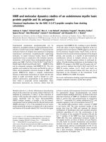
theory and the 6-31 G(d,p) basis set. The optimized structure
of the vinorelbine molecule (C45H54N4O8) is shown in
Figure 1. The labeled wireframe representation of the opti
mized molecular geometry of the vinorelbine molecule is
given in Figure S1 (Supplementary data file). The obtained
bond lengths and bond angles are tabulated in Table 1 and
the dihedral angles are given in Table S1.
As seen in Table 1, the C-C bond lengths of the indolelike rings of vinorelbine were computed in the range of
1.388 1.444 Å. These bond lengths were experimentally
determined between 1.34 and 1.41 Å, in the crystal structure
of the indole molecule (Kaneda & Tanaka, 1976) and
between 1.360-1.412 Å in the crystal structure of the
Bisbenzylisoquinoline alkaloid methylwarifteine molecule
(Borkakoti & Palmer, 1978).
The C-C bond lengths of a phenyl ring are known to be
around 1.4 Å. The mean C-C bond length of benzene crystal
was determined as 1.392 Å (Cox et al., 1958). In substituded
benzene molecules, these bond lengths were experimentally
determined in the 1.378-1.403 range (Campos et al., 1980). In
our study, the C-C bond lengths of the phenyl group of the
vinorelbine molecule were calculated in the range of 1.3891.411 Å, in agreement with the expected values.
The C-N bond lengths (N11-C37 and N11-C48) and the
C-N-C bond angle in the indole-like ring of vinorelbine were
�
computed as 1.374 Å, 1.391 Å, and 110.3 , respectively. In the
crystal structure of the indole molecule, the C-N bond
lengths and C-N-C bond angle were experimentally deter
�
mined as 1.36 Å, 1.38 Å, and 110.5 , respectively (Kaneda &
Tanaka, 1976). Also, the C-C bond lengths (C37-C42, C42-C47,
C47-C48) and C-C-C bond angles (C37-C42-C47 and C42-C47C48) in the indole moiety were computed as 1.388, 1.444,
1.419 Å, and 106.5 and 107.6, respectively, the corresponding
values were experimentally determined as 1.37, 1.41, 1.40 Å,
and 105.8 and 109.8, respectively, in the crystal structure of
ethyladenine-indole complex (Kaneda & Tanaka, 1976).
The results showed that the computed geometrical
parameters of vinorelbine were in conformity with the struc
tural data of similar compounds. An X-ray crystallographic
study on vinorelbine is not available. For this reason, we
compared optimized structure of vinorelbine in gas phase
with the vinorelbine obtained from the crystal structure of
tubulin-vinorelbine complex (PDB ID: 7CNN) (Chengyong
et al., 2021). Figure S2 shows comparatively the geometric
structure of vinorelbine molecule, obtained from the crystal
structure of tubulin-vinorelbine complex (PDB ID: 7CNN) with
that of optimized structure, obtained by using DFT/B3LYP/631G(d,p) level of theory. To quantify the difference between
the optimized geometry of vinorelbine and its experimental
findings, obtained from tubulin-vinorelbine complex, we cal
culated the root-mean-square difrences of the two structures
by using the structures comparer utility of the Chemcraft
program (Zhurko, 2005). Since the H atoms of vinorelbine,
obtained from tubulin-vinorelbine complex, were not present
(PDB ID: 7CNN) (Chengyong et al., 2021). The comparison
between the two structures was made without considering
the H atoms. The weighted RMSD was found to be 6.025.
Moreover, it was found that O atoms gave the largest RMSD
among all kind of atoms of vinorelbine (RMSD ¼ 7.277). The
oxygen and nitrogen atoms of vinorelbine (C45H54N4O8) are
reactive sites that interact with tubulin amino acids, so the
bond angles and bond lengths involving the O and N atoms
of vinorelbine in the tubulin complex are expected to differ
from the calculated values for the single molecule. It must
be considered that the number of oxygen atoms in vinorel
bine is twice that of nitrogen atoms. Sebhaoui et al. (2021)
compared molecular structures of 2-pyrone derivatives
obtained from crystal structures with those of calculated
with B3LYP in the gas phase, by using Chemcraft program.
In that study, it was reported that O atoms gave the largest
RMSD among all kinds of atoms contained by investigated
2-pyrone derivatives.
Molecular electrostatic potential
The molecular electrostatic potential relates with the dipole
moment, electronegativity, and partial charges of a molecule.
It provides a reliable analysis of chemical reactivity and ena
bles to determine electrophilic and nucleophilic regions, as
well as hydrogen-bonding interactions of the molecule (Kaya
€rk et al., 2021; Kutlu et al., 2021; Politzer & Murray,
Kınaytu
2002). The MEP analysis of the optimized structure of vinorel
bine was performed using B3LYP/6-31G(d,p) level of theory.
The electrostatic potential map of a vinorelbine molecule is
shown in Figure 2, along with a legend that shows how
potential varies with color. Dark red color represents the
most negative (electron rich) and dark blue color shows the
most positive (electron poor) regions, whereas, the yellow
JOURNAL OF BIOMOLECULAR STRUCTURE AND DYNAMICS
9669
Table 1. The optimized geometry parameters of vinorelbine, calculated by DFT/B3LYP/6-31G(d,p) level of theory.�
Atoms
O1-C19
O1-C35
O2-C17
O2-H72
O3-C27
O3-C41
O4-C27
O5-C33
O5-C45
O6-C35
O7-C38
O7-C54
O8-C38
N9-C14
N9-C21
N9-C25
N9-H72
N10-C16
N10-C23
N10-C29
Atoms
1.436
1.366
1.410
0.991
1.339
1.439
1.217
1.369
1.419
1.207
1.360
1.438
1.208
1.480
1.467
1.460
1.794
1.486
1.404
1.459
N11-C37
N11-C48
N11-H91
N12-C43
N12-C44
N12-C49
C13-C14
C13-C16
C13-C18
C13-C20
C14-C15
C14-H58
C15-C19
C15-C22
C15-C24
C16-C17
C16-H59
C17-C19
C17-C27
C18-C21
Atoms
1.391
1.374
1.009
1.460
1.479
1.465
1.569
1.571
1.567
1.514
1.553
1.102
1.588
1.565
1.521
1.561
1.100
1.549
1.543
1.533
C18-H60
C18-H61
C19-H62
C20-C23
C20-C28
C21-H63
C21-H64
C22-C31
C22-H65
C22-H66
C23-C30
C24-C26
C24-H67
C25-C26
C25-H68
C25-H69
C26-H70
C28-C32
C28-H71
C29-H73
Atoms
1.092
1.092
1.089
1.395
1.389
1.094
1.105
1.533
1.093
1.098
1.398
1.332
1.088
1.500
1.098
1.109
1.087
1.401
1.085
1.089
C29-H74
C29-H75
C30-C33
C30-H76
C31-H77
C31-H78
C31-H79
C32-C33
C32-C34
C34-C36
C34-C37
C34-C38
C35-C40
C36-C39
C36-H80
C36-H81
C37-C42
C39-C43
C39-C46
C39-H82
Atoms
1.103
1.092
1.401
1.081
1.095
1.094
1.095
1.416
1.544
1.558
1.531
1.557
1.515
1.557
1.090
1.092
1.388
1.542
1.515
1.100
C40-H83
C40-H84
C40-H85
C41-H86
C41-H87
C41-H88
C42-C44
C42-C47
C43-H89
C43-H90
C44-H92
C44-H93
C45-H94
C45-H95
C45-H96
C46-C50
C46-H97
C47-C48
C47-C51
C48-C52
Atoms
1.092
1.094
1.089
1.089
1.093
1.092
1.507
1.444
1.097
1.088
1.096
1.101
1.097
1.091
1.097
1.341
1.091
1.419
1.406
1.400
C49-C50
C49-H98
C49-H99
C50-C53
C51-C55
C51-H100
C52-C56
C52-H101
C53-C57
C53-H102
C53-H103
C54-H104
C54-H105
C54-H106
C55-C56
C55-H107
C56-H108
C57-H109
C57-H110
C57-H111
Atoms
1.525
1.101
1.099
1.512
1.389
1.086
1.389
1.086
1.540
1.096
1.100
1.092
1.090
1.093
1.411
1.086
1.086
1.095
1.095
1.095
C19-O1-C35
C17-O2-H72
C27-O3-C41
C33-O5-C45
C38-O7-C54
C14-N9-C21
C14-N9-C25
C21-N9-C25
C16-N10-C23
C16-N10-C29
C23-N10-C29
C37-N11-C48
C37-N11-H91
C48-N11-H91
C43-N12-C44
C43-N12-C49
C44-N12-C49
C14-C13-C16
C14-C13-C18
C14-C13-C20
124.6
107.7
115.6
119.5
114.9
104.9
113.5
114.2
107.2
117.9
117.7
110.3
122.7
126.5
114.2
109.4
111.6
113.7
103.1
112.2
�
Bond lengths (R) in Å and bond angle (A) in degree (o).
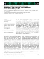
Figure 2. Molecular electrostatic potential (MEP) of vinorelbine obtained by DFT/B3LYP/6- 31 G(d,p) level of theory.
color represents slightly electron-rich and light blue, the
slightly electron-poor regions.The color code of vinorelbine
MEP varies between 1.909 V (dark red) and 1.909 V (dark
blue). Figure 2 shows that regions with positive potential are
predominantly located on hydrogen atoms, where possible
nucleophilic attack sites, and negative potential regions
around oxygen atoms, as possible sites for electro
philic attack.
Vibrational spectral analysis
The Vinorelbine molecule (C45H54N4O8) has 111 atoms in C1
symmetry point group, thus has 327 vibrational modes, both
IR and Raman active. The analysis of the bands and the
assignments of fundamental wavenumbers were made based
on computed potential energy distributions (PED), the mag
nitude and relative intensities of the observed bands and
group frequencies. The wavenumbers of the observed and
calculated fundamental bands in IR and Raman spectra,
along with their proposed assignments were given in
Table 2.
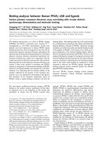
The experimental FT-IR and Raman spectra of vinorelbine
are given in Figures 3 and 4, respectively compared to the
simulated spectra. To resolve the overlapping bands in the IR
and Raman spectra, resolution enhance techniques, such as
curve fitting and second derivative of the experimental spec
trum were used. The Figure S3 represents curve fitted FT-IR
spectrum of vinorelbine. The Figure S4 shows the 1700150 cm 1 region of the Raman spectrum and 1800-400 cm 1
region of the FT-IR spectrum of vinorelbine together with
their second derivative profiles. As seen in Figure S4, the
second derivative minima correspond to the peaks in the ori
ginal spectrum.
Vinorelbine has both aromatic and aliphatic CH groups.
The CH stretching vibrations of the substituted benzene like
molecules are observed in the region 3100–3000 cm 1 (Jiao
et al., 2022; Roeges & Baas, 1994), whereas methylene CH
stretching vibrations are expected in 3000-2800 cm 1
(Hajduchova et al., 2018; Jiao et al., 2022). Methyl CH stretch
ings are observed higher than methylene and around 29802870 cm 1. In our study the observed IR bands at 3078,
3058, 3052 and 3033 cm 1 were assigned to CH stretching
vibrations of the phenyl rings of indole moiety (Silverstein
et al., 1981). The corresponding bands were predicted by
DFT at 3092, 3059, 3048 and 3043 cm 1, respectively as pure
CH stretching vibrations with aromatic CH stretching
9670
S. CELIK ET AL.
Table 2. The experimental and calculated fB3LYP/6-31G(d,p)g wavenumbers
(cm-1) of vinorelbine and the PED distributions of the vibration modes.
Experimental
Calculated
I(IR)
I(Ra)
3438
3454
3117
91
585
8
14
3078
3058
3092
3059
3056
3048
3043
3038
3034
3033
3031
3030
3026
3017
3013
3009
3005
3002
3001
3000
2998
2993
2991
2985
2975
2973
2972
2971
2965
2961
2951
2943
2942
2926
2926
2925
2925
2912
2910
2908
2906
2904
2903
2897
2880
2873
2870
2866
2858
2849
2846
2845
2822
2781
1724
1718
1707
1705
1676
1647
1638
1616
1605
1555
1521
1509
1502
1495
1476
1473
7
30
2
32
26
6
13
8
0
4
13
8
35
17
14
16
12
17
4
11
40
33
33
22
45
25
1
10
33
13
31
6
16
35
38
40
29
25
61
21
23
38
60
34
31
26
51
47
74
37
60
61
367
5
4
237
191
8
154
35
1
7
27
176
14
13
14
10
15
64
59
47
60
61
64
64
60
56
42
44
54
53
48
48
48
48
45
42
40
27
48
57
58
56
38
34
39
35
34
82
82
83
83
79
88
91
90
85
80
48
38
47
45
36
34
32
36
34
12
15
14
19
40
35
6
32
72
13
9
90
7
9
12
15
27
32
Raman
3052
3033
3022
3014
2989
2961
2944
2931
2924
2875
2871
2849
2840
2817
1742
1710
1653
1642
1617
1596
1539
1512
1504
1489
1475
1678
1661
1617
1606
1540
1528
1506
1489
1474
Experimental
IR
mcalS
IR
Table 2. Continued.
Raman
PED%
mNH(100)
mOH(91)ỵ mNH(8)
intermolecular NH
mCH(99) (aromatic)
mCH(100) (aromatic)
mCH(99) (aromatic)
mCH(99) (aromatic)
mCH(100)
mCH(99) (aromatic)
mCH(100) (CH3)
mCH(97) (CH3)
mCH(95) (aromatic)
mCH(99) (CH3)
mCH(100) (CH3)
mCH(99)
mCH(96) (CH2)
mCH(90) (CH3)
mCH(99) (CH2)
mCH(99) (CH3)
mCH(89)
mCH(97) (CH3)
mCH(99) (CH3)
mCH(98) (CH2)
mCH(98)
mCH(96) (CH3)
mCH(96) (CH3)
mCH(100) (CH3)
mCH(99) (CH3)
mCH(99) (CH3)
mCH(95) (CH2)
mCH(100) (CH2)
mCH(98) (CH2)
mCH(95) (CH2)
mCH(100) (CH3)
mCH(95) (CH3)
mCH(98) (CH2)
mCH(99) (CH3)
mCH(100) (CH3)
mCH(91) (CH2)
mCH(99) (CH2)
mCH(90) (CH3)
mCH(85) (CH2)
mCH(95) (CH2)
mCH(89) (CH2)
mCH(98) (CH2)
mCH(100) (CH3)
mCH(98) (CH2)
mCH(95)
mCH(99)
mCH(94) (CH2)
mCH(98)
mCH(93) (CH3)
mCH(92) (CH2)
mCH(96) (CH2)
mCH(99) (CH2)
mCO(81)ỵ mCC(5)
mCC(80)
mCC(76)
mCO(81)
mCO(85)
mCC(58)ỵ mCN(7)
mCC(63)
mCC(63)
mCC(59)
mCC(69)
dCOH(84)
dCCH(24)ỵ mCN(9)ỵ mCC(15)
mCC(27)ỵ mCN(11)ỵdCCH(24)
dHCH(52)
dHCH(57)
mCO(10)ỵ dHCH(18)ỵmCC(7)
(continued)
1458
1460
1451
1431
1431
1414
1412
1372
1374
1360
1348
1333
1321
1307
1298
1298
1271
1271
1268
1251
1244
1232
1242
1228
1226
1204
1206
Calculated
mcalS
I(IR)
I(Ra)
PED%
1468
1466
1464
1463
1462
1459
1458
1457
1455
1454
1452
1449
1448
1446
1445
1444
1444
1442
1441
1436
1434
1432
1430
1422
1418
1417
1413
1387
1385
1379
1372
1371
1369
1366
1362
1354
1348
1347
1344
1343
1342
1341
1336
1329
1327
1319
1319
1318
1313
1310
1304
1300
1291
1288
1286
1277
6
19
6
51
12
8
11
37
6
6
4
4
9
11
5
2
4
2
4
5
7
18
7
33
16
11
12
11
5
4
2
74
9
1
13
4
70
2
4
33
29
8
17
15
28
6
3
32
63
15
19
32
31
3
23
408
52
67
80
84
85
88
89
89
86
85
84
89
91
97
99
100
100
95
91
80
82
78
65
32
31
30
24
24
23
22
40
41
38
28
22
16
22
23
24
23
23
22
18
23
26
46
46
49
57
56
55
40
25
30
31
15
1266
1258
1255
1249
1246
1243
1238
1234
1234
1227
1222
1220
1211
1203
1198
2
36
12
7
38
65
17
128
294
271
177
3
9
8
30
20
17
16
20
24
26
23
19
19
17
18
16
12
13
13
dHCH(67)
dHCH(80)
dHCH(78)
dHCH(6)
dHCH(44)
dHCH(35)
dHCH(14)ỵ mCN(5)
dHCH(78)
dHCH(62)
dHCH(78)
dHCH(89)
dHCH(84)ỵ dCCH(5)
dHCH(71)ỵ dOCH(13)
dHCH(83)
dHCH(65)ỵ dOCH(6)
dHCH(80)
dHCH(90)
dHCH(62)
dHCH(53)ỵ CCCCH(13)ỵ dNCH(6)
dHCH(92)
dHCH(88)
mCN(14)ỵ dCNH(19)ỵ mCC(16)
dHCH(88)
dOCH(46)ỵ dHCH(35)ỵ mCO(6)
dHCH(30)ỵ dOCH(29)
dOCH(50)ỵ dHCH(38)
dHCH(30)ỵ dNCH(34)
mCC(59)
mCC(23)ỵ dCCH(39)
CCCCH(20)ỵdNCH(6)ỵ dCCH(6)
mCC(5)
mCC(7)ỵ mCN(12)
dHCH(27)ỵ dCCH(34)ỵ mCC(9)
dHCH(42)ỵ dCCH(39)
dOCC(8)ỵCCCCH(32)
dCCH(21)ỵ mCC(11)
mCC(11)ỵ dCCH(20)ỵ dHCH(25)
dCCH(20)
mCC(16)ỵ dCCH(26)
mCC(17)
dHCH(6)
dNCH(5)
mCC(17)
dCCH(5)
mCC(8)
dCCH(16)
dCCH(16)
dCCH(13)ỵ mCO(7)
mCO(6)
dCCH(51)ỵ mCC(19)
mCN(30)ỵ mCC(20)
dCCH(33)ỵ mCC(6)
dCCH(35)
dCCH(37)ỵ dNCH(8)
dCCH(5)ỵ CCNCH(5)
mCO(47)ỵ mCC(12)ỵ
dOCO(6)ỵ dCCO(5)
dCCH(18)ỵ mCC(16)ỵ dNCH(9)
mCC(11)
dCCH(36)
dCCH(23)ỵ mCN(7)
mCC(8)ỵ dCCH(6)
mCO(8)
dCCH(41)
mCO(29)ỵ mCC(7)ỵ dCCH(6)
mCO(26)ỵ mCC(6)ỵ dCCH(10)
mCO(19)
mCO(23)ỵdCNH(14)ỵmCC(11)ỵ mCN(6)
dCCH(27)
mCN(6)ỵ mCC(5)
dNCH(12)ỵ mCN(9)ỵ dCCH(8)
mCN(8)
(continued)
JOURNAL OF BIOMOLECULAR STRUCTURE AND DYNAMICS
Table 2. Continued.
Experimental
IR
Raman
1171
1167
1155
1144
1159
1147
1132
1120
1104
1096
1072
1121
1106
1095
1073
1052
1040
1034
1016
1012
1005
989
950
931
986
978
952
947
931
926
905
904
892
890
870
861
850
869
834
851
840
9671
Table 2. Continued.
Calculated
Experimental
mcalS
I(IR)
I(Ra)
PED%
1190
1186
9
19
14
14
1182
1176
1172
1166
1164
1163
1161
1156
1151
1150
1139
1133
1131
1129
1129
1127
1122
1117
1109
1103
1103
1097
1091
1086
1077
1076
1069
1068
1063
1057
1053
1044
1031
1029
1024
1022
1018
1010
1006
134
8
6
4
4
4
19
7
30
33
2
6
1
0
2
12
10
1
62
1
17
45
20
23
84
32
6
7
49
9
12
37
13
8
16
3
40
16
18
15
17
18
25
28
29
28
25
28
28
16
25
28
29
29
27
22
21
17
25
25
20
20
19
25
25
25
25
16
14
16
10
11
13
19
21
19
31
25
1000
995
991
988
985
973
971
962
959
958
956
946
930
922
916
913
910
902
896
884
877
875
859
853
843
30
7
11
12
39
2
4
12
0
4
10
8
6
3
1
1
20
7
14
1
7
10
2
6
4
15
12
11
11
12
22
21
10
10
10
10
7
14
14
12
13
11
11
8
15
14
14
13
24
24
842
835
831
3
5
11
24
20
17
mCN(25)ỵ dNCH(10)ỵ dCCH(6)
mCO(16)ỵ mCC(12)ỵ
dNCH(5)ỵ dCCH(5)
mCO(35)ỵ mCN(9)ỵ mCC(7)ỵ dCNH(6)
mCO(3)
mCC(34)ỵ mCN(5)
dOCH(59)
dOCH(47)
dOCH(57)ỵ dCOC(6)
mCN(27)ỵ dOCH(18)
mCC(26)
mCC(22)
mCN(14)
dCCH(60)
mCC(27)
dOCH(92)
dOCH(93)
dOCH(84)
mCN(7)ỵ mCC(7)ỵ dNCH(6)
dCCH(23)ỵ mCN(23)
dCCH(23)ỵmCC(15)
mCO(25)ỵ dNCH(11)ỵ mCC(10)ỵ mCN(6)
dNCH(37)ỵ mCC(7)
dNCH(13)
mCN(12)ỵ mCC(11)ỵ mCO(10)
mCC(31)ỵ mCN(6)
mCC(27)ỵ mCO(22)
mCO(44)
mCO(19)ỵ mCC(12)
mCC(13)ỵ dCCH(5)
mCC(11)ỵ dCCH(11)
mCO(37)ỵ mCN(5)
mCC(23)ỵ dCCH(14)
mCC(7)
mCO(31)ỵ mCC(10)
mCO(34)ỵ mCC(9)
dCCH(70)ỵ CCOCC(5)
mCO(14)ỵ dCCH(7)
mCC(44)ỵ dCCH(12)
mCC(34)ỵ mCO(13)
mCC(11)
CHCCH(14)ỵ mCO(13)ỵ
dCCH(11)ỵ mCC(8)
mCO(13)ỵ mCN(13)ỵ dNCH(9)ỵ mCC(6)
mCC(18)ỵ mCO(8)ỵ dCCH(6)
CCCCH(43)ỵ CHCCH(22)
mCC(17)
dCCH(45)ỵ mCO(12)
mCO(17)ỵ mCC(8)
mCC(15)ỵ mCO(7)
mCC(10)
CHCCH(63)ỵ CCCCH(6)
mCO(7)ỵ mCC(6)
mCC(12)ỵmCO(9)
mCC(42)
CCCCH(24)ỵ mCO(17)ỵ mCC(10)
mCC(9)ỵ CCCCH(7)
CHCCH(42)ỵ CCCCH(22)ỵ CNCCH(7)
mCC(16)ỵ dCCH(6)
mCO(14)ỵ mCC(6)
mCC(7)
mCC(7)ỵ mCN(5)
dCCC(16)
mCN(5)
CCCCH(48)ỵ CHCCH(8)
mCC(20)ỵ mCO(9)
mCN(27)
mCN(17)ỵ CNCCH(9)ỵ
CHCCH(6)ỵ CCCCH(5)
mCC(22)ỵ mCO(13)ỵ dCOC(8)ỵ dOCO(5)
mCC(32)ỵ mCO(10)ỵ dCOC(9)
mCC(10)
(continued)
Calculated
Raman
mcalS
I(IR)
I(Ra)
PED%
817
818
801
811
804
779
786
778
826
811
809
806
800
791
780
777
773
765
757
754
747
745
26
15
102
22
7
2
16
6
11
4
1
4
55
13
12
9
10
11
12
8
13
14
14
12
27
26
23
23
735
734
717
713
704
686
676
669
655
635
632
624
601
597
587
585
569
565
557
554
537
531
527
521
505
498
490
485
474
469
460
452
446
436
429
422
416
403
398
393
385
376
358
349
342
340
338
2
3
0
1
2
9
0
4
10
5
11
10
27
10
4
8
9
1
3
30
2
4
7
14
6
97
3
9
10
2
4
4
8
4
3
4
4
0
8
1
1
5
7
14
0
6
2
24
24
12
10
7
19
9
7
10
11
12
15
15
12
12
12
18
22
17
17
14
14
15
11
13
17
15
10
10
12
13
13
11
11
16
20
15
11
9
8
8
8
14
12
15
16
16
325
315
314
303
291
288
281
272
270
1
3
2
2
5
3
7
1
3
22
27
26
18
25
28
23
35
36
CCCOH(15)ỵ mCC(6)ỵ mCO(6)
CHCCO(25)ỵ CNCCH(23)ỵ CCCCH(19)
CCCOH(57)ỵ mNH(5)
CCCOH(20)ỵ mCO(6)
mCC(23)ỵ mCN(5)
mCC(7)
CCOCO(20)
dCCH(47)ỵ CCCCH(12)
dOCO(13)ỵ dCOC(11)
dCCH(60)ỵ CHCCH(5)
mCC(7)
CCOCO(11)
CCCCH(46)ỵ CNCCH(14)
CCCCH(17)ỵ dOCO(6)ỵ
CHCCH(5)ỵ mCC(5)
CCCCC(6)ỵ CCCCH(6)ỵ CCOCO(5)
mCC(15)ỵ dCOC(13)ỵmCO(6)
mCC(4)
CCCCC(4)
dCCC(6)ỵ mCC(5)
mCC(7)
mCC(4)
mCC(6)
CCOCO(11)ỵ mCC(6)
dCCO(7)ỵ dCCC(6)ỵ mCN(6)
mCN(7)ỵdCCC(5)
dCCC(11)
dOCO(5)
dOCO(6)
mCO(5)
CCCCC(30)ỵ CCCCH(5)
dCCC(7)
dCOC(5)
CCCOC(36)ỵ dCCH(33)ỵ COCCH(14)
dCCC(4)
mCC(12)ỵ dCCC(6)
dCCN(10)
dCCC(4)
dCOC(11)
mCC(5)
CCCNH(65)
CCNCH(4)
dCOC(10)
CCCCC(14)
dCCO(42)ỵ dCOC(19)ỵ dCCH(6)
CCOCO(13)ỵ dCCO(9)
CCCCC(4)
CCOCO(4)
dCNC(4)
CCCCH(13)ỵ CCCCC(6)
CCNCH(6)
CCCCH(3)
dCOC(10)
dCOC(6)
dCCO(8)ỵ dCOC(7)ỵ dCCC(7)
mNH(25)
dCOC(8)ỵ mNH(6)
dCOC(6)
dCOC(32)ỵ dCCO(6)
CCCNC(3)
dCNC(5)
CCCCH(17)ỵ CHCCH(6)ỵ dCNC(6)ỵ
CCNCH(5)
mCO(6)
CHCCH(6)
dCOC(15)ỵ mCC(8)ỵ CCCCC(5)
CCOCH(21)ỵ dCNC(9)ỵ CCNCH(6)
dCOC(15)
CHCCH(12)ỵ CCCCH(6)ỵ CCCOH(5)
CHCCH(13)ỵ CCCCH(5)
CCOCH(8)ỵ CCOCO(6)
dCOC(3)
(continued)
IR
772
758
741
716
709
690
754
734
713
671
693
677
671
640
640
621
607
622
601
583
563
589
575
564
544
547
526
530
505
506
485
451
488
483
470
462
452
434
432
419
423
418
466
392
375
361
349
338
318
296
288
276
9672
S. CELIK ET AL.
Table 2. Continued.
Experimental
IR
Raman
mcalS
I(IR)
I(Ra)
PED%
262
259
257
250
240
234
231
228
5
1
2
1
1
0
0
54
53
32
35
35
38
39
218
211
209
206
202
197
195
189
180
174
166
163
153
140
138
135
132
120
116
112
108
107
99
92
88
82
76
73
62
60
55
53
46
3
0
0
0
2
1
0
1
1
3
1
2
1
1
1
1
1
1
2
2
2
2
1
1
1
1
1
0
1
1
0
2
0
33
39
39
37
40
55
59
55
42
41
53
55
61
61
65
66
62
144
163
147
145
142
104
142
164
230
309
321
485
528
613
644
916
43
40
0
2
1020
1140
36
26
22
15
13
0
0
1
0
0
1350
2860
3100
2470
2110
dCOC(5)
CCOCH(26)
CCOCH(19)
mNH(9)ỵ mCC(6)
CHCCH(17)ỵ CCCCH(9)
CHCCH(27)ỵ CCCCH(15)ỵ CCOCO(5)
CCOCO(11)ỵ CCCOC(8)ỵ
CCCCH(6)ỵ CHCCH(5)
CHCCH(5)
CHCCH(21)ỵ CCCCH(11)
dCOC(5)
CHCCO(91)
CCOCO(7)ỵ CCOCC(5)
CHCCH(20)ỵ CCCCH(12)ỵ dCCC(9)
CHCCH(22)ỵ CCCCH(7)
CHCCH(22)ỵ CCCCH(9)
CCNCH(6)
CCCOC(34)ỵ CCOCO(29)
CCNCH(29)ỵ CCCOC(9)ỵ CCOCO(7)
dCCO(13)ỵ dCOC(8)
CCNCH(28)ỵ CCCOC(12)ỵ CCOCO(8)
CHCCH(4)
CCOCH(55)
CCOCH(18)ỵ CCOCC(6)
CCOCH(28)
CCOCH(5)
CCOCH(17)ỵ CCOCC(5)
CCCCC(20)ỵ CCCCH(11)
CCCOC(22)ỵ CCOCH(12)
mNH(10)
CCCOC(39)ỵ CCOCH(8)
CCCCO(49)
CCCOC(23)ỵ CCOCO(9)ỵ COCCO(5)
mNH(4)
CCCCO(3)
CCCOC(6)
CCCOC(10)ỵ CCOCO(8)ỵ CCOCH(5)
CCCCC(28)ỵ CCCCH(14)
CCCCC(6)
CCCOC(41)ỵ CCOCO(23)ỵ COCCH(9)
CCCCC(16)ỵ CCCOC(9)ỵ
CCOCO(7)ỵ CCCCH(6)
CCCOC(10)ỵ CCOCO(8)
COCCO(22)ỵ CCCCO(39)ỵ
CCCOC(6)ỵ CCOCO(5)
CCCCC(3)
CCCCO(4)
CCCCO(5)
CCCCC(7)
CCCCC(18)
233
222
219
198
193
179
170
153
135
126
S
Calculated
Scaled wavenumbers. m: stretching, d: in-plane bending, C: torsion.
contributions, according to PED calculations. In the IR spectra
of indole vapour, the CH stretching vibrations were observed
in the range 3128-3041 cm 1 (Klots & Collier, 1995).
Moreover, the aromatic CH stretching vibration of 4-(2-mor
pholinoethanoylamino)-benzenesulfonamide was observed at
3080 cm 1 (Durgun et al., 2016).
In the present work the observed 2989 cm 1 IR band was
attributed to alkene CH stretching motion. The correspond
ing mode was computed at 2991 cm 1. The CH2 and CH3
stretching vibrations were observed at 2961, 2944, 2931,
2924, 2875, 2840, 2817 cm 1 and computed at 2961, 2943,
2926, 2925, 2873, 2845, 2822 cm 1, respectively. The CH
stretching wavenumbers are in accord to those given in the
literature for similar structures (Celik, Yilmaz, et al., 2022;
�lence-Bakır et al., 2021; Mariappan &
Celik et al., 2019; Eg
€
Sundaraganesan, 2015; Mıhc¸ıokur & Ozpozan,
2017;
Pangajavalli et al., 2017; Subramanian et al., 2011;
Thirunavukkarasu et al., 2018).
The N-H stretching motion of indole was observed at
3529 cm 1 in vapor and at 3420 cm 1 in liquid state IR spec
tra of indole (Klots & Collier, 1995). In the FT-IR spectrum of
indole-3-aldehyde, this mode was observed at 3500 cm 1
and was calculated at 3533 cm 1 using DFT/B3LYP/6-31G(d,p)
theory (Muthu et al., 2013). In this study, the N-H stretching
wavenumber of vinorelbine was computed as 3454 cm 1. In
the IR spectrum of solid vinorelbine, the strong band
observed ca. 3438 cm 1 can be attributable to this mode.
However, the contribution of OH stretching motion to this
broad band can not be excluded.
The C ¼ O stretching bands generally fall between 1740
and 1710 cm 1, in aliphatic aldehydes and ketones In many
studies, C ¼ O bond stretching vibrations were observed in
the range of 1755-1630 cm 1 (Celik et al., 2017; Devi &
Gayathri, 2010; Pangajavalli et al., 2017; Thirunavukkarasu
et al., 2018). In this study, C ¼ O stretching vibrations were
calculated at 1724 (PED contribution; 81%), 1705 (81%) and
1676 cm 1 (85%), as almost pure C ¼ O stretching mode. This
mode was observed at 1742, 1710 cm-1 (IR) and 1678 cm 1
(R) in the FT-IR and Raman spectra of vinorelbine. The curve
fitting analysis of the 1800-1700 cm 1 region of the FT-IR
spectrum of vinorelbine resulted 4 band components at
1773, 1743, 1719 and 1710 cm 1 (see Figure S3). The 1743
and 1710 cm 1 band components were attributed to funda
mental C ¼ O stretching motion, in comparison to 1724,
1705 cm 1 computed values, respectively. The 1773 and
1719 cm 1 components were probably overtone or combin
ation bands. In literature, the C ¼ O stretching vibrations
were reported at 1666, 1618 cm 1 (IR) and 1652, 1626 cm 1
€
(R), for anti cancer drug sunitinib (Mıhc¸ıokur & Ozpozan,
2017) and this mode was observed at 1741, 1651 (IR) cm 1
and 1732, 1654 (R) cm 1 in the vibrational spectra of camp
tothecin (Subramanian et al., 2011). Our results are compat
ible with the literature.
The phenyl group carbon–carbon stretching modes are
expected in the range from 1650 to 1200 cm 1 (Socrates,
1980). The CC stretching vibrations of indole was observed
in the range of 1616-1276 cm 1 (Klots & Collier, 1995). In this
study, the CC stretching vibrations of the phenyl ring of the
indole moiety of vinorelbine were observed in the range of
1661-1539 cm 1, and the computed values fall in the range
1647-1555 cm 1, with 58-69% PED contributions. The other
CC stretching vibrations were computed as mixed modes.
The CNH bending vibrational mode was calculated at
1432 cm 1 and observed at 1431 cm 1 in the IR and Raman
spectra of vinorelbine. This mode was observed at
1403 cm 1 in the IR spectrum of indole-3-aldehyde molecule
and calculated at 1403 cm 1 as mixed character with contri
butions of mCC (32%), d(CNC) and mCN (18%) (Muthu et al.,
2013). The CNH bending vibrations were recorded at 1499,
1317 cm 1 and 1501, 1313 cm 1, in the IR and Raman spec
tra of Gly-Tyr (Celik et al., 2017), respectively. In the vibra
tional spectra of flucytosine the CNH bending vibration was
JOURNAL OF BIOMOLECULAR STRUCTURE AND DYNAMICS
9673
Figure 3. The experimental (a) and simulated (b) IR spectra of vinorelbine.
Figure 4. The experimental (a) and simulated (b) Raman spectra of vinorelbine.
observed at 1420 (IR) (Gunasekaran et al., 2006). The CNH
bending wavenumber of isoniazid was calculated at
1466 cm-1 according to DFT(B3LYP)/6-311ỵỵG(d,p) calculaư
tions and observed at 1473.8 and 1470.2 cm 1, in the Ar and
Xe matrixes, respectively (Borba et al., 2009).
The computed aliphatic CH bending vibrations fall in the
range 1489-1413 cm 1. The observed bands at 1489 cm 1 (IR,
R) and 1475 (IR), 1474 (R) cm 1 in the IR and Raman spectra of
vinorelbine were assigned to CH2 bending modes. These vibra
tions were computed at 1489 and 1476 cm 1 as predomin
antly CH2 bending vibrations. The observed bands at 1458 (IR)
and 1460 cm 1 (R), 1451 cm 1 (R), and 1412 (IR) were assigned
to antisymmetric CH3 bending vibrations. The computed val
ues of these bands were as 1457, 1449 and 1413 cm 1, that
found as predominantly daCH3 vibrations. In the study on 4[(2-hydroxy-3-methylbenzylidene)amino]benzene sulfonamide,
the CH3 bending vibration was observed at 1481 cm 1 in the
FT-IR spectrum and calculated at 1512 cm 1 using B3LYP/6311ỵỵG(d,p) level of theory (Ceylan et al., 2015).
The mean absolute deviation, standard deviation, root
mean square and correlation coefficient calculations for the
overall spectrum of vinorelbine show that the theoretically
computed values are in good agreement with experimental
data (see Table 3). The Linear regression analyzes of
9674
S. CELIK ET AL.
calculated IR (a) and Raman (b) wavenumbers versus experi
mental wavenumbers were shown in the supplementary file
Figure S5.
Frontier molecular orbital analysis
The highest energy occupied orbital (HOMO) and the lowest
energy empty orbital (LUMO) are the molecular orbitals that
take part in chemical reactions or interactions with other
species. The HOMO-LUMO energy difference (gap) provides
information about the reactivity of the molecule and the
absorbed/reflected light (Pearson, 1973).
The pictorial illustration of the frontier orbitals of vinorel
bine, calculated by DFT/B3LYP/6-31 G(d,p) level of theory in
DMSO solvent, was given in Figure 5. The HOMO-LUMO
energy gap was predicted to be 4.679 eV. The result indicates
the presence of a charge transfer interaction in the molecule
and reflects the biological activity of the compound. The
HOMO-LUMO energy gap of a tubulin inhibitor anti cancer
peptide Taltobulin was predicted as 6.240 eV using a concep
tual DFT methodology and MN12SX/Def2TZVP/H2O model
chemistry (Flores-Holgu�ın et al., 2019). For anti cancer agents
Cepharanthine (Celik et al., 2022b) and Sunitinib (Mıhc¸ıokur
Table 3. Mean absolute deviation, standard deviation, root mean square and
correlation coefficient (r) between the calculated and observed vibrational
wavenumbers of vinorelbine.
Parameter
Mean absolute deviation
Standard deviation
Root mean square
Correlation coefficient
IR
B3LYP/6-31G(d,p)
Raman
B3LYP/6-31G(d,p)
4.1
2.8
5.6
0.99996
3.3
2.2
4.4
0.99994
€
& Ozpozan,
2017) this gap was calculated as 4.998 eV fDFT/
B3LYP/6-311ỵỵG(d,p)g and 3.31 eV fDFT/B3LYP/6-31G(d,p)g
respectively. Compounds comprising the sulfonamide group
show a large number of biological activities (Maren, 1976;
Scozzafava et al., 2013) and sulfonamides were among the
first drugs to be widely used and used as chemotherapeutic
agents against various diseases (Hansch et al., 1990). The
HOMO-LUMO band gap of a sulfanomide derivative N-(2-((2chloro-4,5-dicyanophenyl)amino)ethyl)-4-methylbenzenesulfo
namide molecule was calculated as 4.3617 eV by DFT/B3LYP/
6-311ỵỵG(d,p) level of theory (Dege et al., 2022). The results
indicate that HOMO-LUMO energy gap of vinorelbine is com
patible with previous findings for bioactive molecules.
To investigate the properties of electronic absorption and
to interpret the UV-VIS spectrum, the lowest singlet, and
spin-allowed excited states of vinorelbine were calculated
using Time-Dependent Density Functional Theory (TD-DFT),
B3LYP functional and 6-31 G(d,p) basis set. The energy differ
ence between the ground state energy level and the first
excited state of vinorelbine was computed as 4.078 eV in gas
phase and as 4.086 in DMSO. The percentage of atomic
orbital contributions to HOMO and LUMO was calculated
using the GaussSum (Version 3.0) tool (O’boyle et al., 2008).
The measured transition energies, oscillator strengths, and
major contributions are listed in Table 4. The experimental
UV-Vis spectrum of Vinorelbine in DMSO solution is shown in
Figure 6.
Molecular docking analysis of vinorelbine with a,b-tubulin
Tubulin is an established target for the binding of anticancer
agents that cause a cytotoxic effect by disrupting
Figure 5. The frontier molecular orbitals of vinorelbine, calculated by DFT/B3LYP/6-31G(d,p) level of theory in DMSO solvent.
JOURNAL OF BIOMOLECULAR STRUCTURE AND DYNAMICS
9675
Table 4. Selected calculated positions of the pure electronic transitions, oscillator strengths (f) and major contributions, calculated by
TD-DFT/B3LYP/6-31G(d,p).
Excited State
In gas phase
1
2
3
In DMSO
1
2
3
Energy (eV)
Wavelength (nm)
Osc. Strength (f)
major contributions (%)
4.078
4.318
4.373
304.05
287.12
283.50
0.0335
0.0559
0.0025
HOMO->LUMO (95%)
H-1->LUMO (86%)
HOMO->L ỵ 1 (80%)
4.086
4.183
4.334
303.46
296.43
286.07
0.064
0.0099
0.0074
HOMO->LUMO (89%)
H-1->LUMO (88%)
H-1->L þ 1 (18%), HOMO->L þ 1 (77%)
Figure 6. The UV-Vis spectrum of DMSO solution of vinorelbine. Upper frame is the curve fitted 205-270 nm region of the spectrum.
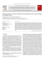
Figure 7. The 3 D docked view of vinorelbine in Tubulin (4O2B). The ligand interaction diagrams of receptor ligand complex are shown.
microtubular dynamics. Vinorelbine, a vinca alcoloid, func
tions as an anti-tubulin agent (Gregory & Smith, 2000).
However, the mechanism of action needs to be clarified in
detail. To enlight the vinorelbine-tubulin interaction and to
determine the binding modes and binding affinities of vinor
elbine towards tubulin, in this study, the docking studies of
vinorelbine into a,b-tubulin (PDB IDs: 4O2B and 1SA0) (Prota
et al., 2014; Ravelli et al., 2004) were performed. Vinorelbine
9676
S. CELIK ET AL.
Figure 8. The 3 D docked view of vinorelbine in tubulin (1SA0). The ligand interaction diagrams of receptor ligand complex are shown.
Table 5. The binding affinities and probable interactions of ligands with tubulin (1SA0) target�.
Ligand
Binding Affinity
(kcal/mol)
Binding free
Energy (kcal/mol)
Interacting Residues
FFSA
(Liao et al., 2015)
7.18
FFSA
(This study)
8.3
DAMA- Colchicine
(El-Naggar et al., 2020)
DAMA- Colchicine
(This study)
8.5
13.08
Thr179a (H-bonding)
Val181a (H-bonding)
Asn258b
Met259b(hydrophobic)
Val315b
Ala316b(hydrophobic)
Val318b(hydrophobic)
Asn350b
Lys352b(hydrophobic)
Ala354b
Asn101a (H-bonding)
Val181a (H-bonding)
Leu248b (Pi-Alkyl)
Ala250b (Pi-Alkyl)
Lys254b (Pi-Alkyl)
Asn258b (H-bonding)
Ala316b (Pi-sigma)
Ala317b
(Halogen-Fluorine)
Ala354b (Pi-Alkyl)
Ser178 (H-bonding)
Cys241 (H-bonding
and hydrophobic)
Leu248 (H-bonding)
Ala250 (Hydrophobic)
Leu255 (Hydrophobic)
Lys352 (H-bonding
and hydrophobic)
Vinorelbine
(This study)
8.0
17.59
Asn101a (H-bonding)
Ala180a (Carbon
H-bonding)
Val181a (H-bonding)
Cys241b (H-bonding
and pi-sulfur)
Leu248b (Pi-Alkyl)
Ala250b (Pi-Alkyl)
Leu255b (Alkyl
and pi-sigma)
Met259b (pi-sulfur)
Val315b (Carbon
H-bonding)
Lys352b (Carbon
H-bonding
and Pi-Alkyl)
Ser178a (H-bonding
and Carbon H-bonding)
Arg221a (Pi-Alkyl)
Pro222a (Carbon
H-bonding)
Tyr224a (H-bonding
and Pi-Alkyl)
Gln247b (Carbon
H-bonding)
Arg322b (Alkyl)
Met325b (Alkyl)
�
The same residues that found to interact in this study and in references were marked bold.
was docked into the colchicine binding site of tubulin (PDB
ID: 4O2B) to explore the interaction of vinorelbine in the
catalytic site of tubulin. In parallel, the molecular docking
simulation was also carried out with the X-ray crystal struc
ture of tubulin-colchicine: stathmin-like domain complex
(PDB ID: 1SA0). The active sites of the target proteins were
JOURNAL OF BIOMOLECULAR STRUCTURE AND DYNAMICS
9677
� hydrogen bond interactions with Arg66, at 2.64 and 2.67
Å bond lengths,
� alkyl interaction with Ala385, at 4.73 Å bond length,
� alkyl (5.24 Å) and pi-alkyl (3.89 Å) interactions with
Ala 389,
� alkyl interaction (4.46 Å) with Ala 426 and
� alkyl interaction (4.6 Å) with Lys430.
Figure 9. The 3 D docked view of vinorelbine (red circle) and 3-(4Fluorophenyl)-N-((4-fluorophenyl)sulphonyl)acrylamide (FFSA) (black circle) in
tubulin (1SA0).
Figure 10. The superposition of the docked views of vinorelbine, in tubulin tar
get, obtained by optimized structure (blue stick), obtained by the published
vinorelbine X-ray conformation (pink). The experimental location of vinorelbine
in vinorelbine tubulin complex is also shown (yellowish green).
searched using CAVER program (Jurcik et al., 2018) and were
defined in the grid size of 40 Å � 40 Å � 40 Å. A semi flexible
docking protocole was chosen, where the structure of ligand
molecule is flexible but targed protein is fixed.
The crystal structure of the tubulin-colchicine complex
was obtained from the protein data bank (PDB ID: 4O2B)
(Prota et al., 2014). The colchicine ligand and water mole
cules were removed, and polar hydrogens were added. The
optimized in gas phase structure of vinorelbine was adapted
for docking. The partial charges of the vinorelbine molecule
were computed using the Geistenger technique.
The docking results are shown in Figure 7. The binding
affinity is found as 9.4 kcal/mol. The docking into 4O2B
results indicate that vinorelbine interacts with residues
Arg46F, Leu47F, Arg51F, Arg66F, Ala385A, Ala389A, Ala426A,
and Lys430A in the A and F chains of tubulin.
Vinorelbine makes:
� Pi-cation; Pi-Donor hydrogen bond interaction with
Arg46F, at 4.04 Å bond length,
� carbon hydrogen bond interaction with Leu47, at 3.28 Å
bond length,
� unfavorable donor-donor and hydrogen bond interactions
with Arg51, at 1.99 Å and 2.45 Å bond lengths,
respectively.
In addition, we also docked the vinorelbine into the bind
ing site of tubulin (4O2B), determined for the hexa substi
tuted cyclotriphosphazenes CP 2-11-tubulun complexes
� an et al., 2022). In this case, vinorelbine interacted with
(Dog
the amino acids Arg61E, Leu68E, Arg158B, Glu196B, Asp199B,
Pro263B and Gly410A in the A, B and E chains of tubulin
with 7.9 kcal/mol binding affinity. Since the obtained bind
ing affinity was decreased in comparison to that of the previ
ous binding site, the tubuline-vinerolbine complex was
found more stable in docking into the active site found
using the CAVER program, with a binding affinity of
9.4 kcal/mol.
For the molecular docking simulation of vinorelbine into
tubulin-colchicine: stathmin-like domain complex (PDB code:
1SA0) (Ravelli et al., 2004), The crystal structure of target pro
tein was taken from Protein data bank (PDB ID: 1SA0) and,
was confirmed to the docking by removing ligand and water
molecules and adding polar hydrogens in it. The active sites
of 1SA0 were searched by the CAVER program (Jurcik et al.,
2018). As a result of the docking simulations, binding affinity
is found as 8.0 kcal/mol. The 3 D docked view of vinorel
bine in tubulin (1SA0) is shown in Figure 8.
Liao et al. (2015) investigated the interaction between
potential tubulin polimerisation inhibitor 3-(4-Fluorophenyl)N-((4-fluorophenyl)sulphonyl)acrylamide (FFSA) and tubulin
(PDB ID 1SA0) by docking simulations. Three binding modes
were determined, and it was reported that the binding mode
2 was the most favourable, with the lowest binding free
energy. Recently El-Naggar et al. (2020) docked the cocrystal
lized
ligand,
N-deacetyl-N-(2-mercaptoacetyl)-colchicine
(DAMA-colchicine) into tubulin (1SA0) and determined the
binding free energy as 13.08 kcal/mol. In our study, the
results of the docking protocol were validated by re-docking
of the reference molecules FFSA and DAMA-colchicine inside
the active sites of target (1SA0) and docking of vinorelbine
and reference molecules into 1SA0 were comparatively investi
gated. In Table 5 the binding affinities and ligand-target pro
tein interactions of vinorelbine, FFSA and DAMA-colchicine are
given in comparison with those of FFSA-tubulin (Liao et al.,
2015) and DAMA-colchicine (El-Naggar et al., 2020) complexes.
The docked FFSA was found to interact with Val181a,
Asn258b, Ala316b and Ala354b as the same as determined by
Liao et al. (2015). Moreover, the binding site of vinorelbine in
tubulin (1SA0) was found to close to that of FFSA in binding
mode 2, which was determined as the best binding mode for
FFSA (Liao et al., 2015). In this study the obtained vinorelbine
and FFSA binding sites into tubulin (1SA0), were shown com
paratively in Figure 9. Moreover, the binding free energy of
DAMA and colchicine docked into 1SA0 was calculated as
17.50 kcal/mol by using MM/PB(GB)SA approach.
9678
S. CELIK ET AL.
Table 6. Interactions of vinorelbine with CYP2D6 and CYP3A4 enzymes�.
PDB ID
Bonding
Affinity
(kcal/mol)
Interacted Residues and Interactions (bond lengths Å)
Hydrogen
bond
Phe481(1.85)
Carbon-Hydrogen
bond
Phe481(3.65)
Alky
Pi-Alky
Cys57(4.21,
4.36, 4.84)
Arg106(5.09)
Pro107(4.36)
Pro227(5.42, 5.49)
Pro107(4.18, 4.30)
Pro227(4.89)
Val376(4.52, 5.20)
Pro107(4.49)
Pro227(4.69)
Leu61(3.70, 4.36)
Arg64(5.39)
Pro107(3.91, 4.64)
Pro227(4.17)
Val376(4.82)
Tyr25(5.17)
Pro107(5.32)
Pro227(4.30, 4.68)
His28(5.43)
Pro107(4.11, 4.98)
Pro227(4.00)
Val376(4.77)
His28(5.43)
Pro107(4.02, 4.96)
Pro227(4.16)
Val376(4.65)
Phe108(5.23)
Leu211(5.11)
Phe57(5.33)
Phe108(5.43)
Phe213(4.48)
Phe241(4.56, 5.26)
Leu482(4.63)
Ile369(5.43)
Leu482(4.63)
2F9Q
9.2
1TQN
7.3
1W0E
7.1
1W0F
9.3
1W0G
7.9
Gln78(3.10)
Pro107(3.47)
Pro107(4.21)
Pro227(5.25, 5.40)
2V0M
10.6
3NXU
10.1
Glu374(3.67)
Gly481(3.52)
Arg105(3.32)
Arg106(3.10)
Pro107(3.50)
Ser119(3.33)
Thr224(3.59)
Gly481(2.34)
Ile223(5.36)
Leu482(4.85)
Met114(5.30)
Leu210(4.64)
Leu211(5.45)
Ile301(5.03)
Gln78(3.4)
Pro107(3.34)
Asn104(2.91)
Lys390(3.08)
Arg106(2.59)
Ser119(2.99)
Arg372(2.27)
Gln78(3.50)
Pro227(3.72)
Lys390(2.86)
Pro107(3.03)
Pi-Sigma
Pi-Pi
Unfavorable
D-D or A-A
Tyr25(1.46)
(D-D)
Phe220(3.96)
Phe57(3.91,5.0)
Phe108(4.25)
Glu374(2.87)
(A-A)
�Interaction bond lengths are given in parentheses in Å. A-A ¼ unfavorable acceptor-acceptor; D-D ¼ unfavorable Donor-Donor interactions. The binding residues
previously reported are marked as bold (Mannu et al., 2011; Marechal et al., 2006; Subhani & Jamil, 2015).
Figure 11. The 3 D docked view of vinorelbine in CYP2D6 (PDB ID: 2F9Q) (a), the interaction diagrams of vinorelbine-CYP2D6 complex (b-c) and the distance of
docked ligand from Heme groups (d).
Finally, vinorelbine was re-docked into the active site,
determined by the crystal structure of vinorelbine tubulin
complex (PFB ID 7CNN), by using the two initial structures o
the ligand: 1) The published vinorelbine X-ray conformation
(Chengyong et al., 2021) and 2) the optimized geometry of
vinorelbine obtained in this study. When the published vinor
elbine X-ray conformation was used as initial ligand geom
etry, the binding affinity was found to be 9.2 kcal/mol,
whereas, when optimized vinorelbine geometry was used,
9.5 kcal/mol binding affinity was revealed in the docking
JOURNAL OF BIOMOLECULAR STRUCTURE AND DYNAMICS
9679
Figure 12. The docking results of vinorelbine into CYP3A4 enzyme structures of 1TQN (a-d) and 1W0E (e-h).
simulations. The increase in binding affinity when the opti
mized structure is used instead of the published vinorelbine
X-ray conformation indicates the importance of the initial
structure of the ligand. In Figure 10, the superposition of the
3 D docked view of vinorelbine in optimized geometry (blue
stick) and in the published vinorelbine X-ray conformation
(pink stick) are shown in comparison with that of obtained
experimentally in the X-ray structure of the vinorelbine-tubu
lin complex (yellowish green stick). As seen in Figure 10, the
docked view of the optimized structure of vinorelbine (blue
molecule) is compatible to those found experimentally in
vinorelbine-tubulin complex (yellowish green molecule).
The approaches of molecular mechanical energies com
bined with Poisson-Boltzmann or generalized Born and surface
area continuous solving (MM/PBSA and MM/GBSA) methods
which are based on molecular dynamics simulations of the
receptor-ligand complex, are widely used to estimate the bind
ing free energy of small ligands to proteins (Genheden & Ryde,
2015). Since both MM/PBSA and MM/GBSA approaches are
important for binding free energy calculations, Wang et al.
(2019) developed a program by the combination of both
methods, called MM/PB(GB)SA approach. In this study the
binding free energy of vinorelbine with 4O2B, 1SA0 and 7CNN
were calculated by using MM/PB(GB)SA approach with the
General Amber Force Field2 (GAFF2) and Force Field14 Stony
Brook (ff14SB) force field combination and the GB6 procedure
(Wang et al., 2019), and are obtained as 28.5, 17.59 and
50.39 kcal/mol, respectively.
The lowest binding free energy (-50.39 kcal/mol) and best
binding affinity (-9.5 kcal/mol) of vinorelbine were estimated by
9680
S. CELIK ET AL.
Figure 13. The docking results of vinorelbine into CYP3A4 enzyme of 1W0F (a-d) and 1W0G (e-h) structures.
modelling studies for vinorelbine-7CNN system, when optimized
geometry of vinorelbine was used as initial geometry for docking.
Recently Boichuk et al. (2021) calculated free binding
energies of various ligands docked into the colchicine bind
ing site of tubulin (PDB ID: 4O2B) by using MM/GBSA
approach. The binding free energy of BAL27862 (Avanpulin),
a microtubule-destabilizing agent that binds to the colchi
cine site of tubulin (Prota et al., 2014), was also calculated
for comparison. It was reported that the estimated binding
free energy of BAL27862-4O2B system as 32.60 kcal/mol,
whereas the binding free energy of the various ligands were
span from 6.22 to 16.82 kcal/mol. In our study, the bind
ing free energy of vinorelbine-4O2B system was found as
28.5 kcal/mol, indicating that vinorelbine could compete
with BAL27862 in colchicine binding site of 4O2B.
The modeling studies of vinorelbine against tubulin indi
cated that the alkyl, pi-alkyl interactions and hydrogen bond
ing with the protein residues helps to stabilize the binding
of vinorelbine in the colchicine-binding domain of
a,b-tubulin interface.
JOURNAL OF BIOMOLECULAR STRUCTURE AND DYNAMICS
9681
Figure 14. The docking results of vinorelbine into CYP3A4 enzyme of 2V0M (a-d) and 3NXU(e-h) structures.
Molecular docking of vinorelbine into cytochorome P450
CYP3A4 and CYP2D6 enzymes
The side effects of vinca alkaloids are known to be associ
ated with their metabolism (Lokwani et al., 2020). Most
important drugs metabolized by cytochrome P450 enzymes
and the CYP2D6 and CYP3A4 are the two most significant
cytochrome enzymes. Beulz-Riche et al. (2005) investigated
in vitro metabolism of vinorelbine on liver microsomes tested
for their CYP activities and it was determined that CYP3A4
was the main enzyme involved in the hepatic metabolism
and bitransformation of vinorelbine in human, and CYP2D6
is not involved in metabolism of vinorelbine. Topletz et al.
(2013) investigated the metabolism of vinorelbine using
cDNA-expressed human cytochrome P450s and human liver
9682
S. CELIK ET AL.
microsomes. It was reported that depending on in vitro find
ings CYP3A enzymes are predicted to metabolize vinorelbine.
To enlighten the metabolic behavior of vinorelbine, dock
ing simulations against cytochrome P450, CYP2D6 and
CYP3A4 enzymes were performed, docking modes, and dock
ing affinities were determined.
The crystal structure of substrate free Human Cytochrome
P450 2D6 (CYP2D6) (Rowland et al., 2006) was obtained from
Protein Data Bank ( PDB ID: 2F9Q) .
By eliminating water molecules from CYP2D6 and replacing
them with polar hydrogens, the protein was conformed to
the docking. The optimized in gas phase structure of vinorel
bine molecule was adapted. The Geistenger technique was
used to compute the partial charges of the vinorelbine mol
ecule. The active site of CYP2D6 was determined using a
40 � 40 � 40 Å3 grid size. Vinorelbine bound to CYP2D6
(2F9Q) target with intermolecular hydrogen, carbon hydro
gen, alkyl, and Pi-alkyl interactions with Cys57, Leu61, Arg64
and Phe481 residues of CYP2D6. The binding affinity (DG)
was determined as 9.2 kcal/mol. In Table 6 the interactions
between vinorelbine and 2F9Q target were tabulated. In
Figure 11 the 3 D docked view of vinorelbine, its interactions
with CYP2D6 (PDB ID: 2F9Q) and the distance of docked lig
and from heme groups were shown. The docking simulations
revealed that the best binding site of vinorelbine was quite
far from Heme. Our findings are in accord with the in vitro
experimental results indicating that CYP2D6 enzyme is not
involve in vinorelbine metabolism (Beulz-Riche et al., 2005).
Although vinorelbine bound to 2F9Q with high binding affin
ity, since its binding site is far away from Fe atom of heme,
it can not undergo enzymatic oxidation.
CYP3A4 is a highly flexible enzyme and undergoes con
formational changes depending on the ligand with which it
is being co-crystallized (Lokwani et al., 2020). There are many
available X-ray structures in the protein database of the
CYP3A4 enzyme, as apo (ligand unbound) protein structures
(PDB IDs: 1TQN, 1W0E) (Williams et al., 2004; Yano et al.,
2004) and ligand-bound crystal structures fPDB IDs: 1W0F
(Progesterone), 1W0G (Metyrapone), 2V0M (Ketoconazole),
€gren, 2006; Sevrioukova
and 3NXU (Ritonavir)g (Ekroos & Sjo
& Poulos, 2010; Williams et al., 2004). In the available two
apo CYPA4 crystal structures (PDB IDs 1TQN and 1W0E) the
main difference was found to be in the orientation of
Arg212. In 1W0E, Arg 212 is orientated away from the heme
group, and in another structure 1TQN, it occupies the orien
tation toward the heme group (Fishelovitch et al., 2007). On
the other hand, when all available X-ray models of ligand
bound structures of CYP3A4 were superimposed, it was
shown that the conformational changes took place primarily
in the F-G- and C-terminal loop regions, the I-helix adjacent
to the heme and the 369–371 peptide (Sevrioukova &
Poulos, 2015).
In this study the CYP3A4 crystal structures, those with
PDB IDs 1TQN (Yano et al., 2004), 1W0E, 1W0F, 1W0G
€gren, 2006), and
(Williams et al., 2004), 2V0M (Ekroos & Sjo
3NXU (Sevrioukova & Poulos, 2010) are used in molecular
docking studies and the crystal structures were obtained
from Protein Data Bank ( The
binding affinities, interactions and interacted residues were
tabulated in Table 6. The docking results of vinorelbine into
CYP3A4 structures were shown in Figures 12–14. The func
tional sites of CYP3A4 enzyme include Arg 105, Arg 106, Phe
108, Ser 119, Arg 212, Phe 215, Phe 303, Ala 304, Glu 307,
Thr 308, Ala 369, Met 370, Arg 371, Leu 372, Glu 373 and
Arg 374 as binding residues (Mannu et al., 2011; Marechal
et al., 2006; Subhani & Jamil, 2015). As seen in Table 6, the
interacted residues of CYP3A4 structures with vinorelbine are
in accord to pevious findings.
In the case of docking of vinorelbine into the ligand
unbound structure of CYP3A4 (1TQN), binding affinity was
found as 7.1 kcal/mol and interacted with Gln78, Arg106,
Pro107, Pro227 and Val376 residues of target protein (see
Table 6). Subhani and Jamil (2015) investigated the interac
tions of the gemcitabine, carboplatin and cisplatin anticancer
molecules with CYP3A4 (1TQN) by molecular docking simula
tions. It was reported that gemcitabine and carboplatin inter
acted with Arg 106 and Pro107 molecules with which
vinorelbine interacted. These results indicate that the binding
site of vinorelbine to the 1TQN target is close to the binding
site of gemcitabine and carboplatin (Subhani & Jamil, 2015).
The in silico docking investigations shown that vinorelbine
binds most strongly to 2V0M among CYP 3A4 enzyme struc
tures (DG ¼ 10.6 kcal/mol), followed by the 3NXU structure
(DG ¼ 10.1 kcal/mol). The distance smallest distance
between the catalytic site of vinorelbine and the Fe atom of
heme in both structures was 6.92 Å (2V0M) and
7.75 Å (3NXU).
Conclusions
In this study, conformational preferencies of vinorelbine mol
ecule, a vinca alkaloid, that widely used as a chemometric
drug were investigated and the obtained most stable con
former was then optimized at DFT/B3LYP/6-31G(d,p) level of
theory. The equilibrium geomety parameters, harmonic
wavenumbers, HOMO–LUMO and MEP calculations of vinor
elbine molecule were carried out for the first time using the
same level of theory. The recorded experimental IR, Raman
and UV-Vis spectra were computed to those corresponding
theoretical results and found that these theoretical results
are in good agreement with the experimental data.
The in silico screening of vinorelbine against the target
protein, a,b-tubulin (PDB IDs: 4O2B; 1SA0), revealed high
binding affinities ffor PDB ID: 4O2B; DG ¼ 9.4 kcal/mol and
PDB ID: 1SA0; DG ¼ 8.0 kcal/molg, indicating that vinorel
bine has good anti-tumor properties.
In addition, to enlighten the hematic metabolism of vinor
elbine, the interaction of vinorelbine with the cytochrome
P450 enzymes (CYP), was investigated by docking studies
against CYP2D6 and CYP3A4 targets. High binding affinities
to CYP2D6 (-9.2 kcal/mol) and to CYP3A4 (from 7.1 to
10.6 kcal/mol) indicate that cytochrome P450 enzymes are
good targets for vinorelbine.
We hope that our results provide further insights into the
bioactivity and hematic metabolism of the anticancer drug
vinorelbine.
JOURNAL OF BIOMOLECULAR STRUCTURE AND DYNAMICS
Acknowledgement
We thank to G. A. Zhurko for allowing us to use Chemcraft
demo software.
Disclosure statement
No potential conflict of interest was reported by the authors.
Funding
This work was supported by the Scientific Research
€
Coordination Unit of Istanbul University [ONAP-2423].
Projects
References
Altinoz, M. A., Ozpinar, A., Alturfan, E. E., & Elmaci, I. (2018). Vinorelbine’s
anti-tumor actions may depend on the mitotic apoptosis, autophagy
and inflammation: Hypotheses with implications for chemo-immuno
therapy of advanced cancers and pediatric gliomas. Journal of
Chemotherapy (Florence, Italy), 30(4), 203–212.
Aronson, J. K. (Ed.). (2015). Meyler’s side effects of drugs: The international
encyclopedia of adverse drug reactions and interactions. Elsevier.
Becke, A. (1993). Density-functional thermochemistry. III. The role of
exact exchange. Journal of Chemical Physics, 98(7), 5648–5652. https://
doi.org/10.1063/1.464913
Beulz-Riche, D., Grude�, P., Puozzo, C., Sautel, F., Filaquier, C., Rich�
e, C., &
Ratanasavanh, D. (2005). Characterization of human cytochrome P450
isoenzymes involved in the metabolism of vinorelbine. Fundamental
and Clinical Pharmacology, 19(5), 545–553. />1472-8206.2005.00367.x
Boichuk, S., Galembikova, A., Syuzov, K., Dunaev, P., Bikinieva, P.,
Aukhadieva, A., Zykova, S., Igidov, N., Gankova, K., Novikova, M., &
Kopnin, P. (2021). The design, synthesis, and biological activities of
pyrrole-based carboxamides: The novel tubulin ınhibitors targeting
the colchicine-binding site. Molecules, 26(19), 5780. />3390/molecules26195780
�mez-Zavaglia, A., & Fausto, R. (2009). Molecular structure,
Borba, A., Go
infrared spectra, and photochemistry of isoniazid under cryogenic
conditions. The Journal of Physical Chemistry. A, 113(32), 9220–9230.
Borkakoti, N., & Palmer, R. A. (1978). The structure of the bisbenzylisoqui
noline alkaloid methylwarifteine. Acta Crystallographica Section B
Structural Crystallography and Crystal Chemistry, 34(2), 490–495.
/>�mezBueno, O., Gallego, J. E., Martins, S., Prota, A. E., Gago, F., Go
SanJuan, A., Camarasa, M.-J., Barasoain, I., Steinmetz, M. O., D�ıaz, J. F.,
P�
erez-P�erez, M.-J., Liekens, S., & Priego, E.-M. (2018). High-afnity
ligands of the colchicine domain in tubulin based on a structureguided design. Scientific Reports, 8(1), 4242. />s41598-018-22382-x
Campos, J. A., Casado, J., & Rios, M. A. (1980). The geometry of the phe
nyl ring in substituted benzene molecules. A molecular orbital
approach. Journal of the American Chemical Society, 102(5),
1501–1504. />Celik, S., Ozel, A. E., & Akyuz, S. (2016). Comparative study of antitumor
active cyclo (Gly-Leu) dipeptide: A computational and molecular mod
eling study. Vibrational Spectroscopy, 83, 57–69. />1016/j.vibspec.2016.01.007
Celik, S., Akyuz, S., & Ozel, A. E. (2022a). Molecular modeling, DFT quan
tum chemical analysis, and molecular docking on edotecarin, an indo
locarbazole anticancer agent. Molecular Crystals and Liquid Crystals,
1–23. />Celik, S., Vagifli, F., Akyuz, S., Ozkok, F., E. Ozel, A., Dosler, S., & Onul, N.
(2022). Synthesis, vibrational spectroscopic investigation, molecular
docking, antibacterial and antimicrobial studies of a new anthraquin
one derivative compound. Spectroscopy Letters, 55(4), 259–277.
/>
9683
Celik, S., Yilmaz, G., Ozel, A. E., & Akyuz, S. (2022). Structural and spectral
analysis of anticancer active cyclo (Ala–His) dipeptide. Journal of
Biomolecular Structure & Dynamics, 40(2), 660–672. />1080/07391102.2020.1817150
Celik, S., Akyuz, S., & Ozel, A. E. (2022b). Vibrational spectroscopic charac
terization and structural investigations of Cepharanthine, a natural
alkaloid. Journal of Molecular Structure, 1258, 132693. />10.1016/j.molstruc.2022.132693
Celik, S., Ozkok, F., Ozel, A. E., Cakir, E., & Akyuz, S. (2021). Synthesis, FTIR and NMR characterization, antibacterial and antioxidant activities,
and DNA docking analysis of a new vanillin-derived imine compound.
Journal of Molecular Structure, 1236, 130288. />molstruc.2021.130288
Celik, S., Akyuz, S., & Ozel, A. E. (2017). Vibrational spectroscopic and
structural investigations of bioactive molecule Glycyl-Tyrosine (GlyTyr). Vibrational Spectroscopy, 92, 287–297. />vibspec.2017.08.007
Celik, S., Albayrak, A. T., Akyuz, S., E., & Ozel, A. (2019). Molecular model
ling and vibrational investigations of ammonium-based ionic liquid
(CLTOAB). Journal of Biomolecular Structure & Dynamics, 37(10),
2515–2526.
€
€ Durgun, M., T€
Ceylan, U.,
urkmen, H., Yalc¸ın, S¸. P., Kilic, A., & Ozdemir,
N.
(2015). Theoretical and experimental investigation of 4-[(2-hydroxy-3methylbenzylidene) amino] benzenesulfonamide: Structural and spec
troscopic properties, NBO, NLO and NPA analysis. Journal of Molecular
Structure, 1089, 222–232. />042
Chengyong, W., Jinghong, X., Yanyan, W., Qing-Jie, X., Lingling, M.,
Yuyan, L., Hai, C., Qian, L., Quan, Z., Bo, S., & Yuxi, W. (2021). The
high-resolution X-ray structure of vinca-domain inhibitors of microtu
bules provides a rational approach for drug design. FEBS Letters,
595(2), 195–205. />Chi, S., Xie, W., Zhang, J., & Xu, S. (2015). Theoretical insight into the
structural mechanism for the binding of vinblastine with tubulin.
Journal of Biomolecular Structure & Dynamics, 33(10), 2234–2254., &,
()., (), -. />Cox, E. G., Cruickshank, D. W. J., & Smith, J. A. S. (1958). The crystal struc
ture of benzene at -3 � C. Proceedings of the Royal Society of London.
Series A. Mathematical and Physical Sciences, 247(1248), 1–21.
Debernardis, A., Alfonsi, G., Santacroce, L., Bottalico, L., Sabatini, R., Flace,
P., Vermesan, D., Auteri, P., & Cagiano, R. (2009). Antiviral properties
of antineoplastic drugs. From Herpes simplex-1 disappearance to a
wide antiviral action: A serendipity case report. Acta Bio-Medica:
Atenei Parmensis, 80(3), 265–267.
€kce, H., Dog
�an, O. E., Alpaslan, G., Ag
� ar, T., Muthu, S., & Sert,
Dege, N., Go
Y. (2022). Quantum computational, spectroscopic investigations on N(2-((2-chloro-4, 5-dicyanophenyl) amino) ethyl)-4-methylbenzenesulfo
namide by DFT/TD-DFT with different solvents, molecular docking
and drug-likeness researches. Colloids and Surfaces A: Physicochemical
and Engineering Aspects, 638, 128311. />surfa.2022.128311
Devi, T., & R., Gayathri, S. (2010). FTIR and FT-Raman spectral analysis of
paclitaxel drugs. International Journal of Pharmaceutical Sciences
Review and Research, 2(2), 106–110.
� an, H., Bahar, M. R., C¸alıs¸kan, E., Tekin, S., Uslu, H., Akman, F., Koran,
Dog
€rg€
K., Sandal, S., & Go
ul€
u, A. O. (2022). Synthesis and spectroscopic
characterizations of hexakis [(1-(40 -oxyphenyl)-3-(substituted-phenyl)
prop-2-en-1-one)] cyclotriphosphazenes: Their in vitro cytotoxic activ
ity, theoretical analysis and molecular docking studies. Journal of
Biomolecular Structure and Dynamics, 40(7), 3258–3272. https://doi.
org/10.1080/07391102.2020.1846621
� lu, E. (2016).
Durgun, M., Yalc¸ın, S¸. P., T€
urkmen, H., Akkurt, M., & Erog
Structural study of 4-(2-morpholinoethanoylamino)-benzenesulfona
mide by X-ray diffraction technique and DFT calculations. Bulgarian
Chemical Communications, 48(1), 5–12.
€ useven,
� lence-Bakır, S., Celik, S., S¸ahin, M., Ozel, A. E., Akyuz, S., & Ulk€
Eg
B. (2021). Synthesis, molecular modelling, FT-IR, Raman and NMR
characterization, molecular docking and ADMET study of new nickel
(II) complex with an N4-tetradentate thiosemicarbazone. Journal of
Biomolecular Structure & Dynamics, 39(12), 4212–4224.
9684
S. CELIK ET AL.
El-Naggar, A. M., Eissa, I. H., Belal, A., & El-Sayed, A. A. (2020). Design,
eco-friendly synthesis, molecular modeling and anticancer evaluation
of thiazol-5 (4 H)-ones as potential tubulin polymerization inhibitors
targeting the colchicine binding site. RSC Advances, 10(5), 2791–2811.
/>€gren, T. (2006). Structural basis for ligand promiscuity in
Ekroos, M., & Sjo
cytochrome P450 3A4. Proceedings of the National Academy of
Sciences,
103(37),
13682–13687.
/>0603236103
Fishelovitch, D., Hazan, C., Shaik, S., Wolfson, H. J., & Nussinov, R. (2007).
Structural dynamics of the cooperative binding of organic molecules
in the human cytochrome P450 3A4. Journal of the American
Chemical Society, 129(6), 1602–1611.
Flores-Holgu�ın, N., Frau, J., & Glossman-Mitnik, D. (2019). Conceptual DFT
as a chemoinformatics tool for the study of the Taltobulin anticancer
peptide. BMC Research Notes, 12(1), 1–4. />s13104-019-4478-7
Frisch, M. J., Trucks, G. W., Schlegel, H. B., Scuseria, G. E., Robb, M. A.,
Cheeseman, J. R., Scalmani, G., Barone, V., Petersson, G. A., Nakatsuji,
H., Li, X., Caricato, M., Marenich, A. V., Bloino, J., Janesko, B. G.,
Gomperts, R., Mennucci, B., Hratchian, H. P., Ortiz, J. V., … Fox, D. J.
(2016). Gaussian 16, Revision C.01. Gaussian, Inc.
Genheden, S., & Ryde, U. (2015). The MM/PBSA and MM/GBSA methods
to estimate ligand-binding affinities. Expert Opinion on Drug Discovery,
10(5), 449–461. />Gregory, R. K., & Smith, I. E. (2000). Vinorelbine–A clinical review. British
Journal of Cancer, 82(12), 1907–1913.
Gunasekaran, S., Seshadri, S., & Muthu, S. (2006). Vibrational spectra and
normal coordinate analysis of flucytosine. Indian Journal of Pure &
Applied Physics, 44, 581–586.
Hajduchova, Z., Pach, L., & Lokaj, J. (2018). Adsorption of dodecylbenze
nesulfonic acid on the alumina particles in the preparation of alumina
�ty, 62(2), 138–145.
foam. Ceramics–Silika
Hansch, C., Sammes, P. G., & Taylor, J. B. (1990). Comprehensive medicinal
chemistry: The rational design, mechanistic study & therapeutic applica
tions of chemical compounds (Vol. 5). Pergamon Press.
Istvan, K. (2002). Simirra, A program for simulation of IR and Raman
Spectra. Chemical Research Center. (It was obtained from Dr. Gabor
Keresztury in Chemical Research Center in Budapest).
Jiao, A., Tian, S., & Lin, H. (2022). Analysis of outburst coal structure char
acteristics in Sanjia coal mine based on FTIR and XRD. Energies, 15(6),
1956. />Jurcik, A., Bednar, D., Byska, J., Marques, S. M., Furmanova, K., Daniel, L.,
Kokkonen, P., Brezovsky, J., Strnad, O., Stourac, J., Pavelka, A., Manak,
M., Damborsky, J., & Kozlikova, B. (2018). CAVER Analyst 2.0: Analysis
and visualization of channels and tunnels in protein structures and
molecular dynamics trajectories. Bioinformatics (Oxford, England),
34(20), 3586–3588. />Kaneda, T., & Tanaka, J. (1976). The crystal structure of the
ıntermolecular complex between 9-ethyladenine and ındole. Bulletin
of the Chemical Society of Japan, 49(7), 1799–1804. />1246/bcsj.49.1799
Kaya Kınayt€
urk, N., Kalaycı, T., & Tunalı, B. (2021). Experimental and com
putational investigations on the molecular structure, vibrational spec
tra, electronic properties, and molecular electrostatic potential
analysis of phenylenediamine isomers. Spectroscopy Letters, 54(9),
693–706. />Klots, T. D., & Collier, W. B. (1995). Heteroatom derivatives of indene Part
3. Vibrational spectra of benzoxazole, benzofuran, and indole.
Spectrochimica Acta Part A: Molecular and Biomolecular Spectroscopy,
51(8), 1291–1316. />Kutlu, E., Emen, F. M., Kismali, G., Kınayt€
urk, N. K., Kılıc¸, D., Karacolak,
A. I., & Demirdogen, R. E. (2021). Pyridine derivative platinum com
plexes: Synthesis, molecular structure, DFT and initial anticancer activ
ity studies. Journal of Molecular Structure, 1234, 130191. https://doi.
org/10.1016/j.molstruc.2021.130191
Laurent, G. J., & Shapiro, S. D. (Eds.). (2006). Encyclopedia of respiratory
medicine (1st ed.). Academic Press.
Liao, S. Y., Chen, J. C., Mo, G. Q., Zhang, C., & Zheng, K. C. (2015). A com
putational study of binding between 3-(4-fluorophenyl)-N-((4-
fluorophenyl) sulphonyl) acrylamide and tubulin. Molecular Simulation,
41(4), 356–364. />Lokwani, D. K., Sarkate, A. P., Karnik, K. S., Nikalje, A. P. G., & Seijas, J. A.
(2020). Structure-based site of metabolism (SOM) prediction of ligand
for CYP3A4 enzyme: Comparison of Glide XP and Induced Fit Docking
(IFD).
Molecules,
25(7),
1622.
/>molecules25071622
Mannu, J., Jenardhanan, P., & Mathur, P. P. (2011). A computational study
of CYP3A4 mediated drug interaction profiles for anti-HIV drugs.
Journal of Molecular Modeling, 17(8), 1847–1854. />1007/s00894-010-0890-6
Marechal, J.-D., Yu, J., Brown, S., Kapelioukh, I., Rankin, E. M., Wolf, C. R.,
Roberts, G. C. K., Paine, M. J. I., & Sutcliffe, M. J. (2006). In silico and
in vitro screening for inhibition of cytochrome P450 CYP3A4 by come
dications commonly used by patients with cancer. Drug Metabolism
and Disposition: The Biological Fate of Chemicals, 34(4), 534–538.
Maren, T. H. (1976). Relations between structure and biological activity
of sulfonamides. Annual Review of Pharmacology and Toxicology, 16(1),
309–327. />Mariappan, G., & Sundaraganesan, N. (2015). Structural and vibrational
spectroscopic analysis of anticancer drug mitotane using density
functional theory. Materials Today: Proceedings, 2(3), 965–968.
McQueen, C. (2010). Comprehensive toxicology(2nd Ed.). Elsevier.
€ & Ozpozan,
€
Mıhc¸ıokur, O.,
T. (2017). Molecular structure, vibrational
spectroscopic analysis (IR & Raman), HOMO-LUMO and NBO analysis
of anti-cancer drug sunitinib using DFT method. Journal of Molecular
Structure, 1149, 27–41. />Muthu, S., Maheswari, J. U., & Sundius, T. (2013). Molecular structural,
non-linear optical, second order perturbation and Fukui studies of
Indole-3-Aldehyde
using
density
functional
calculations.
Spectrochimica Acta Part A: Molecular and Biomolecular Spectroscopy,
106, 299–309. />Ngan, V. K., Bellman, K., Panda, D., Hill, B. T., Jordan, M. A., & Wilson, L.
(2000). Novel actions of the antitumor drugs vinflunine and vinorel
bine on microtubules. Cancer Research, 60(18), 5045–5051.
O’boyle, N. M., Tenderholt, A. L., & Langner, K. M. (2008). Cclib: A library
for package-independent computational chemistry algorithms. Journal
of Computational Chemistry, 29(5), 839–845.
Pangajavalli, S., Ranjithkumar, R., Sridhar, B., & Ramaswamy, S. (2017).
Hirshfeld and vibrational analysis of 5-Benzyl-7a-Hydroxy-1-Methyl2,3,5,6,7,7a-Hexahydro-1h-3a, 7-Methanoindeno [2,1-d]Pyrrolo[3,2c]Azepine-12,13(4h)-Dione. Mechanics, Materials Science & Engineering
Journal, Magnolithe, 12, 1–9.
Pearson, R. G. (1973). Hard and soft acids and bases (Vol. 2). Van
Nostrand Reinhold.
Politzer, P., & Murray, J. S. (2002). The fundamental nature and role of
the electrostatic potential in atoms and molecules. Theoretical
Chemistry Accounts, 108(3), 134–142. />Prota, A. E., Danel, F., Bachmann, F., Bargsten, K., Buey, R. M., Pohlmann,
J., Reinelt, S., Lane, H., & Steinmetz, M. O. (2014). The novel micro
tubule-destabilizing drug BAL27862 binds to the colchicine site of
tubulin with distinct effects on microtubule organization. Journal of
Molecular Biology, 426(8), 1848–1860. />2014.02.005
Pulay, P., Fogarasi, G., Pongor, G., Boggs, J. E., & Vargha, A. (1983).
Combination of theoretical ab initio and experimental information to
obtain reliable harmonic force constants. Scaled quantum mechanical
(QM) force fields for glyoxal, acrolein, butadiene, formaldehyde, and
ethylene. Journal of the American Chemical Society, 105(24),
7037–7047. />Ravelli, R. B., Gigant, B., Curmi, P. A., Jourdain, I., Lachkar, S., Sobel, A., &
Knossow, M. (2004). Insight into tubulin regulation from a complex
with colchicine and a stathmin-like domain. Nature, 428(6979),
198–202. />Roeges, N. P., & Baas, J. M. A. (1994). A guide to the complete interpret
ation of infrared spectra of organic structures (pp. 1–340). Wiley.
Rowland, P., Blaney, F. E., Smyth, M. G., Jones, J. J., Leydon, V. R.,
Oxbrow, A. K., Lewis, C. J., Tennant, M. G., Modi, S., Eggleston, D. S.,
Chenery, R. J., & Bridges, A. M. (2006). Crystal structure of human