Advanced technologies and polymer materials for surgical sutures
Bạn đang xem bản rút gọn của tài liệu. Xem và tải ngay bản đầy đủ của tài liệu tại đây (11.84 MB, 312 trang )
CHAPTER 1
Advances in biopolymer based
surgical sutures
Blessy Joseph1, Jemy James2, Nandakumar Kalarikkal3 and
Sabu Thomas3
1Business Innovation and Incubation (BIIC), Mahatma Gandhi University, Kottayam, Kerala, India;
2University Bretagne Sud, Lorient, France; 3International and Inter University Centre for Nanoscience
and Nanotechnology, Mahatma Gandhi University, Kottayam, Kerala, India
1.1 Introduction
Over the years, there has been a dramatic growth of the wound closure
market. Traditionally materials like silk, cotton, horsehair, animal tendons
and intestines, and wire made of precious metals were in operative pro-
cedures. The limitations and risks associated with such wound closure
devices demanded the need for efficient and cost-effective techniques for
wound healing. Although there have been significant advances in tissue
adhesives and other mechanical wound closure devices, sutures have been
the preferred choice for surgeons. Sutures can be defined as the materials
used to uphold tissues together normally after a trauma or surgery [1]. They
can be natural or synthetic materials that can provide adequate mechanical
strength during tissue fixation. The art of suturing can be found in the
Egyptian mummified resins, in which they have used woolen threads, plant
fibers, hair, and tendons. Suturing techniques were documented in 500
BCE (Before Common Era) by Indian surgeon Sushruta in “Sushruta
Samhita [2].” Metal wires were first applied in the human body by French
physicists Lapayode and Sicre in 1775 to set a broken humerus (upper arm
bone)[3].
A fundamental change was witnessed following Second World War,
after which polymer sutures and stainless steel became superior. The se-
lection of suture material is dependent on the physical and biological
characteristics of the suture as well as the type of tissue to be healed. Sutures
are made from synthetic or natural polymers. Synthetic polymers are not
readily degradable. They accumulate and can have a long-term detrimental
effect on ecosystems. The tunable physical characteristics of biopolymers
make them a reliable material for the fabrication of sutures. Biopolymers
can be obtained from natural sources or synthesized chemically from
Advanced Technologies and Polymer Materials for Surgical Sutures © 2023 Elsevier Ltd.
ISBN 978-0-12-819750-9
All rights reserved. 1
2 Advanced Technologies and Polymer Materials for Surgical Sutures
biological material or entirely biosynthesized by living organisms[4]. They
are easily biodegradable as they are obtained from renewable sources. The
term “biodegradation” generally refers to degradation by microorganisms.
The polymer is broken down into carbon dioxide and water which forms
food for microorganisms[5]. Biopolymers as surgical sutures have gained
considerable attention because of their unique properties like biocompat-
ibility and biodegradability. Biopolymers can adopt more precise and
defined 3D shapes and structures when compared to synthetic polymers
having more simple and random organization[6]. This makes biopolymers
attractive for in vivo applications. They are generally classified into three
categories based on the nature of repeating units they are composed of
(i) polysaccharides, often carbohydrate structures (cellulose, chitin, starch,
alginate, etc.); (ii) polypeptides made of amino acids (collagen, actin), and
(iii) polynucleotides deoxynucleic acid (DNA) and ribonucleic acid (RNA))
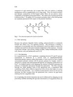
(Fig. 1.1). This chapter intends to provide an overview of the biopolymers
used for suture fabrication, their physical and biological properties, and how
these properties facilitate wound repair. Sterilization techniques used for
sutures have also been discussed in this chapter.
1.2 Polymers as suture materials
In the past sutures made of natural materials like dried animal gut, animal
hair (e.g., horse hair), silk, tendons, and plant fibers (e.g., linen, cotton)
were widely used [7,8]. The technological advancements in polymer sci-
ence paved way for the development of sutures with diverse materials
having excellent mechanical and physical properties. There has been a
large-scale expansion and evolution of the research and business in the area
of materials for biomedical applications. Still, sutures and staples are the
most used material in the biomedical industry. Sutures are to be used in
many cases where natural wound closure is difficult and external
Figure 1.1 Classification of biopolymers according to their structure.
Advances in biopolymer based surgical sutures 3
reinforcement is highly essential. Biopolymer based absorbable sutures are
much preferred to nonabsorbable sutures. Sutures are generally classified as
absorbable and nonabsorbable based on whether they degrade or not after
performing the intended function. Nonabsorbable sutures need to be
removed by doctors, hence causing additional discomfort to patients.
Whereas absorbable sutures degrade within the body usually by hydrolysis
or with the aid of proteolytic enzymes[9].
1.3 Biopolymers
Environmental problems arise with the continued use of synthetic poly-
mers. Intensive research has been carried out in this direction, possibly
replacing synthetic polymers with natural ones. As mentioned above,
biopolymers are obtained from biological sources. Hence, the use of bio-
polymers offers an eco-friendly approach. They are decomposed by mi-
croorganisms or natural processes like availability of moisture, sunlight, etc.
which is environmentally friendly when compared to petroleum based
synthetic polymers releasing toxic byproducts into the surroundings. Bio-
polymers are employed in diversified fields such as food packaging, drug
delivery, tissue engineering, etc. Although they are biocompatible, many of
them lack sufficient mechanical properties desired for medical applications.
Most often they are crosslinked or modified with materials like glutaral-
dehyde, citric acid, poly (carboxylic acids), and so forth[10]. Crosslinkers
like glutaraldehyde can be cytotoxic hence greener approaches are also
being explored. Nanoparticles are also used to enhance the properties of
biopolymers. The interaction between biopolymers and nanoparticles re-
sults in nanocomposites with improved functionalities like antimicrobial
property, tensile strength, thermal stability, or water resistance. Many re-
searchers have investigated the ability of silver nanoparticles (AgNPs ) to
improve the antimicrobial properties of biopolymers, wherein cost-effective
methodologies could be formulated for developing wound dressings or
food packaging films. Cellulose paper coated with silver-gold nanoparticles
displayed improved antibacterial activity against E.coli [11]. Another work
reported the synthesis of silver-cellulose hybrids which showed excellent
antibacterial activity against E.coli and S.aureus whereas pure cellulose
(Microcrystalline cellulose) didn’t exhibit any activity against the respective
microbial strains[12]. Although several biopolymers find promising appli-
cations in the biomedical sector, we will be concentrating on polymers like
cellulose, collagen, silk, chitosan, chitin, polyhydroxyalkanoates (PHA), and
PLA which particularly fit well for the suture industry.
4 Advanced Technologies and Polymer Materials for Surgical Sutures
Cellulose is the naturally occurring homopolymer consisting of b-1, 4
linked glucan chains. Being inherently biodegradable and low-cost material,
cellulose finds immense application in healthcare [13]. Cellulose materials
try to self-assemble and form an extended network by both intramolecular
and intermolecular hydrogen bonds, which makes them relatively stable.
Chitin is a sustainable biopolymer due to its abundance. Structurally,
chitin is N-acetyl glucosamine and the main source are crustaceans like
crabs, shellfish, etc. The deacylated form of chitin known as chitosan
consists of N-acetyl glucosamine and glucosamine moieties. Both chitin and
chitosan are versatile enough to be processed to any form like sponges, gels,
or scaffolds, thereby finding many applications in tissue engineering and
drug delivery[14]. Natural silk fibers are produced by arthropods like
silkworms or spider. Mulberry silkworms (Bombyx mori) are most
commonly reared to produce silk. They have a core-shell structure con-
sisting of 3 components, a heavy chain fibroin, a light chain fibroin, and a
third small glycoprotein, known as the P25 protein. These proteins are
coated with hydrophilic sericins. Silk materials are used as sponges, films, or
sutures for applications like ligament tissue engineering, hepatic tissue en-
gineering, cartilage tissue engineering, and so on [15e17]. Poly(lactic acid)
(PLA) is a biodegradable polyester produced from the monomer, lactic acid
(LA) by mechanisms like direct polycondensation (DP) and ring-opening
polymerization (ROP). The tunable physicochemical properties and
biocompatibility of PLA make it suitable for biomedical applications.
Collagen is a major structural protein in animals and forms a vital part of the
extracellular matrix. It provides tensile strength to tendons and ligaments
and also elasticity to the skin. It has a 3D architecture comprising of a right-
handed bundle of three parallel, left-handed poly proline II-type helices
[18]. Source of collagen includes bovine skin and tendons, porcine skin,
marine organisms like sponges, fish, and jelly fish. It is used for soft tissue
repair, dental applications, and as scaffolds for tissue engineering [19,20].
Polyhydroxyalkanoates (PHA) are naturally synthesized polyesters accu-
mulated as energy storage material inside the cellular structure of various
microorganisms.
1.4 Biopolymers for sutures
1.4.1 Collagen
Collagen nanofibrils (CoNF) have a great potential for being mechanically
strong but biodegradable sutures. They play a major role in tissue
Advances in biopolymer based surgical sutures 5
engineering as being the key component of the extracellular matrix. It is the
most abundant protein in the human body and imparts structural integrity
and strength to the tissues[21]. The use of collagen as a modern biomaterial
began in 1881. Joseph Lister and William Macewen (Fig. 1.2) reported the
advantages of catgut, a collagen-rich biomaterial prepared from the small
intestine of sheep[23]. Untreated catgut sutures are often processed from
dead animal tissue, hence causing infections[24]. They are often used in the
case of subcutaneous or fatty tissue[25]. Collagen sutures were modified
with heparin for sustained release of platelet-derived growth factor-BB
(PDGF-BB). Tendon-derived cells seeded on PDGF-BB incorporated
collagen sutures showed 50% greater proliferation than untreated collagen
sutures[26]. This could be because collagen provides active chemical sites
for conjugating growth factors. Collagen has also been used to coat surgical
sutures to improve their functionalities. Polyester/polyethylene sutures
coated with collagen were evaluated for their response to bone and tendon
cells[27]. Collagen coating was found to stimulate proliferation and adhe-
sion of cells in collagen coated sutures when compared to uncoated one.
1.4.2 Polylactic acid (PLA)
PLA is one of the most popular biodegradable and bio-based polymers.
PLA is used to prepare biodegradable polymer sutures [28]. The biocom-
patibility of the polymers has been extensively studied, and it has been
proven to be one of the best biopolymers for biomedical applications like
sutures etc. [29]. PLA is a polymer derived from LA and its structure makes
it easily breakable during metabolism and thereby making it easier to be
excreted from the body [30]. Degradation occurs through enzymatic or
hydrolytic scission of ester bonds. The degradation of PLA depends on its
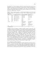
Figure 1.2 SEM images of PLA suture loaded with PM-Ds: (A) 100 Â times, (B) 1000Â
times. (Reproduced with permission from Ref. [22].)
6 Advanced Technologies and Polymer Materials for Surgical Sutures
molecular weight, crystallinity, presence of fillers, etc. Recently, Liu et al.
reported the fabrication of PLA sutures loaded with PLA microspheres
containing drug[22]. Initially PLA microspheres containing drug genta-
micin sulfate was prepared (PM-Ds). Further, this drug loaded microspheres
were loaded onto the PLA sutures (PM-Ds/PLA). The mechanical prop-
erties were analyzed which showed an increase in the properties of the drug
loaded suture when compared to the neat suture. A sustained release of the
drug up to 8 days could be achieved. As evident from the scanning electron
microscopy images, the microspheres entered the gaps of the suture fibers,
and stuck to them firmly which could have resulted in the prolonged
release of the drug (Fig. 1.2).
In another study, biopolymers like chitosan, alginate, and the blends of
these polymers were coated on the surface of PLA sutures. The mechanical
studies were carried out. Some of the drugs based on antibiotic sensitivity
was chosen and was introduced into the sutures using surface treatment
method like dip coating. The drug release studies and antimicrobial activity
proved that the drug-coated bio polymeric sutures were effective in wound
closing and wound healing [31]. Poor biocompatibility and cellular affinity
are major problem encountered with PLA sutures. To improve the surface
hydrophilicity, PLA sutures were initially treated with lipase followed by
grafting with chitosan [32]. It’s evident from the SEM images that initially
the untreated sutures had a smooth surface. Once grafted with chitosan, in
some places chitosan united and led to a rougher surface and large friction
coefficient. However, hydrophilicity was greatly improved.
Blends of PLA and polycaprolactone compatibilized with Ethyl Ester L-
Lysine Triisocyanate (LTI) were melt-spun to produce suture threads of
diameter 0.3 mm 1.0 phr of LTI was found to be the most suitable
composition for producing sutures, at higher loadings the sutures were too
rigid. The suture threads didn’t induce any bacterial growth [33].
1.4.3 Silk
Silk is a protein polymer whose characteristics are slow degradation and
good mechanical strength. Silk is preferred for cardiovascular, neurological,
and ophthalmic procedures [34]. The ease of handling and improved knot
security properties makes silk superior among other sutures. But their use is
hindered due to the high inflammatory reactions posed by them [35,36].
Bacterial attachment to silk sutures was compared to commercially available
Monocryl Plus suture [37]. From Fig. 1.3 it is evident that the
Advances in biopolymer based surgical sutures 7
Figure 1.3 Scanning electron microscope images of (A) silk suture knot material and
(B) Monocryl Plus suture knot material. Microorganisms and cellular detritus are highly
visible in silk sutures. (Reproduced with permission from Ref. [37].)
microorganisms were highly colonized around the suture knot of silk suture
when compared to that of Monocryl Plus suture.
Maintaining sterile conditions in the wound has always been a hurdle
after suturing. Medical devices and sutures contribute about 45% of
nosocomial infections or hospital-acquired infections [38]. Antibacterial
sutures play a pivotal role in combating surgical site infections[39]. Once a
biofilm is formed on the surface of a suture, it becomes resistant to tradi-
tional antimicrobials.
Once bacteria colonize a suture, local methods to treat bacterial in-
fections become inadequate. Hence, several strategies to prevent bacterial
adherence have been proposed by researchers including the addition of
antibiotics, nanoparticles, biomaterials, etc. Sutures impregnated with an-
tibiotics have been found to prevent the adherence of bacteria and biofilm
formation [40].
Tetracycline hydrochloride (TCH), a bacteriostatic drug is found to
exhibit activity against a wide range of gram-positive and gram-negative
microorganisms[41]. The efficacy of TCH-treated sutures was studied by
Viju and Thilagavathi [42]. As was expected, untreated silk sutures promote
the growth of E.coli and S.aureus.
Synergistic chitosan and TCH drug was exploited to develop antimi-
crobial silk sutures for preventing microbial infections [43]. Such combi-
nations can provide a prolonged antibacterial effect. AgNPs have been
widely used as an antibacterial agent[44,45]. AgNPs exhibits their antimi-
crobial potential through various mechanisms. The anchoring of AgNPs to
8 Advanced Technologies and Polymer Materials for Surgical Sutures
microbial cells, followed by penetration into the cells, reactive oxygen
species and free radical generation, and modulation of microbial signal
transduction pathways have been recognized as the most prominent ways of
antimicrobial action [46]. AgNPs were coated on silk sutures to impart
antibacterial properties [47]. Mechanical strength was retained after the
addition of AgNPs; however, a significant reduction in bacterial growth
was achieved. Cytotoxicity studies using 3T3 mouse embryonic fibroblast
cells showed 82% cell viability for silver treated samples. This showed that
the silver treatment did not affect their proliferative capacity.
Surface modification of silk fibroin suture, AASF (antheraea assama,
popularly known as golden silk; found only in certain parts of Assam) was
achieved by grafting polypropylene (PP) onto silk fibroin sutures[48]. Here
the sutures were first sterilized using argon and then low-temperature
plasma grafting of PP onto sterilized sutures was done to achieve the
desired biofunctionalities. Here the modified suture showed more
biocompatibility and improved wound healing when compared to the
untreated ones. In vivo studies were conducted in three groups. The first
group was sutured with AASF, the second with argon plasma-treated AASF
(AASFAr) and the third group with PP grafted AASF sutures (PP-AASF).
The histopathology studies on the 14th postoperative day show the pres-
ence of inflammatory cells in group A characterized by lesser collagen
formation (Fig. 1.4). Group B shows a considerably fair amount of collagen
formation with slight infiltration in and around hair follicles. Whereas PP-
AASF sutured group (Group C) shows highly accelerated wound healing
activity. Moreover, a greater amount of hair follicles was also present when
compared to the other groups.
Figure 1.4 Histologic evaluation of wound healing on 14th postoperative day. His-
topathological section of the sample collected from the incised wound of (A) group A,
(B) group B, and (C) group C animals shows inflammatory cell inflammation (IN) in and
around the hair follicle (HF) as well as subepidermal tissue. Proliferation of fibrous
connective tissue (CT) indicates faster healing of group B and group C as compared to
group A animals. (Reproduced with permission from Ref. [48].)
Advances in biopolymer based surgical sutures 9
1.4.4 Chitin & chitosan
Chitin and chitosan are polymers derived from marine animals and is some
of the most available biopolymers other than cellulose. However, some of
the challenges make its usage cumbersome [49]. Though chitin is highly
biocompatible, nontoxic, and biodegradable, along with its antimicrobial
effect, there are still more challenges to overcome to exploit its huge po-
tential for prospective applications [50]. Chitosan is a potent antimicrobial
agent and its antimicrobial activity can be attributed to its cationic nature
[51]. The positively charged chitosan molecules interact with negatively
charged microbial cell membranes leading to the disruption of the microbial
membrane [52]. Sutures were fabricated from chitin having good me-
chanical strength [53]. No allergic reactions or inflammation was seen. The
chitin suture was absorbed in about 4 months in rat muscles. The accel-
erated degradation can be mainly due to the action of lysozyme. Chitin
nanofibrils are used as nanofillers for reinforcing polymers to obtain
nanocomposites with enhanced stability, especially in the case of bio-
resorbable sutures [54]. Chitosan stimulates tissue regeneration and prevents
scar formation. The mechanical strength of chitosan is very low; hence, it is
mainly exploited as suture coatings. Chitosan has been used for coating silk
sutures [55]. Silk sutures coated with chitosan also showed excellent anti-
bacterial efficacy [56]. A modified derivative of chitosan known as hydroxyl
propyl trimethyl ammonium chloride (HACC) chitosan coated on Vicryl
suture showed excellent antibacterial activity and also displayed good
biocompatibility [57]. HACC is a water-soluble modified derivative of
chitosan that exhibits good antibacterial activity [58,59]. HACC coated
sutures effectively prevented biofilm formation when compared to
triclosan-coated sutures. Prabha et al. showed that extracted chitosan (EC)
from crab shells showed higher inhibition of biofilm formed by mixed
species[60]. The antibacterial and antifungal effects of Vicryl absorbable
sutures coated with chitosan, uncoated sutures, and commercially available
triclosan-coated sutures were studied against S. epidermidis and C. albicans
(Fig. 1.5). The uncoated suture (control) and sutures soaked with acetic acid
(Vehicle control) did not show any antibacterial or anticandidal activity.
The commercial triclosan-coated sutures exhibited only antibacterial ac-
tivity and did not show any anticandidal activity. EC immobilized sutures
exhibited good antimicrobial activity against both strains compared to
commercially available chitosan (CC).
10 Advanced Technologies and Polymer Materials for Surgical Sutures
Figure 1.5 Antimicrobial activities of the impregnated suture against S. epidermidis
and C. albicans: (A) Control; (B) VC; (C) EC (400 mg/mL); (D) CC (400 & 450 mg/mL); (E)
Triclosan coated Reproduced with permission from Ref. [60].
1.4.5 Polyhydroxyalkanoate (PHA)
PHA is a microbial polyester having excellent biocompatibility and
biodegradability. Poly(3-hydroxybutyrate) (PHB) is the most widespread
member of the polyhydroxyalkanoate family and is produced under un-
balanced growth conditions like depletion of essential nutrients such as
nitrogen, phosphorus, or magnesium [61]. Poly (4-hydroxybutyrate)
(P4HB) is a typical PHA type used for the fabrication of surgical materials.
The most well-known product, and the first approved by the US Food and
Drug Administration, is the TephaFLEX suture fabricated from P4HB [62].
In vivo studies of PHA sutures implanted intramuscularly over 1 year
showed that animals that received the sutures were in good health condi-
tion during the period of study. No adverse reactions were observed, and
functional characteristics of the animals were also not affected [63]. Poly-
hydroxyalkanoate sutures decreased tendency to curl were fabricated by
extrusion and orientation of the fibers [64]. The resulting fibers had an
elongation to break from about 17% to about 85% and Young’s modulus of
less than 350,000 psi. He et al. evaluated the biocompatibility of mono-
filament made from poly (3-hydroxybutyrate-co-3-hydroxyhexanoate)
(PHBHHx) and a multifilament made from poly(3-hydroxybutyrate-co-3-
hydroxyvalerate) (PHBV) and PLA blend [65]. The PHBHHx fiber and the
PHBV/PLA fiber showed remarkable biocompatibility to be used as sur-
gical sutures.
Advances in biopolymer based surgical sutures 11
1.4.6 Cellulose
Plants are the major source of cellulose, the most abundant and easily
available carbohydrate polymer on earth. Bacterial cellulose, an alternate
and highly purified form of cellulose is produced by aerobic bacteria,
mainly of the genus Acetobacter [66]. Their unique nanostructure, excel-
lent water retaining capacity, good mechanical strength, and high crystal-
linity makes them preferred choice in biomedical applications [67].
Bacterial cellulose nanocrystals (BCNC) were used to reinforce chitin (RC)
fibers to form BCNC/RC yarns[68]. The fibers were produced by wet
spinning technology for application as surgical sutures. In vitro studies
showed good biocompatibility and in vivo studies revealed good wound
healing with BC coated yarns. However, the knot pull tensile strength of all
coated yarns was lower than uncoated ones.
Oxidized cellulose is highly biocompatible and has great antibacterial
properties against a variety of pathogens. The ability of oxidized cellulose as
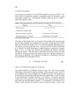
suture material was studied by Li et al. who explored the effect of tempo
oxidation treatment on the physical and mechanical properties of TORC
(TEMPO-mediated oxidation of regenerated cellulose) sutures [69]. The
carboxyl content in the suture materials was controlled by varied oxidation
times. It could be seen that TEMPO oxidation significantly influenced the
degradation of sutures as evaluated from the hydrolysis test performed by
immersing the sutures in physiological saline for 7, 14, 21, and 28 days
(Fig. 1.6). The carboxyl groups introduced in the sutures due to TEMPO
Figure 1.6 The in the vitro degradation rate of TORC and different TORC at different
oxidation times (15, 30, 45, 60, and 90 min after PBS impregnation (mean ặ S.D.,
n ẳ 10)). (Reproduced with permission from [69].)
12 Advanced Technologies and Polymer Materials for Surgical Sutures
oxidation leads to increased molecular chain spacing and reduction in
molecular interatomic force whereby water easily penetrates the fiber
resulting in breakage.
1.5 Sterilization of sutures
Sterilization technology plays a prominent role in the biomedical field
because bacterial colonization remains a big problem with medical implants or
devices. Every surgical procedure is associated with a certain risk of
contamination and hence the sutures must be well sterilized to prevent
bacterial adherence. Despite the widespread use of sutures in the 19th century,
suture associated infections were a major concern. Lord Joseph Lister made
remarkable contributions to the history of sterilization [2]. He pointed out
that whatever be the cause of wound infection, carbolic acid could prevent or
halt its further progress [70].In 1869, he developed aseptic silk sutures treated
with carbolic acid followed by sutures from sheep intestine known as catgut
sutures(catgut sutures treated with 5% chromic acid).
Claudius introduced the concept of using potassium iodide for suture
sterilization in 1902, and the infection rate was further reduced [71]. A process
for sterilizing catgut sutures and ligatures using heat was invented in 1958 [72].
Here catgut sutures and ligatures sealed in a container in the presence of
aqueous isopropanol solution were sterilized by heat. Ethicon Inc. started
using electron beam accelerators for suture sterilization in 1957 [73].
According to the European Norm 556 sterility is defined as the state of
being free from viable microorganisms ( 1 Â 10e6)[74]. Generally, sterili-
zation techniques can be classified as physical and chemical. Physical steriliza-
tion involves sterilization using heat and radiations whereas chemical
sterilization involves the use of chemicals like ethylene oxide, hydrogen
peroxide, formaldehyde, b-propiolactone, etc. Certain sterilization procedures
result in stiffening of sutures and hence the selection of appropriate sterilization
techniques is critical. The tensile strength of Virgin silk suture treated by
thermal methods of sterilization was found to decrease [75]. Sterilization of
collagen sutures with b-propiolactone showed no significant loss of strength of
the finished sutures, hence can be used as an alternative to heat sterilization[76].
1.6 Conclusion and future perspectives
The growing environmental concerns have led to increased research in the
field of biopolymers. Natural polymers or biopolymers are derived from
Advances in biopolymer based surgical sutures 13
living organisms. These have the added advantage of being biodegradable,
biocompatible, renewable, and reduced antigenicity. Sutures are materials
of immense importance in the biomedical field. Along with ease of
handling and biocompatibility, the mechanical properties of suture materials
are a major factor that affects the overall suture quality.
Despite the modern technological advancements in the materials in the
methodology perspectives, biopolymeric sutures have an important role in
wound healing. The progressive techniques like onsite evaluation of the
wound healing, easy to use sutures or wound closure methods, smart sutures,
and other wound closure devices and products are to be looked up in the
future. No sutures can be called ideal as such. The vital concern is surgical site
infection after surgery. Although antibacterial sutures delivering antibiotics
have been developed, maintaining all desirable biological and morphological
features in a single suture is still a matter of research. The growing misuse of
antibiotics requires more alternatives to combat surgical site infections.
The merging of the biopolymeric sutures and nanotechnology will give
a boost to the surgical industry where properties like better antimicrobial
activity, faster wound healing, etc. could be achieved. The inventions and
innovations in suture fabrication have a huge potential to be applied for the
betterment of the patients and making their lives better.
References
[1] C. Dennis, S. Sethu, S. Nayak, L. Mohan, Y.Y. Morsi, G. Manivasagam, Suture
materials - current and emerging trends, J. Biomed. Mater. Res. A 104 (2016)
1544e1559, />
[2] T.M. Muffly, A.P. Tizzano, M.D. Walters, The history and evolution of sutures in
pelvic surgery, J. R. Soc. Med. 104 (2011) 107e112, /> jrsm.2010.100243.
[3] J.M. Seitz, M. Durisin, J. Goldman, J.W. Drelich, Recent advances in biodegradable
metals for medical sutures: a critical review, Adv. Healthc. Mater. 4 (2015) 1915e1936,
/>
[4] A.M. Smith, S. Moxon, G.A. Morris, Biopolymers as wound healing materials, in: Wound
Heal. Biomater, 2016, />
[5] N.R. Nair, V.C. Sekhar, K.M. Nampoothiri, A. Pandey, Biodegradation of bio-
polymers, Curr. Dev. Biotechnol. Bioeng. (2016), /> 444-63662-1.00032-4.
[6] S. Mohan, O.S. Oluwafemi, N. Kalarikkal, S. Thomas, S.P. Songca, Biopolymers e
application in nanoscience and nanotechnology, in: Recent Adv. Biopolym, 2016,
/>
[7] P.P. Dattilo Jr., P.P. Dattilo Jr., M.W. King, M.W. King, N.L. Cassill, N.L. Cassill,
J.C. Leung, J.C. Leung, Medical textiles: application of an absorbable barbed bi-
directional surgical suture, J. Text. Appar. Technol. Manang. 2 (2002).
14 Advanced Technologies and Polymer Materials for Surgical Sutures
[8] A. Goel, Surgical sutures - a review, Delhi J. Ophthalmol. (2016), /> 10.7869/djo.161.
[9] B. Joseph, A. George, S. Gopi, N. Kalarikkal, S. Thomas, Polymer sutures for
simultaneous wound healing and drug delivery e a review, Int. J. Pharm. 524 (2017),
/>
[10] N. Reddy, R. Reddy, Q. Jiang, Crosslinking biopolymers for biomedical applications,
Trends Biotechnol. (2015), />
[11] T.T. Tsai, T.H. Huang, C.J. Chang, N. Yi-Ju Ho, Y.T. Tseng, C.F. Chen, Anti-
bacterial cellulose paper made with silver-coated gold nanoparticles, Sci. Rep. 7 (2017)
1e10, />
[12] Y.Y. Dong, L.H. Fu, S. Liu, M.G. Ma, B. Wang, Silver-reinforced cellulose hybrids
with enhanced antibacterial activity: synthesis, characterization, and mechanism, RSC
Adv. (2015), />
[13] N.K. Blessy Joseph, H.J. Maria, S. Thomas, Nanocellulose: health care applications, in:
M. Mishra (Ed.), Encycl. Polym. Appl, CRC Press, 2018, pp. 1829e1852.
[14] A. Anitha, S. Sowmya, P.T.S. Kumar, S. Deepthi, K.P. Chennazhi, H. Ehrlich,
M. Tsurkan, R. Jayakumar, Chitin and chitosan in selected biomedical applications,
Prog. Polym. Sci. (2014), />
[15] Y. Wang, D.J. Blasioli, H.J. Kim, H.S. Kim, D.L. Kaplan, Cartilage tissue engineering
with silk scaffolds and human articular chondrocytes, Biomaterials (2006), https://
doi.org/10.1016/j.biomaterials.2006.03.050.
[16] J. Chen, G.H. Altman, V. Karageorgiou, R. Horan, A. Collette, V. Volloch,
T. Colabro, D.L. Kaplan, Human bone marrow stromal cell and ligament fibroblast
responses on RGD-modified silk fibers, J. Biomed. Mater. Res. 67A (2003) 559e570,
/>
[17] L. Meinel, S. Hofmann, V. Karageorgiou, L. Zichner, R. Langer, D. Kaplan,
G. Vunjak-Novakovic, Engineering cartilage-like tissue using human mesenchymal
stem cells and silk protein scaffolds, Biotechnol. Bioeng. 88 (2004) 379e391, https://
doi.org/10.1002/bit.20252.
[18] M.D. Shoulders, R.T. Raines, Collagen structure and stability, Annu. Rev. Biochem.
(2009), />
[19] R. Parenteau-Bareil, R. Gauvin, F. Berthod, Collagen-based biomaterials for tissue
engineering applications, Materials (2010), />
[20] R. Khan, M.H. Khan, Use of collagen as a biomaterial: an update, J. Indian Soc.
Periodontol. (2013), />
[21] S.A. Sell, M.J. McClure, K. Garg, P.S. Wolfe, G.L. Bowlin, Electrospinning of
collagen/biopolymers for regenerative medicine and cardiovascular tissue engineering,
Adv. Drug Deliv. Rev. 61 (2009) 1007e1019, /> j.addr.2009.07.012.
[22] S. Liu, G. Wu, X. Zhang, J. Yu, M. Liu, Y. Zhang, P. Wang, X. Yin, J. Zhang, F. Li,
M. Zhang, Preparation and properties of poly (lactic acid) (PLA) suture loaded with
PLA microspheres enclosed drugs (PM-Ds), J. Text. Inst. (2019), /> 10.1080/00405000.2019.1610999.
[23] S. Chattopadhyay, R.T. Raines, Collagen-based biomaterials for wound healing,
October 141 (2008) 520e529, />
[24] R.G.C. Gerard, V. Yu, Sutures and anchoring devices, in: McGlamry’s Compr. Textb.
Foot Ankle Surg, 2001, pp. 139e174.
[25] E.J. Holder, The story of catgut, Postgrad. Med. (1949), /> pgmj.25.287.427.
[26] M. Younesi, B.O. Donmez, A. Islam, O. Akkus, Heparinized collagen sutures for
sustained delivery of PDGF-BB: delivery profile and effects on tendon-derived cells in-
vitro, Acta Biomater. (2016), />
Advances in biopolymer based surgical sutures 15
[27] A.D. Mazzocca, M.B. McCarthy, C. Arciero, A. Jhaveri, E. Obopilwe, L. Rincon,
J. Wyman, G.A. Gronowicz, R.A. Arciero, Tendon and bone responses to a collagen-
coated suture material, J. Shoulder Elbow Surg. (2007), />
j.jse.2007.02.113.
[28] K. Hamad, M. Kaseem, H.W. Yang, F. Deri, Y.G. Ko, Properties and medical ap-
plications of polylactic acid: a review, Express Polym. Lett. 9 (2015).
[29] L. Avérous, Synthesis, properties, environmental and biomedical applications of pol-
ylactic acid, in: Handb. Biopolym. Biodegrad. Plast. Prop. Process. Appl, 2012, https://
doi.org/10.1016/B978-1-4557-2834-3.00009-4.
[30] R. Langer, A. Basu, A.J. Domb, Special issue: polylactide (PLA) based biopolymers,
Adv. Drug Deliv. Rev. (2016), />
[31] O.L. Shanmugasundaram, R.V.M. Gowda, D. Saravanan, Drug release and antimi-
crobial studies on polylactic acid suture, Int. J. Biotechnol. Mol. Biol. Res. 2 (2011)
80e89. />
7BDBFE141300.
[32] S. Liu, M. Liu, G. Wu, X. Zhang, J. Yu, Y. Zhang, P. Wang, X. Yin, Enhanced
surface hydrophilicity of polylactic acid sutures treated by lipase and chitosan, Textil.
Res. J. (2018), />
[33] A. Visco, C. Scolaro, A. Giamporcaro, S. De Caro, E. Tranquillo, M. Catauro, Threads
made with blended biopolymers: mechanical, physical and biological features, Poly-
mers (2019), />
[34] G.H. Altman, F. Diaz, C. Jakuba, T. Calabro, R.L. Horan, J. Chen, H. Lu,
J. Richmond, D.L. Kaplan, Silk-based biomaterials, Biomaterials 24 (2003) 401e416,
/>
[35] R.H.H. Tan, R.J.W. Bell, B.A. Dowling, A.J. Dart, Suture materials: composition and
applications in veterinary wound repair, Aust. Vet. J. (2003), />
j.1751-0813.2003.tb11075.x.
[36] F. Javed, M. Al-Askar, K. Almas, G.E. Romanos, K. Al-Hezaimi, Tissue reactions to
various suture materials used in oral surgical interventions, ISRN Dent. (2012), https://
doi.org/10.5402/2012/762095.
[37] S. Sala-Pérez, M. López-Ramírez, M. Quinteros-Borgarello, E. Valmaseda-Castellón,
C. Gay-Escoda, Antibacterial suture vs silk for the surgical removal of impacted lower
third molars. A randomized clinical study, Med. Oral Patol. Oral Cir. Bucal. 21 (2016)
e95ee102, />
[38] C.L. He, Z.M. Huang, X.J. Han, Fabrication of drug-loaded electrospun aligned
fibrous threads for suture applications, J. Biomed. Mater. Res. A (2009), https://
doi.org/10.1002/jbm.a.32004.
[39] E. Laas, C. Poilroux, C. Bézu, C. Coutant, S. Uzan, R. Rouzier, E. Chéreau,
Antibacterial-coated suture in reducing surgical site infection in breast surgery: a
prospective study, Int. J. Breast Cancer (2012), />
[40] D. Leaper, P. Wilson, O. Assadian, C. Edmiston, M. Kiernan, A. Miller, G. Bond-
Smith, J. Yap, The role of antimicrobial sutures in preventing surgical site infection,
Ann. R. Coll. Surg. Engl. 99 (2017) 439e443, />
rcsann.2017.0071.
[41] I. Chopra, M. Roberts, Tetracycline antibiotics: mode of action, applications, mo-
lecular biology, and epidemiology of bacterial resistance, Microbiol. Mol. Biol. Rev. 65
(2001) 232e260, />
[42] S. Viju, G. Thilagavathi, Characterization of tetracycline hydrochloride drug incor-
porated silk sutures, J. Text. Inst. (2013), />
00405000.2012.720758.
[43] S. Viju, G. Thilagavathi, Fabrication and characterization of silk braided sutures, Fibers
Polym. 13 (2012) 782e789, />
16 Advanced Technologies and Polymer Materials for Surgical Sutures
[44] R. Najafi-taher, B. Ghaemi, S. Kharazi, S. Rasoulikoohi, A. Amani, Promising anti-
bacterial effects of silver nanoparticle-loaded tea tree oil nanoemulsion: a synergistic
combination against resistance threat, AAPS PharmSciTech (2017), /> 10.1208/s12249-017-0922-y.
[45] A.J. Kora, R.B. Sashidhar, Biogenic silver nanoparticles synthesized with rhamnoga-
lacturonan gum: antibacterial activity, cytotoxicity and its mode of action, Arab. J.
Chem. (2018), />
[46] T.C. Dakal, A. Kumar, R.S. Majumdar, V. Yadav, Mechanistic basis of antimicrobial
actions of silver nanoparticles, Front. Microbiol. (2016), /> fmicb.2016.01831.
[47] S. De Simone, A.L. Gallo, F. Paladini, A. Sannino, M. Pollini, Development of silver
nano-coatings on silk sutures as a novel approach against surgical infections, J. Mater.
Sci. Mater. Med. 25 (2014) 2205e2214, />
[48] D. Gogoi, A.J. Choudhury, J. Chutia, A.R. Pal, M. Khan, M. Choudhury, P. Pathak,
G. Das, D.S. Patil, Development of advanced antimicrobial and sterilized plasma
polypropylene grafted MUGA (antheraea assama) silk as suture biomaterial, Bio-
polymers 101 (2014) 355e365, />
[49] D. Elieh-Ali-Komi, M.R. Hamblin, Chitin and chitosan: production and application
of versatile biomedical nanomaterials, Int. J. Adv. Res. 4 (2016) 411e427.
[50] C.K.S. Pillai, W. Paul, C.P. Sharma, Chitin and chitosan polymers: chemistry, solu-
bility and fiber formation, Prog. Polym. Sci. (2009), /> j.progpolymsci.2009.04.001.
[51] S. Ahmed, S. Ikram, Chitosan based scaffolds and their applications in wound healing,
Achiev. Life Sci. 10 (2016) 27e37, />
[52] T. Dai, M. Tanaka, Y.Y. Huang, M.R. Hamblin, Chitosan preparations for wounds
and burns: antimicrobial and wound-healing effects, Expert Rev. Anti Infect. Ther.
(2011), />
[53] M. Nakajima, K. Atsumi, K. Kifune, K. Miura, H. Kanamaru, Chitin is an effective
material for sutures, Jpn. J. Surg. (1986), />
[54] I.P. Dobrovol’skaya, I.A. Kasatkin, V.E. Yudin, E.M. Ivan’kova, V.Y. Elokhovskii,
Supramolecular structure of chitin nanofibrils, Polym. Sci. A 57 (2015) 52e57, https://
doi.org/10.1134/S0965545X15010022.
[55] D. Sudha, B. Dhurai, T. Ponthangam, Development of herbal drug loaded antimi-
crobial silk suture, Indian J. Fibre Text. Res. 42 (2017) 286e290.
[56] S. Viju, G. Thilagavathi, Effect of chitosan coating on the characteristics of silk-braided
sutures, J. Ind. Textil. (2013), />
[57] Y. Yang, S.B. Yang, Y.G. Wang, S.H. Zhang, Z.F. Yu, T.T. Tang, Bacterial inhibition
potential of quaternised chitosan-coated VICRYL absorbable suture: an in vitro and
in vivo study, J. Orthop. Transl. 8 (2017) 49e61, /> j.jot.2016.10.001.
[58] L. Marcotte, J. Barbeau, M. Lafleur, Permeability and thermodynamics study of qua-
ternary ammonium surfactants - phosphocholine vesicle system, J. Colloid Interface
Sci. (2005), />
[59] M. Crismaru, L.A.T.W. Asri, T.J.A. Loontjens, B.P. Krom, J. De Vries, H.C. Van Der
Mei, H.J. Busscher, Survival of adhering staphylococci during exposure to a quaternary
ammonium compound evaluated by using atomic force microscopy imaging, Anti-
microb. Agents Chemother. (2011), />
[60] S. Prabha, J. Sowndarya, P.J.V.S. Ram, D. Rubini, B. Hari, W. Aruni, P. Nithyanand,
Chitosan-coated surgical sutures prevent adherence and biofilms of mixed microbial
communities, Curr. Microbiol. 78 (2021) 502e512.
[61] J. Chee, S. Yoga, N. Lau, S. Ling, R.M.M. Abed, Bacterially produced poly-
hydroxyalkanoate ( PHA ): converting renewable resources into bioplastics, in: Current
Advances in biopolymer based surgical sutures 17
Research, Technology and Education Topics in Applied Microbiology and Microbial
Biotechnology, 2010, pp. 1395e1404.
[62] Brigham, Sinskey, Applications of polyhydroxyalkanoates in the medical industry, Int. J.
Biotechnol. Wellness Ind. (2012), />[63] E.I. Shishatskaya, T.G. Volova, A.P. Puzyr, O.A. Mogilnaya, S.N. Efremov, Tissue
response to the implantation of biodegradable polyhydroxyalkanoate sutures, J. Mater.
Sci. Mater. Med. (2004), />[64] S. Rizk, Non-curling Polyhydroxyalkanoate Sutures, US8084125, 2011.
[65] Y. He, Z. Hu, M. Ren, C. Ding, P. Chen, Q. Gu, Q. Wu, Evaluation of PHBHHx
and PHBV/PLA fibers used as medical sutures, J. Mater. Sci. Mater. Med. 25 (2014)
561e571.
[66] M. Moniri, A. Boroumand Moghaddam, S. Azizi, R. Abdul Rahim, A. Bin Ariff,
W. Zuhainis Saad, M. Navaderi, R. Mohamad, Production and status of bacterial
cellulose in biomedical engineering, Nanomaterials 7 (2017) 257, /> 10.3390/nano7090257.
[67] F. Esa, S.M. Tasirin, N.A. Rahman, Overview of bacterial cellulose production and
application, Agric. Agric. Sci. Procedia 2 (2014) 113e119, /> j.aaspro.2014.11.017.
[68] H. Wu, G.R. Williams, J. Wu, J. Wu, S. Niu, H. Li, H. Wang, L. Zhu, Regenerated
chitin fibers reinforced with bacterial cellulose nanocrystals as suture biomaterials,
Carbohydr. Polym. 180 (2018) 304e313, /> j.carbpol.2017.10.022.
[69] H. Li, F. Cheng, C. Chávez-Madero, J. Choi, X. Wei, X. Yi, T. Zheng, J. He,
Manufacturing and physical characterization of absorbable oxidized regenerated cel-
lulose braided surgical sutures, Int. J. Biol. Macromol. (2019), /> j.ijbiomac.2019.05.030.
[70] M. Worboys, Joseph Lister and the performance of antiseptic surgery, Notes Record
Roy. Soc. Lond. (2013), />[71] A. Davey, C.S. Ince, Fundamentals of Operating Department Practice, 2015, https://
doi.org/10.1017/CBO9781316529874.
[72] B. Alfred, Sterilization of Surgical Catgut Sutures and Ligatures, 2832664, 1958.
[73] R. Singh, D. Singh, A. Singh, Radiation sterilization of tissue allografts: a review,
World J. Radiol. (2016), />[74] M.J. Abreu, M.E. Silva, L. Schacher, D. Adolphe, Recycling of textiles used in the
operating theatre, Recycl. Textil. (2006) 183e202, /> 9781845691424.4.183.
[75] G.N. Shuttleworth, L.F. Vaughn, H.B. Hoh, Material properties of ophthalmic sutures
after sterilization and disinfection, J. Cataract Refract. Surg. 25 (1999) 1270e1274,
/>[76] E.L. Ball, A.C. Dornbush, G.M. Sieger, F.E. Stirn, J.C. Vitucci, J.F. Weidenheimer,
E.L.L. Laboratories, P. River, A.C. Dornbush, G.M. Sieger, F.E. Stirn, J.C. Vitucci,
J.F. Weiden, Sterilization of Regenerated Collagen Sutures with f3-Propiolactone,
1960, pp. 269e272.
CHAPTER 2
Functionalization of sutures
Felipe López-Saucedo1, Alejandro Ramos-Ballesteros2 and
Emilio Bucio1
1Departamento de Química de Radiaciones y Radioquímica, Instituto de Ciencias Nucleares,
Universidad Nacional Autónoma de México, Circuito Exterior, Ciudad Universitaria, CDMX, Mexico;
2Notre Dame Radiation Laboratory, University of Notre Dame, Indiana, United States
2.1 Introduction
It is well known that postoperative infections, and complications derived
from them, are among the main causes that delay and lengthen the healing
process. Postoperative infections are recurrent and responsible for a pro-
longed hospital stay, extra intake of antibiotics, and, if the infection
progress, additional surgeries and treatments that may cause death. This
issue is a worldwide concern to solve, and several efforts have been made to
optimize the postoperative phase since it is as important as the surgery itself.
Although it is known that the causes of infection are diverse, suturing is
particularly relevant because the zone exposed to the intervention is sus-
ceptible to pathogenic attacks hosted on the suture surface [1]. Biohazard
usually occurs at the time of insertion by contact with opportunistic skin
microorganisms, but also for the migration of microorganisms from pre-
existing foci of infection in the patient.
A suture thread is a biomedical device (natural or synthetic) allocated to
connect blood vessels or to approximate tissues to accelerate healing. Su-
tures are widely used in surgery above other methods such as staples, tapes,
or laser cautery [2] due to easy sterilization, multipurpose, flexibility,
handling, strong knotting, strain at rupture, elastic modulus, hypoallergenic,
and ability to avoid the formation of biofilms around suture [3].
In previous sections were also summarized the desirable characteristics
that materials for suture threads should comply like:
❖ High biocompatibility and nontoxicity
❖ Easy manipulation for the surgeon (folding, knotting, etc.)
❖ Easy sterilization without compromising material integrity
❖ Hypoallergenic
❖ Absorbable upon completion of its function (preferably)
❖ Inhibit bacterial growth
Advanced Technologies and Polymer Materials for Surgical Sutures © 2023 Elsevier Ltd.
ISBN 978-0-12-819750-9
All rights reserved. 19
20 Advanced Technologies and Polymer Materials for Surgical Sutures
It is precisely the latter feature (antibacterial activity), where a high
volume of research has been focused lately on some researches where it is
achieved.
One of the possible solutions to decrease bacterial growth (and therefore
infections) is using suture threads with an agent to inhibit bacterial growth
in a localized and long-lasting manner [4,5]. The inclusion of superficial
agents adds new properties to the existing ones, enhancing efficiency and
safety, and fortunately, the functionalization achieves excellent results
without compromising the integrity of the polymeric suture threads.
Currently, there is no ideal material able to cover all the required properties,
therefore, the surgeon must choose among the assortment of suture ma-
terials according to the type of wound, length, organ, exposition, and
patient condition [6]. In addition to this, surgical interventions expose the
skin tissue to damage, and, consequently, marks and/or scars can be per-
manent, so the aesthetic variable must also be considered.
The objectives of suture modification are based on finding and stan-
dardizing experimental methodologies, as well as comparing characteristics
such as biocompatibility, susceptibility to biofilm proliferation and toxicity
of materials before and after their processing [7]. Suture functionalization
includes various strategies going from impregnation and coatings to those
methods where the surface is modified using high-energy radiation, such as
plasma treatment or gamma radiation.
2.2 Suture materials: from hairs to antibacterial
biopolymers
Since ancient times, humanity has used cotton ties, hair, and other natural
fibers to approximate tissues. Egyptian civilization at the dawn of 3000
B.C., employed different types of cords for mummification, but it is not
discarded the use of threads for medical purposes [8]. In Arabia, around 900
B.C., surgical procedures were perfectioned and animal-origin absorbable
sutures equivalent to modern catgut were used. Meanwhile in India,
around 600 and 500 B.C., the Sushruta Samhita (Sanskrit text on medicine
and surgery) already recommended the use of different suture materials
including cotton, leather, and even horsehair; as well as other suture
techniques that were compiled and described in one of the first medicine
manuscripts ever [9]. In Europe, Galen of Pergamum (129e201 A.D.),
who lived in the region of present-day Turkey, wrote several books on the
use of sutures in surgical procedures, which turned into the annals of the
Functionalization of sutures 21
occidental medicine in the Classical Age. By the Middle Age, silk sutures
began to be used, this is a natural fiber formed by a nonabsorbable polymer.
Already in contemporary times, in the early 20th century, Dr. William
Halstead (1852e1922), who was a pioneer of modern surgery in the
United States, recommended the use of silk sutures and even tested with
silver sutures threads in hernia surgeries [10].
Starting the first half of the 20th century, during “the boom” of the
exploration and exploitation of oil derivatives, many polymer materials
began to be developed and were proved in a variety of products, including
the first synthetic sutures. At this time in history, it was forward in the
design of the first synthetic suture polymers; for example, the first methods
for obtaining polyamides and polyesters were established, moreover,
increasing demand for materials such as polypropylene (PP) allowed the
production of strong suture monofilaments [11].
Undoubtedly, necessity is the mother of invention, and with humanity’s
progress, the optimization of sutures has become evident. There is still a
long way to go, but thanks to the new techniques of surface modification, it
seems that the path is traced. With functionalization, not only the inherent
properties of the original material are preserved, like hardness, flexibility,
thermal and chemical resistance, but also new features are added including
hydrophilicity, charge, surface area, and antimicrobial properties.
2.3 Suture types
Surface modification and specific functionalization of sutures depend on
composition and application. If the suture is natural or synthetic, the
functional groups at the surface will be susceptible to different reactions;
therefore, different methods are required. Suture threads are classified ac-
cording to several criteria, but the most common are biodegradability,
origin, and macroscopic structure (Table 2.1).
Table 2.1 Suture classification.
Suture thread
Degradability Absorbable Nonabsorbable
Origin Natural Synthetic
Metallic No metallic (organics)
Structure Monofilament Multifilament