Transcoronary Transplantation of Progenitor Cells after Myocardial Infarction ppt
Bạn đang xem bản rút gọn của tài liệu. Xem và tải ngay bản đầy đủ của tài liệu tại đây (351.78 KB, 15 trang )
Transcoronary
Transplantation of
Progenitor Cells after
Myocardial Infarction
original article
The
new england journal of medicine
n engl j med 355;12 www.nejm.org september 21, 2006
1222
Transcoronary Transplantation of Progenitor
Cells after Myocardial Infarction
Birgit Assmus, M.D., Jörg Honold, M.D., Volker Schächinger, M.D.,
Martina B. Britten, M.D., Ulrich Fischer-Rasokat, M.D., Ralf Lehmann, M.D.,
Claudius Teupe, M.D., Katrin Pistorius, M.D., Hans Martin, M.D.,
Nasreddin D. Abolmaali, M.D., Torsten Tonn, M.D., Stefanie Dimmeler, Ph.D.,
and Andreas M. Zeiher, M.D.
From the Division of Cardiology and Mo-
lecular Cardiology, Department of Medi-
cine III (B.A., J.H., V.S., M.B.B., U.F R., R.L.,
C.T., K.P., S.D., A.M.Z.), Division of He-
matology, Department of Medicine II
(H.M.), and the Department of Diagnos-
tic and Interventional Radiology (N.D.A.),
Johann Wolfgang Goethe University; and
the Institute for Transfusion Medicine
and Immunohematology, Red Cross
Blood Donor Service, Baden–Württem-
berg–Hessen (T.T.) — both in Frankfurt,
Germany. Address reprint requests to Dr.
Zeiher at the Department of Medicine III,
J.W. Goethe University, Theodor-Stern-
Kai 7, 60590 Frankfurt, Germany, or at
Drs. Assmus and Honold contributed equal-
ly to the article.
N Engl J Med 2006;355:1222-32.
Copyright © 2006 Massachusetts Medical Society.
Abstract
Background
Pilot studies suggest that intracoronary transplantation of progenitor cells derived
from bone marrow (BMC) or circulating blood (CPC) may improve left ventricular
function after acute myocardial infarction. The effects of cell transplantation in
patients with healed myocardial infarction are unknown.
Methods
After an initial pilot trial involving 17 patients, we randomly assigned, in a controlled
crossover study, 75 patients with stable ischemic heart disease who had had a myo-
cardial infarction at least 3 months previously to receive either no cell infusion (23
patients) or infusion of CPC (24 patients) or BMC (28 patients) into the patent coro-
nary artery supplying the most dyskinetic left ventricular area. The patients in the
control group were subsequently randomly assigned to receive CPC or BMC, and the
patients who initially received BMC or CPC crossed over to receive CPC or BMC,
respectively, at 3 months’ follow-up.
Results
The absolute change in left ventricular ejection fraction was significantly greater
among patients receiving BMC (+2.9 percentage points) than among those receiving
CPC (−0.4 percentage point, P = 0.003) or no infusion (−1.2 percentage points,
P<0.001). The increase in global cardiac function was related to significantly en-
hanced regional contractility in the area targeted by intracoronary infusion of BMC.
The crossover phase of the study revealed that intracoronary infusion of BMC was
associated with a significant increase in global and regional left ventricular func-
tion, regardless of whether patients crossed over from control to BMC or from CPC
to BMC.
Conclusions
Intracoronary infusion of progenitor cells is safe and feasible in patients with healed
myocardial infarction. Transplantation of BMC is associated with moderate but
significant improvement in the left ventricular ejection fraction after 3 months.
(ClinicalTrials.gov number, NCT00289822.)
Copyright © 2006 Massachusetts Medical Society. All rights reserved.
Downloaded from www.nejm.org at RIKSHOSPITALET HF on February 18, 2008 .
progenitor-cell infusion in ischemic heart disease
n engl j med 355;12 www.nejm.org september 21, 2006
1223
C
hronic heart failure is common,
and its prevalence continues to increase.
1
Ischemic heart disease is the principal cause
of heart failure.
2
Although myocardial salvage due
to early reperfusion therapy has significantly re-
duced early mortality rates,
3
postinfarction heart
failure resulting from ventricular remodeling re-
mains a problem.
4
One possible approach to re-
versing postinfarction heart failure is enhance-
ment of the regeneration of cardiac myocytes as
well as stimulation of neovascularization within
the infarcted area. Initial clinical pilot studies
have suggested that intracoronary infusion of pro-
genitor cells is feasible and may beneficially af-
fect postinfarction remodeling processes in pa-
tients with acute myocardial infarction.
5-9
However,
it is currently unknown whether such a treatment
strategy may also be associated with improvements
in cardiac function in patients with persistent left
ventricular dysfunction due to healed myocardial
infarction with established scar formation.
Therefore, in the prospective TOPCARE-CHD
(Transplantation of Progenitor Cells and Recovery
of LV [Left Ventricular] Function in Patients with
Chronic Ischemic Heart Disease) trial, we inves-
tigated whether intracoronary infusion of pro-
genitor cells into the infarct-related artery at least
3 months after myocardial infarction improves
global and regional left ventricular function.
Methods
Patients
Between January 2002 and December 2004, a total
of 92 patients who had had a myocardial infarc-
tion at least 3 months previously were recruited
into the study at a single center. Patients be-
tween 18 and 80 years of age were eligible for
inclusion in the study if they had had a document-
ed myocardial infarction at least 3 months before
inclusion and had a well-demarcated region of
left ventricular dysfunction and a patent infarct-
related artery. Exclusion criteria were the pres-
ence of acutely decompensated heart failure with
a New York Heart Association (NYHA) class of
IV, a history of other severe chronic diseases or
cancer, or unwillingness to participate. The ethics
review board of the Johann Wolfgang Goethe Uni-
versity in Frankfurt, Germany, approved the
protocol; the trial was registered according to the
German Drug Law (accession numbers, 0703/01
and 0704/01); and the study was conducted in
accordance with the Declaration of Helsinki. Writ-
ten informed consent was obtained from each
patient.
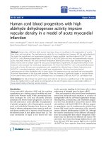
Study Design
The study consisted of three phases: a pilot trial
comprising 17 patients (7 receiving progenitor cells
derived from bone marrow [BMC] and 10 receiv-
ing progenitor cells derived from circulating blood
[CPC]); a second phase, in which 75 patients were
randomly assigned to receive intracoronary infu-
sion of BMC (28 patients) or CPC (24) or no cell
infusion (23); and a third phase, in which the 75
randomly assigned patients crossed over to one
of the active treatments if they had originally been
in the control group or to the alternate cell type if
they had initially received intracoronary cell infu-
sion (Fig. 1).
The primary end point of the study was the
absolute change in global left ventricular ejection
fraction (LVEF) as measured by quantitative left
ventricular angiography 3 months after cell infu-
sion. Secondary end points included quantitative
variables relating to the regional left ventricular
function of the target area, as well as left ven-
tricular volumes derived from serial left ventric-
ular angiograms. In addition, functional status
was assessed by NYHA classification. Finally,
event-free survival was defined as freedom from
death, myocardial infarction, stroke, or rehospi-
talization for worsening heart failure. Causes of
rehospitalization during follow-up were verified
by review of the discharge letters or charts of
hospital stays.
Preparation and Transplantation
of Progenitor Cells
For patients assigned to receive CPC, mononuclear
cells were isolated by Ficoll density-gradient cen-
trifugation of 270 ml of venous blood and cultured
for 3 days ex vivo, as previously reported.
6,7,9-12
A mean of 22×10
6
±11×10
6
CPC were infused. For
patients assigned to receive BMC, 50 ml of bone
marrow aspirate was obtained while the pa-
tients were under local anesthesia on the morn-
ing of cell-transplantation day. BMC were iso-
lated by Ficoll density-gradient centrifugation,
as previously reported.
6,7,9
We infused a mean of
205×10
6
±110×10
6
BMC, of which on average less
than 1% were positive for the hematopoietic
progenitor-cell marker CD34.
For cell transplantation, arterial puncture
Copyright © 2006 Massachusetts Medical Society. All rights reserved.
Downloaded from www.nejm.org at RIKSHOSPITALET HF on February 18, 2008 .
The new england journal of medicine
n engl j med 355;12 www.nejm.org september 21, 2006
1224
was followed by the administration of 7500 to
10,000 U of heparin and (in 89% of the cell-treated
patients) a bolus of abciximab (0.25 mg per kilo-
gram of body weight). Cells were infused into
the vessel supplying the most dyskinetic left ven-
tricular area by means of a balloon catheter with
a stop-flow technique, as previously described.
6
Evaluation of Safety and Feasibility
Clinical, laboratory, and safety-related data were
prospectively collected. Follow-up visits after
3 months were performed by physicians. Proce-
dural complications were defined as any ventric-
ular arrhythmia, visible thrombus formation, dis-
tal embolization, or injury of the coronary artery
associated with the cell-infusion catheterization
procedure. For patients undergoing bone mar-
row aspiration, potential bleeding complications
were assessed. During hospitalization, telemetry
was routinely performed for 24 hours after the
procedure in all patients.
Left Ventricular Angiography
Left ventricular angiograms were obtained at the
time of the baseline procedure and at 3 months’
follow-up. Quantitative analysis of paired left ven-
tricular angiograms recorded in identical projec-
tions was performed by an investigator who was
blinded to the individual patients’ treatments; the
analysis was performed with QCA-CMS software
(version 5.2, Medis), as described elsewhere.
6,7,9
Magnetic Resonance Imaging
In a subgroup of 35 patients who did not have
implanted defibrillators or pacemakers and who
consented to and tolerated the imaging proce-
dure, cardiac magnetic resonance imaging (MRI)
(a 1.5-T system; Magnetom Sonata, Siemens Med-
ical Solutions) was performed at baseline and at
3 months’ follow-up. The results were analyzed
as previously described
7
by an experienced inves-
tigator who was blinded to the type of cells in-
fused.
Eligible patients
(
N=17)
CPC (N=10) BMC (N=7)
Eligible patients (N=75)
1st LVA
CPC (N=24)
2nd LVA
BMC (N=28)
2nd LVA
CPC (N=10)
3rd LVA
BMC (N=11)
3rd LVA
BMC (N=21)
3rd LVA
CPC (N=24)
3rd LVA
Control (N=23)
2nd LVA
Phase 1: Pilot Trial
Phase 2: Randomized,
Controlled Trial
Phase 3: Crossover
Phase
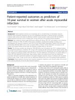
Figure 1. Design of the Trial.
Eligible patients with chronic ischemic cardiomyopathy had a severely hypokinetic area on the baseline left ventricu-
lar angiogram (LVA) and had had a myocardial infarction at least 3 months previously.
Copyright © 2006 Massachusetts Medical Society. All rights reserved.
Downloaded from www.nejm.org at RIKSHOSPITALET HF on February 18, 2008 .
progenitor-cell infusion in ischemic heart disease
n engl j med 355;12 www.nejm.org september 21, 2006
1225
Detection of Viable Myocardium
All patients underwent low-dose dobutamine
stress echocardiography, combined thallium sin-
gle-photon-emission computed tomography and
[
18
F]fluorodeoxyglucose positron-emission tomog-
raphy, or both, as previously described.
6
It was pos-
sible to analyze regional left ventricular viability
in 80 patients (87%).
Statistical Analysis
Continuous variables are presented as means
(±SD), unless otherwise noted. Categorical vari-
ables were compared with use of the chi-square
test or Fisher’s exact test. Statistical comparisons
between initial and follow-up data were performed
in a nonparametric, paired fashion with use of
the Wilcoxon signed-rank test. Nonparametric
Mann–Whitney U tests and Kruskal–Wallis tests
were used to compare continuous variables with
categorical variables as well as to compare the
results between treatment groups. Bonferroni-
adjusted analysis-of-variance testing was used for
between-group analysis of quantitative left ven-
tricular angiographic results in phases 1 and 2 (the
pilot phase and first randomized phase). For multi-
variate analysis, the treatment groups were cate-
gorized as follows: control, 0; CPC, 1; and BMC, 2.
The multivariate analysis was performed with use
of a stepwise linear regression model with a for-
ward-entry stepping algorithm; variables with a
P value of ≤0.05 on univariate analysis were en-
tered in the model. Statistical significance was
assumed for P values of less than 0.05. All statis-
tical analyses were performed with SPSS software
(version 12.0).
Results
Baseline Characteristics of the Patients
A total of 92 patients were enrolled in the study.
Of these, 35 patients received BMC as their ini-
tial treatment (in phases 1 and 2 of the trial), 34
patients received CPC (in phases 1 and 2), and
23 patients received no intracoronary cell infu-
sion (in phase 2, as the control group).
Table 1
illustrates that the three groups of patients were
well matched.
Effects of Progenitor-Cell Infusion
Quantitative Characteristics of Left Ventricular
Function
Patients with an adverse clinical event (six), sub-
total stenosis of the target vessel at follow-up
(three), an intraventricular thrombus precluding
performance of left ventricular angiography (one),
or atrial flutter or fibrillation at follow-up (one)
were excluded from the exploratory analysis. In
addition, of the 81 eligible patients, left ventricu-
lar angiograms could not be quantitatively ana-
lyzed in 4 because of inadequate contrast opaci-
fication, in 1 because of ventricular extrasystoles,
and in 4 because of the patients’ refusal to un-
dergo invasive follow-up. Thus, a total of 72 of 81
serial paired left ventricular angiograms were
available for quantitative analysis (28 in the BMC
group, 26 in the CPC group, and 18 in the control
group).
Table 2
summarizes the angiographic charac-
teristics of the 75 patients included in the ran-
domized phase of the study. At baseline, the three
groups did not differ with respect to global LVEF,
the extent or magnitude of regional left ventricu-
lar dysfunction, left ventricular volumes, or stroke
volumes.
The absolute change in global LVEF from base-
line to 3 months did significantly differ among the
three groups of patients. Patients receiving BMC
had a significantly larger change in LVEF than
patients receiving CPC (P = 0.003) and those in the
control group (P<0.001). Similar results were ob-
tained when patients from the first two phases
of the study (the pilot phase and the randomized
phase) were pooled. The results did not differ
when patients without evidence of viable myo-
cardium before inclusion were analyzed sepa-
rately. The change in LVEF was −0.3±3.4 percent-
age points in the control group (9 patients),
+0.4±3.0 percentage points in the CPC group (18
patients), and +3.7±4.0 percentage points in the
BMC group (18 patients) (P = 0.02 for the com-
parison with the control group and P = 0.02 for
the comparison with the CPC group).
In the subgroup of 35 patients who underwent
serial assessment of left ventricular function by
MRI, MRI-derived global LVEF increased signifi-
cantly, by 4.8±6.0% (P = 0.03) among those receiv-
ing BMC (11 patients) and by 2.8±5.2% (P = 0.02)
among those receiving CPC (20 patients), where-
as no change was observed in 4 control patients
(P = 0.14). Thus, MRI-derived assessment of left
ventricular function further corroborated the re-
sults obtained from the total patient population.
Analysis of regional left ventricular function
revealed that BMC treatment significantly in-
creased contractility in the center of the left ven-
tricular target area (
Table 2
). Likewise, MRI-derived
Copyright © 2006 Massachusetts Medical Society. All rights reserved.
Downloaded from www.nejm.org at RIKSHOSPITALET HF on February 18, 2008 .
The new england journal of medicine
n engl j med 355;12 www.nejm.org september 21, 2006
1226
regional analysis of left ventricular function re-
vealed that the number of hypocontractile seg-
ments was significantly reduced, from 10.1±3.6
to 8.7±3.6 segments (P = 0.02), and the number
of normocontractile segments significantly in-
creased, from 3.8±4.5 to 5.4±4.6 segments (P =
0.01), in the BMC group, whereas no significant
changes were observed in the CPC group. MRI-
derived infarct size, as measured by late enhance-
ment volume normalized to left ventricular mass,
remained constant both in the CPC group (25±
18% at baseline and 23±14% at 3 months, 13
patients) and in the BMC group (20±10% at both
time points, 9 patients). Thus, taken together, the
data suggest that intracoronary infusion of BMC
is associated with significant improvements in
global and regional left ventricular contractile
function among patients with persistent left ven-
tricular dysfunction due to prior myocardial in-
farction.
To identify independent predictors of improved
global LVEF, a stepwise multivariate regression
analysis was performed; it included classic deter-
minants of LVEF as well as various baseline char-
Table 1. Baseline Characteristics of the Patients.*
Characteristic
Control Group
(N = 23)
CPC Group
(N = 34)
BMC Group
(N = 35) P Value
Demographic and laboratory characteristics
Age — yr 61±9 56±12 60±11 0.32
Female sex — no. (%) 0 6 (18) 4 (11) 0.11
Blood pressure — mm Hg
Systolic 117±21 109±20 109±22 0.28
Diastolic 66±11 63±10 62±14 0.48
Heart rate — beats/min 68±12 65±8 64±11 0.56
Body-mass index† 28±3 27±4 27±4 0.37
NYHA class — no. (%) 0.34
1 7 (30) 7 (21) 5 (14)
2 11 (48) 13 (38) 18 (51)
3 5 (22) 12 (35) 12 (34)
4 0 2 (6) 0
Serum creatinine — mg/dl‡ 1.1±0.3 1.1±0.2 1.1±0.4 0.71
Risk factors
Hypertension — no. (%) 16 (70) 19 (56) 21 (60) 0.58
Diabetes mellitus — no. (%) 5 (22) 9 (26) 10 (29) 0.84
Current or former smoking — no. (%) 19 (83) 28 (82) 27 (77) 0.78
Hypercholesterolemia — no. (%)§ 20 (87) 28 (82) 27 (77) 0.31
Family history of coronary artery disease — no. (%) 10 (43) 24 (71) 21 (60) 0.12
Medical history
Previous MI — no. (%) 0.34
Anterior 9 (39) 22 (65) 20 (57)
Inferior 12 (52) 8 (24) 13 (37)
Lateral 0 1 (3) 0
Anterior and inferior 2 (9) 3 (9) 2 (6)
Time since most recent MI — mo 0.59
Mean 81±101 77±76 81±72
Median 24 50 60
Range 3–358 3–276 4–300
Copyright © 2006 Massachusetts Medical Society. All rights reserved.
Downloaded from www.nejm.org at RIKSHOSPITALET HF on February 18, 2008 .
progenitor-cell infusion in ischemic heart disease
n engl j med 355;12 www.nejm.org september 21, 2006
1227
acteristics of the three groups (
Table 3
). The
multivariate analysis identified the type of pro-
genitor cell infused and the baseline stroke vol-
ume as the only statistically significant indepen-
dent predictors of LVEF recovery.
Functional Status
The functional status of the patients, as assessed
by NYHA classification, improved significantly
in the BMC group (from 2.23±0.6 to 1.97±0.7,
P = 0.005). It did not improve significantly either
in the CPC group (class, 2.16±0.8 at baseline and
1.93±0.8 at 3 months; P = 0.13) or in the control
group (class, 1.91±0.7 and 2.09±0.9, respectively;
P = 0.27).
Randomized Crossover Phase
Of the 24 patients who initially were randomly
assigned to CPC infusion, 21 received BMC at the
time of their first follow-up examination. Likewise,
of the 28 patients who initially were randomly
assigned to BMC infusion, 24 received CPC after
3 months. Of the 23 patients of the control
group, 10 patients received CPC and 11 received
BMC at their reexamination at 3 months (Fig. 1).
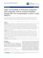
As illustrated in Figure 2, regardless of whether
patients received BMC as initial treatment, as
crossover treatment after CPC infusion, or as
crossover treatment after no cell infusion, glob-
al LVEF increased significantly after infusion of
BMC. In contrast, CPC treatment did not sig-
Table 1. (Continued.)
Characteristic
Control Group
(N = 23)
CPC Group
(N = 34)
BMC Group
(N = 35) P Value
Extent of coronary artery disease — no. (%) 0.07
One vessel 2 (9) 10 (29) 10 (29)
Two vessels 9 (39) 5 (15) 15 (43)
Three vessels 12 (52) 19 (56) 10 (29)
Target vessel — no. (%) 0.35
Left anterior descending artery 13 (57) 22 (65) 17 (49)
Left circumflex artery 5 (22) 3 (9) 4 (11)
Right coronary artery 5 (22) 9 (26) 14 (40)
Coronary-artery bypass grafting — no. (%) 5 (22) 10 (29) 7 (20) 0.63
Concomitant PCI — no. (%)
Target vessel 4 (17) 9 (26) 10 (29) 0.61
Vessel other than the target vessel 1 (4) 2 (6) 3 (9) 0.80
Pacemaker or implantable cardioverter–defibrillator
— no. (%)
5 (22) 7 (21) 12 (34) 0.37
Evidence of viable myocardium — no. (%)¶ 5 (28) 9 (28) 8 (27) 0.99
Current medication
Antiplatelet therapy: aspirin, clopidogrel, or both — no. (%) 22 (96) 32 (94) 31 (89) 0.54
Beta-blocker — no. (%) 22 (96) 33 (97) 33 (94) 0.85
ACE inhibitor or angiotensin-receptor blocker — no. (%) 21 (91) 33 (97) 33 (94) 0.64
Spironolactone — no. (%) 6 (26) 12 (35) 11 (31) 0.76
Diuretics — no. (%) 17 (74) 25 (74) 28 (80) 0.79
Warfarin — no. (%) 6 (26) 7 (21) 13 (37) 0.30
Statin — no. (%) 21 (91) 32 (94) 30 (86) 0.49
* Plus–minus values are means ±SD. MI denotes myocardial infarction, PCI percutaneous coronary intervention, and
ACE angiotensin-converting enzyme.
† Body-mass index is calculated as the weight in kilograms divided by the square of the height in meters.
‡ To convert the values for creatinine to micromoles per liter, multiply by 88.4.
§ Hypercholesterolemia was defined by a low-density lipoprotein level of more than 130 mg per deciliter (3.4 mmol per li-
ter) or the use of lipid-lowering therapy.
¶ Viability could be analyzed in 80 patients (18 in the control group, 32 in the CPC group, and 30 in the BMC group).
Copyright © 2006 Massachusetts Medical Society. All rights reserved.
Downloaded from www.nejm.org at RIKSHOSPITALET HF on February 18, 2008 .
The new england journal of medicine
n engl j med 355;12 www.nejm.org september 21, 2006
1228
Table 2. Quantitative Variables Pertaining to Left Ventricular Function, as Assessed by Left Ventricular Angiography.*
Variable Baseline 3 Months’ Follow-up Absolute Change P Value
Global LVEF (%)
Control group 43±13 42±13 −1.2±3.0 0.12
CPC group 39±10 39±10 −0.4±2.2 0.60
BMC group 41±11 43±10 +2.9±3.6 0.001
P value for all 3 groups 0.68 0.31 0.001
Regional contractility in central target
area (SD from normal/chord)
Control group −1.55±0.40 −1.50 ±0.47 −0.06±0.33 0.62
CPC group −1.72±0.36 −1.75±0.41 −0.03±0.30 0.70
BMC group −1.63±0.40 −1.38±0.42 +0.26±0.43 0.006
P value for all 3 groups 0.44 0.03 0.09
Extent of regional left ventricular dysfunction
(% circumference)
Control group 45±24 45±22 0±5 0.41
CPC group 52±18 50±19 −3±6 0.15
BMC group 45±18 42±19 −3±10 0.31
P value for all 3 groups 0.51 0.50 0.37
End-diastolic volume (ml/m
2
of BSA)
Control group 90±38 87±33 −3±17 0.45
CPC group 96±34 93±30 −3±18 0.47
BMC group 79±29 79±29 0±10 0.95
P value for all 3 groups 0.14 0.26 0.62
End-systolic volume (ml/m
2
of BSA)
Control group 55±36 55±32 −1±12 0.91
CPC group 62±31 60±26 −2±13 0.57
BMC group 49±26 47±26 −2±5 0.09
P value for all 3 groups 0.21 0.26 0.83
Stroke volume (ml/m
2
of BSA)
Control group 34±7 32±4 −2±7 0.22
CPC group 35±8 34±8 −1±7 0.31
BMC group 30±9 32±8 +2±7 0.21
P value for all 3 groups 0.08 0.78 0.09
Left ventricular end-diastolic pressure
(mm Hg)
Control group 14±9 12±6 −2±7 0.15
CPC group 12±7 12±6 0±6 0.84
BMC group 12±8 12±7 0±7 0.91
P value for all 3 groups 0.64 0.61 0.42
* Plus–minus values are means ±SD. BSA denotes body-surface area.
Copyright © 2006 Massachusetts Medical Society. All rights reserved.
Downloaded from www.nejm.org at RIKSHOSPITALET HF on February 18, 2008 .
progenitor-cell infusion in ischemic heart disease
n engl j med 355;12 www.nejm.org september 21, 2006
1229
nificantly alter LVEF when given either before or
after BMC.
Thus, the intrapatient comparison of the dif-
ferent treatment strategies not only documents
the superiority of intracoronary infusion of BMC
over the infusion of CPC for improving global
left ventricular function, but also corroborates
our findings in the analysis of data according to
initial treatment assignment. The preserved im-
provement in cardiac function observed among
patients who initially received BMC treatment
and then crossed over to CPC treatment demon-
strates that the initially achieved differences in
cardiac function persisted for at least 6 months
after intracoronary infusion of BMC.
Procedural Safety and Clinical Outcomes
In 3 of the 135 intracoronary progenitor-cell–
infusion procedures (pooled data from all study
phases), local dissection of the coronary arterial
wall was angiographically visible after inflation
of the balloon during cell infusion; in these cases
the dissection was successfully treated with im-
mediate stent implantation. However, two of these
three patients had subsequent elevations in cre-
atine kinase (Table 4). The further clinical course
of these three patients was uneventful. One ad-
ditional patient required defibrillation from his
implanted defibrillator for ventricular fibrilla-
tion during induction of myocardial ischemia by
transient balloon occlusion for cell infusion. The
clinical events before and after discharge from
the hospital are listed in
Table 4
.
Discussion
Using a randomized, controlled trial design, we
examined the effects of intracoronary infusion of
adult progenitor cells on global and regional left
ventricular function in patients with chronic
ischemic heart disease who had had a myocar-
dial infarction at least 3 months previously. Our
results demonstrate that infusion of BMC into
the infarct-related artery is associated with mod-
erate but significant improvements in both glob-
al and regional left ventricular contractile func-
tion. These improvements were observed in the
presence of full conventional pharmacologic
treatment and lasted at least 6 months.
The application procedures, infusion media,
and infused volumes of cell suspension were iden-
tical in the two intracoronary-infusion groups.
Therefore, potential confounding effects relat-
ing to ischemic preconditioning or microvascu-
lar activation can be ruled out in accounting for
the improved cardiac function observed in the
group treated with BMC. Moreover, intrapatient
comparison in the crossover phase of the trial
rules out the possibility that differences in the
patient populations studied may have affected
outcomes. However, the mechanisms involved in
mediating improved contractile function after
intracoronary progenitor-cell infusion are not
well understood.
Experimentally, although there is no definitive
proof that cardiac myocytes may be regenerated,
BMC were shown to contribute to functional re-
covery of left ventricular contraction when in-
jected into freshly infarcted hearts,
13-15
whereas
CPC profoundly stimulated ischemia-induced
neovascularization.
16,17
Bot h cell t ypes were shown
to prevent cardiomyocyte apoptosis and reduce
the development of myocardial fibrosis and there-
by improve cardiac function after acute myocar-
dial infarction.
18,19
Indeed, in our TOPCARE-AMI
(Transplantation of Progenitor Cells and Regen-
eration Enhancement in Acute Myocardial Infarc-
tion) studies,
6,7,9
intracoronary infusion of CPC
was associated with functional improvements
similar to those found with the use of BMC im-
mediately after myocardial infarction. In the cur-
rent study, however, which involved patients
who had had a myocardial infarction at least
3 months before therapy, transcoronary adminis-
tration of CPC was significantly inferior to ad-
Table 3. Stepwise Linear Regression Analysis for Predictors of Improvement
in Global Left Ventricular Ejection Fraction.*
Variable
Nonstandardized
Coefficient B 95% CI for B P Value
Treatment group 1.49 0.53 to 2.46 0.003
Baseline stroke volume −0.13 −0.22 to –0.05 0.002
No. of cardiovascular risk factors 0.76
Time since most recent MI 0.48
Concomitant PCI 0.60
Age 0.82
Baseline ejection fraction 0.72
Baseline end-diastolic volume 0.88
* Values are shown only for significant differences. MI denotes myocardial infarc-
tion, and PCI percutaneous coronary intervention. For the overall model, the ad-
justed R
2
was 0.29; P<0.001 by analysis of variance.
Copyright © 2006 Massachusetts Medical Society. All rights reserved.
Downloaded from www.nejm.org at RIKSHOSPITALET HF on February 18, 2008 .
The new england journal of medicine
n engl j med 355;12 www.nejm.org september 21, 2006
1230
ministration of BMC in altering global left ven-
tricular function.
This study does not explain the cellular mecha-
nisms associated with the significantly improved
left ventricular function in the patients treated
with BMC, nor does it explain the responses to
CPC infusion, which were of only borderline sig-
nificance. It is likely that the smaller number of
progenitor cells derived from 270 ml of venous
blood, which was 1/10 the number of monocytic
cells obtained from 50 ml of bone marrow aspi-
rate, may have contributed to the smaller effects
of CPC in improving left ventricular contractile
function. Moreover, CPC obtained from patients
with chronic ischemic heart disease show pro-
found functional impairments,
20,21
which might
limit their recruitment, after intracoronary infu-
sion, into chronically reperfused scar tissue many
months or years after myocardial infarction. Thus,
additional studies in which larger numbers of
functionally enhanced CPC are used will be re-
quired to increase the response to intracoronary
infusion of CPC.
The magnitude of the improvement after in-
tracoronary infusion of BMC, with absolute
increases in global LVEF of approximately 2.9
percentage points according to left ventricular
angiography and 4.8 percentage points accord-
ing to MRI, was modest. However, it should be
noted that the improvement in LVEF occurred in
the setting of full conventional pharmacologic
treatment: more than 90% of the patients were
receiving beta-blocker and angiotensin-convert-
ing–enzyme inhibitor treatment. Moreover, results
from trials of contemporary reperfusion for the
treatment of acute myocardial infarction, which
is regarded as the most effective treatment strat-
egy for improving left ventricular contractile per-
formance after ischemic injury, have reported in-
creases in global LVEF of 2.8% (in the CADILLAC
[Controlled Abciximab and Device Investigation
to Lower Late Angioplasty Complications] trial)
and 4.1% (in the ADMIRAL [Abciximab before
Direct Angioplasty and Stenting in Myocardial
Infarction Regarding Acute and Long-Term Fol-
low-up] trial).
22,23
The number of patients, as well as the dura-
tion of follow-up, is not sufficient to address the
question of whether the moderate improvement
in LVEF associated with one-time intracoronary
BMC infusion is associated with reduced mortal-
ity and morbidity among patients with heart fail-
ure secondary to previous myocardial infarction.
We conclude that intracoronary infusion of BMC
Mean Absolute Change in LVEF (percentage points)
2
3
1
0
¡1
¡2
Baseline ˚ BMC BMC ˚ CPC Baseline ˚ CPC CPC ˚ BMC Baseline ˚ control Control ˚ CPC Baseline ˚ control Control ˚ BMC
Crossover: CPC to BMC Crossover: Control to CPC Crossover: Control to BMCCrossover: BMC to CPC
4
P=0.06 P=0.01 P=0.06 P=0.03
Figure 2. Absolute Change in Quantitative Global Left Ventricular Ejection Fraction (LVEF) during the Crossover Phase of the Trial.
Data at 3 and 6 months are shown for all patients crossing over from BMC to CPC infusion (18 patients), from CPC to BMC infusion
(18 patients), and from no cell infusion to either CPC infusion (10 patients) or BMC infusion (11 patients). I bars represent standard
errors.
Copyright © 2006 Massachusetts Medical Society. All rights reserved.
Downloaded from www.nejm.org at RIKSHOSPITALET HF on February 18, 2008 .
progenitor-cell infusion in ischemic heart disease
n engl j med 355;12 www.nejm.org september 21, 2006
1231
is associated with persistent improvements in
regional and global left ventricular function
and improved functional status among patients
who have had a myocardial infarction at least
3 months previously. Given the reasonable
short-term safety profile of this therapeutic ap-
proach, studies on a larger scale are warranted
to examine its potential effects on morbidity
and mortality among patients with postinfarc-
tion heart failure.
Supported by the Deutsche Forschungsgemeinschaft (FOR
501-1: WA 146/2-1), the Foundation Leducq Transatlantic Net-
work of Excellence for Cardiac Regeneration, the European
Union European Vascular Genomics Network (contract no.
LSHM-CT-2003-503254), and the Alfried Krupp Stiftung (to Dr.
Dimmeler).
Dr. Schächinger reports having received consulting fees from
Guidant and AstraZeneca and lecture fees from AstraZeneca,
Merck Sharp & Dohme, Pfizer, Novartis, Guidant, Boston Scien-
tific, Boehringer Ingelheim, Sanofi-Aventis, and Lilly. Dr. Dim-
meler reports being a member of the scientific advisory board of
Guidant. Dr. Zeiher reports having received consulting fees from
Guidant. Drs. Dimmeler and Zeiher report that they are co-
founders of t2cure, a for-profit company focused on regenera-
tive therapies for cardiovascular disease. They serve as scientific
advisors and are shareholders. No other potential conflict of
interest relevant to this article was reported.
We are indebted to the staff of our catheterization laborato-
ries; to Beate Mantz, Isabel Geweyer, and Heike Braun (study
nurses); to Tina Rasper (biologic technician); and to Arne Koch
(MRI staff member).
Table 4. Clinical Events during the 3-Month Follow-up Period.*
Event
Control Group
(N = 23)
CPC Group
(N = 34)
BMC Group
(N = 35) P Value
number (percent)
In-hospital events
Death 0 0 0
MI 0 2 0
Infarct-vessel stent thrombosis 0 1 0
Stent thrombosis at a site other than the target
vessel
000
Cerebral infarction 0 0 0
Ventricular arrhythmia (detected during monitoring) 1 1 0
Cumulative total 1 (4) 3 (9) 0 0.20
Events after discharge
Death 1 0 0
MI 0 0 0
Rehospitalization for heart failure 1 1 0
Stent thrombosis after hospitalization 0 0 0
Infarct-vessel revascularization† 0 2 4
Coronary bypass surgery 0 0 0
Cerebral infarction 1 1 0
Syncope 0 2 0
Documented ventricular arrhythmia 0 0 0
Cumulative total 2 (9) 5 (15) 4 (11) 0.79
Cumulative events, before or after discharge
Death or MI 1 (4) 2 (6) 0 0.37
Death, MI, or rehospitalization for heart failure 1 (4) 3 (9) 0 0.20
Death, MI, stroke, rehospitalization for heart failure,
or ventricular tachycardia
3 (13) 5 (15) 0 0.07
* For analysis of cumulative events, the first event per patient was counted. The number of events may exceed the cumu-
lative total because some events may have occurred in the same patient. MI denotes myocardial infarction.
† This category includes revascularization due to in-hospital stent thrombosis as well as that due to restenosis.
Copyright © 2006 Massachusetts Medical Society. All rights reserved.
Downloaded from www.nejm.org at RIKSHOSPITALET HF on February 18, 2008 .
n engl j med 355;12 www.nejm.org september 21, 2006
1232
progenitor-cell infusion in ischemic heart disease
References
2001 Heart and stroke statistical up-
date. Dallas: American Heart Association,
2000.
Braunwald E. Cardiovascular medicine
at the turn of the millennium: triumphs,
concerns, and opportunities. N Engl J Med
1997;337:1360-9.
Lange RA, Hillis LD. Reperfusion ther-
apy in acute myocardial infarction. N Engl
J Med 2002;346:954-5.
Sutton MG, Sharpe N. Left ventricular
remodeling after myocardial infarction:
pathophysiology and therapy. Circulation
2000;101:2981-8.
Strauer BE, Brehm M, Zeus T, et al. Re-
pair of infarcted myocardium by autologous
intracoronary mononuclear bone marrow
cell transplantation in humans. Circula-
tion 2002;106:1913-8.
Assmus B, Schachinger V, Teupe C, et
al. Transplantation of Progenitor Cells and
Regeneration Enhancement in Acute Myo-
cardial Infarction (TOPCARE-AMI). Circu-
lation 2002;106:3009-17.
Britten MB, Abolmaali ND, Assmus
B, et al. Infarct remodeling after intra-
coronary progenitor cell treatment in pa-
tients with acute myocardial infarction
(TOPCARE-AMI): mechanistic insights
from serial contrast-enhanced magnetic
resonance imaging. Circulation 2003;108:
2212-8.
Wollert KC, Meyer GP, Lotz J, et al. In-
tracoronary autologous bone-marrow cell
transfer after myocardial infarction: the
BOOST randomised controlled clinical
trial. Lancet 2004;364:141-8.
Schachinger V, Assmus B, Britten MB,
1.
2.
3.
4.
5.
6.
7.
8.
9.
et al. Transplantation of progenitor cells
and regeneration enhancement in acute
myocardial infarction: final one-year re-
sults of the TOPCARE-AMI Trial. J Am Coll
Cardiol 2004;44:1690-9.
Dimmeler S, Aicher A, Vasa M, et al.
HMG-CoA reductase inhibitors (statins)
increase endothelial progenitor cells via
the PI 3-kinase/Akt pathway. J Clin Invest
2001;108:391-7.
Vasa M, Fichtlscherer S, Adler K, et al.
Increase in circulating endothelial pro-
genitor cells by statin therapy in patients
with stable coronary artery disease. Circu-
lation 2001;103:2885-90.
Vasa M, Fichtlscherer S, Aicher A, et al.
Number and migratory activity of circulat-
ing endothelial progenitor cells inversely
correlate with risk factors for coronary
artery disease. Circ Res 2001;89:E1-E7.
Balsam LB, Wagers AJ, Christensen JL,
Kofidis T, Weissman IL, Robbins RC. Hae-
matopoietic stem cells adopt mature hae-
matopoietic fates in ischaemic myocardi-
um. Nature 2004;428:668-73.
Orlic D, Kajstura J, Chimenti S, et al.
Bone marrow cells regenerate infarcted
myocardium. Nature 2001;410:701-5.
Mangi AA, Noiseux N, Kong D, et al.
Mesenchymal stem cells modified with
Akt prevent remodeling and restore perfor-
mance of infarcted hearts. Nat Med 2003;
9:1195-201.
Kalka C, Masuda H, Takahashi T, et
al. Transplantation of ex vivo expanded
endothelial progenitor cells for therapeu-
tic neovascularization. Proc Natl Acad Sci
U S A 2000;97:3422-7.
10.
11.
12.
13.
14.
15.
16.
Murohara T, Ikeda H, Duan J, et al.
Transplanted cord blood-derived endothe-
lial precursor cells augment postnatal
neovascularization. J Clin Invest 2000;105:
1527-36.
Kawamoto A, Gwon HC, Iwaguro H, et
al. Therapeutic potential of ex vivo expand-
ed endothelial progenitor cells for myo-
cardial ischemia. Circulation 2001;103:
634-7.
Kocher AA, Schuster MD, Szabolcs MJ,
et al. Neovascularization of ischemic myo-
cardium by human bone-marrow-derived
angioblasts prevents cardiomyocyte apop-
tosis, reduces remodeling and improves
cardiac function. Nat Med 2001;7:430-6.
Rupp S, Badorff C, Koyanagi M, et al.
Statin therapy in patients with coronary
artery disease improves the impaired en-
dothelial progenitor cell differentiation
into cardiomyogenic cells. Basic Res Car-
diol 2004;99:61-8.
Valgimigli M, Rigolin GM, Fucili A, et
al. CD34+ and endothelial progenitor cells
in patients with various degrees of con-
gestive heart failure. Circulation 2004;
110:1209-12.
Montalescot G, Barragan P, Witten-
berg O, et al. Platelet glycoprotein IIb/IIIa
inhibition with coronary stenting for acute
myocardial infarction. N Engl J Med 2001;
344:1895-903.
Stone GW, Grines CL, Cox DA, et al.
Comparison of angioplasty with stenting,
with or without abciximab, in acute myo-
cardial infarction. N Engl J Med 2002;346:
957-66.
Copyright © 2006 Massachusetts Medical Society.
17.
18.
19.
20.
21.
22.
23.
JOURNAL
EDITORIAL
FELLOW
The Journal’s editorial office invites applications for a one-year
research fellowship beginning in July 2007 from individuals at any
stage of training. The editorial fellow will work on Journal projects
and will participate in the day-to-day editorial activities of the Journal
but is expected in addition to have his or her own independent
projects. Please send curriculum vitae and research interests
to the Editor-in-Chief, 10 Shattuck St., Boston, MA 02115
(fax, 617-739-9864), by October 1, 2006.
Copyright © 2006 Massachusetts Medical Society. All rights reserved.
Downloaded from www.nejm.org at RIKSHOSPITALET HF on February 18, 2008 .
n engl j med 351;17 www.nejm.org october 21, 2004
1716
PERSPECTIVE
ingly run up against the historical reluctance of
American voters to allocate much more than 18
percent of the GDP to federal spending. Since 1946
the federal government’s share of the GDP has
stayed remarkably close to 18 percent, going below
16 percent in only 2 of the 57 years and above 20
percent in only 1.
One response, of course, is to ignore this de facto
ceiling on federal revenues and assume that an in-
creasingly graying society will want to spend a
greater share of its money on pensions and health
care for the elderly. But Medicare and Social Security
both rely on a substantial component of payroll-tax
financing, the burden of which falls primarily on
nonelderly workers. Although many of these work-
ers have elderly parents and are anticipating their
own age of eligibility, it is unclear whether there
would be political support for such a large transfer
of resources from the nonelderly to the elderly.
The late Senator Daniel P. Moynihan (D-N.Y.) fa-
mously characterized Social Security as the third
rail of American politics. Since he made that re-
mark, the dollars spent on both Social Security and
Medicare have increased, raising that third rail’s
voltage. As a result, an enormous amount of polit-
ical capital is required to address the issue of long-
term financing, making it highly tempting for the
next administration simply to leave the matter to its
successors. Unfortunately, deferring the issue will
only exacerbate the problem for future administra-
tions and taxpayers.
Dr. Newhouse reports serving on the board of directors of and
holding equity in Aetna.
From the Department of Health Care Policy, Harvard Medical
School; and the Department of Health Policy and Management,
Harvard School of Public Health — both in Boston; and the
Kennedy School of Government, Harvard University, Cambridge,
Mass.
1. CBO’s March baseline projections. In: CBO’s current budget
projections. Washington, D.C.: Congressional Budget Office,
March 2004. (Accessed October 1, 2004, at />showdoc.cfm?index=1944&sequence=0#table1.)
2. Blendon RJ, Benson JM, Brodie M, et al. Voters and health
care in the 1996 election. JAMA 1997;277:1253-8.
3. Jamieson A, Shin HB, Day J. Voting and registration in the
election of November 2000. Current population reports. P20-542.
Washington, D.C.: Census Bureau, February 2002.
4. Congressional Budget Office. The budget and economic out-
look, September 2004. (Accessed October 1, 2004, at http://www.
cbo.gov/showdoc.cfm?index=5773&sequence=2.)
5. Organisation for Economic Co-operation and Development.
Health data 2000. (Accessed October 1, 2004, at d.
org/document/30/0,2340,en_2649_33929_12968734_119656_1_
1_1,00.html.
Enormous progress made during the past few de-
cades has dramatically enhanced our understanding
of the pathobiology and pathophysiology responsi-
ble for acute myocardial infarction. Investigations
in vascular biology have elucidated the critical role
of growth factors, the proliferation of smooth-
muscle cells, and the central role of inflammation
in the initiation and progression of atherosclero-
sis.
1
Research has also focused on the initiating
events or “triggers” that qualitatively alter the sta-
ble or quiescent phase of coronary atherosclerosis
and initiate a cascade of events that culminates in
acute myocardial infarction. Some triggering phe-
nomena may exert a single, transient effect on the
pathophysiologic process, such as a surge of sym-
pathetic activity, whereas others exert a more varied
and pervasive effect, amplifying risk at multiple
points and over a longer period. In this issue of the
Journal, Peters et al. (pages 1721–1730) provide
compelling epidemiologic evidence that particulate
air pollution from traffic may trigger the abrupt
onset of acute myocardial infarction. An under-
standing of air pollution in the larger context of
triggering of the entire process of atherosclerosis
suggests, in addition, that air pollution plays a more
complex and multifaceted role in the development
of cardiovascular disease over the longer term.
As initially described 15 years ago, the trigger-
ing of acute myocardial infarction typically begins
with the so-called vulnerable or high-risk coronary
atherosclerotic plaque, a focal lesion in jeopardy of
plaque disruption.
2
The vulnerable plaque is usually
an inflamed, thin-cap fibroatheroma, characterized
by a lipid-rich, atheromatous core with cholesterol
crystals and necrotic debris, a thin fibrous cap with
an infiltration of macrophages and lymphocytes,
T
riggering Myocardial Infarction
Peter H. Stone, M.D.
Related article, page 1721
Financing Medicare in the Next Administration
Downloaded from www.nejm.org on February 18, 2008 . Copyright © 2004 Massachusetts Medical Society. All rights reserved.
n engl j med 351;17 www.nejm.org october 21, 2004
1717
PERSPECTIVE
and decreased smooth-muscle-cell content, and
associated with expansive remodeling of the outer
vessel wall.
3
The inflammatory cells associated with
this type of high-risk plaque express a variety of cy-
tokines and chemokines that contribute to inflam-
mation and oxidative stress, as well as matrix me-
talloproteinases that can degrade the extracellular
matrix, thereby weakening the plaque’s fibrous cap
and rendering it prone to rupture. Other, less com-
mon, coronary plaques that are prone to disruption
may be characterized by extensive proliferation of
smooth-muscle cells in a proteoglycan-rich matrix
without the accompanying intense inflammation
and thin fibrous cap; in such cases, a thrombus may
form from a superficial erosion of the endothelial
surface.
In persons with such a pathobiologic substrate
of vulnerable plaque, the initiating event or trigger
that may lead to the disruption of the plaque is
often an external activity associated with increased
sympathetic stimulation, such as physical or emo-
tional stress or vasoconstriction. This trigger may
lead very rapidly to the rupture of the vulnerable
plaque, exposing the bloodstream to the thrombo-
genic contents of the plaque or the denuded endo-
thelial surface, leading to rapid thrombus formation
and, consequently, acute myocardial infarction. An
additional trigger or initiating process, such as a
transient increase in coagulability, inflammation,
viscosity, or vasoconstriction, may further predis-
pose to the formation of a thrombus.
Recently, it has been suggested that the sites at
which triggers contribute to the development of
acute myocardial infarction can be extended proxi-
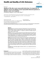
Triggering Myocardial Infarction
Figure. Cascade of Triggers Culminating in Acute Myocardial Infarction.
There are multiple stages in the atherosclerotic process culminating in thrombosis and acute myocardial infarction: initial formation of
plaque in a focal area of the artery; progression of some plaques to become vulnerable to disruption; precipitation of plaque disruption
through either rupture or erosion; and transient exacerbation of a prothrombotic environment. Identifiable factors or determinants, referred
to as triggers, are responsible for the development of each of these stages. These factors may be transient, such as an abrupt event that rap-
idly leads to plaque disruption or a brief exacerbation of prothrombotic conditions, or they may be longer-term determinants, such as system-
ic inflammation, a local vascular response to plaque formation leading to focal plaque inflammation, or a local area of flow disturbance
leading to the initiation of atherosclerosis. CAD denotes coronary artery disease.
Triggers leading to
atherosclerosis:
Risk factors for CAD
Local hemodynamic factors
(shear stress)
Focal atherosclerotic
plaque formation
Inflamed vulnerable
plaque
Rupture of vulnerable
plaque
Erosion of vulnerable
plaque without
inflammation
Coronary thrombosis and
acute myocardial infarction
Triggers probably leading to focal
inflammation:
Local hemodynamic factors
Local vascular-remodeling
response to plaque formation
Exacerbation by systemic
inflammation
Triggers leading to plaque
disruption:
Increased sympathetic
stimulation
Increased physical or emotional
stress
Increased local or systemic
inflammation
Increased vasoconstriction
Triggers predisposing to
thrombosis:
Increased coagulability
Increased inflammation
Increased viscosity
Increased vasoconstriction
Downloaded from www.nejm.org on February 18, 2008 . Copyright © 2004 Massachusetts Medical Society. All rights reserved.
n engl j med 351;17 www.nejm.org october 21, 2004
1718
PERSPECTIVE
mally in the pathophysiologic process
4
(see Figure).
Although atherosclerosis may affect the coronary
tree diffusely, the principal manifestations of coro-
nary plaque are highly focal. In a susceptible person
with an adverse risk-factor profile, local hemody-
namic disturbances, such as small areas of dis-
turbed coronary blood flow and low shear stress,
constitute the initiating event or trigger leading to
focal atherosclerotic plaque formation and progres-
sion, which may continue for years. Although there
may be many such areas of intimal thickening in
early stages of atherosclerosis, only a subgroup, and
perhaps a very small subgroup, of these coronary
plaques become inflamed or vulnerable at any one
time. Other plaques may acquire characteristics of
fibrosis and scarring, and still others may remain
quiescent.
The triggering factors that determine which of
those early plaques will progress and become in-
flamed are unknown, but it is likely that local he-
modynamic factors, local vascular-remodeling re-
sponses, and the degree of systemic inflammation
all contribute. Plaques that are inflamed and prone
to rupture are those in which there is expansive or
outward remodeling of the arterial wall, whereas
those that are more fibrotic and scarred, without
active inflammation, are associated with constric-
tive or inward remodeling. The divergent vascular-
remodeling characteristics most likely reflect the
balance in the dynamic regulation of collagen syn-
thesis and breakdown.
Epidemiologic studies have long demonstrated
the increased cardiac morbidity and mortality as-
sociated with particulate air pollution, and recent
investigations have focused on the mechanistic role
of air pollution in cardiovascular disease.
5
Inhala-
tion of particulate air pollution into the lungs leads
to both pulmonary and systemic inflammation, with
induction of cytokines and chemokines and gener-
ation of oxidative species. These injurious mole-
cules create and exacerbate inflammation and oxi-
dative stress, which lead to direct vascular injury,
atherosclerosis, and autonomic dysfunction. Partic-
ulate air pollution also rapidly leads to a significant
increase in fibrinogen, plasma viscosity, and platelet
activation, as well as the release of endothelins, a
family of potent vasoconstrictor molecules. Studies
in animals have clearly documented the short- and
long-term adverse effects of particulate air pollution
on each step of the triggering cascade of coronary
disease culminating in acute myocardial infarction:
accelerated atherosclerosis, vasoconstriction, and
increased thrombogenesis.
The association between exposure to traffic and
the abrupt onset of myocardial infarction described
by Peters et al. suggests that particulate air pollution
from traffic may have led to abrupt plaque disrup-
tion and perhaps to the exacerbation of a thrombo-
genic environment. Transient, intense inflamma-
tion, vasoconstriction, and increased coagulability,
alone or in combination, are potential culprits in
this process.
In addition to these extremely short-term effects
of particulate air pollution, its deleterious longer-
term effects on the entire gamut of atherosclerotic
triggers cannot be overemphasized. Decades of ep-
idemiologic evidence underscore the cardiovascu-
lar morbidity and mortality related to air pollution.
The proinflammatory, proatherosclerotic, and pro-
thrombotic effects of particulate air pollution are
compelling. As both epidemiologic and now mech-
anistic evidence mounts, there is greater urgency to
accelerate our efforts to reduce particulate air pol-
lution and to improve cardiovascular health.
Dr. Stone reports having received grant support from Boston Sci-
entific and Pfizer.
From the Cardiovascular Division, Brigham and Women's Hospi-
tal, Harvard Medical School, Boston.
1. Libby P. Inflammation in atherosclerosis. Nature 2002;420:
868-74.
2. Muller JE, Tofler GH, Stone PH. Circadian variation and trig-
gers of onset of acute cardiovascular disease. Circulation 1989;
79:733-43.
3. Virmani R, Kolodgie FD, Burke AP, Farb A, Schwartz SM. Les-
sons from sudden coronary death: a comprehensive morpholog-
ical classification scheme for atherosclerotic lesions. Arterioscler
Thromb Vasc Biol 2000;20:1262-75.
4. Stone PH, Coskun AU, Yeghiazarians Y, et al. Prediction of
sites of coronary atherosclerosis progression: in vivo profiling of
endothelial shear stress, lumen, and outer vessel wall character-
istics to predict vascular behavior. Curr Opin Cardiol 2003;18:
458-70.
5. Brook RD, Franklin B, Cascio W, et al. Air pollution and car-
diovascular disease: a statement for healthcare professionals
from the Expert Panel on Population and Prevention Science of
the American Heart Association. Circulation 2004;109:2655-71.
Triggering Myocardial Infarction
Downloaded from www.nejm.org on February 18, 2008 . Copyright © 2004 Massachusetts Medical Society. All rights reserved.