Statin Therapy, LDL Cholesterol, C-Reactive Protein, and Coronary Artery Disease pot
Bạn đang xem bản rút gọn của tài liệu. Xem và tải ngay bản đầy đủ của tài liệu tại đây (494.37 KB, 18 trang )
Statin Therapy, LDL
Cholesterol, C-Reactive
Protein, and Coronary
Artery Disease
n engl j med
352;1
www.nejm.org january
6, 2005
The
new england journal
of
medicine
29
original article
Statin Therapy, LDL Cholesterol,
C-Reactive Protein, and Coronary Artery Disease
Steven E. Nissen, M.D., E. Murat Tuzcu, M.D., Paul Schoenhagen, M.D.,
Tim Crowe, B.S., William J. Sasiela, Ph.D., John Tsai, M.D., John Orazem, Ph.D.,
Raymond D. Magorien, M.D., Charles O’Shaughnessy, M.D.,
and Peter Ganz, M.D., for the Reversal of Atherosclerosis
with Aggressive Lipid Lowering (REVERSAL) Investigators*
From the Cleveland Clinic Foundation,
Cleveland (S.E.N., E.M.T., P.S., T.C.); Pfizer,
New York (W.J.S., J.T., J.O.); Ohio State Uni-
versity Medical Center, Columbus (R.D.M.);
North Ohio Heart Care, Elyria (C.O.); and
Brigham and Women’s Hospital, Boston
(P.G.). Address reprint requests to Dr. Nis-
sen at the Department of Cardiovascular
Medicine, Cleveland Clinic Foundation, 9500
Euclid Ave., Cleveland, OH 44195, or at
*The REVERSAL Investigators are listed in
the Appendix.
N Engl J Med 2005;352:29-38.
Copyright © 2005 Massachusetts Medical Society.
background
Recent trials have demonstrated better outcomes with intensive than with moderate
statin treatment. Intensive treatment produced greater reductions in both low-density
lipoprotein (LDL) cholesterol and C-reactive protein (CRP), suggesting a relationship
between these two biomarkers and disease progression.
methods
We performed intravascular ultrasonography in 502 patients with angiographically doc-
umented coronary disease. Patients were randomly assigned to receive moderate treat-
ment (40 mg of pravastatin orally per day) or intensive treatment (80 mg of atorvastatin
orally per day). Ultrasonography was repeated after 18 months to measure the pro-
gression of atherosclerosis. Lipoprotein and CRP levels were measured at baseline and
follow-up.
results
In the group as a whole, the mean LDL cholesterol level was reduced from 150.2 mg per
deciliter (3.88 mmol per liter) at baseline to 94.5 mg per deciliter (2.44 mmol per liter) at
18 months (P<0.001), and the geometric mean CRP level decreased from 2.9 to 2.3 mg
per liter (P<0.001). The correlation between the reduction in LDL cholesterol levels and
that in CRP levels was weak but significant in the group as a whole (r=0.13, P=0.005),
but not in either treatment group alone. In univariate analyses, the percent change in the
levels of LDL cholesterol, CRP, apolipoprotein B-100, and non–high-density lipoprotein
cholesterol were related to the rate of progression of atherosclerosis. After adjustment
for the reduction in these lipid levels, the decrease in CRP levels was independently and
significantly correlated with the rate of progression. Patients with reductions in both
LDL cholesterol and CRP that were greater than the median had significantly slower rates
of progression than patients with reductions in both biomarkers that were less than the
median (P=0.001).
conclusions
For patients with coronary artery disease, the reduced rate of progression of athero-
sclerosis associated with intensive statin treatment, as compared with moderate statin
treatment, is significantly related to greater reductions in the levels of both atherogenic
lipoproteins and CRP.
abstract
Copyright © 2005 Massachusetts Medical Society. All rights reserved.
Downloaded from www.nejm.org at RIKSHOSPITALET HF on February 18, 2008 .
n engl j med
352;1
www.nejm.org january
6
,
2005
The
new england journal
of
medicine
30
wo recent trials demonstrated
that intensive lipid-lowering therapy with
statins improved clinical outcomes
1
and
reduced the progression of atherosclerosis.
2
Many
authorities attributed the greater benefits of inten-
sive statin therapy, as compared with moderate
statin therapy, to greater reductions in the levels of
atherogenic lipoproteins, particularly low-density
lipoprotein (LDL) cholesterol.
3
However, statins
have a wide range of biologic effects in addition to
lipid lowering, including reductions in the levels of
C-reactive protein (CRP), a phenomenon common-
ly termed a “pleiotropic effect.”
4-6
In both recent
comparisons, at the conclusion of the trials, CRP
levels were 30 to 40 percent lower after intensive
statin therapy than after moderate treatment.
4
This
finding raises a provocative scientific question: Do
reductions in CRP represent an independent factor
influencing the benefits of more intensive statin
therapy?
Large observational studies have established a
strong relationship between CRP levels and the
morbidity and mortality associated with coronary
disease.
7-9
However, the precise mechanism under-
lying the association between CRP levels and ad-
verse outcomes remains incompletely described.
Theoretically, by decreasing the levels of atherogen-
ic lipoproteins, statins could decrease systemic in-
flammation, thereby reducing CRP levels. An alter-
native hypothesis proposes that statins have direct
antiinflammatory effects, independent of their lip-
id-lowering capabilities. In this model, CRP plays a
more direct role in the pathogenesis of atheroscle-
rosis, and a statin-mediated reduction in inflamma-
tion contributes directly to reduced disease activity.
Because statins decrease the levels of both LDL cho-
lesterol and CRP, it is difficult to determine whether
CRP is an indirect biomarker reflecting the benefits
of statins or a direct participant in atherogenesis.
Intravascular ultrasonography is a useful tech-
nique for assessing the effect of therapies on the
vascular wall, providing a precise and continuous
measure of the progression of atherosclerosis. In
the Reversal of Atherosclerosis with Aggressive Lip-
id Lowering (REVERSAL) trial, intensive therapy
with 80 mg of atorvastatin per day slowed the pro-
t
Table 1. Laboratory Values at Baseline and Follow-up and Change in Values from Baseline.*
Characteristic
Both Groups
(N=502)
Pravastatin Group
(N=249)
Atorvastatin Group
(N=253) P Value†
Baseline
Total cholesterol (mg/dl)
232.2±34.2 232.6±34.1 231.8±34.2 0.80
LDL cholesterol (mg/dl) 150.2±26.9 150.2±25.9 150.2±27.9 0.99
HDL cholesterol (mg/dl) 42.6±10.7 42.9±11.4 42.3±9.9 0.51
Non-HDL cholesterol (mg/dl) 189.6±32.5 189.7±32.3 189.5±32.7 0.96
Triglycerides (mg/dl) 197.4±100.6 197.7±105.6 197.2±95.7 0.96
Apo B-100 (mg/dl) 152.7±23.4 153.0±22.4 152.4±24.3 0.79
CRP (mg/liter)‡ 0.46
Geometric mean 2.9 3.0 2.8
Interquartile range 1.4 to 6.1 1.4 to 6.1 1.3 to 6.3
18-Mo follow-up
Total cholesterol (mg/dl)
169.2±40.0 187.5±32.2 151.3±38.9 <0.001
LDL cholesterol (mg/dl) 94.5±32.2 110.4±25.8 78.9±30.2 <0.001
HDL cholesterol (mg/dl) 43.8±11.3 44.6±11.3 43.1±11.3 0.15
Non-HDL cholesterol (mg/dl) 125.4±39.6 142.9±32.2 108.1±38.6 <0.001
Triglycerides (mg/dl) 157.0±93.8 165.8±92.1 148.4±94.9 0.04
Apo B-100 (mg/dl) 104.8±29.1 118.1±24.0 91.8±27.9 <0.001
CRP (mg/liter)‡ <0.001
Geometric mean 2.3 2.9 1.8
Interquartile range 0.9 to 5.4 1.3 to 6.2 0.8 to 4.3
Copyright © 2005 Massachusetts Medical Society. All rights reserved.
Downloaded from www.nejm.org at RIKSHOSPITALET HF on February 18, 2008 .
n engl j med
352;1
www.nejm.org january
6, 2005
ldl cholesterol, c-reactive protein, and atherosclerosis progression
31
gression of atherosclerosis more than did moder-
ate treatment with 40 mg of pravastatin per day.
2
We applied statistical methods to examine the rela-
tionship between the reductions in LDL cholesterol
and CRP levels and the rate of disease progression
measured by intravascular ultrasonography.
study design
The institutional review board of each participat-
ing center approved the protocol, and all patients
provided written informed consent. Intravascular
ultrasonography was performed in a single vessel
in patients who had a clinical indication for coro-
nary angiography and had stenosis of at least 20
percent on angiography. Eligible patients had to
have an LDL cholesterol level of 125 to 210 mg per
deciliter (3.23 to 5.43 mmol per liter) after a statin-
free washout period of 4 to 10 weeks. Patients were
randomly assigned to receive either 40 mg of prav-
astatin or 80 mg of atorvastatin orally daily. The pa-
tients and all study personnel were unaware of the
treatment assignments or the results of laboratory
measurements.
intravascular ultrasonography
Investigators performed intravascular ultrasonogra-
phy in the longest and least angulated target vessel
that met the inclusion criteria. After the adminis-
methods
* Plus–minus values are means ±SD. To convert values for cholesterol to millimoles per liter, multiply by 0.02586. To con-
vert values for triglycerides to millimoles per liter, multiply by 0.01129.
† P values were calculated by means of the two-sample t-test.
‡ CRP levels were not available for six patients at baseline or follow-up (one in the pravastatin group and five in the atorva-
statin group).
Table 1. (Continued.)
Characteristic
Both Groups
(N=502)
Pravastatin Group
(N=249)
Atorvastatin Group
(N=253) P Value†
Change from baseline
Total cholesterol
<0.001
Mean (mg/dl) ¡63±44 ¡45±37 ¡81±43
Percent ¡26.3 ¡18.4 ¡34.1
LDL cholesterol <0.001
Mean (mg/dl) ¡56±37 ¡40±29 ¡71±37
Percent ¡35.8 ¡25.2 ¡46.3
HDL cholesterol 0.11
Mean (mg/dl) 1.2±7.9 1.6±7.7 0.8±8.0
Percent 4.2 5.6 2.9
Non-HDL cholesterol <0.001
Mean (mg/dl) ¡64±43 ¡47±35 ¡81±43
Percent ¡33.0 ¡23.6 ¡42.2
Triglycerides 0.002
Mean (mg/dl) ¡40±96 ¡32±94 ¡49±98
Percent ¡13.5 ¡6.8 ¡20.0
Apo B-100 <0.001
Mean (mg/dl) ¡48±30 ¡35±25 ¡61±30
Percent ¡30.6 ¡22.0 ¡39.1
CRP‡ <0.001
Geometric mean (mg/liter) ¡0.2 0.2 ¡0.7
Interquartile range (mg/liter) ¡1.9 to 0.8 ¡1.5 to 1.6 ¡2.8 to 0.1
Percent ¡22.4 –5.2 ¡36.4
Copyright © 2005 Massachusetts Medical Society. All rights reserved.
Downloaded from www.nejm.org at RIKSHOSPITALET HF on February 18, 2008 .
n engl j med
352;1
www.nejm.org january
6
,
2005
The
new england journal
of
medicine
32
tration of intracoronary nitroglycerin, the transduc-
er was positioned in the distal vessel and withdrawn
at a rate of 0.5 mm per second (the “pullback”) with
the use of a motor drive. A core laboratory evaluat-
ed the image quality of each ultrasonogram, and
only patients whose ultrasonograms met prespeci-
fied image-quality requirements were eligible for
randomization. After an 18-month treatment peri-
od, patients again underwent intravascular ultraso-
nography under identical conditions. This method
of intravascular ultrasonography has been described
previously in detail.
2,10,11
core laboratory measurements
Personnel who were unaware of the patients’ clin-
ical characteristics and treatment assignments
used manual planimetry to measure, on a computer
screen, a series of cross-sections of ultrasonograph-
ic images selected exactly 1.0 mm apart along the
long axis of the vessel. Measurements were per-
formed in accordance with the standards of the
American College of Cardiology and the European
Society of Cardiology.
12
For each cross-section ana-
lyzed, the operator measured the area of the exter-
nal elastic membrane and the lumen. The accuracy
and reproducibility of this method have been report-
ed previously.
2,13
calculation of end points
The average area of atheroma per cross-section was
calculated as follows:
S
(EEM
CSA
– LUMEN
CSA
),
n
where EEM
CSA
is the cross-sectional area of the
external elastic membrane, LUMEN
CSA
is the cross-
sectional area of the lumen, and n is the number of
cross-sections in the pullback. To compensate for
pullbacks of differing lengths, the total atheroma
volume for each patient was calculated as the av-
erage area of atheroma multiplied by the median
number of cross-sections in the pullbacks for all
patients in the study. The efficacy variable “change
in normalized total atheroma volume” (TAV) was
calculated as TAV
18
months
¡TAV
baseline
. The percent
atheroma volume (PAV) was calculated with the use
of the following formula:
S
(EEM
CSA
– LUMEN
CSA
)
¬100.
S
EEM
CSA
The efficacy variable “change in PAV” was calculat-
ed as PAV
18
months
–PAV
baseline
.
laboratory tests
A central laboratory performed all biochemical de-
terminations (Medical Research Laboratory, High-
land Heights, Ky.).
statistical analysis
For continuous variables with a normal distribution,
means
±
SD are reported. For CRP levels, the geo-
metric means and interquartile ranges are report-
ed. Because the ultrasonographic end points were
not normally distributed, we applied an analysis-of-
covariance model to rank-transformed data to deter-
mine P values. Correlations between variables are
described with the use of Spearman rank-correla-
tion coefficients, and multivariate regression analy-
ses based on rank-transformed data were used to
obtain partial correlation coefficients adjusted for
the effects of covariates.
14
The ultrasonographic
variable served as the dependent variable; the inde-
pendent variables consisted of the change in CRP
coupled with the change in non–high-density lipo-
protein (non-HDL) cholesterol, LDL cholesterol, or
apolipoprotein B-100 (apo B-100). For a further de-
scription of bivariate relationships with ultrasono-
graphic end points, we used the locally weighted
scatterplot smoothing (LOWESS) technique.
15
This
technique is designed to produce a smooth fit to
the data and reduces the influence of extreme out-
liers. Analyses were performed with the use of SAS
software, version 6.12.
patient population
Between June 1999 and September 2001, 502 pa-
tients were enrolled at 34 U.S. centers and under-
went intravascular ultrasonography at both base-
line and 18 months of follow-up that could be
evaluated (249 in the pravastatin group and 253 in
the atorvastatin group). The average age was 56
years, 72 percent were men, 89 percent were white
(race was recorded by the study coordinators on the
case-report form), 26 percent were current smok-
ers, 69 percent had a history of hypertension, and
19 percent had a history of diabetes.
2
laboratory findings and results
of intravascular ultrasonography
Table 1 summarizes laboratory values at baseline
and at the completion of the study (18 months) for
the entire population and each treatment group. For
results
Copyright © 2005 Massachusetts Medical Society. All rights reserved.
Downloaded from www.nejm.org at RIKSHOSPITALET HF on February 18, 2008 .
n engl j med
352;1
www.nejm.org january
6, 2005
ldl cholesterol, c-reactive protein, and atherosclerosis progression
33
all 502 patients, the mean baseline LDL cholester-
ol level was 150.2 mg per deciliter (3.88 mmol per
liter), the non-HDL cholesterol level was 189.6 mg
per deciliter (4.90 mmol per liter), and the geomet-
ric mean CRP level was 2.9 mg per liter. After 18
months of treatment, the mean LDL cholesterol lev-
el was 94.5 mg per deciliter (2.44 mmol per liter),
the non-HDL cholesterol level was 125.4 mg per
deciliter (3.24 mmol per liter), and the geometric
mean CRP level was 2.3 mg per liter. There were
greater reductions in LDL cholesterol, non-HDL
cholesterol, and CRP levels in the atorvastatin group
than in the pravastatin group (P<0.001 for each
comparison).
2
Table 2 summarizes measures of disease burden
as determined by intravascular ultrasonography at
baseline and the completion of the study for the en-
tire population and the two treatment groups. Both
measures of the progression of atherosclerosis —
total atheroma volume and percent atheroma vol-
ume — reflected a slower rate of progression in the
group that received intensive treatment with ator-
vastatin than in the group that received moderate
treatment with pravastatin.
correlation between reductions
in lipoprotein and crp
There was a weak but significant correlation be-
tween the percent reductions in LDL cholesterol and
in CRP levels only for the study group as a whole
(r=0.13, P=0.005) — not for the pravastatin group
alone (r=¡0.008, P=0.90) or the atorvastatin group
alone (r=0.09, P=0.17). Changes in other athero-
genic lipoproteins, such as apo B-100 and non-HDL
cholesterol, had similarly weak correlations with the
reduction in CRP levels in the regression analysis.
effect of changes in crp and lipids
on progression
Table 3 summarizes the correlations between the
changes in the levels of atherogenic lipoproteins,
CRP, and HDL cholesterol and the rate of progres-
* Values in parentheses are interquartile ranges.
† P values were calculated with the use of the Wilcoxon rank-sum test.
‡ Values were adjusted for pullbacks of different lengths by multiplying the average area of atheroma volume for each patient by the median
number of cross-sections in the pullbacks for all patients in the study.
Table 2. Baseline and Follow-up Values for Intravascular Ultrasonographic End Points and Change in Values from Baseline.*
Atheroma Volume Both Groups (N=502) Pravastatin Group (N=249) Atorvastatin Group (N=253) P Value†
Mean ±SD Median Mean ±SD Median Mean ±SD Median
Baseline
Total (mm
3
)
189.4±115.3 165.9
(113.8 to 238.9)
194.5±114.8 168.6
(117.4 to 246.2)
184.4±115.7 161.9
(111.0 to 228.2)
0.20
Normalized
total (mm
3
)‡ 184.1±83.1 174.5
(122.1 to 232.3)
189.1±86.5 187.2
(122.1 to 239.1)
179.1±79.4 166.6
(122.4 to 226.6)
0.26
Percent 38.9±11.0 38.9
(32.2 to 46.2)
39.5±10.8 40.0
(32.5 to 46.3)
38.4±11.3 38.2
(31.7 to 45.8)
0.18
18-Mo follow-up
Total (mm
3
)
191.7±110.7 169.9
(113.3 to 244.0)
199.6±112.3 180.0
(125.5 to 255.3)
183.9±108.8 160.9
(107.4 to 240.3)
0.04
Normalized
total (mm
3
)‡ 186.5±81.5 175.7
(124.5 to 239.2)
194.2±86.0 179.7
(128.9 to 248.2)
178.9±76.2 170.5
(119.8 to 222.2)
0.08
Percent 40.2±10.5 39.9
(33.8 to 47.1)
41.4±10.0 41.8
(35.0 to 47.7)
39.0±10.8 38.7
(31.6 to 45.7)
<0.001
Change from baseline
Total (mm
3
)
2.3±31.7 1.4
(¡14.4 to 19.5)
5.1±31.4 4.4
(¡13.3 to 21.9)
¡0.4±31.8 ¡0.9
(¡14.5 to 13.8)
0.04
Normalized
total (mm
3
)‡ 2.4±29.4 1.5
(¡15.3 to 20.1)
5.1±27.6 4.1
(¡13.2 to 23.5)
¡0.2±31.0 ¡0.9
(¡17.9 to 15.3)
0.03
Percent 1.3±5.1 0.9
(¡1.9 to 4.4)
1.9±4.9 1.6
(¡1.6 to 4.7)
0.6±5.1 0.2
(¡2.5 to 3.9)
0.002
Copyright © 2005 Massachusetts Medical Society. All rights reserved.
Downloaded from www.nejm.org at RIKSHOSPITALET HF on February 18, 2008 .
n engl j med
352;1
www.nejm.org january
6
,
2005
The
new england journal
of
medicine
34
sion of atherosclerosis for both end points assessed
by means of intravascular ultrasonography. Univar-
iate analysis revealed significant correlations be-
tween ultrasonographic measures of disease pro-
gression and laboratory measures of atherogenic
lipoproteins, including LDL cholesterol, apo B-100,
and non-HDL cholesterol. The percent change in
the LDL cholesterol level had the closest correlation
with progression, with a correlation coefficient of
0.12 for total atheroma volume (P=0.005) and of
0.14 for percent atheroma volume (P=0.002).
The correlations between the reduction in CRP
levels and the rates of progression on intravascular
ultrasonography were also significant and similar
in strength to the relationships observed for the
atherogenic lipoproteins. Univariate analysis yield-
ed a correlation coefficient of 0.11 for both total and
percent atheroma volume (P=0.02 and P=0.01, re-
spectively). Most correlations between the rates of
progression on ultrasonography and the percent
change in non-HDL cholesterol, LDL cholesterol,
and CRP levels remained significant on multivariate
analysis but were weaker than those obtained by
univariate analyses (Table 3).
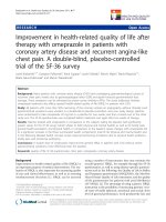
As shown in Figure 1, greater reductions in LDL
cholesterol levels were associated with slower rates
of progression on intravascular ultrasonography.
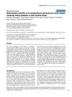
Figure 2 shows this same relationship for the re-
duction in CRP levels. Patients with the largest re-
ductions in CRP levels had regression of atheroma,
as evidenced by progression rates of less than zero.
Table 4 shows the rates of progression of ath-
erosclerosis on ultrasonography for subgroups de-
fined according to whether the reductions in LDL
cholesterol or CRP levels were greater than or less
than the median decreases. For both efficacy mea-
sures, the highest rates of progression were in the
subgroup in which decreases in both LDL choles-
terol and CRP levels were less than the median. Sig-
nificantly lower progression rates were observed in
the subgroup with decreases in both LDL cholester-
ol and CRP levels that were greater than the median
(P=0.001 for both efficacy measures).
Epidemiologic evidence has established a strong
relationship between elevated levels of atherogenic
lipoproteins, particularly LDL cholesterol, and the
risk of death and complications from cardiovascu-
lar causes. Placebo-controlled trials of statins have
demonstrated that pharmacologic therapies that re-
duce LDL cholesterol levels also proportionally de-
crease cardiovascular risk.
16-19
Accordingly, the
clinical benefits of statin therapy have largely been
attributed to reductions in the levels of atherogenic
lipoproteins. However, observational studies have
also established a strong relationship between the
levels of CRP, the most stable and reliable labora-
tory measure of systemic inflammation, and adverse
discussion
* Values are Spearman rank-correlation coefficients.
Table 3. Relationships between Changes in Laboratory Measures
and Intravascular Ultrasonographic End Points.
Laboratory Measure
Percent Atheroma
Volume
Total Atheroma
Volume
Correlation
Coefficient*
P
Value
Correlation
Coefficient*
P
Value
Univariate analysis
LDL cholesterol
Change 0.10 0.03 0.09 0.04
Percent change 0.14 0.002 0.12 0.005
HDL cholesterol
Change ¡0.04 0.40 ¡0.01 0.84
Percent change ¡0.04 0.42 ¡0.01 0.78
Triglycerides
Change 0.05 0.23 0.06 0.19
Percent change 0.08 0.08 0.08 0.09
Non-HDL cholesterol
Change 0.09 0.05 0.07 0.10
Percent change 0.13 0.004 0.10 0.02
apo B-100
Change 0.09 0.05 0.08 0.06
Percent change 0.13 0.004 0.12 0.008
CRP
Change 0.11 0.01 0.11 0.02
Percent change 0.11 0.01 0.11 0.02
Multivariate analysis (adjusted for changes
in CRP and non-HDL cholesterol)
Percent change in non-HDL
cholesterol
0.11 0.01 0.08 0.06
Percent change in CRP 0.09 0.04 0.09 0.05
Multivariate analysis (adjusted for changes
in CRP and LDL cholesterol)
Percent change in LDL
cholesterol
0.12 0.008 0.11 0.02
Percent change in CRP 0.09 0.04 0.08 0.06
Multivariate analysis (adjusted for changes
in CRP and apo B-100)
Percent change in apo B-100
0.11 0.01 0.10 0.03
Percent change in CRP 0.09 0.05 0.08 0.07
Copyright © 2005 Massachusetts Medical Society. All rights reserved.
Downloaded from www.nejm.org at RIKSHOSPITALET HF on February 18, 2008 .
n engl j med
352;1
www.nejm.org january
6, 2005
ldl cholesterol, c-reactive protein, and atherosclerosis progression
35
cardiovascular outcomes. Statins have a variety of
pleiotropic properties, including their ability to in-
duce dose-dependent decreases in the levels of CRP
and other inflammatory biomarkers.
5,6
Since stat-
ins reduce the levels of both LDL cholesterol and
CRP, it is difficult to determine the relative contribu-
tion of the reduction in each of these biomarkers to
the observed clinical benefits.
We sought to close this gap in knowledge by an-
alyzing the correlation among lipid levels, CRP lev-
els, and the rate of progression of atherosclerosis,
using intravascular ultrasonography to measure dis-
ease progression in patients who were being treat-
ed with statins.
2
Intravascular ultrasonography is
a useful technique for assessing the effect of thera-
pies on the vascular wall, providing a precise and
continuous measure of disease progression.
20
In
the REVERSAL trial, intensive therapy with 80 mg
of atorvastatin per day slowed the rate of progres-
sion of atherosclerosis more than did moderate
treatment with 40 mg of pravastatin per day. Be-
cause we studied two different intensities of statin
therapy, we evaluated a broad range of reductions
in LDL cholesterol and CRP, permitting a post hoc
analysis of the relationship between these two bio-
markers and the rate of progression of atheroscle-
rosis across a clinically important range of values.
Correlation analysis revealed that reductions
in the levels of atherogenic lipoproteins were not
closely correlated with reductions in CRP levels.
There was a weak but significant correlation be-
tween the reduction in LDL cholesterol levels and
the reduction in CRP levels for the overall group of
502 patients (r=0.13, P=0.005), but not in either
treatment group alone. These data demonstrate that
statin-mediated reductions in CRP are largely unre-
lated to the decrease in LDL cholesterol levels. These
findings confirm the work of other investigators
and strongly suggest that the statin-mediated re-
duction in CRP is unlikely to be a secondary conse-
quence of a reduction in LDL cholesterol but, rather,
is potentially mediated by independent pathways.
21
Analysis of the relationship among lipopro-
tein levels, CRP levels, and the rate of progression
of atherosclerosis yielded particularly informative
results. Reductions in both LDL cholesterol and
CRP levels were significantly correlated to the rate
of progression. In univariate analyses, both ultra-
sonographic measures of progression — the change
in the normalized total atheroma volume and the
change in percent atheroma volume — correlated
significantly with the reduction in the levels of ath-
erogenic lipoproteins, including LDL cholesterol,
non-HDL cholesterol, and apo B-100. The clos-
est correlation was between the LDL cholesterol
level and the percent atheroma volume (r=0.14,
P=0.002). However, similar correlations were ob-
served for the relationship between the reduction
in CRP levels and the rate of progression on intra-
vascular ultrasonography (r=0.11, P=0.01). Substi-
tuting non-HDL cholesterol for LDL cholesterol,
to account for the broad range of atherogenic lipo-
proteins, did not increase the correlation. Since the
levels of both CRP and LDL cholesterol showed rel-
atively weak correlations with the ultrasonographic
end points (r values of 0.11 to 0.14), this analysis
Figure 1. Locally Weighted Smoothed Scatterplots Showing the Relationship
between the Change in LDL Cholesterol Levels and the Rate of Progression
of Atherosclerosis in the Entire Group of 502 Patients.
In each plot, the solid line represents the point estimates and the upper
and lower lines the 95 percent confidence intervals. To convert values for LDL
cholesterol to millimoles per liter, multiply by 0.02586.
Change in Percent Atheroma Volume
2.5
3.0
2.0
1.5
0.5
1.0
0.0
¡100 ¡75 ¡50 ¡25 0
25
Change in LDL Cholesterol (mg/dl)
3.5
Change in Total Atheroma Volume (mm
3
)
8
12
10
6
4
¡2
0
2
¡4
¡100 ¡75 ¡50 ¡25 0
25
Change in LDL Cholesterol (mg/dl)
14
Copyright © 2005 Massachusetts Medical Society. All rights reserved.
Downloaded from www.nejm.org at RIKSHOSPITALET HF on February 18, 2008 .
n engl j med
352;1
www.nejm.org january
6
,
2005
The
new england journal
of
medicine
36
demonstrates that biomarkers can account for only
a small fraction of the observed progression rate.
To determine whether the reduction in CRP lev-
els represented an independent factor influencing
the progression of atherosclerosis, we adjusted the
CRP correlations for the effects of atherogenic lipo-
proteins. In this multivariate analysis, CRP remained
significant in most analyses, regardless of which
measure of atherogenic lipoproteins was used —
LDL cholesterol, apo B-100, or non-HDL cholester-
ol. Patients with reductions in the levels of both LDL
cholesterol and CRP that were greater than the me-
dian reduction had significantly lower progression
rates than patients in whom the reductions were
less than the median decrease (P=0.001). These data
provide evidence that the reduction in CRP levels
plays an independent role in the beneficial effects
of statins on the progression of coronary athero-
sclerosis.
Since measures of progression reflected by intra-
vascular ultrasonography are not normally distrib-
uted, we used LOWESS methods to illustrate the
relationships between the reductions in LDL cho-
lesterol and CRP levels and the rates of progres-
sion determined by ultrasonography (Fig. 1 and 2).
These plots demonstrated a continuous relationship
between the magnitude of reduction in either LDL
cholesterol or CRP levels and the rates of progres-
sion of atherosclerosis for both measures of effi-
cacy. Atherosclerosis regressed in patients with the
greatest reduction in CRP levels, but not in those
with the greatest reduction in LDL cholesterol lev-
els. Although the data are not provided in this arti-
cle, LOWESS plots showed slower rates of progres-
sion in the intensively treated atorvastatin subgroup
across a broad range of reductions in lipids and CRP.
The slower rate of progression in the atorvastatin
group for any magnitude of reduction in LDL cho-
lesterol levels can be partially explained by the ad-
ditional effects of treatment on the reduction in CRP
levels, just as the differences in the CRP plots can
be partially explained by the additional reduction
in LDL cholesterol levels effected by atorvastatin
therapy. Thus, the effects of the reductions in both
LDL cholesterol and CRP levels must be considered
to explain the observed differences in progression
between atorvastatin and pravastatin treatment.
Our findings have important implications for
understanding the pathogenesis of the progression
of atherosclerosis and the mechanism of benefit
of statin therapy. The Pravastatin or Atorvastatin
and Infection Therapy (PROVE IT) trial demon-
strated improved outcomes
1
and the REVERSAL tri-
al demonstrated reduced rates of progression of
atherosclerosis
2
after intensive, as compared with
moderate, statin therapy. Although a single trial had
previously shown that the effects of statins are evi-
dent within 16 weeks,
22
the rapidity of the diver-
gence in results between the treatment groups in
both trials was unexpected.
4
In most earlier place-
bo-controlled trials, differences between statins
and placebo were not evident for the first two years
after randomization.
16-18
However, in both the
REVERSAL and PROVE IT trials, CRP levels were
30 to 40 percent lower at the conclusion of the trial
in the intensively treated patients than in the group
that received moderate treatment, which may ex-
Figure 2. Locally Weighted Smoothed Scatterplots Showing the Relationship
between the Changes in CRP Levels and the Rate of Progression of Athero-
sclerosis in the Entire Group of 502 Patients.
In each plot, the solid line represents the point estimates and the upper
and lower lines the 95 percent confidence intervals.
Change in Percent Atheroma Volume
2.5
1.5
¡1.5
0.5
¡0.5
¡2.5
0.0
¡14 0
642¡2¡4¡6¡8¡10¡12
¡14 0
642¡2¡4¡6¡8¡10¡12
Change in CRP (mg/liter)
3.5
Change in Total Atheroma Volume (mm
3
)
5
10
0
¡5
¡10
¡15
Change in CRP (mg/liter)
15
Copyright © 2005 Massachusetts Medical Society. All rights reserved.
Downloaded from www.nejm.org at RIKSHOSPITALET HF on February 18, 2008 .
n engl j med
352;1
www.nejm.org january
6, 2005
ldl cholesterol, c-reactive protein, and atherosclerosis progression
37
plain the magnitude and unexpectedly rapid diver-
gence of outcomes reported by Ridker et al. else-
where in this issue of the
Journal
.
23
Our findings are consistent with a variety of ex-
perimental observations that suggest a direct role
for CRP in the pathogenesis of atherosclerosis.
CRP renders oxidized LDL more susceptible to
uptake by macrophages, induces the expression
of vascular-cell adhesion molecules, stimulates the
production of tissue factor, and impairs the produc-
tion of nitric oxide.
24-27
Children with elevated CRP
levels have increased carotid intimal medial thick-
ness and reduced vasodilatation mediated by bra-
chial-artery flow.
28
A recent study suggested that
the presence of above-average levels of CRP attenu-
ates the benefits of intensive statin therapy with re-
spect to the carotid intimal media thickness.
29
Evidence of a dual mechanism of benefit for
statins — lipid lowering and a reduction in inflam-
mation — has important implications for current
and future treatment of atherosclerosis. Current
guidelines emphasize the use of lipid-lowering ther-
apies to reach target levels of LDL cholesterol, non-
HDL cholesterol, or both. However, individual agents
differ in their ability to reduce the levels of inflam-
matory biomarkers. Accordingly, our study raises
the provocative question of whether the effects of
statins on CRP, as well as LDL cholesterol, should
be considered in decisions regarding therapy.
Our study has important limitations. It is a hy-
pothesis-generating post hoc analysis examining
the effect of a single inflammatory marker on dis-
ease progression, not morbidity or mortality. None-
theless, our findings suggest that the level of CRP
may ultimately represent an important therapeutic
target. We do not believe that these data are suffi-
cient to recommend routine serial measurement of
CRP in order to modulate statin therapy, but further
study is warranted. An ongoing clinical trial is as-
sessing the use of CRP levels to guide therapy in pa-
tients who do not have elevated LDL cholesterol
levels.
30
Since approaches to the reduction of LDL
cholesterol levels that do not involve statins have
uncertain antiinflammatory effects, the ability of
such therapies to improve the outcome requires
testing in clinical trials.
31
Funded by Pfizer.
Dr. Nissen reports having served as a consultant to AstraZeneca,
Atherogenics, Lipid Sciences, Wyeth, Novartis, Pfizer, Sankyo, Take-
da, Kowa, Sanofi, Novo-Nordisk, Eli Lilly, Kos Pharmaceuticals,
GlaxoSmithKline, Forbes Medi-tech, and Merck–Schering Plough;
having served as a lecturer for AstraZeneca and Pfizer; and having
received funding from AstraZeneca, Takeda, Sankyo, Pfizer, Athero-
genics, and Lipid Sciences for ongoing clinical trials. Dr. Tuzcu re-
ports having received lecture fees from AstraZeneca, Merck, Pfizer,
and Takeda and grant support from Pfizer. Mr. Crowe reports own-
ing Pfizer stock. Drs. Sasiela, Tsai, and Orazem are employees of
Pfizer. Dr. Magorien reports having served as a consultant to Bristol-
Myers Squibb and owning stock in Merck. Dr. Ganz reports having
served as a consultant for AstraZeneca and Pfizer and a lecturer for
Pfizer.
* CRP levels were not available for six patients at baseline or follow-up. The subgroups were formed on the basis of the median percent change
in LDL cholesterol of ¡37.1 percent and the median percent change in CRP of ¡21.4 percent.
† Values in parentheses are interquartile ranges. Confidence intervals (CIs) are for the medians.
‡ P=0.001 for the comparison with the subgroup in which the reduction in the levels of both LDL cholesterol and CRP was less than the median
reduction (by Wilcoxon’s rank-sum test).
Table 4. Rates of Progression According to the Change in LDL Cholesterol and CRP Levels.*
Subgroup
No. of
Patients Percent Atheroma Volume† Total Atheroma Volume (mm
3
)†
Median 95% CI Mean ±SD Median 95% CI Mean ±SD
Reduction in LDL cholesterol and
CRP both greater than median
141 0.24 (¡2.8 to 3.5)‡ ¡0.77 to 0.54 0.33±5.3 ¡1.98 (¡23.0 to 10.8)‡ ¡6.26 to 3.67 ¡2.41±31.6
Reduction in LDL cholesterol
greater than median, reduc-
tion in CRP less than median
106 0.81 (¡2.0 to 4.8) ¡0.32 to 1.81 1.62±4.7 2.06 (¡12.8 to 21.5) ¡3.26 to 6.41 4.04±28.7
Reduction in LDL cholesterol less
than median, reduction in
CRP greater than median
108 1.21 (¡2.0 to 4.0) ¡0.31 to 2.08 0.91±4.9 ¡1.04 (¡18.6 to 22.5) ¡6.78 to 8.74 1.42±29.2
Reduction in LDL cholesterol and
CRP both less than median
141 1.82 (¡1.5 to 5.1) 1.0 to 2.84 2.25±5.0 8.21 (¡11.8 to 27.5) 0.40 to 13.05 7.49±27.5
Copyright © 2005 Massachusetts Medical Society. All rights reserved.
Downloaded from www.nejm.org at RIKSHOSPITALET HF on February 18, 2008 .
n engl j med
352;1
www.nejm.org january
6
,
2005
38
ldl cholesterol, c-reactive protein, and atherosclerosis progression
appendix
In addition to the authors, the following investigators participated in this study: Wake Forest University, Winston-Salem, N.C., M. Kutcher;
University of Colorado Health Sciences Center, Denver, J. Burchenal; University of Texas–San Antonio, San Antonio, S. Bailey; Heart Insti-
tute at Borgess, Kalamazoo, Mich., T. Fischell; University of Florida, Gainesville, R. Kerensky; Heart Care Center, Blue Island, Ill., R. Iaf-
faldano; University of Chicago, Chicago, J. Lopez; William Beaumont Hospital, Royal Oak, Mich., C. Grines; University of California, San
Diego, San Diego, A. DeMaria; UCLA Medical Center for Health Sciences, Los Angeles, J. Tobis; LeBauer Cardiovascular Research Founda-
tion, Greensboro, N.C., B. Brodie; University of Washington Medical Center, Seattle, D. Linker; Cedars-Sinai Medical Center, Los Angeles,
J. Forrester; University of North Carolina, Chapel Hill, S. Smith; Androscoggin Cardiology Research, Auburn, Me., R. Weiss; Medical Col-
lege of Ohio, Toledo, C. Cooper; Rhode Island Hospital, Providence, B. Sharaf; East Carolina University, Greenville, N.C., M. Miller; Buffalo
Cardiology and Pulmonary Associates, Buffalo, N.Y., J. Corbelli; Heart Care Group, Allentown, Pa., J. Kleaveland; University of Arkansas for
Medical Sciences, Little Rock, L. Garza; University of Louisville, Louisville, Ky., M. Leesar; Capital Cardiology Associates, Albany, N.Y.,
A. DeLago; Cardiology of Georgia–Piedmont Hospital, Atlanta, C. Wickliffe; New England Medical Center, Boston, J. Kuvin; Kramer &
Crouse Cardiology, Kansas City, Mo., P. Kramer; Miriam Hospital, Providence, R.I., P. Gordon; Mount Sinai Hospital, New York, S. Sharma;
Oklahoma Heart Institute, Tulsa, W. Leimbach; Eastlake Cardiovascular Associates, St. Clair Shores, Mich., R. Cleary, Jr.; University Hospi-
tals of Cleveland, R. Nair.
references
1.
Cannon CP, Braunwald E, McCabe CH,
et al. Intensive versus moderate lipid lower-
ing with statins after acute coronary syn-
dromes. N Engl J Med 2004;350:1495-504.
2.
Nissen SE, Tuzcu EM, Schoenhagen P,
et al. Effect of intensive compared with mod-
erate lipid-lowering therapy on progression
of coronary atherosclerosis: a randomized
controlled trial. JAMA 2004;291:1071-80.
3.
Sacks FM. High-intensity statin treat-
ment for coronary heart disease. JAMA 2004;
291:1132-4.
4.
Topol EJ. Intensive statin therapy —
a sea change in cardiovascular prevention.
N Engl J Med 2004;350:1562-4.
5.
Liao JK. Isoprenoids as mediators of
the biological effects of statins. J Clin Invest
2002;110:285-8.
6.
Albert MA, Danielson E, Rifai N, Ridker
PM. Effect of statin therapy on C-reactive
protein levels: the pravastatin inflammation/
CRP evaluation (PRINCE): a randomized tri-
al and cohort study. JAMA 2001;286:64-70.
7. Ridker PM, Rifai N, Rose L, Buring JE,
Cook NR. Comparison of C-reactive protein
and low-density lipoprotein cholesterol lev-
els in the prediction of first cardiovascular
events. N Engl J Med 2002;347:1557-65.
8. Ridker PM. Clinical application of
C-reactive protein for cardiovascular disease
detection and prevention. Circulation 2003;
107:363-9.
9. Ridker PM, Hennekens CH, Buring JE,
Rifai N. C-reactive protein and other mark-
ers of inflammation in the prediction of car-
diovascular disease in women. N Engl J Med
2000;342:836-43.
10. Nissen SE, Tsunoda T, Tuzcu EM, et al.
Effect of recombinant ApoA-I Milano on cor-
onary atherosclerosis in patients with acute
coronary syndromes: a randomized con-
trolled trial. JAMA 2003;290:2292-300.
11. Eisen HJ, Tuzcu EM, Dorent R, et al.
Everolimus for the prevention of allograft
rejection and vasculopathy in cardiac-trans-
plant recipients. N Engl J Med 2003;349:847-
58.
12. Mintz GS, Nissen SE, Anderson WD, et
al. American College of Cardiology clinical
expert consensus document on standards for
acquisition, measurement and reporting of
intravascular ultrasound studies (IVUS): a
report of the American College of Cardiolo-
gy Task Force on Clinical Expert Consensus
Documents. J Am Coll Cardiol 2001;37:1478-
92.
13. Schoenhagen P, Sapp SK, Tuzcu EM, et
al. Variability of area measurements obtained
with different intravascular ultrasound cath-
eter systems: impact on clinical trials and a
method for accurate calibration. J Am Soc
Echocardiogr 2003;16:277-84.
14. Neter J, Wasserman W, Kutner MH. Ap-
plied linear regression models. Homewood,
Ill.: R.D. Irwin, 1983.
15. Chambers JM, Cleveland WS, Kleiner B,
Tukey PA. Graphical methods for data analy-
sis. Boston: Duxbury Press, 1983.
16. Sacks FM, Pfeffer MA, Moye LA, et al.
The effect of pravastatin on coronary events
after myocardial infarction in patients with
average cholesterol levels. N Engl J Med 1996;
335:1001-9.
17. Randomised trial of cholesterol lower-
ing in 4444 patients with coronary heart dis-
ease: the Scandinavian Simvastatin Survival
Study (4S). Lancet 1994;344:1383-9.
18. Heart Protection Study Collaborative
Group. MRC/BHF Heart Protection Study of
cholesterol lowering with simvastatin in
20,536 high-risk individuals: a randomised
placebo-controlled trial. Lancet 2002;360:7-
22.
19. Sever PS, Dahlof B, Poulter NR, et al.
Prevention of coronary and stroke events
with atorvastatin in hypertensive patients
who have average or lower-than-average
cholesterol concentrations, in the Anglo-
Scandinavian Cardiac Outcomes Trial-Lipid
Lowering Arm (ASCOT-LLA): a multicentre
randomised controlled trial. Lancet 2003;
361:1149-58.
20. Nissen SE, Yock P. Intravascular ultra-
sound: novel pathophysiological insights
and current clinical applications. Circula-
tion 2001;103:604-16.
21. Plenge JK, Hernandez TL, Weil KM, et al.
Simvastatin lowers C-reactive protein within
14 days: an effect independent of low-den-
sity lipoprotein cholesterol reduction. Cir-
culation 2002;106:1447-52.
22. Schwartz GG, Olsson AG, Ezekowitz
MD, et al. Effects of atorvastatin on early re-
current ischemic events in acute coronary
syndromes: the MIRACL study: a random-
ized controlled trial. JAMA 2001;285:1711-
8.
23. Ridker PM, Cannon CP, Morrow D, et al.
C-reactive protein levels and outcomes after
statin therapy. N Engl J Med 2005;352:20-8.
24. Zouki C, Beauchamp M, Baron C, Filep
JG. Prevention of in vitro neutrophil adhe-
sion to endothelial cells through shedding
of L-selectin by C-reactive protein and pep-
tides derived from C-reactive protein. J Clin
Invest 1997;100:522-9.
25. Torzewski M, Rist C, Mortensen RF, et
al. C-reactive protein in the arterial intima:
role of C-reactive protein receptor-depen-
dent monocyte recruitment in atherogene-
sis. Arterioscler Thromb Vasc Biol 2000;20:
2094-9.
26. Cermak J, Key NS, Bach RR, Balla J, Jacob
HS, Vercellotti GM. C-reactive protein induc-
es human peripheral blood monocytes to
synthesize tissue factor. Blood 1993;82:513-
20.
27. Verma S, Wang CH, Li SH, et al. A self-
fulfilling prophecy: C-reactive protein atten-
uates nitric oxide production and inhibits an-
giogenesis. Circulation 2002;106:913-9.
28. Jarvisalo MJ, Harmoinen A, Hakanen M,
et al. Elevated serum C-reactive protein lev-
els and early arterial changes in healthy chil-
dren. Arterioscler Thromb Vasc Biol 2002;
22:1323-8.
29. Kent SM, Taylor AJ. Usefulness of low-
ering low-density lipoprotein cholesterol to
<70 mg/dl and usefulness of C-reactive pro-
tein in patient selection. Am J Cardiol 2003;
92:1224-7.
30. Ridker PM. Rosuvastatin in the primary
prevention of cardiovascular disease among
patients with low levels of low-density lipo-
protein cholesterol and elevated high-sensi-
tivity C-reactive protein: rationale and design
of the JUPITER trial. Circulation 2003;108:
2292-7.
31. Abramowicz M. Cholesterol rethink for
high-risk patients. Med Lett Drugs Ther 2004;
46:37-8.
Copyright © 2005 Massachusetts Medical Society.
Copyright © 2005 Massachusetts Medical Society. All rights reserved.
Downloaded from www.nejm.org at RIKSHOSPITALET HF on February 18, 2008 .
n engl j med
352;1
www.nejm.org january
6, 2005
editorials
73
Statins for Atherosclerosis — As Good as It Gets?
Michael R. Ehrenstein, Ph.D., F.R.C.P., Elizabeth C. Jury, Ph.D., and Claudia Mauri, Ph.D.
If ever there were a perfect marriage of drug with
disease it might be between statins and atheroscle-
rosis. At first the relationship was simple: statins
inhibited synthesis of the cholesterol that contrib-
uted to atheroma, and less cholesterol meant less
atheroma. Just as married couples often adapt to
each other, so it is with statins and atheroma, or
to be more precise, an increased understanding of
their relationship has revealed an apparent adap-
tation. Atherosclerosis is now recognized to have a
notable inflammatory component, and in parallel,
statins appear to inhibit inflammatory processes
directly. Rheumatologists pondering this phenom-
enon from the outside (and wondering whether
they would eventually be asked to treat cardiovas-
cular disease with immunosuppressive drugs) be-
gan to recognize distinct but related connections
closer to home. Patients with rheumatoid arthritis
and systemic lupus erythematosus (SLE) have a sig-
nificantly increased risk of cardiovascular disease
and therefore might benefit from statin therapy.
The other side of the coin is the possibility that the
antiinflammatory actions of statins can also im-
prove the autoimmune aspect of the disease itself.
Indeed, the list of disorders for which statins might
prove beneficial is growing and now extends from
multiple sclerosis and neurodegenerative disorders
to rheumatoid arthritis and SLE.
The action of statins turns out to be more com-
plex and broader than was originally suspected, and
recent studies have revealed their multiple immu-
nologic actions. Among the first reports of the im-
munologic effects of statins was the finding that this
class of drug inhibits the increase in cell-surface pro-
teins of major histocompatibility complex class II
induced by interferon-
g
.
1
Such proteins are central
in presenting antigen and activating T cells, and
their expression is often increased in inflammation.
Increased production of interferon-
g
by activated
T cells is characteristic of a number of autoimmune
diseases in humans, including collagen-induced ar-
thritis, and experimental autoimmune encephalo-
myelitis, a murine model of multiple sclerosis. Stat-
in therapy proved effective in both these diseases
as well as in murine lupus, in which the cytokine
dysregulation is more complex but includes in-
creased production of interferon-
g
.
2
In fact, SLE provides an apt illustration of the
complex interaction among statins, atheroma, and
the autoimmune disease itself. Interferon-
g
is now
generally accepted as directly promoting athero-
sclerosis, and therefore, the increased production
of interferon-
g
associated with active lupus would
accelerate the formation of atheroma. Equally, there
is now evidence from a murine model of lupus sug-
gesting that elevated plasma cholesterol levels ex-
acerbate SLE.
3
These results suggest that the use
of statins could have several benefits for patients
with SLE. Statin-induced reductions in cholester-
ol decrease atherosclerosis but may also ameliorate
the disease itself. Moreover, statins mitigate auto-
immunity, thereby opening the possibility of dimin-
ishing disease activity and, as a direct consequence,
lowering the risk of atherosclerosis.
What about cardiovascular disease? The immu-
nomodulatory effects of statins in various autoim-
mune diseases apply equally well to cardiovascu-
lar disease, in which surprisingly similar immune
dysregulation is observed. Indeed, the inflamma-
tory component of atherosclerosis, characterized by
increased production of interferon-
g
by T cells, has
led immunologists to suggest that atherosclerosis
should be added to the list of organ-specific auto-
immune diseases.
Two articles in this issue of the
Journal
, one by
Nissen et al.
4
and one by Ridker et al.,
5
confirm that
reducing the inflammatory component of cardio-
vascular disease through the use of statin therapy
improves the clinical outcome independently of the
reduction in serum cholesterol levels. Critical to the
conclusions of both articles is the finding that a de-
crease in C-reactive protein, a marker of inflamma-
tion, is only weakly correlated with changes in se-
rum lipid levels. However, from an immunologic
perspective, the association between cholesterol lev-
els and immunologic regulation may be closer than
previously realized. Cholesterol is a key component
of the structure and function of cell membranes.
The response of lymphocytes to exogenous signals
such as antigen is orchestrated by a number of mol-
ecules that cluster in cholesterol-rich areas of the
cell membrane known as lipid rafts. Lipid rafts act
as platforms, bringing together molecules essen-
tial for the activation of immune cells, but also sep-
arating such molecules when the conditions for
activation are not appropriate. Several strands of
Downloaded from www.nejm.org on February 18, 2008 . Copyright © 2005 Massachusetts Medical Society. All rights reserved.
The
new england journal
of
medicine
74
n engl j med
352;1
www.nejm.org january
6, 2005
Downloaded from www.nejm.org on February 18, 2008 . Copyright © 2005 Massachusetts Medical Society. All rights reserved.
n engl j med
352;1
www.nejm.org january
6, 2005
editorials
75
evidence suggest that the inhibition of cholesterol
synthesis by statins disrupts these lipid rafts and
thereby influences the function of lymphocytes
(Fig. 1).
Could all the immunomodulation by statins be
ascribed to their ability to reduce cholesterol levels
in the cell membrane? Serum and membrane cho-
lesterol may be differentially affected by statin treat-
ment, and only by assaying both could the full ef-
fects of statins be identified. Although the changes
in membrane cholesterol levels may be relevant only
at sites of lymphocyte activation, as in atheroma or
autoimmune diseases, the possibility that lympho-
cyte function may be generally impaired in the
many patients who are taking statins raises a note
of caution. However, even in the area of infection,
there are suggestions that statin therapy has a fa-
vorable effect on sepsis and can reduce the repli-
cation of the human immunodeficiency virus.
6
The notion that the antiinflammatory effects of
statins ameliorate cardiovascular disease suggests
that it should be possible to create other antiin-
flammatory agents, perhaps tailored to the specific
immunologic abnormalities in atheroma. Deter-
mining the mechanisms of action of statins and
their relative importance will help to rationalize the
design of such therapies.
From the Centre for Rheumatology, Department of Medicine, Uni-
versity College, London.
1.
Kwak B, Mulhaupt F, Myit S, Mach F. Statins as a newly recog-
nized type of immunomodulator. Nat Med 2000;6:1399-402.
2.
Lawman S, Mauri C, Jury EC, Cook HT, Ehrenstein MR. Ator-
vastatin inhibits autoreactive B cell activation and delays lupus devel-
opment in New Zealand black/white F1 mice. J Immunol 2004;173:
7641-6.
3.
Aprahamian T, Rifkin I, Bonegio R, et al. Impaired clearance of
apoptotic cells promotes synergy between atherogenesis and auto-
immune disease. J Exp Med 2004;199:1121-31.
4.
Nissen SE, Tuzcu EM, Schoenhagen P, et al. Effects of statin
therapy on LDL cholesterol, C-reactive protein, and the progres-
sion of coronary artery disease. N Engl J Med 2005;352:29-38.
5.
Ridker PM, Cannon CP, Morrow D, et al. Clinical relevance of
C-reactive protein levels after statin therapy. N Engl J Med 2005;
352:20-8.
6.
del Real G, Jimenez-Baranda S, Mira E, et al. Statins inhibit
HIV-1 infection by down-regulating Rho activity. J Exp Med 2004;
200:541-7.
Copyright © 2005 Massachusetts Medical Society.
Bacterial Infections — A Major Cause of Death
among Children in Africa
E. Kim Mulholland, M.D., and Richard A. Adegbola, Ph.D.
For the past 25 years, since the United Nations
Children’s Fund (UNICEF) has been publishing
estimates of mortality among children worldwide,
the international medical community has been
aware of the appalling burden of early deaths among
African children. Early studies indicated that, in the
absence of any effective medical care, children born
in a rural African village had a probability of death
before the age of five years of 30 to 50 percent.
1
From
the outset, it was understood that many of these
deaths result from the combined effect of poverty
and malnutrition.
2
Since 1980, mortality rates have
fallen but remain high by global standards. Twelve
African countries still report official death rates for
children under the age of five of more than 20 per-
cent. Community-based studies of death among
children have been able to attribute these deaths
to a number of common causes, either syndromes
or specific diseases (Table 1).
These studies have suggested that the most im-
portant cause of death among children in Africa is
malaria. The studies are based on the administration
of a questionnaire in an interview with the family,
conducted after the child’s death, usually by a health
Figure 1 (facing page). Effects of Statins on T-Cell
Activation.
Panel A shows Jurkat T cells that were incubated with
10 µM atorvastatin or medium. The cells were then fixed
with 4 percent paraformaldehyde and incubated with
cholera toxin B, labeled with phycoerythrin. Cholera toxin
B binds to glycosphingolipid GM1, which is a marker for
lipid-raft domains. T cells exposed to atorvastatin show
alterations in the expression and distribution of lipid-raft
domains. In Panel B, the T-cell receptor (TCR) and co-
stimulatory molecules, including lymphocyte function–
associated antigen 1 (LFA-1), CD28, CD4, and CD40 lig-
and (CD40L), are recruited to lipid rafts after activation.
Statins interfere with the activation of T cells by deplet-
ing membrane cholesterol and disrupting the integrity of
lipid rafts. Statin treatment causes the exclusion from lip-
id microdomains of raft-associated molecules such as
the Lck protein tyrosine kinase, the inhibition of actin po-
lymerization, and the formation of a stable immunologic
synapse and therefore disrupts T-cell activation.
Downloaded from www.nejm.org on February 18, 2008 . Copyright © 2005 Massachusetts Medical Society. All rights reserved.
editorials
n engl j med 357;22 www.nejm.org november 29, 2007
2301
etin, stimulates the growth of thrombopoietin-
dependent cell lines in vitro, and raises the plate-
let count in normal volunteers. In this phase 1
trial of eltrombopag in patients with chronic ITP
who did not have a response to at least one pre-
vious type of treatment, the drug raised the plate-
let count to 50,000 or more per cubic millimeter
in 21 of 26 patients who received 75 mg per day,
in 19 of 27 who received 50 mg per day, and in
8 of 29 who received 30 mg per day. As with
AMG 531, the durability of the response and the
long-term safety of the compound are unknown.
In a companion paper in this issue of the Journal,
McHutchison et al. report their results regarding
eltrombopag in the treatment of thrombocyto-
penia associated with cirrhosis due to hepatitis C
infection.
12
In this small trial, treatment with
eltrombopag raised the platelet count to 100,000
or more per cubic millimeter in most patients
who received the highest dose of the compound,
thereby enabling the initiation of antiviral ther-
apy. Notably, during the 12-week period of anti-
viral treatment, platelet counts fell despite the con-
tinuation of eltrombopag therapy, but the levels
remained above the baseline. Whether this obser-
vation has implications for the durability of the
response to eltrombopag in patients with ITP is
not known.
The results reported for thrombopoietin-recep-
tor agonists are too preliminary for any definitive
statement about applications in clinical practice,
but they surely encourage further work in this
direction. Hematologists everywhere are thwart-
ed by patients with ITP in whom every available
treatment has failed to improve the platelet count.
A new, safe way of treating the disease would be
a considerable advance.
Harrington WJ, Minnich V, Hollingsworth JW, Moore CV.
Demonstration of a thrombocytopenic factor in the blood of pa-
tients with thrombocytopenic purpura. J Lab Clin Med 1951;38:
1-10.
Tomer A, Koziol J, McMillan R. Autoimmune thrombocyto-
penia: flow cytometric determination of platelet-associated auto-
antibodies against platelet-specific receptors. J Thromb Haemost
2005;3:74-8.
Roark JH, Bussel JB, Cines DB, Siegel DL. Genetic analysis of
autoantibodies in idiopathic thrombocytopenic purpura reveals
evidence of clonal expansion and somatic mutation. Blood 2002;
100:1388-98.
Cines DB, Blanchette VS. Immune thrombocytopenic pur-
pura. N Engl J Med 2002;346:995-1008.
George JN, Woolf SH, Raskob GE, et al. Idiopathic thrombo-
cytopenic purpura: a practice guideline developed by explicit
methods for the American Society of Hematology. Blood 1996;
88:3-40.
McMillan R, Wang L, Tomer A, Nichol J, Pistillo J. Suppres-
sion of in vitro megakaryocyte production by antiplatelet auto-
antibodies from adult patients with chronic ITP. Blood 2004;103:
1364-9.
Houwerzijl EJ, Blom NR, van der Want JJL, et al. Ultrastruc-
tural study shows morphologic features of apoptosis and para-
apoptosis in megakaryocytes from patients with idiopathic throm-
bocytopenic purpura. Blood 2004;103:500-6.
Emmons RV, Reid DM, Cohen RL, et al. Human thrombo-
poietin levels are high when thrombocytopenia is due to mega-
karyocyte deficiency and low when due to increased platelet de-
struction. Blood 1996;87:4068-71.
Li J, Yang C, Xia Y, et al. Thrombocytopenia caused by the
development of antibodies to thrombopoietin. Blood 2001;98:
3241-8.
Bussel JB, Kuter DJ, George JN, et al. AMG 531, a thrombo-
poiesis-stimulating protein, for chronic ITP. N Engl J Med 2006;
355:1672-81. [Erratum, N Engl J Med 2006;355:2054.]
Bussel JB, Cheng G, Saleh MN, et al. Eltrombopag for the
treatment of chronic idiopathic thrombocytopenic purpura.
N Engl J Med 2007;357:2237-47.
McHutchison JG, Dusheiko G, Shiffman ML, et al. Eltrom-
bopag for thrombocytopenia in patients with cirrhosis associ-
ated with hepatitis C. N Engl J Med 2007;357:2227-36.
Copyright © 2007 Massachusetts Medical Society.
1.
2.
3.
4.
5.
6.
7.
8.
9.
10.
11.
12.
Statins for Ischemic Systolic Heart Failure
Frederick A. Masoudi, M.D., M.S.P.H.
Hydroxymethylglutaryl–coenzyme A reductase in-
hibitors (statins) represent one of the most impor-
tant pharmacologic advances in the prevention of
cardiovascular disease in decades. Since the pub-
lication of the Scandinavian Simvastatin Survival
Study in 1994,
1
several trials have demonstrated
important benefits of statins in patients with es-
tablished coronary disease. These findings have
resulted in strong recommendations for the use
of statins in clinical-practice guidelines.
2
Statins
are one of the few classes of drugs that are em-
bedded in clinical-performance measures for cor-
onary artery disease, which indicates that clini-
cians should be considered remiss if they do not
prescribe these agents for all their eligible pa-
tients.
3
In the context of the strong evidence base and
recommendations supporting the use of statins
for secondary prevention of cardiovascular dis-
ease, in this issue of the Journal Kjekshus et al.
4
Downloaded from www.nejm.org on February 18, 2008 . Copyright © 2007 Massachusetts Medical Society. All rights reserved.
T he n e w eng l a nd j ou r n al of m edi cin e
n engl j med 357;22 www.nejm.org november 29, 2007
2302
report on a study assessing the efficacy of 10 mg
of rosuvastatin daily in patients with heart fail-
ure and left ventricular systolic dysfunction attri-
buted to coronary artery disease. The study, called
the Controlled Rosuvastatin Multinational Trial
in Heart Failure (CORONA), was a randomized,
placebo-controlled trial involving patients who
were at least 60 years of age (mean, 73 years)
who were receiving high rates of evidence-based
therapy for left ventricular systolic dysfunction,
including angiotensin-converting–enzyme inhibi-
tors or angiotensin-receptor blockers and beta-
blockers. As compared with placebo, treatment
with rosuvastatin resulted in no significant dif-
ference in the primary composite outcome of
death from cardiovascular causes, nonfatal myo-
cardial infarction, or nonfatal stroke, even though
the drug was associated with substantial reduc-
tions in levels of low-density lipoprotein (LDL)
cholesterol and high-sensitivity C-reactive protein.
Patients in the rosuvastatin group had signifi-
cantly fewer hospitalizations for cardiovascular
causes, including heart failure; rates of adverse
drug events did not differ between the two study
groups. Rosuvastatin therapy had no effect on the
health status of patients, as assessed on the basis
of New York Heart Association class and the
McMaster Overall Treatment Evaluation question-
naire, which were designated as tertiary outcomes.
Results aside, one might ask whether a study
of a statin for secondary prevention in this pop-
ulation was warranted. Although the numbers of
patients with systolic heart failure who have been
enrolled in previous secondary-prevention trials
have been inadequate to generate robust evidence,
observational studies have suggested benefits of
statin therapy on morbidity and mortality in this
population.
5
Statins also have a favorable effect on
surrogate end points (e.g., endothelial function),
which in theory would be beneficial for patients
with heart failure.
Given these facts, it might be tempting to as-
sume that patients with ischemic left ventricular
systolic dysfunction would accrue benefits from
statins similar to those identified in previous tri-
als. However, there are several reasons to resist this
temptation. First, the limitations of assumptions
based on observational data
6
and surrogates
7
are
well documented. Furthermore, the need to under-
stand specifically the balance of risks and bene-
fits of drug therapy in patients with heart failure
is magnified by particular characteristics of this
population. Although patients with ischemic left
ventricular systolic dysfunction have high rates of
adverse outcomes, their risk of ischemic cardio-
vascular events — outcomes that statins seem most
likely to prevent — may occur less frequently than
in other patients with coronary disease. Moreover,
heart failure disproportionately affects older per-
sons, who often have a substantial risk of coex-
isting illnesses, a factor that raises questions
about the applicability of evidence from clinical
trials involving younger patients with a single,
dominant clinical problem.
8
Finally, typical reg-
imens for this population involve multiple drugs,
both because of the burden of coexisting illness-
es and the number of drugs used to treat heart
failure.
9
The addition of a new drug to an already
complex regimen increases not only the cost but
also the risk of adverse drug interactions. When
coupled with a theoretical concern about possi-
ble adverse drug effects from statins specific to
patients with heart failure,
10
such factors amplify
the need to understand the safety and efficacy of
this therapy.
How, then, can the clinical findings of the
CORONA study be reconciled with the existing
randomized trials of statins in patients with es-
tablished coronary artery disease? First, statins as
a class may not be efficacious in patients with
ischemic left ventricular systolic dysfunction who
are already receiving evidence-based therapy for
heart failure. An attenuated effect of statins could
reflect the distribution of the causes of outcomes
in this population. For example, among patients
in the CORONA study, rates of nonfatal myocardi-
al infarction were about one quarter of the rates
reported in patients in the Prospective Study of
Pravastatin in the Elderly at Risk (PROSPER)
study,
11
a statin trial that enrolled patients whose
mean age was about 75 years and who had a mean
follow-up of about 38 months (as compared with
32.8 months in the CORONA study). It is also im-
portant to point out that the confidence intervals
around the primary end point in the CORONA
study are consistent with as much as a 17% rela-
tive reduction in risk or an absolute risk reduction
of approximately 2%. An absolute benefit of this
magnitude would be clinically significant and is
similar to that identified in PROSPER. Second, it
is possible that even though rosuvastatin low-
ered levels of LDL cholesterol and high-sensitivity
C-reactive protein, the drug does not share the
same benefits regarding important health out-
Downloaded from www.nejm.org on February 18, 2008 . Copyright © 2007 Massachusetts Medical Society. All rights reserved.
editorials
n engl j med 357;22 www.nejm.org november 29, 2007
2303
comes with other statins. Although several stat-
ins have proven clinical efficacy, supporting the
assumption of a class effect, experience with
cerivastatin has shown that such assumptions
can lead us astray. It is reassuring that in the
CORONA study, patients in the rosuvastatin
group had fewer hospitalizations for cardiovascu-
lar causes and no greater risk of adverse events
than did those in the placebo group. Finally, stat-
ins may have less incremental benefit in a popu-
lation of older patients who are at higher risk
for competing events, which could reduce the
likelihood of ascertaining a benefit for specific
cardiovascular outcomes. Although only a minor-
ity of deaths in the CORONA study were desig-
nated as having noncardiovascular causes, deaths
that did not have a clear cause were presumed
to be cardiovascular in nature, potentially lim-
iting the quantification of the magnitude of com-
peting risks.
Future trials may shed light on some of these
unresolved questions. The Justification for the
Use of Statins in Primary Prevention: An Inter-
vention Trial Evaluating Rosuvastatin (JUPITER)
(ClinicalTrials.gov number, NCT00239681) trial
12
should provide additional perspective on the
general effect of rosuvastatin on important
health outcomes in patients without estab-
lished cardiovascular disease. The results of
the Gruppo Italiano per lo Studio della Soprav-
vivenza nell’Insufficienza Cardiaca Heart Fail-
ure Study (GISSI-HF) (ClinicalTrials.gov number,
NCT00336336),
13
a randomized trial in which
patients with heart failure are receiving either
rosuvastatin or placebo, will complement the
findings of the CORONA study. The GISSI-HF
study is also enrolling patients with nonischemic
cardiomyopathies and those with preserved left
ventricular systolic function, both important sub-
groups of the population with heart failure who
were not evaluated in the CORONA study.
The results of the CORONA study highlight
issues that are central to the conduct of trials
involving patients with heart failure. When im-
portant questions are raised about the benefits
and risks of a therapy that is well established in
other populations, it may still be essential to es-
tablish treatment effects in the population with
heart failure. Admittedly, enrolling subjects in
trials that challenge well-established treatment
paradigms may be difficult despite equipoise on
an intellectual level. Second, trials simply must
focus more attention on including patients who
are representative of those seen in clinical prac-
tice. In enrolling older patients, the CORONA
study made important strides, although the pro-
portion of women who were enrolled (less than
25%) was no higher than that in previous heart
failure trials. Finally, because health status (in-
cluding symptom burden and quality of life) pro-
vides a patient-centered understanding of the ef-
fect of any treatment, it should be included as an
outcome in all studies of heart failure. Ideally,
health status outcomes would not be consigned
to tertiary status and would be assessed with
valid, reliable, and clinically sensitive instruments
designed specifically for use in populations with
heart failure.
14
Trials enrolling more representa-
tive populations and assessing a broader range
of outcomes are instrumental to informed deci-
sion making.
15
Meanwhile, enough uncertainty exists about
the mechanisms underlying the primary results of
the CORONA study that clinicians should con-
tinue to prescribe statins for patients with is-
chemic heart failure and left ventricular systolic
dysfunction. Until further evidence accumulates,
we cannot tell to what extent the CORONA study
reflects the limitations of the use of statins for
patients with heart failure, the problems associ-
ated with a particular drug, or the intrinsic chal-
lenges of treating older patients with complex
coexisting illnesses.
Dr. Masoudi reports receiving consulting fees from Amgen,
UnitedHealthcare, and Takeda and grant support from Amgen.
No other potential conflict of interest relevant to this article was
reported.
From the Department of Medicine, Division of Cardiology, Den-
ver Health Medical Center, Denver, and the Department of Med-
icine, Division of Cardiology, University of Colorado at Denver
and Health Sciences Center, Aurora, CO.
This article (10.1056/NEJMe0707221) was published at www.
nejm.org on November 5, 2007.
Randomised trial of cholesterol lowering in 4444 patients
with coronary heart disease: the Scandinavian Simvastatin Sur-
vival Study (4S). Lancet 1994;344:1383-9.
Smith SC Jr, Allen J, Blair SN, et al. AHA/ACC guidelines for
secondary prevention for patients with coronary and other ath-
erosclerotic vascular disease: 2006 update: endorsed by the Na-
tional Heart, Lung, and Blood Institute. Circulation 2006;113:
2363-72. [Erratum, Circulation 2006;113:e847.]
Krumholz HM, Anderson JL, Brooks NH, et al. ACC/AHA
clinical performance measures for adults with ST-elevation and
non-ST-elevation myocardial infarction: a report of the Ameri-
can College of Cardiology/American Heart Association Task
Force on Performance Measures (Writing Committee to Develop
Performance Measures on ST-Elevation and Non-ST-Elevation
Myocardial Infarction). Circulation 2006;113:732-61.
1.
2.
3.
Downloaded from www.nejm.org on February 18, 2008 . Copyright © 2007 Massachusetts Medical Society. All rights reserved.
editorials
n engl j med 357;22 www.nejm.org november 29, 2007
2304
Kjekshus J, Apetrei E, Barrios V, et al. Rosuvastatin in older
patients with systolic heart failure. N Engl J Med 2007;357:2248-
61.
Foody JM, Shah R, Galusha D, Masoudi FA, Havranek EP,
Krumholz HM. Statins and mortality among elderly patients
hospitalized with heart failure. Circulation 2006;113:1086-
92.
Rossouw JE, Anderson GL, Prentice RL, et al. Risks and ben-
efits of estrogen plus progestin in healthy postmenopausal
women: principal results from the Women’s Health Initiative
randomized controlled trial. JAMA 2002;288:321-33.
Nissen SE, Tardif JC, Nicholls SJ, et al. Effect of torcetrapib
on the progression of coronary atherosclerosis. N Engl J Med
2007;356:1304-16. [Erratum, N Engl J Med 2007;357:835.]
Masoudi FA, Havranek EP, Wolfe P, et al. Most hospitalized
older persons do not meet the enrollment criteria for clinical
trials in heart failure. Am Heart J 2003;146:250-7.
Masoudi FA, Baillie CA, Wang Y, et al. The complexity and
cost of drug regimens of older patients hospitalized with heart
failure in the United States, 1998-2001. Arch Intern Med 2005;
165:2069-76.
Laufs U, Custodis F, Bohm M. HMG-CoA reductase inhibi-
4.
5.
6.
7.
8.
9.
10.
tors in chronic heart failure: potential mechanisms of benefit
and risk. Drugs 2006;66:145-54.
Shepherd J, Blauw GJ, Murphy MB, et al. Pravastatin in elderly
individuals at risk of vascular disease (PROSPER): a randomised
controlled trial. Lancet 2002;360:1623-30.
Ridker PM. Rosuvastatin in the primary prevention of cardio-
vascular disease among patients with low levels of low-density
lipoprotein cholesterol and elevated high-sensitivity C-reactive
protein: rationale and design of the JUPITER trial. Circulation
2003;108:2292-7.
Tavazzi L, Tognoni G, Franzosi MG, et al. Rationale and de-
sign of the GISSI heart failure trial: a large trial to assess the ef-
fects of n-3 polyunsaturated fatty acids and rosuvastatin in symp-
tomatic congestive heart failure. Eur J Heart Fail 2004;6:635-41.
Green CP, Porter CB, Bresnahan DR, Spertus JA. Develop-
ment and evaluation of the Kansas City Cardiomyopathy Ques-
tionnaire: a new health status measure for heart failure. J Am
Coll Cardiol 2000;35:1245-55.
Tunis SR, Stryer DB, Clancy CM. Practical clinical trials:
increasing the value of clinical research for decision making in
clinical and health policy. JAMA 2003;290:1624-32.
Copyright © 2007 Massachusetts Medical Society.
11.
12.
13.
14.
15.
receive
immediate
notification
when
a
journal
article
is
released
early
To be notified when an article is released early
on the Web and to receive the table of contents
of the Journal by e-mail every Wednesday evening,
sign up through our Web site at
www.nejm.org
Downloaded from www.nejm.org on February 18, 2008 . Copyright © 2007 Massachusetts Medical Society. All rights reserved.