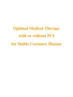
Optimal Medical Therapy with or without PCI for Stable Coronary Disease potx
Bạn đang xem bản rút gọn của tài liệu. Xem và tải ngay bản đầy đủ của tài liệu tại đây (312.54 KB, 15 trang )
Optimal Medical Therapy
with or without PCI
for Stable Coronary Disease
n engl j med 356;15 www.nejm.org april 12, 2007
1503
The new england
journal of medicine
established in 1812 april 12, 2007 vol. 356 no. 15
Optimal Medical Therapy with or without PCI
for Stable Coronary Disease
William E. Boden, M.D., Robert A. O’Rourke, M.D., Koon K. Teo, M.B., B.Ch., Ph.D., Pamela M. Hartigan, Ph.D.,
David J. Maron, M.D., William J. Kostuk, M.D., Merril Knudtson, M.D., Marcin Dada, M.D., Paul Casperson, Ph.D.,
Crystal L. Harris, Pharm.D., Bernard R. Chaitman, M.D., Leslee Shaw, Ph.D., Gilbert Gosselin, M.D.,
Shah Nawaz, M.D., Lawrence M. Title, M.D., Gerald Gau, M.D., Alvin S. Blaustein, M.D., David C. Booth, M.D.,
Eric R. Bates, M.D., John A. Spertus, M.D., M.P.H., Daniel S. Berman, M.D., G.B. John Mancini, M.D.,
and William S. Weintraub, M.D., for the COURAGE Trial Research Group*
A BS TR A C T
Affiliations for all authors are listed in the
Appendix. Address reprint requests to Dr.
Boden at the Division of Cardiology, Buf-
falo General Hospital, 100 High St., Buffalo,
NY 14203, or at wboden@kaleidahealth.
org.
*Members of the Clinical Outcomes Uti-
lizing Revascularization and Aggressive
Drug Evaluation (COURAGE) trial are list-
ed in the Appendix and in the Supplemen-
tary Appendix, available with the full text
of this article at www.nejm.org.
This article (10.1056/NEJMoa070829) was
published at www.nejm.org on March 26,
2007.
N Engl J Med 2007;356:1503-16.
Copyright © 2007 Massachusetts Medical Society.
Background
In patients with stable coronary artery disease, it remains unclear whether an initial
management strategy of percutaneous coronary intervention (PCI) with intensive
pharmacologic therapy and lifestyle intervention (optimal medical therapy) is superior
to optimal medical therapy alone in reducing the risk of cardiovascular events.
Methods
We conducted a randomized trial involving 2287 patients who had objective evidence
of myocardial ischemia and significant coronary artery disease at 50 U.S. and Cana-
dian centers. Between 1999 and 2004, we assigned 1149 patients to undergo PCI with
optimal medical therapy (PCI group) and 1138 to receive optimal medical therapy alone
(medical-therapy group). The primary outcome was death from any cause and non-
fatal myocardial infarction during a follow-up period of 2.5 to 7.0 years (median, 4.6).
Results
There were 211 primary events in the PCI group and 202 events in the medical-
therapy group. The 4.6-year cumulative primary-event rates were 19.0% in the PCI
group and 18.5% in the medical-therapy group (hazard ratio for the PCI group,
1.05; 95% confidence interval [CI], 0.87 to 1.27; P = 0.62). There were no significant
differences between the PCI group and the medical-therapy group in the composite
of death, myocardial infarction, and stroke (20.0% vs. 19.5%; hazard ratio, 1.05;
95% CI, 0.87 to 1.27; P = 0.62); hospitalization for acute coronary syndrome (12.4% vs.
11.8%; hazard ratio, 1.07; 95% CI, 0.84 to 1.37; P = 0.56); or myocardial infarction
(13.2% vs. 12.3%; hazard ratio, 1.13; 95% CI, 0.89 to 1.43; P = 0.33).
Conclusions
As an initial management strategy in patients with stable coronary artery disease,
PCI did not reduce the risk of death, myocardial infarction, or other major cardio-
vascular events when added to optimal medical therapy. (ClinicalTrials.gov number,
NCT00007657.)
Copyright © 2007 Massachusetts Medical Society. All rights reserved.
Downloaded from www.nejm.org at RIKSHOSPITALET HF on February 18, 2008 .
T h e ne w e ng l a n d j o u r na l o f m e di c ine
n engl j med 356;15 www.nejm.org april 12, 2007
1504
D
uring the past 30 years, the use of
percutaneous coronary intervention (PCI)
has become common in the initial man-
agement strategy for patients with stable coronary
artery disease in North America, even though treat-
ment guidelines advocate an initial approach with
intensive medical therapy, a reduction of risk fac-
tors, and lifestyle intervention (known as optimal
medical therapy).
1,2
In 2004, more than 1 million
coronary stent procedures were performed in the
United States,
3
and recent registry data indicate
that approximately 85% of all PCI procedures are
undertaken electively in patients with stable cor-
onary artery disease.
4
PCI reduces the incidence of
death and myocardial infarction in patients who
present with acute coronary syndromes,
5-10
but
similar benefit has not been shown in patients with
stable coronary artery disease.
11-15
This issue has
been studied in fewer than 3000 patients,
16
many
of whom were treated before the widespread use
of intracoronary stents and current standards of
medical management.
17-28
Although successful PCI of flow-limiting ste-
noses might be expected to reduce the rate of
death, myocardial infarction, and hospitalization
for acute coronary syndromes, previous studies
have shown only that PCI decreases the frequency
of angina and improves short-term exercise per-
formance.
11,12,15
Thus, the long-term prognostic
effect of PCI on cardiovascular events in patients
with stable coronary artery disease remains un-
certain. Our study, the Clinical Outcomes Utiliz-
ing Revascularization and Aggressive Drug Evalu-
ation (COURAGE) trial, was designed to determine
whether PCI coupled with optimal medical ther-
apy reduces the risk of death and nonfatal myo-
cardial infarction in patients with stable coro-
nary artery disease, as compared with optimal
medical therapy alone.
Me t hods
Study Design
The methods we used in the trial have been de-
scribed previously.
29,30
Sponsorship and oversight
of the trial were provided by the Department of
Veterans Affairs Cooperative Studies Program.
Additional funding was provided by the Canadian
Institutes of Health Research. Supplemental cor-
porate support from several pharmaceutical com-
panies included funding and in-kind support. All
support from the pharmaceutical industry con-
sisted of unrestricted research grants payable to
the Department of Veterans Affairs.
The study protocol was approved by the hu-
man rights committee at the coordinating center
and by the local institutional review board at each
participating center. An independent data and
safety monitoring board oversaw the conduct, safe-
ty, and efficacy of the trial. Data management and
statistical analyses were performed solely by the
data coordinating center with oversight by the trial
executive committee, whose members, after un-
blinding, had full access to the data and vouch
for the accuracy and completeness of the data and
the analyses. The companies that provided finan-
cial support, products, or both had no role in the
design, analysis, or interpretation of the study.
Study Population
Patients with stable coronary artery disease and
those in whom initial Canadian Cardiovascular
Society (CCS) class IV angina subsequently stabi-
lized medically were included in the study. Entry
criteria included stenosis of at least 70% in at least
one proximal epicardial coronary artery and ob-
jective evidence of myocardial ischemia (substan-
tial changes in ST-segment depression or T-wave
inversion on the resting electrocardiogram or in-
ducible ischemia with either exercise or pharma-
cologic vasodilator stress) or at least one coronary
stenosis of at least 80% and classic angina with-
out provocative testing. Exclusion criteria included
persistent CCS class IV angina, a markedly posi-
tive stress test (substantial ST-segment depression
or hypotensive response during stage 1 of the
Bruce protocol), refractory heart failure or cardio-
genic shock, an ejection fraction of less than 30%,
revascularization within the previous 6 months,
and coronary anatomy not suitable for PCI. A de-
tailed description of the inclusion and exclusion
criteria is included in the Supplementary Appen-
dix (available with the full text of this article at
www.nejm.org). Patients who were eligible for the
study underwent randomization after providing
written informed consent.
Treatment
Patients were randomly assigned to undergo PCI
and optimal medical therapy (PCI group) or opti-
mal medical therapy alone (medical-therapy group).
A permuted-block design was used to generate
Copyright © 2007 Massachusetts Medical Society. All rights reserved.
Downloaded from www.nejm.org at RIKSHOSPITALET HF on February 18, 2008 .
Optim a l Medical Ther apy w ith or w ithout PCI for Sta ble Coronary Dise ase
n engl j med 356;15 www.nejm.org april 12, 2007
1505
random assignments within each study site along
with previous coronary-artery bypass grafting
(CABG) as a stratifying variable. All patients re-
ceived antiplatelet therapy with aspirin at a dose
of 81 to 325 mg per day or 75 mg of clopidogrel
per day, if aspirin intolerance was present. Patients
undergoing PCI received aspirin and clopidogrel,
in accordance with accepted treatment guidelines
and established practice standards. Medical anti-
ischemic therapy in both groups included long-
acting metoprolol, amlodipine, and isosorbide
mononitrate, alone or in combination, along with
either lisinopril or losartan as standard second-
ary prevention. All patients received aggressive
therapy to lower low-density lipoprotein (LDL)
cholesterol levels (simvastatin alone or in combi-
nation with ezetimibe) with a target level of 60 to
85 mg per deciliter (1.55 to 2.20 mmol per liter).
After the LDL cholesterol target was achieved, an
attempt was made to raise the level of high-den-
sity lipoprotein (HDL) cholesterol to a level above
40 mg per deciliter (1.03 mmol per liter) and lower
triglyceride to a level below 150 mg per deciliter
(1.69 mmol per liter) with exercise, extended-release
niacin, or fibrates, alone or in combination.
In patients undergoing PCI, target-lesion revas-
cularization was always attempted, and complete
revascularization was performed as clinically ap-
propriate. Success after PCI as seen on angiogra-
phy was defined as normal coronary-artery flow
and less than 50% stenosis in the luminal diam-
eter after balloon angioplasty and less than 20%
after coronary stent implantation, as assessed by
visual estimation of the angiograms before and
after the procedure. Clinical success was defined
as angiographic success plus the absence of in-
hospital myocardial infarction, emergency CABG,
or death. Drug-eluting stents were not approved
for clinical use until the final 6 months of the
study, so few patients received these intracoronary
devices.
Clinical Outcome
Clinical outcome was adjudicated by an indepen-
dent committee whose members were unaware of
treatment assignments. The primary outcome mea-
sure was a composite of death from any cause
and nonfatal myocardial infarction. Secondary out-
comes included a composite of death, myocardial
infarction, and stroke and hospitalization for un-
stable angina with negative biomarkers. The an-
gina status of patients was assessed according to
the CCS classification during each visit. Further
analyses of other secondary outcomes — includ-
ing quality of life, the use of resources, and cost-
effectiveness — are being conducted but have not
yet been completed.
The prespecified definition of myocardial in-
farction (whether periprocedural or spontaneous)
required a clinical presentation consistent with
an acute coronary syndrome and either new ab-
normal Q waves in two or more electrocardio-
graphic leads or positive results in cardiac bio-
markers. Silent myocardial infarction, as detected
by abnormal Q waves, was confirmed by a core
laboratory and was also included as an outcome
of myocardial infarction.
Statistical Analysis
We projected composite 3-year event rates of 21.0%
in the medical-therapy group and 16.4% in the PCI
group (relative difference, 22%) during a follow-
up period of 2.5 to 7.0 years. We also incorporated
assumptions about crossover between study groups
and loss to follow-up.
31
We estimated that the en-
rollment of 2270 patients would provide a power
of 85% to detect the anticipated difference in the
primary outcome at the 5% two-sided level of
significance. A detailed description of the sam-
ple-size calculation is included in the Supplemen-
tary Appendix.
Estimates of the cumulative event rate were
calculated by the Kaplan–Meier method,
32
and
the primary efficacy of PCI, as compared with
optimal medical therapy, was assessed by the
stratified log-rank statistic.
33
The treatment ef-
fect, as measured by the hazard ratio and its
associated 95% confidence interval (CI), was esti-
mated with the use of the Cox proportional-haz-
ards model.
34
Data for patients who were lost to
follow-up were censored at the time of the last
contact. Analyses were performed according to the
intention-to-treat principle. Categorical variables
were compared by use of the chi-square test or
the Wilcoxon rank-sum test, and continuous vari-
ables were compared by use of the Student t-test.
Adjusted analysis of the primary outcome was
performed with the use of a Cox proportional-
hazards regression model with eight preidentified
covariates of interest — age, sex, race, previous
myocardial infarction, extent or distribution of
angiographic coronary artery disease, ejection frac-
Copyright © 2007 Massachusetts Medical Society. All rights reserved.
Downloaded from www.nejm.org at RIKSHOSPITALET HF on February 18, 2008 .
T h e ne w e ng l a n d j o u r na l o f m e di c ine
n engl j med 356;15 www.nejm.org april 12, 2007
1506
tion, presence or absence of diabetes, and health
care system (Veterans Affairs or non–Veterans
Affairs facility in the United States, or a Canadian
facility) — as well as the stratifying variable of
previous CABG. All other comparisons were un-
adjusted. A level of significance of less than 0.01
was used for all subgroup analyses and interac-
tions.
R e s ult s
Baseline Characteristics and Angiographic
Data
Between June 1999 and January 2004, a total of
2287 patients were enrolled in the trial at 50 U.S.
and Canadian centers (
Fig. 1
). Of these patients,
1149 were randomly assigned to the PCI group
and 1138 to the medical-therapy group. The base-
line characteristics of the patients were recently
published
35
and were similar in the two groups
(
Table 1
). The median time from the first episode
of angina before randomization was 5 months
(median, three episodes per week, with exertion
or at rest), and 58% of patients had CCS class II or
III angina. A total of 2168 patients (95%) had ob-
jective evidence of myocardial ischemia, whereas
the remaining 119 patients with classic angina
(CCS class III) and severe coronary stenoses did
not undergo ischemia testing (56 in the PCI group
and 63 in the medical-therapy group). Among pa-
tients who underwent myocardial perfusion im-
aging at baseline, 90% had either single (23%) or
multiple (67%) reversible defects for inducible is-
chemia. Two thirds of the patients had multivessel
coronary artery disease.
Of the 1149 patients in the PCI group, 46 never
underwent a procedure because the patient either
declined treatment or had coronary anatomy un-
suitable for PCI, as determined on clinical reas-
sessment. In 27 patients (2%), the operator was
unable to cross any lesions. PCI was attempted for
1688 lesions in 1077 patients, of whom 1006 (94%)
received at least one stent. In the stent group,
590 patients (59%) received one stent and 416
(41%) more than one stent. Drug-eluting stents
were used in 31 patients. On average, stenosis
in the luminal diameter, as evaluated on visual
assessment of angiograms, was reduced from a
mean (±SD) of 83±14% to 31±34% in the 244
lesions not treated with stents and from 82±12%
to 1.9±8% in the 1444 lesions treated with stents.
After PCI, successful treatment as seen on angi-
ography was achieved in 1576 of 1688 lesions
(93%), and clinical success (i.e., all lesions success-
fully dilated and no in-hospital complications)
was achieved in 958 of 1077 patients (89%).
Medication and Treatment Targets
Patients had a high rate of receiving multiple,
evidence-based therapies after randomization and
during follow-up, with similar rates in both study
groups (
Table 2
). At the 5-year follow-up visit,
70% of subjects had an LDL cholesterol level of
less than 85 mg per deciliter (2.20 mmol per liter)
(median, 71±1.3 mg per deciliter [1.84±0.03 mmol
per liter]); 65% and 94% had systolic and diastolic
blood pressure targets of less than 130 mm Hg
and 85 mm Hg, respectively; and 45% of patients
with diabetes had a glycated hemoglobin level of
no more than 7.0% (
Table 2
). Patients had high
rates of adherence to the regimen of diet, regular
exercise, and smoking cessation as recommended
by clinical practice guidelines,
1,2
although the
mean body-mass index did not decrease.
Follow-up Period
The median follow-up period was 4.6 years (inter-
quartile range, 3.3 to 5.7) and was similar in the
two study groups, with a total of 120,895 patient-
months at risk. Only 9% of patients were lost to
follow-up in the two groups (107 in the PCI group
and 97 in the medical-therapy group, P = 0.51) be-
fore the occurrence of a primary outcome or the
end of follow-up. Vital status was not ascertained
in 194 patients (99 in the PCI group and 95 in the
medical-therapy group, P = 0.81).
Primary Outcome
The primary outcome (a composite of death from
any cause and nonfatal myocardial infarction) oc-
curred in 211 patients in the PCI group and 202
patients in the medical-therapy group (Table 3).
The estimated 4.6-year cumulative primary event
rates were 19.0% in the PCI group and 18.5% in
the medical-therapy group (unadjusted hazard ra-
tio for the PCI group, 1.05; 95% CI, 0.87 to 1.27;
P = 0.62) (
Fig. 2
).
Secondary Outcomes
For the prespecified composite outcome of death,
nonfatal myocardial infarction, and stroke, the
event rate was 20.0% in the PCI group and 19.5%
Copyright © 2007 Massachusetts Medical Society. All rights reserved.
Downloaded from www.nejm.org at RIKSHOSPITALET HF on February 18, 2008 .
Optim a l Medical Ther apy w ith or w ithout PCI for Sta ble Coronary Dise ase
n engl j med 356;15 www.nejm.org april 12, 2007
1507
33p9
3071 Met eligibility criteria
2287 Consented to participate
(74% of patients with protocol eligibility)
35,539 Patients underwent assessment
32,468 Were excluded
8677 Did not meet inclusion criteria
5155 Had undocumented ischemia
3961 Did not meet protocol for vessels
6554 Were excluded for logistic reasons
18,360 Had one or more exclusions
4513 Had undergone recent (<6 mo) revascu-
larization
4939 Had an inadequate ejection fraction
2987 Had a contraindication to PCI
2542 Had a serious coexisting illness
1285 Had concomitant valvular disease
1203 Had class IV angina
1071 Had a failure of medical therapy
947 Had left main coronary artery stenosis
>50%
722 Had only PCI restenosis (no new lesions)
528 Had complications after myocardial
infarction
784 Did not provide consent
450 Did not receive physician’s
approval
237 Declined to give permission
97 Had an unknown reason
1149 Were assigned to PCI group
46 Did not undergo PCI
27 Had a lesion that could not be dilated
1006 Received at least one stent
107 Were lost to follow-up
1138 Were assigned to medical-therapy group
97 Were lost to follow-up
1149 Were included in the primary analysis 1138 Were included in the primary analysis
AUTHOR:
FIGURE:
JOB: ISSUE:
4-C
H/T
RETAKE
SIZE
ICM
CASE
Line
H/T
Combo
Revised
AUTHOR, PLEASE NOTE:
Figure has been redrawn and type has been reset.
Please check carefully.
REG F
Enon
1st
2nd
3rd
Boden
1 of 3
04-12-07
ARTIST: ts
35615
Figure 1. Enrollment and Outcomes.
Of 35,539 patients who were assessed for eligibility in the trial, 32,468 were excluded for a variety of reasons (patients
could have more than one reason for exclusion). A total of 3071 patients met all inclusion criteria. Of these, 2287
(74%) consented to participate in the study (932 in Canada, 968 in U.S. Veterans Affairs facilities, and 387 in U.S.
facilities other than Veterans Affairs hospitals). Of these patients, 1149 were randomly assigned to the PCI group
and 1138 to the medical-therapy group. The median follow-up was 4.6 years for both study groups.
Copyright © 2007 Massachusetts Medical Society. All rights reserved.
Downloaded from www.nejm.org at RIKSHOSPITALET HF on February 18, 2008 .
T h e ne w e ng l a n d j o u r na l o f m e di c ine
n engl j med 356;15 www.nejm.org april 12, 2007
1508
Table 1. Baseline Clinical and Angiographic Characteristics.*
Characteristic
PCI Group
(N = 1149)
Medical-Therapy
Group (N = 1138) P Value
Demographic
Age — yr 61.5±10.1 61.8±9.7 0.54
Sex — no. (%) 0.95
Male 979 (85) 968 (85)
Female 169 (15) 169 (15)
Race or ethnic group — no. (%)† 0.64
White 988 (86) 975 (86)
Black 57 (5) 57 (5)
Hispanic 68 (6) 58 (5)
Other 35 (3) 47 (4)
Clinical
Angina (CCS class) — no. (%) 0.24
0 135 (12) 148 (13)
I 340 (30) 341 (30)
II 409 (36) 425 (37)
III 261 (23) 221 (19)
Missing data 3 (<1) 2 (<1)
Duration of angina — mo 0.53
Median 5 5
Interquartile range 1–15 1–15
Episodes/wk with exertion or at rest within last mo 0.83
Median 3 3
Interquartile range 1–6 1–6
History — no. (%)
Diabetes 367 (32) 399 (35) 0.12
Hypertension 757 (66) 764 (67) 0.53
Congestive heart failure 57 (5) 51 (4) 0.59
Cerebrovascular disease 100 (9) 102 (9) 0.83
Myocardial infarction 437 (38) 439 (39) 0.80
Previous PCI 174 (15) 185 (16) 0.49
CABG 124 (11) 124 (11) 0.94
Stress test‡
Total patients — no. (%) 972 (85) 977 (86) 0.84
Treadmill test — no. (%) 555 (57) 553 (57)
Duration of treadmill test — min 7.0±2.7 6.9±2.3 0.43
Pharmacologic stress — no. (%) 417 (43) 424 (43)
Echocardiography — no. (%) 63 (6) 54 (6)
Nuclear imaging — no. (%) 685 (70) 708 (72) 0.59
Single reversible defect§ 154 (22) 161 (23) 0.09
Multiple reversible defects§ 444 (65) 483 (68) 0.09
Copyright © 2007 Massachusetts Medical Society. All rights reserved.
Downloaded from www.nejm.org at RIKSHOSPITALET HF on February 18, 2008 .
Optim a l Medical Ther apy w ith or w ithout PCI for Sta ble Coronary Dise ase
n engl j med 356;15 www.nejm.org april 12, 2007
1509
in the medical-therapy group (hazard ratio, 1.05;
95% CI, 0.87 to 1.27; P = 0.62) (Table 3 and
Fig. 2
).
The rates of hospitalization for acute coronary syn-
dromes were 12.4% in the PCI group and 11.8%
in the medical-therapy group (hazard ratio, 1.07;
95% CI, 0.84 to 1.37; P = 0.56), and adjudicated
rates of myocardial infarction were 13.2% and
12.3%, respectively (hazard ratio, 1.13; 95% CI,
0.89 to 1.43; P = 0.33). For death alone, the rates
were 7.6% and 8.3%, respectively (hazard ratio,
0.87; 95% CI, 0.65 to 1.16); the mortality curves
for the two groups were virtually identical during
the initial 4.6 years of the study. For stroke alone,
the rate was 2.1% in the PCI group and 1.8% in the
medical-therapy group (hazard ratio, 1.56; 95% CI,
0.80 to 3.04; P = 0.19). When the primary end point
was calculated with the exclusion of periproce-
dural myocardial infarction, the event rates were
16.2% and 17.9% (hazard ratio, 0.90; 95% CI, 0.73
to 1.10; P = 0.29).
At a median follow-up of 4.6 years, 21.1% of
patients in the PCI group had additional revascu-
larization, as compared with 32.6% of those in
the medical-therapy group (hazard ratio, 0.60;
95% CI, 0.51 to 0.71; P<0.001). In the PCI group,
77 patients subsequently underwent CABG, as com-
pared with 81 patients in the medical-therapy
group. Revascularization was performed for an-
gina that was unresponsive to maximal medical
therapy or when there was objective evidence of
worsening ischemia on noninvasive testing, at the
discretion of the patient’s physician. The median
time to subsequent revascularization was 10.0
months (interquartile range, 4.5 to 28.0) in the
PCI group and 10.8 months (interquartile range,
3.2 to 30.7) in the medical-therapy group.
There was a substantial reduction in the preva-
lence of angina in both groups during follow-up.
There was a statistically significant difference in
the rates of freedom from angina throughout
most of the follow-up period, in favor of the PCI
group (
Table 2
). At 5 years, 74% of patients in
the PCI group and 72% of those in the medical-
therapy group were free of angina (P = 0.35).
Subgroup Analyses
There was no significant interaction (P<0.01) be-
tween treatment effect and any predefined sub-
group variable (
Fig. 3
). Of note, among patients
with multivessel coronary artery disease, previous
myocardial infarction, and diabetes, the rate of
the primary end point was similar for both groups.
When subgroup variables were included in a multi-
variate analysis, the hazard ratio for treatment
was essentially unchanged (1.09; 95% CI, 0.90 to
1.33; P = 0.77).
Di s cus sio n
As an initial management strategy, PCI added to
optimal medical therapy did not reduce the pri-
mary composite end point of death and nonfatal
Table 1. (Continued.)
Characteristic
PCI Group
(N = 1149)
Medical-Therapy
Group (N = 1138) P Value
Angiographic
Vessels with disease — no. (%) 0.72
1 361 (31) 343 (30)
2 446 (39) 439 (39)
3 341 (30) 355 (31)
Disease in graft¶ 77 (62) 85 (69) 0.36
Proximal LAD disease 360 (31) 417 (37) 0.01
Ejection fraction 60.8±11.2 60.9±10.3 0.86
* Plus–minus values are means ±SD. Baseline data were missing for one patient in each study group. CCS denotes
Canadian Cardiovascular Society, CABG coronary-artery bypass grafting, and LAD left anterior descending artery.
† Race or ethnicity was reported by the patient at enrollment.
‡ Nuclear imaging could have been performed after either an exercise treadmill test or pharmacologic stress.
§ The percentage in this category is the proportion of patients who underwent imaging.
¶ The percentage in this category is the proportion of patients who had undergone previous CABG.
Copyright © 2007 Massachusetts Medical Society. All rights reserved.
Downloaded from www.nejm.org at RIKSHOSPITALET HF on February 18, 2008 .
T h e ne w e ng l a n d j o u r na l o f m e di c ine
n engl j med 356;15 www.nejm.org april 12, 2007
1510
Table 2. Clinical Status, Risk and Lifestyle Factors, and Use of Medication.*
Variable PCI Group (N = 1149) Medical-Therapy Group (N = 1138)
Baseline 1 Yr 3 Yr 5 Yr Baseline 1 Yr 3 Yr 5 Yr
median ±SE
Clinical status
No. evaluated 1148 1031 820 423 1137 1010 824 406
Blood pressure — mm Hg
Systolic 131±0.77 126±0.64 125±0.68 124±0.81 130±0.66 124±0.73 123±0.78 122±0.92
Diastolic 74±0.33 72±0.35 70±0.52 70±0.81 74±0.33 70±0.43 70±0.52 70±0.65
Cholesterol — mg/dl
Total 172±1.37 156±1.17 148±1.13 143±1.74 177±1.41 150±1.10 145±1.30 140±1.64
HDL 39±0.39 42±0.39 43±0.47 41±0.67 39±0.37 41±0.42 42±0.49 41±0.75
LDL 100±1.17 84±0.97 76±0.85 71±1.33 102±1.22 81±0.86 74±0.92 72±1.21
Triglycerides — mg/dl 143±2.96 129±2.74 124±2.79 123±4.13 149±3.03 133±2.90 126±2.84 131±4.70
Body-mass index
28.7±0.18 28.5±0.19 29.0±0.21 29.0±0.34 28.9±0.17 29.0±0.19 29.3±0.21 29.5±0.31
Angina-free — no. (%)†
135 (12) 680 (66) 602 (72) 316 (74) 148 (13) 595 (58) 558 (67) 296 (72)
Risk or lifestyle factor
Current smoker — no. (%) 260 (23) 206 (20) 156 (19) 74 (17) 259 (23) 206 (20) 160 (19) 80 (20)
AHA Step 2 diet — no. (%) 626 (55) 803 (78) 631 (77) 326 (77) 613 (54) 800 (79) 660 (80) 312 (77)
Moderate activity — no. (%)‡ 290 (25) 473 (46) 351 (42) 179 (42) 279 (25) 433 (43) 330 (40) 146 (36)
Glycated hemoglobin in patients
with diabetes
No. evaluated 319 239 197 97 336 286 233 123
Level — % 6.9±0.1 7.1±0.1 7.1±0.1 7.1±0.1 7.1±0.1 7.0±0.1 7.1±0.1 7.1±0.1
Medication
No. evaluated 1147 1044 837 428 1138 1028 838 417
ACE inhibitor — no. (%) 669 (58) 668 (64) 536 (64) 284 (66) 680 (60) 633 (62) 522 (62) 260 (62)
ARB — no. (%) 48 (4) 93 (9) 104 (12) 49 (11) 54 (5) 99 (10) 108 (13) 67 (16)
Statin — no. (%) 992 (86) 972 (93) 780 (93) 398 (93) 1014 (89) 972 (95) 769 (92) 386 (93)
Other antilipid — no. (%) 89 (8) 236 (23) 324 (39) 211 (49) 94 (8) 253 (25) 321 (38) 224 (54)
Aspirin — no. (%) 1097 (96) 995 (95) 792 (95) 408 (95) 1077 (95) 977 (95) 796 (95) 391 (94)
Beta-blocker — no. (%)
975 (85) 887 (85) 705 (84) 363 (85) 1008 (89) 916 (89) 724 (86) 357 (86)
Calcium-channel blocker — no. (%)§
459 (40) 415 (40) 360 (43) 180 (42) 488 (43) 501 (49) 418 (50) 217 (52)
Nitrates — no. (%)¶ 714 (62) 553 (53) 396 (47) 173 (40) 825 (72) 690 (67) 511 (61) 237 (57)
* Plus–minus values are medians ±SE, with the SE calculated with the use of the interquartile range. To convert cholesterol values to milli
-
moles per liter, multiply by 0.02586. To convert triglyceride values to millimoles per liter, multiply by 0.01129. ACE denotes angiotensin-
converting enzyme, and ARB angiotensin-receptor blocker.
† The comparison between the PCI group and the medical-therapy group was significant at 1 year (P<0.001) and 3 years (P = 0.02) but not at
baseline or at 5 years.
‡ This category includes at least 30 to 45 minutes of moderate activity five times per week or vigorous activity three times per week.
§ The comparison between the PCI group and the medical-therapy group was significant at 1 year (P<0.001), 3 years (P = 0.005), and 5 years
(P = 0.003).
¶ The comparison between the PCI group and the medical-therapy group was significant at all time points (P<0.001).
Copyright © 2007 Massachusetts Medical Society. All rights reserved.
Downloaded from www.nejm.org at RIKSHOSPITALET HF on February 18, 2008 .
Optim a l Medical Ther apy w ith or w ithout PCI for Sta ble Coronary Dise ase
n engl j med 356;15 www.nejm.org april 12, 2007
1511
myocardial infarction or reduce major cardiovas-
cular events, as compared with optimal medical
therapy alone, during follow-up of 2.5 to 7.0 years,
despite a high baseline prevalence of clinical co-
existing illnesses, objective evidence of ischemia,
and extensive coronary artery disease as seen on
angiography. Although the degree of angina re-
lief was significantly higher in the PCI group
than in the medical-therapy group, there was also
substantial improvement in the medical-therapy
group. All secondary outcomes and individual com-
ponents of the primary outcome showed no sig-
nificant differences between the study groups,
nor was there a significant interaction between
treatment effect and any prespecified subgroup
variable. For the primary outcome, the 95% CI
excludes a relative benefit of more than 13% in
the PCI group. Thus, it is highly unlikely that we
missed a prognostically important treatment ben-
efit in favor of the initial PCI strategy.
Table 3. Primary and Secondary Outcomes.*
Outcome Number of Events Hazard Ratio (95% CI)† P Value† Cumulative Rate at 4.6 Years
PCI Group
Medical-Therapy
Group PCI Group
Medical-Therapy
Group
%
Death and nonfatal myocardial
infarction‡
211 202 1.05 (0.87–1.27) 0.62 19.0 18.5
Death§ 68 74
Periprocedural myocardial
infarction
35 9
Spontaneous myocardial infarction 108 119
Death, myocardial infarction, and
stroke
222 213 1.05 (0.87–1.27) 0.62 20.0 19.5
Hospitalization for ACS 135 125 1.07 (0.84–1.37) 0.56 12.4 11.8
Death§ 85 95 0.87 (0.65–1.16) 0.38 7.6 8.3
Cardiac 23 25
Other 45 51
Unknown 17 19
Total nonfatal myocardial infarction 143 128 1.13 (0.89–1.43) 0.33 13.2 12.3
Periprocedural myocardial
infarction
35 9
Spontaneous myocardial infarction 108 119
Death, myocardial infarction, and ACS 294 288 1.05 (0.90–1.24) 0.52 27.6 27.0
Stroke 22 14 1.56 (0.80–3.04) 0.19 2.1 1.8
Revascularization (PCI or CABG)¶ 228 348 0.60 (0.51–0.71) <0.001 21.1 32.6
* ACS denotes acute coronary syndrome, PCI percutaneous coronary intervention, and CABG coronary-artery bypass grafting.
† The hazard ratio is for the PCI group as compared with the medical-therapy group, and P values were calculated by the log-rank test and are
unadjusted for multiple variables.
‡ The definition of myocardial infarction was the finding of new Q waves at any time; a spontaneous creatine kinase MB fraction of at least
1.5 times the upper limit of normal or a troponin T or I level of at least 2.0 times the upper limit of normal; during a PCI procedure, a cre-
atine kinase MB fraction of at least 3 times the upper limit of normal or a troponin T or I level of at least 5.0 times the upper limit of nor-
mal, associated with new ischemic symptoms; and after CABG, a creatine kinase MB fraction or a troponin T or I level of at least 10.0 times
the upper limit of normal. If periprocedural myocardial infarction is excluded from the primary outcome, the hazard ratio is 0.90 (95% CI,
0.73 to 1.10; P = 0.29).
§ Some patients had a nonfatal myocardial infarction before their subsequent death so that the number of deaths overall is greater than the
number of deaths in the primary outcome analysis, which includes the time until the first event.
¶ Values exclude the initial PCI procedure in patients who were originally assigned to the PCI group.
Copyright © 2007 Massachusetts Medical Society. All rights reserved.
Downloaded from www.nejm.org at RIKSHOSPITALET HF on February 18, 2008 .
T h e ne w e ng l a n d j o u r na l o f m e di c ine
n engl j med 356;15 www.nejm.org april 12, 2007
1512
Our findings may be explained, in part, by dif-
ferences in atherosclerotic plaque morphology and
vascular remodeling associated with acute coro-
nary syndromes, as compared with stable coronary
artery disease. Vulnerable plaques (precursors of
acute coronary syndromes) tend to have thin
fibrous caps, large lipid cores, fewer smooth-
muscle cells, more macrophages, and less colla-
gen, as compared with stable plaques, and are
associated with outward (expansive) remodeling
of the coronary-artery wall, causing less stenosis
of the coronary lumen.
36
As a result, vulnerable
plaques do not usually cause significant stenosis
before rupture and the precipitation of an acute
coronary syndrome.
36
By contrast, stable plaques
tend to have thick fibrous caps, small lipid cores,
more smooth-muscle cells, fewer macrophages,
and more collagen and are ultimately associated
with inward (constrictive) remodeling that nar-
rows the coronary lumen. These lesions produce
ischemia and anginal symptoms and are easily
detected by coronary angiography but are less like-
ly to result in an acute coronary syndrome.
37,38
Thus, unstable coronary lesions that lead to
myocardial infarction are not necessarily severely
stenotic, and severely stenotic lesions are not nec-
essarily unstable. Focal management of even
severely stenotic coronary lesions with PCI in our
study did not reduce the rate of death and myo-
cardial infarction, presumably because the treated
stenoses were not likely to trigger an acute coro-
nary event. Furthermore, our lower-than-projected
AUTHOR:
FIGURE:
JOB: ISSUE:
4-C
H/T
RETAKE
SIZE
ICM
CASE
Line
H/T
Combo
Revised
AUTHOR, PLEASE NOTE:
Figure has been redrawn and type has been reset.
Please check carefully.
REG F
Enon
1st
2nd
3rd
Boden
2 of 3
04-12-07
ARTIST: ts
35615
39p6
No. at Risk
Medical therapy
PCI
30
35
192
200
408
417
638
637
834
833
959
952
1017
1013
1138
1149
Survival Free of Death from
Any Cause and Myocardial
Infarction
1.0
0.9
0.7
0.6
0.5
0.8
0
0 1 2 3 4 75 6
Years
Medical therapy
Medical therapy
Medical therapy
Medical therapy
PCI
PCI
Hazard ratio, 1.05; 95% CI (0.87–1.27); P=0.62
PCI
Hazard ratio, 0.87; 95% CI (0.65–1.16); P=0.38
Hazard ratio, 1.07; 95% CI (0.84–1.37); P=0.56
PCI
Hazard ratio, 1.13; 95% CI (0.89–1.43); P=0.33
A B
C
D
No. at Risk
Medical therapy
PCI
38
44
302
312
468
488
717
733
917
929
1029
1051
1073
1094
1138
1149
Overall Survival
1.0
0.9
0.7
0.6
0.5
0.8
0
0 1 2 3 4 75 6
Years
No. at Risk
Medical therapy
PCI
127
134
236
246
418
431
662
667
833
835
956
957
1025
1027
1138
1149
Survival Free of ACS
1.0
0.9
0.7
0.6
0.5
0.8
0
0 1 2 3 4 75 6
Years
No. at Risk
Medical therapy
PCI
120
134
192
200
409
418
638
637
834
833
962
954
1019
1015
1138
1149
Survival Free of Myocardial
Infarction
1.0
0.9
0.7
0.6
0.5
0.8
0
0 1 2 3 4 75 6
Years
Figure 2. Kaplan–Meier Survival Curves.
In Panel A, the estimated 4.6-year rate of the composite primary outcome of death from any cause and nonfatal myocardial infarction
was 19.0% in the PCI group and 18.5% in the medical-therapy group. In Panel B, the estimated 4.6-year rate of death from any cause
was 7.6% in the PCI group and 8.3% in the medical-therapy group. In Panel C, the estimated 4.6-year rate of hospitalization for acute
coronary syndrome (ACS) was 12.4% in the PCI group and 11.8% in the medical-therapy group. In Panel D, the estimated 4.6-year rate
of acute myocardial infarction was 13.2% in the PCI group and 12.3% in the medical-therapy group.
Copyright © 2007 Massachusetts Medical Society. All rights reserved.
Downloaded from www.nejm.org at RIKSHOSPITALET HF on February 18, 2008 .
Optim a l Medical Ther apy w ith or w ithout PCI for Sta ble Coronary Dise ase
n engl j med 356;15 www.nejm.org april 12, 2007
1513
event rate in the medical-therapy group may be
explained by systemic therapy that reduced plaque
vulnerability through aggressive intervention for
multiple risk factors and evidence-based use of
medication.
Rates of angina were consistently lower in the
PCI group than in the medical-therapy group dur-
ing follow-up, and rates of subsequent revascu-
larization were likewise lower. However, there
was a substantial increase in freedom from an-
gina in patients in the medical-therapy group as
well, most of which had taken place at 1 year but
with a further improvement at 5 years. To what
extent this finding reflects a benefit of specific
39p6
0.25 0.50 1.00 2.001.751.50
Medical Therapy
Better
PCI Better
Overall
Sex
Male
Female
Myocardial infarction
Yes
No
Extent of CAD
Multivessel disease
Single-vessel disease
Smoking
Current
Not current
Diabetes
Yes
No
CCS angina class
0 or I
II or III
Ejection fraction
≤50%
>50%
Age
>65 yr
≤65 yr
Previous CABG
No
Yes
Race
White
Nonwhite
Health care system
Canadian
U.S. non-VA
U.S. VA
No. of
Patients Hazard Ratio (95% CI)
Medical Therapy
Event Rate for the Primary
Outcome
Baseline Characteristics
0.19
0.18
0.26
0.25
0.14
0.21
0.12
0.21
0.18
0.24
0.15
0.20
0.18
0.26
0.16
0.22
0.16
0.17
0.29
0.18
0.24
0.14
0.21
0.22
1.15 (0.93–1.42)
1.05 (0.87–1.27)
0.87 (0.54–1.42)
1.27 (0.90–1.78)
0.71 (0.44–1.14)
1.06 (0.80–1.38)
1.08 (0.87–1.34)
0.98 (0.52–1.82)
1.04 (0.84–1.29)
1.00 (0.77–1.32)
1.10 (0.83–1.46)
1.05 (0.84–1.32)
1.14 (0.77–1.70)
1.09 (0.85–1.40)
1.01 (0.75–1.38)
1.20 (0.92–1.56)
0.99 (0.73–1.32)
1.08 (0.86–1.36)
1.00 (0.71–1.41)
1.17 (0.76–1.80)
1.04 (0.84–1.30)
1.22 (0.93–1.60)
0.91 (0.69–1.21)
0.65 (0.40–1.06)
P Value
0.19
0.19
0.18
0.23
0.17
0.21
0.15
0.20
0.19
0.25
0.17
0.17
0.20
0.28
0.17
0.24
0.16
0.17
0.34
0.19
0.19
0.17
0.15
0.22
PCI
2287
1947
338
876
1371
1581
700
653
1631
766
1468
964
1371
406
1848
904
1381
2039
248
1963
322
932
387
968
0.03
0.15
0.65
0.71
0.33
0.73
0.72
0.62
0.81
0.43
0.17
AUTHOR:
FIGURE:
JOB: ISSUE:
4-C
H/T
RETAKE
SIZE
ICM
CASE
Line
H/T
Combo
Revised
AUTHOR, PLEASE NOTE:
Figure has been redrawn and type has been reset.
Please check carefully.
REG F
Enon
1st
2nd
3rd
Boden
3 of 3
04-12-07
ARTIST: ts
35615
Figure 3. Subgroup Analyses.
The chart shows hazard ratios (black squares, sized in proportion to the number of subjects in a group), 95% CIs (horizontal lines), cumu-
lative 4.6-year event rates for the composite primary outcome (death from any cause and nonfatal myocardial infarction) for the PCI group
versus the medical-therapy group for the specified subgroups, and P values for the interaction between the treatment effects and sub-
group variables. P values were calculated with the use of the Wald statistic. There was no significant interaction between treatment and
subgroup variables as defined according to the prespecified value for interaction (P<0.01), although there was a trend for interaction with
respect to sex (P = 0.03). PCI denotes percutaneous coronary intervention, CAD coronary artery disease, CCS Canadian Cardiovascular
Society, CABG coronary-artery bypass grafting, and VA Veterans Affairs.
Copyright © 2007 Massachusetts Medical Society. All rights reserved.
Downloaded from www.nejm.org at RIKSHOSPITALET HF on February 18, 2008 .
T h e ne w e ng l a n d j o u r na l o f m e di c ine
n engl j med 356;15 www.nejm.org april 12, 2007
1514
antianginal medications (e.g., nitrates and beta-
blockers) or a favorable effect of therapies such
as statins on endothelial function and atheroscle-
rosis is unclear.
Our findings parallel those reported in recent
trials,
39,40
in which observed clinical-event rates
that were associated with optimal medical ther-
apy were lower than projected in the trial design.
These results are also concordant with a meta-
analysis of all previous trials involving PCI ver-
sus medical management.
16
In the aggregate,
these studies, including our own, include out-
come data on more than 5000 patients and show
that PCI has no effect in reducing major cardio-
vascular events.
The preponderance of male patients (85%) is
a limitation of our study, as is the lack of ethnic
diversity (14% of the patients were nonwhite). We
used bare-metal stents, since drug-eluting stents
were not available until late during accrual. Al-
though the latter factor may be perceived as a
limitation, published data indicate no benefit
(either short-term or long-term) with respect to
death and myocardial infarction in patients with
stable coronary artery disease who receive drug-
eluting stents, as compared with those who re-
ceive bare-metal stents.
41-46
Our findings reinforce existing clinical prac-
tice guidelines, which state that PCI can be safely
deferred in patients with stable coronary artery
disease, even in those with extensive, multivessel
involvement and inducible ischemia, provided that
intensive, multifaceted medical therapy is institut-
ed and maintained.
1,2
As an initial management
approach, optimal medical therapy without rou-
tine PCI can be implemented safely in the major-
ity of patients with stable coronary artery disease.
However, approximately one third of these pa-
tients may subsequently require revascularization
for symptom control or for subsequent develop-
ment of an acute coronary syndrome.
In summary, our trial compared optimal med-
ical therapy alone or in combination with PCI as
an initial management strategy in patients with
stable coronary artery disease. Although the ad-
dition of PCI to optimal medical therapy reduced
the prevalence of angina, it did not reduce long-
term rates of death, nonfatal myocardial infarc-
tion, and hospitalization for acute coronary syn-
dromes.
Supported by the Cooperative Studies Program of the U.S. De-
partment of Veterans Affairs Office of Research and Development,
in collaboration with the Canadian Institutes of Health Research;
and by unrestricted research grants from Merck, Pfizer, Bristol-
Myers Squibb, Fujisawa, Kos Pharmaceuticals, Datascope, Astra-
Zeneca, Key Pharmaceutical, Sanofi-Aventis, First Horizon, and
GE Healthcare. All industrial funding in support of the trial was
directed through the U.S. Department of Veterans Affairs.
Dr. Boden reports receiving consulting fees and lecture fees
from Kos Pharmaceuticals, PDL BioPharma, Pfizer, CV Thera-
peutics, and Sanofi-Aventis, and grant support from Merck and
Abbott Laboratories; Dr. O’Rourke, consulting fees from King
Pharmaceuticals, Lilly, and CV Therapeutics; Dr. Teo, grant sup-
port from Boehringer Ingelheim; Dr. Knudtson, lecture fees from
Medtronic and Lilly; Dr. Harris, having equity ownership in
Amgen; Dr. Chaitman, receiving consulting fees from CV Thera-
peutics, Merck, and Bayer, lecture fees from Pfizer, AstraZeneca,
and CV Therapeutics, and grant support from Pfizer, CV Thera-
peutics, and Sanofi-Aventis; Dr. Shaw, grant support from Bristol-
Myers Squibb and Astellas Healthcare; Dr. Booth, grant support
from Actelion; Dr. Bates, consulting fees from Sanofi-Aventis
and AstraZeneca and lecture fees from Sanofi-Aventis; Dr. Sper-
tus, consulting fees from Amgen and United Healthcare and
grant support from Amgen, Roche Diagnostics, and Lilly (and in
the past, consulting fees and grant support from CV Therapeu-
tics and owning the copyright for the Seattle Angina Question-
naire, the Peripheral Artery Questionnaire, and the Kansas City
Cardiomyopathy Questionnaire); Dr. Berman, consulting fees
and lecture fees from Bristol-Myers Squibb, Astellas, Tyco, and
Siemens and grant support from Bristol-Myers Squibb and As-
tellas; Dr. Mancini, consulting and lecture fees from Pfizer,
Abbott, and GlaxoSmithKline, lecture fees from Merck and
Sanofi-Aventis, and grant support from Cordis and GlaxoSmith-
Kline; and Dr. Weintraub, consulting fees from Sanofi-Aventis
and Bristol-Myers Squibb and grant support from Sanofi-Aventis.
No other potential conflict of interest relevant to this article was
reported.
Appendix
The authors’ affiliations are as follows: Western New York Veterans Affairs (VA) Healthcare Network and Buffalo General Hospital–
SUNY, Buffalo, NY (W.E.B.); South Texas Veterans Health Care System–Audie Murphy Campus, San Antonio, TX (R.A.O., P.C.); Mc-
Master University Medical Center, Hamilton, ON, Canada (K.K.T.); VA Cooperative Studies Program Coordinating Center, VA Con-
necticut Healthcare System, West Haven, CT (P.M.H); Vanderbilt University Medical Center, Nashville (D.J.M); London Health Sciences
Centre, London, ON, Canada (W.J.K.); Foothills Hospital, Calgary, AB, Canada (M.K.); Hartford Hospital, Hartford, CT (M.D.); VA
Cooperative Studies Program Clinical Research Pharmacy Coordinating Center, Albuquerque, NM (C.L.H.); Saint Louis University, St.
Louis (B.R.C.); Cedars–Sinai Medical Center, Los Angeles (L.S., D.S.B.); Montreal Heart Institute, Montreal (G. Gosselin); Sudbury
Regional Hospital, Sudbury, ON, Canada (S.N.); Queen Elizabeth Health Sciences Centre, Halifax, NS, Canada (L.M.T.); Mayo Clinic,
Rochester, MN (G. Gau); Houston VA Medical Center, Houston (A.S.B.); Lexington VA Medical Center, Lexington, KY (D.C.B.); Uni-
versity of Michigan Medical Center, Ann Arbor (E.R.B.); Mid America Heart Institute, Kansas City, MO (J.A.S.); Vancouver Hospital and
Health Sciences Centre, Vancouver, BC, Canada (G.B.J.M.); and Christiana Care Health System, Newark, DE (W.S.W.).
The members of the COURAGE trial were as follows: Writing Committee: W. Boden (study cochair), R. O’Rourke (study cochair) K. Teo
(study cochair), P. Hartigan. W. Weintraub, D. Maron, J. Mancini; Executive Committee: W. Weintraub (chair), W. Boden, R. O’Rourke,
K. Teo, P. Hartigan, M. Knudtson, D. Maron, E. Bates, A. Blaustein, D. Booth, R. Carere, S. Ellis, G. Gosselin, G. Gau, A. Jacobs,
Copyright © 2007 Massachusetts Medical Society. All rights reserved.
Downloaded from www.nejm.org at RIKSHOSPITALET HF on February 18, 2008 .
Optim a l Medical Ther apy w ith or w ithout PCI for Sta ble Coronary Dise ase
n engl j med 356;15 www.nejm.org april 12, 2007
1515
S. King, III, W. Kostuk, C. Harris, J. Spertus; P. Peduzzi (ex officio); Data and Safety Monitoring Board: T. Ryan (chair), B. Turnbull, T.
Feldman, R. Bonow, W. Haskell, P. Diehr, P. Lachenbruch, D. Waters, D. Johnstone; Adjudication Committee: L. Cohen (chair), B.
Cantin, W. Hager, F. Samaha, J. Januzzi, J. Arrighi, B. Chaitman; Economics Committee: W. Weintraub (chair), P. Hartigan, R.
O’Rourke, W. Boden, P. Barnett, J. Spertus, R. Goeree; Optimal Medical Therapy Committee: D. Maron (chair), W. Boden, R. O’Rourke,
K. Teo, W. Weintraub.
The following members assisted in coordination of the study: VA Cooperative Studies Program Coordinating Center, VA Connecticut
Healthcare System, West Haven, CT — P. Peduzzi (director); M. Antonelli, (associate director of operations); J. Smith (project manager);
R. Kilstrom, B. Hunter (coordinators); L. Durant (quality assurance officer); S. O’Neil (end points coordinator); T. Economou, J. Nabors
(programmers); A. Kossack (data clerk); VA Cooperative Studies Program Clinical Research Pharmacy Coordinating Center, Albuquer-
que, NM — M. Sather (director), C. Harris (assistant director), W. Gagne (project manager), C. Fye (pharmacist); VA Cooperative Stud-
ies Human Rights Committee, West Haven, CT — R. Marottoli (chair), H. Allore, D. Beckwith, W. Farrell, R. Feldman, R. Mehta, J.
Neiderman, E. Perry, S. Kasl, M. Zeman; VA Office of Research and Development, Clinical Science Research and Development, Wash-
ington, DC — T. O’Leary (acting director), G. Huang (deputy director, Cooperative Studies Program); Study Chairs Offices — Western
New York VA Healthcare Network and Buffalo General Hospital–SUNY, Buffalo, NY — W. Boden (study cochair), M. Dada, K. Potter
(national coordinators), T. Rivera (program assistant); South Texas Veterans Health Care System, San Antonio, TX — R. O’Rourke
(study cochair), P. Casperson (national coordinator), A. O’Shea (program assistant); McMaster University Medical Center, Hamilton,
ON, Canada — K. Teo (study cochair), G. Woodcock (coordinator); Laboratories: Christiana Care Center for Outcomes Research,
Newark, DE, and Emory University, Atlanta — W. Weintraub; Health Economics Research Center, Menlo Park, CA — P. Barnett; Pro-
gram for Assessment of Technology in Health, Hamilton, ON, Canada — R. Goeree, B. O’Brien; Vancouver Hospital, Cardiovascular
Imaging Research Core Laboratory, Vancouver, BC, Canada — G.B.J. Mancini, E. Yeoh; Washington University Central Lipid Core
Laboratory, St. Louis — J. Ladenson, V. Thompson; Saint Louis University ECG Core Laboratory, St. Louis — B. Chaitman, T. Bertran;
Cedars–Sinai Medical Center Nuclear Core Laboratory, Los Angeles — D. Berman, J. Gerlach, R. Littman, L. Shaw; San Diego State
University PACE Program, San Diego, CA — K. Calfas, J. Sallis.
The following investigators are listed according to their clinical study sites: VA: South Texas Veterans Health Care System, San Antonio, TX
— R. O’Rourke, P. Baker, J. Bolton; VA Medical Center, Houston — A. Blaustein, C. Rowe; VA Medical Center, Durham, NC — K. Morris, S.
Hoffman; VA Health Care System, New York — S. Sedlis, M. Keary; VA Health Care System, Ann Arbor, MI — C. Duvernoy, C. Majors; VA Medical
Center, Lexington, KY — Booth, M. Shockey; James A. Haley Veterans Hospital, Tampa, FL — R. Zoble, I. Fernandez; VA Health Care System, Puget
Sound, WA — K. Lehmann, A. Sorley, M. Abel; VA Health Care System, Albuquerque, NM — M. Sheldon, K. Wagoner; Portland VA Medical
Center, Portland, OR — E. Murphy, K. Avalos; Iowa City VA Medical Center, Iowa City — J. Rossen, K. Schneider; Central Arkansas Veterans Health
Care System, Little Rock, AR — B. Molavi, L. Garza, P. Barton; VA Medical Center, Atlanta — K. Mavromatis, Z. Forghani; Tennessee Valley
Health Care System, Nashville — R. Smith, C. Mitchell; VA Medical Center, Memphis, TN — K. Ramanathan, T. Touchstone. Canada: London
Health Sciences Centre, London, ON — W. Kostuk, K. Sridhar, S. Carr, D. Wiseman; Sudbury Regional Hospital, Sudbury, ON — S. Nawaz, C.
Dion; Montreal Heart Institute, Montreal — G. Gosselin, J. Theberge, M. Cuso; Queen Elizabeth II Health Care Center, Halifax, NS — L. Title, P.
Simon, L. Carroll, K. Courtney-Cox; Sunnybrook Health Care Centre, Toronto — E. Cohen, E. Hsu; University Health Network–Toronto Hospital,
Toronto — V. Dzavik, J. Lan; Foothills Hospital, Calgary, AB — M. Knudtson, D. Lundberg; Hamilton General Hospital–McMaster Clinic, Hamilton,
ON — M. Natarajan, G. Cappelli; St. Michael’s Hospital, Toronto — M. Kutryk, A. DiMarco, B. Strauss; Vancouver Hospital, Vancouver, BC — A.
Fung, J. Chow; Saint John Regional Hospital, Saint John, NB — D. Marr, F. Fitzgerald; St. Paul’s Hospital, Vancouver, BC — R. Carere, T. Nacario;
University of Alberta Hospital, Edmonton — W. Tymchak, L. Harris; Trillium Health Care, Newmarket, ON — C. Lazzam, A. Carter; Hôpital du Sacre
Coeur de Montreal, Montreal — D. Palisaitis, C. Mercure. U.S. Non-VA: Mayo Clinic, Rochester, MN — M. Bell, M. Peterson; MIMA Century
Research Associates, Melbourne, FL — R. Vicari, M. Carroll; University of Michigan Medical Center, Ann Arbor — E. Bates, A. Luciano; Southern
California Kaiser Permanente Medical Group, CA — P. Mahrer; S. Reyes; University of Oklahoma, Oklahoma City — J. Saucedo, D. vanWieren; Mid
America Heart Institute, St. Louis — J. O’Keefe, P. Kennedy; Boston Medical Center, Boston — A. Jacobs. C. Berger, S. Mayo; Emory University
Hospital, Atlanta — J. Miller, T. Arnold; Hartford Hospital, Hartford, CT — F. Kiernan, D. Murphy; Henry Ford Health System, Detroit — A.
Kugelmass, R. Pangilinan; University of Rochester Medical Center, Rochester, NY — R. Schwartz, L. Caufield; Vanderbilt University Hospital, Nash-
ville — D. Hansen, C. Mitchell; SUNY University Hospital, Syracuse, NY — R. Carhart, A. Pennella; Cleveland Clinic, Cleveland — S. Ellis, C.
Stevenson; Barnes–Jewish Hospital, St. Louis — R. Krone, J. Humphrey; Mayo Clinic, Scottsdale, AZ — C. Appleton, J. Wisbey; Christiana
Care Health Systems, Wilmington, DE — M. Stillabower, A. DiSabatino; Rush–Presbyterian–St. Luke’s Medical Center, Chicago — M. Davidson,
J. Mathien.
References
Gibbons RJ, Abrams J, Chatterjee K,
et al. ACC/AHA 2002 guideline update for
the management of patients with chronic
stable angina — summary article: a report
of the American College of Cardiology/
American Heart Association Task Force
on practice guidelines (Committee on the
Management of Patients with Chronic Sta-
ble Angina). J Am Coll Cardiol 2003;41:
159-68.
Smith SC Jr, Feldman TE, Hirshfeld
JW Jr, et al. ACC/AHA/SCAI 2005 guide-
line update for percutaneous coronary in-
tervention — summary article: a report of
the American College of Cardiology/Amer-
ican Heart Association Task Force on Prac-
tice Guidelines (ACC/AHA/SCAI Writing
Committee to Update the 2001 Guidelines
1.
2.
for Percutaneous Coronary Intervention).
Circulation 2006;113:156-75.
Rosamond W, Flegal K, Friday G, et al.
Heart disease and stroke statistics —
2007 update: a report from the American
Heart Association Statistics Committee
and Stroke Statistics Subcommittee. Cir-
culation 2007;115(5):e69-e171.
Feldman DN, Gade CL, Slotwiner AJ,
et al. Comparison of outcomes of percuta-
neous coronary interventions in patients
of three age groups (<60, 60 to 80, and
>80 years) (from the New York State Angio-
plasty Registry). Am J Cardiol 2006;98:
1334-9.
Antman EM, Anbe DT, Armstrong PW,
et al. ACC/AHA guidelines for the man-
agement of patients with ST-elevation myo-
3.
4.
5.
cardial infarction — executive summary:
a report of the American College of Cardi-
ology/American Heart Association Task
Force on Practice Guidelines (Writing Com-
mittee to Revise the 1999 Guidelines for
the Management of Patients with Acute
Myocardial Infarction). Circulation 2004;
110:588-636. [Erratum, Circulation 2005;
111:2013.]
Keeley EC, Boura JA, Grines CL. Prima-
ry angioplasty versus intravenous throm-
bolytic therapy for acute myocardial infarc-
tion: a quantitative review of 23 randomised
trials. Lancet 2003;361:13-20.
Fragmin and Fast Revascularisation
during InStability in Coronary artery dis-
ease Investigators. Invasive compared with
non-invasive treatment in unstable coro-
6.
7.
Copyright © 2007 Massachusetts Medical Society. All rights reserved.
Downloaded from www.nejm.org at RIKSHOSPITALET HF on February 18, 2008 .
n engl j med 356;15 www.nejm.org april 12, 2007
1516
Optim a l Medical Ther apy w ith or w ithout PCI for Sta ble Coronary Dise ase
nary-artery disease: FRISC II prospective
randomised multicentre study. Lancet 1999;
354:708-15.
Cannon CP, Weintraub WS, Demopou-
los LA, et al. Comparison of early invasive
and conservative strategies in patients with
unstable coronary syndromes treated with
the glycoprotein IIb/IIIa inhibitor tirofi-
ban. N Engl J Med 2001;344:1879-87.
Fox KA, Poole-Wilson PA, Henderson
RA, et al. Interventional versus conserva-
tive treatment for patients with unstable
angina or non-ST-elevation myocardial
infarction: the British Heart Foundation
RITA 3 randomised trial: Randomized In-
tervention Trial of unstable Angina. Lancet
2002;360:743-51.
Mehta SR, Cannon CP, Fox KA, et al.
Routine vs selective invasive strategies in
patients with acute coronary syndromes:
a collaborative meta-analysis of random-
ized trials. JAMA 2005;293:2908-17.
Parisi AF, Folland ED, Hartigan P.
A comparison of angioplasty with medi-
cal therapy in the treatment of single-ves-
sel coronary artery disease. N Engl J Med
1992;326:10-6.
Coronary angioplasty versus medical
therapy for angina: the second Randomised
Intervention Treatment of Angina (RITA-2)
trial. Lancet 1997;350:461-8.
Pitt B, Waters D, Brown WV, et al. Ag-
gressive lipid-lowering therapy compared
with angioplasty in stable coronary artery
disease. N Engl J Med 1999;341:70-6.
Hueb W, Soares PR, Gersh BJ, et al.
The Medicine, Angioplasty, or Surgery
Study (MASS-II): a randomized, controlled
clinical trial of three therapeutic strate-
gies for multivessel coronary artery dis-
ease: one-year results. J Am Coll Cardiol
2004;43:1743-51.
Henderson RA, Pocock SJ, Clayton TC,
et al. Seven-year outcome in the RITA-2
trial: coronary angioplasty versus medical
therapy. J Am Coll Cardiol 2003;42:1161-
70.
Katritsis DG, Ioannidis JP. Percutane-
ous coronary intervention versus conser-
vative therapy in nonacute coronary artery
disease: a meta-analysis. Circulation 2005;
111:2906-12.
Yusuf S, Wittes J, Friedman L. Overview
of results of randomized clinical trials in
heart disease. I. Treatments following myo-
cardial infarction. JAMA 1988;260:2088-
93.
A randomized trial of propranolol in
patients with acute myocardial infarction.
I. Mortality results. JAMA 1982;247:1707-
14.
Pfeffer MA, Braunwald E, Moyé LA,
et al. Effect of captopril on mortality and
morbidity in patients with left ventricular
dysfunction after myocardial infarction:
results of the Survival and Ventricular En-
largement trial. N Engl J Med 1992;327:
669-77.
8.
9.
10.
11.
12.
13.
14.
15.
16.
17.
18.
19.
The SOLVD Investigators. Effect of
enalapril on survival in patients with re-
duced left ventricular ejection fractions
and congestive heart failure. N Engl J Med
1991;325:293-302.
The Heart Outcomes Prevention Eval-
uation Study Investigators. Effects of an
angiotensin-converting–enzyme inhibitor,
ramipril, on cardiovascular events in high-
risk patients. N Engl J Med 2000;342:145-
53. [Errata, N Engl J Med 2000;342:748,
1376.]
Fox KM. Efficacy of perindopril in re-
duction of cardiovascular events among
patients with stable coronary artery dis-
ease: randomised, double-blind, placebo-
controlled, multicentre trial (the EUROPA
study). Lancet 2003;362:782-8.
Randomised trial of cholesterol low-
ering in 4444 patients with coronary heart
disease: the Scandinavian Simvastatin Sur-
vival Study (4S). Lancet 1994;344:1383-
9.
Heart Protection Study Collaborative
Group. MRC/BHF Heart Protection Study
of cholesterol lowering with simvastatin
in 20,536 high-risk individuals: a ran-
domised placebo-controlled trial. Lancet
2002;360:7-22.
LaRosa JC, Grundy SM, Waters DD,
et al. Intensive lipid lowering with atorvas-
tatin in patients with stable coronary dis-
ease. N Engl J Med 2005;352:1425-35.
The Clopidogrel in Unstable Angina
to Prevent Events Trial Investigators. Ef-
fects of clopidogrel in addition to aspirin
in patients with acute coronary syndromes
without ST-segment elevation. N Engl J
Med 2001;345:494-502. [Errata, N Engl J
Med 2001;345:1506, 1716.]
de Lorgeril M, Salen P, Martin JL,
Monjaud I, Delaye J, Mamelle N. Mediter-
ranean diet, traditional risk factors, and
the rate of cardiovascular complications
after myocardial infarction: final report
of the Lyon Diet Heart Study. Circulation
1999;99:779-85.
O’Connor GT, Buring JE, Yusuf S, et al.
An overview of randomized trials of reha-
bilitation with exercise after myocardial
infarction. Circulation 1989;80:234-44.
Boden WE, O’Rourke RA, Teo KK, et al.
Design and rationale of the Clinical Out-
comes Utilizing Revascularization and Ag-
gressive DruG Evaluation (COURAGE) trial
Veterans Affairs Cooperative Studies Pro-
gram no. 424. Am Heart J 2006;151:1173-9.
Weintraub WS, Barnett P, Chen S, et al.
Economics methods in the Clinical Out-
comes Utilizing percutaneous coronary
Revascularization and Aggressive Guide-
line-driven drug Evaluation (COURAGE)
trial. Am Heart J 2006;151:1180-5.
Shih JH. Sample size calculation for
complex clinical trials with survival end-
points. Control Clin Trials 1995;16:395-
407.
Kaplan EL, Meier P. Nonparametric es-
20.
21.
22.
23.
24.
25.
26.
27.
28.
29.
30.
31.
32.
timation from incomplete observations.
J Am Stat Assoc 1958;53:457-81.
Schoenfeld DA, Tsiatis AA. A modified
log rank test for highly stratified data.
Biometrika 1987;74:167-75.
Cox DR. Regression models and life-
tables. J R Stat Soc [B] 1972;34:187-220.
Boden WE, O’Rourke RA, Teo KK, et
al. The evolving pattern of symptomatic
coronary artery disease in the United States
and Canada: baseline characteristics of the
Clinical Outcomes Utilizing Revascular-
ization and Aggressive DruG Evaluation
(COURAGE) trial. Am J Cardiol 2007;99:
208-12.
Naghavi M, Libby P, Falk E, et al. From
vulnerable plaque to vulnerable patient:
a call for new definitions and risk assess-
ment strategies. Circulation 2003;108:1664-
72.
Ambrose JA, Tannenbaum MA, Alexo-
poulos D, et al. Angiographic progression
of coronary artery disease and the devel-
opment of myocardial infarction. J Am
Coll Cardiol 1988;12:56-62.
Little WC, Constantinescu M, Apple-
gate RJ, et al. Can coronary angiography
predict the site of a subsequent myocar-
dial infarction in patients with mild-to-
moderate coronary artery disease? Circu-
lation 1988;78:1157-66.
Hochman JS, Lamas GA, Buller CE,
et al. Coronary intervention for persistent
occlusion after myocardial infarction.
N Engl J Med 2006;355:2395-407.
de Winter RJ, Windhausen F, Cornel
JH, et al. Early invasive versus selectively
invasive management for acute coronary
syndromes. N Engl J Med 2005;353:1095-
104.
Curfman GD, Morrissey S, Jarcho JA,
Drazen JM. Drug-eluting coronary stents:
promise and uncertainty. N Engl J Med
2007;356:1059-60.
Spaulding C, Daemen J, Boersma E,
Cutlip DE, Serruys PW. A pooled analysis
of data comparing sirolimus-eluting stents
with bare-metal stents. N Engl J Med 2007;
356:989-97.
Stone GW, Moses JW, Ellis SG, et al.
Safety and efficacy of sirolimus- and pacli-
taxel-eluting coronary stents. N Engl J Med
2007;356:998-1008.
Kastrati A, Mehilli J, Pache J, et al.
Analysis of 14 trials comparing sirolimus-
eluting stents with bare-metal stents.
N Engl J Med 2007;356:1030-9.
Lagerqvist B, James SK, Stenestrand U,
Lindbäck J, Nilsson T, Wallentin L. Long-
term outcomes with drug-eluting stents
versus bare-metal stents in Sweden. N Engl
J Med 2007;356:1009-19.
Mauri L, Hsieh W, Massaro JM, Ho
KKL, D’Agostino R, Cutlip DE. Stent throm-
bosis in randomized clinical trials of drug-
eluting stents. N Engl J Med 2007;356:1020-
9.
Copyright © 2007 Massachusetts Medical Society.
33.
34.
35.
36.
37.
38.
39.
40.
41.
42.
43.
44.
45.
46.
Copyright © 2007 Massachusetts Medical Society. All rights reserved.
Downloaded from www.nejm.org at RIKSHOSPITALET HF on February 18, 2008 .