Abnormal Brain Development in Newborns with Congenital Heart Disease doc
Bạn đang xem bản rút gọn của tài liệu. Xem và tải ngay bản đầy đủ của tài liệu tại đây (391.16 KB, 12 trang )
Abnormal Brain
Development in Newborns
with Congenital Heart
Disease
original article
T h e
ne w e ngl a nd j o u r n a l o f m e d icine
n engl j med 357;19 www.nejm.org november 8, 2007
1928
Abnormal Brain Development in Newborns
with Congenital Heart Disease
Steven P. Miller, M.D., C.M., Patrick S. McQuillen, M.D., Shannon Hamrick, M.D.,
Duan Xu, Ph.D., David V. Glidden, Ph.D., Natalie Charlton, B.S., Tom Karl, M.D.,
Anthony Azakie, M.D., Donna M. Ferriero, M.D., A. James Barkovich, M.D.,
and Daniel B. Vigneron, Ph.D.
From the Departments of Neurology
(S.P.M., D.M.F., A.J.B.), Pediatrics (P.S.M.,
D.M.F., A.J.B.), Radiology (D.X., N.C.,
A.J.B., D.B.V.), Epidemiology and Biosta-
tistics (D.V.G.), and Surgery (T.K., A.A.),
University of California at San Francisco,
San Francisco; the Department of Pediat-
rics, University of British Columbia, Van-
couver, Canada (S.P.M.); and the Depart-
ment of Pediatrics, Emory University,
Atlanta (S.H.). Address reprint requests
to Dr. Miller at the Division of Neurology,
BC Children’s Hospital, K3-180, 4480 Oak
St., Vancouver, BC V6H 3V4, Canada, or
at
N Engl J Med 2007;357:1928-38.
Copyright © 2007 Massachusetts Medical Society.
A b s t r a c t
Background
Congenital heart disease in newborns is associated with global impairment in devel-
opment. We characterized brain metabolism and microstructure, as measures of
brain maturation, in newborns with congenital heart disease before they underwent
heart surgery.
Methods
We studied 41 term newborns with congenital heart disease — 29 who had trans-
position of the great arteries and 12 who had single-ventricle physiology — with
the use of magnetic resonance imaging (MRI), magnetic resonance spectroscopy
(MRS), and diffusion tensor imaging (DTI) before cardiac surgery. We calculated
the ratio of N-acetylaspartate to choline (which increases with brain maturation), the
ratio of lactate to choline (which decreases with maturation), average diffusivity
(which decreases with maturation), and fractional anisotropy of white-matter tracts
(which increases with maturation). We compared these findings with those in 16
control newborns of a similar gestational age.
Results
As compared with control newborns, those with congenital heart disease had a de-
crease of 10% in the ratio of N-acetylaspartate to choline (P = 0.003), an increase of
28% in the ratio of lactate to choline (P = 0.08), an increase of 4% in average diffusiv-
ity (P<0.001), and a decrease of 12% in white-matter fractional anisotropy (P<0.001).
Preoperative brain injury, as seen on MRI, was not significantly associated with find-
ings on MRS or DTI. White-matter injury was observed in 13 newborns with con-
genital heart disease (32%) and in no control newborns.
Conclusions
Term newborns with congenital heart disease have widespread brain abnormalities
before they undergo cardiac surgery. The imaging findings in such newborns are
similar to those in premature newborns and may reflect abnormal brain develop-
ment in utero.
Downloaded from www.nejm.org on February 18, 2008 . Copyright © 2007 Massachusetts Medical Society. All rights reserved.
Br ain Development in Newbor ns with Congenital Heart Dise ase
n engl j med 357;19 www.nejm.org november 8, 2007
1929
I
n the united states, severe congeni-
tal heart disease is a common cause of child-
hood morbidity, occurring in 6 to 8 infants
per 1000 live births.
1
Although most forms of con-
genital heart disease are now amenable to early
surgical repair, deficits that impair widespread
neurodevelopmental domains are identified in up
to half of childhood survivors: fine motor skills,
visuospatial skills, and cognition, including mem-
ory, attention, and higher-order language skills.
2‑5
Despite the importance of these functional im-
pairments at a public health level, the underlying
basis of the deficits is largely unknown.
Although studies of brain injury in newborns
with congenital heart disease have focused large-
ly on factors related to surgery and cardiopulmo-
nary bypass, a substantial percentage of children
are found to have cognitive impairments regard-
less of the type of cardiopulmonary-bypass treat-
ment.
2,3,6
Indeed, more than half of newborns
with congenital heart disease have neurologic ab-
normalities before surgery.
7
Although magnetic
resonance imaging (MRI) shows focal brain in-
juries acquired before or after heart surgery,
8‑10
the extent of these lesions may not account for
global impairments in development that are seen
later in childhood.
Advanced MRI techniques, such as magnetic
resonance spectroscopy (MRS) and diffusion ten-
sor imaging (DTI), now provide an unprecedented
window into neonatal brain development in vivo.
MRS measures regional brain biochemistry. Of
the compounds measured by MRS, N-acetylaspar-
tate and lactate are useful in assessing metabolic
changes associated with brain development and
injury. Levels of N-acetylaspartate, an acetylated
amino acid found in high concentrations in neu-
rons, increase with advancing cerebral maturity.
11
Although lactate levels are elevated with distur-
bances in the delivery of cerebral energy sub-
strates and oxidative metabolism,
12
elevated lac-
tate levels are observed in premature newborns
in the absence of overt brain injury.
11
Changes in
metabolite ratios are predictive of neurodevelop-
mental outcomes after hypoxia–ischemia — for
example, higher ratios of N-acetylaspartate to
choline and lower ratios of lactate to choline are
associated with better outcomes.
13
DTI characterizes the three-dimensional spa-
tial distribution of water diffusion in each voxel
of the MRI scan,
14
providing a sensitive measure
of regional brain microstructural development.
With increasing maturity, average diffusivity de-
creases,
14,15
presumably owing to a decrease in
water content and to the development of mem-
branes in neuronal and glial cells, changes that
restrict water diffusion.
14,16
In gray matter of the
cerebral cortex, fractional anisotropy, a measure
of the directionality of water diffusion, is high
early in the third trimester,
17,18
reflecting the ra-
dial organization of the cerebral cortex, and be-
comes undetectable by term.
17,18
However, fraction-
al anisotropy increases with the maturation of
white matter, particularly with the maturation
of the oligodendrocyte lineage and early events of
myelination.
15,19,20
White-matter injury is the characteristic pat-
tern of brain injury in premature newborns.
21,22
Yet full-term infants with congenital heart dis-
ease have a strikingly high incidence of white-
matter injury.
10,23‑25
We hypothesized that this
shared selective vulnerability reflects impaired
brain development, possibly caused by impaired
cerebral oxygen delivery in utero.
26‑28
There is in-
creasing evidence in support of this hypothesis,
particularly in newborns with two forms of con-
genital heart disease: transposition of the great
arteries and single-ventricle physiology, especially
the hypoplastic left heart syndrome. To investi-
gate whether brain development is impaired be-
fore neonatal cardiac surgery and whether such
impairment might be the basis for widespread
developmental deficits in newborns with congen-
ital heart disease, we studied a prospective cohort
of term newborns with transposition of the great
arteries and single-ventricle physiology, using MRI
techniques to measure brain development, as rep-
resented by microstructure and metabolism, and
compared these infants with a group of normal
term newborns.
Me t hods
Patients
Between September 2001 and July 2005, we
screened newborns with transposition of the
great arteries or single-ventricle physiology who
had been born in or transferred to the University
of California, San Francisco, Children’s Hospital
for inclusion in our study. Neonates were exclud-
ed if their gestational age at birth was less than
36 weeks or if there was a suspected congenital
infection or a genetic malformation syndrome.
We prospectively studied 16 normal term neo-
Downloaded from www.nejm.org on February 18, 2008 . Copyright © 2007 Massachusetts Medical Society. All rights reserved.
T h e n e w e ngl a n d j o u r n a l o f m e d icine
n engl j med 357;19 www.nejm.org november 8, 2007
1930
nates with the same methods, permitting direct
comparison of brain development. Term newborns
with no signs of perinatal illness or major mal-
formations (e.g., congenital heart disease) were
enrolled as normal control subjects through a
complementary study.
29
All of the infants were
admitted to our hospital’s well-baby nursery af-
ter an examination by the attending pediatrician
showed no abnormalities.
Preoperative clinical data were prospectively
collected from the medical records and reviewed
by a pediatric intensivist who was unaware of the
neuroimaging findings.
9
We calculated the over-
all severity of illness in newborns with congenital
heart disease with the use of the Score for Neo-
natal Acute Physiology–Perinatal Extension (SNAP–
PE), in which scores range from 0 to 70, with
higher scores indicating a greater severity of ill-
ness.
30
Newborns were enrolled after their parents
had provided informed written consent. The eth-
ics review board of our institution approved the
study protocol.
MRI Studies
Preoperatively, MRI studies were performed as
soon as the baby could be safely transported to
the MRI scanner with the use of a specialized
MRI-compatible isolette, which included a dedi-
cated neonatal head coil.
31
A repeat MRI scan was
obtained postoperatively in 36 of 41 newborns
with congenital heart disease. No adverse events
occurred with this protocol. A neuroradiologist
who was unaware of all clinical information ex-
cept for age and cardiac diagnosis scored each
MRI scan for acquired focal, multifocal, or global
changes, as reported previously.
8,9
Three-Dimensional MRS imaging
Three-dimensional MRS imaging (MRSI) with
specialized lactate editing overcomes the limita-
tions of conventional, single-voxel MRS with the
use of a point-resolved spectroscopic sequence to
acquire spatially resolved MRS data over most of
the brain with a spatial resolution of 1 cm
3
.
32,33
The lactate-editing MRSI technique allows the de-
tection of lactate, independent of lipid, in addition
to N-acetylaspartate, choline, and creatine. All
spectra were analyzed off-line with the use of
automated routines developed by our group,
32,34,35
with voxels (1 cm
3
) centered bilaterally on seven
anatomical regions of gray and white matter with
the use of prespecified anatomical references
(Fig. 1). Each voxel is reviewed to ensure an ade-
quate ratio of signal intensity to noise (SNR), or
peak height divided by noise height, with ratios
reported only for voxels with a choline SNR of
more than 5 (seen in all newborns with congenital
heart disease and in 14 control newborns).
32,35
Since absolute quantitation of individual me-
tabolite concentrations is not possible with this
MRSI technique, ratios of N-acetylaspartate and
lactate to choline were calculated bilaterally in each
region.
DTI
DTI was performed with the use of a sequence
developed by our group specifically for neonatal
brain imaging. Images were acquired in 4.8 min-
utes with the use of a multirepetition, single-shot
echo planar sequence with six gradient directions,
with a diffusion weighting of 700 seconds per
square millimeter (b value) and an image without
diffusion weighting. The sequence resulted in an
in-plane resolution of 1.4 mm, as reported previ-
ously.
17,35,36
The diffusion tensor describes an el-
lipsoid in space, with size, shape, and orientation
given by the “maximum,” “intermediate” and
“minimum” eigenvalues and their corresponding
eigenvectors. The maximum eigenvalue reflects
axial diffusion, such as that parallel to organized
white-matter tracts. In contrast, the intermediate
and minimum eigenvalues reflect radial diffusion,
perpendicular to white-matter tracts. Average dif-
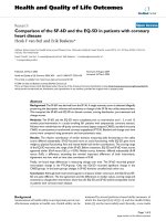
Figure 1 (facing page). Magnetic Resonance Spectro-
scopic Images in a Newborn with Transposition
of the Great Arteries.
In Panel A, a metabolite map shows lactate (red color)
laid over choline (green); more intense yellow and or-
ange indicate a higher ratio of lactate to choline. Ele-
vated ratios of lactate to choline are diffusely distribut-
ed but are most prominent in the periventricular white
matter (arrows). In Panel B, proton spectra are measured
bilaterally from the following 1-cm
3
regions of interest
(clear boxes) overlaid on T
2
-weighted images (with yellow
boxes for orientation only): frontal, perirolandic, and
posterior white matter (image 1); basal ganglia and thal-
amus (image 2); and optic radiations and the calcarine
region (image 3). In Panel C, diffusion tensor imaging
shows water-diffusion measures bilaterally from the follow-
ing regions of interest (measuring 5 mm × 5 mm × 3 mm
unless otherwise noted): perirolandic white matter
(image 1), posterior and frontal white matter (image 2),
and basal ganglia, thalamus, optic radiations (3 mm ×
10 mm × 3 mm), and the calcarine region.
Downloaded from www.nejm.org on February 18, 2008 . Copyright © 2007 Massachusetts Medical Society. All rights reserved.
Br ain Development in Newbor ns with Congenital Heart Dise ase
n engl j med 357;19 www.nejm.org november 8, 2007
1931
fusivity reflects the mean of these eigenvalues,
expressed as 10
−3
millimeters squared per sec-
ond, whereas fractional anisotropy reflects their
variance (higher fractional anisotropy with in-
creasing variance).
We then generated parametric maps for aver-
age diffusivity, fractional anisotropy, and the three
eigenvalues.
17,35‑37
Average diffusivity was calcu-
lated for the same regions assessed by MRSI,
with fractional anisotropy calculated from white-
matter regions. Given the high spatial resolution,
some regions of interest were smaller than those
used for MRSI (Fig. 1) to separate white and gray
matter as much as possible.
36
33p9
AUTHOR
FIGURE
JOB: ISSUE:
4-C
H/T
RETAKE 1st
2nd
SIZE
ICM
CASE
Line
H/T
Combo
Revised
AUTHOR, PLEASE NOTE:
Figure has been redrawn and type has been reset.
Please check carefully.
REG F
FILL
TITLE
3rd
Enon
ARTIST:
Miller
1a-c
11-8-07
mst
35719
A
B
C
1 2
3
1 2
3
Downloaded from www.nejm.org on February 18, 2008 . Copyright © 2007 Massachusetts Medical Society. All rights reserved.
T h e n e w e ngl a n d j o u r n a l o f m e d icine
n engl j med 357;19 www.nejm.org november 8, 2007
1932
Statistical Analysis
We compared clinical variables in newborns with
congenital heart disease and in control newborns
with the use of the Mann–Whitney U test for con-
tinuous data, Cuzick’s test for ordinal variables,
38
and Fisher’s exact test for categorical variables,
using Stata Software, version 9 (Stata). Unadjusted
mean values for ratios of N-acetylaspartate and
lactate to choline, average diffusivity, and fraction-
al anisotropy are presented for newborns with
congenital heart disease and control newborns.
We used linear regression for repeated measures
(generalized estimating equations) to compare ra-
tios of N-acetylaspartate and lactate to choline,
average diffusivity, and fractional anisotropy (as
outcomes) in newborns with congenital heart dis-
ease, as compared with the values in control new-
borns (as predictors), accounting for multiple re-
gions of interest in each infant and adjusting for
gestational age at the time that MRI was per-
formed.
39
Region-specific effects were explored by
inclusion of an interaction term. We then tested
the effect of cardiac lesions with the control new-
borns as the reference group and with infants who
had transposition of the great arteries and those
who had single-ventricle physiology as compari-
son groups.
Among newborns with congenital heart dis-
ease, we explored whether preoperative brain in-
jury (as seen on MRI), SNAP–PE rating, or critical
illness (requiring mechanical ventilation or ino-
tropes) predicted the ratios of N-acetylaspartate
and lactate to choline, average diffusivity, and
fractional anisotropy, using linear regression for
repeated measures and adjusting for age at the
time of MRI and region of interest. A log-trans-
formed outcome variable was used in all regres-
sions. We calculated the relative percent differ-
ences in ratios of N-acetylaspartate and lactate to
choline, average diffusivity, and fractional anisot-
ropy between newborns with congenital heart
disease and control newborns by the exponentia-
tion of the mean differences of the log-trans-
formed values from the regression model.
40
All
reported P values are two-sided and have not
been adjusted for multiple testing.
R e s ults
Clinical Condition and MRI
Of the 58 eligible newborns with congenital heart
disease, the parents of 41 infants (71%) provided
consent for participation in the study. Of these
newborns, 29 had transposition of the great ar-
teries, and 12 had single-ventricle physiology, with
associated aortic-arch obstruction in 10 newborns.
As compared with control newborns, those with
congenital heart disease had a slightly lower ges-
tational age at birth (median difference, approxi-
mately 3 days) (
Table 1
), although they underwent
MRI at a similar gestational age. Newborns with
congenital heart disease were also smaller in
weight, length, and head circumference. Although
5-minute Apgar scores were lower in newborns
with congenital heart disease, none had a score
of less than 6. Although this cohort of patients
by definition is cyanotic and most of the new-
borns required stabilization with prostaglandins
and mechanical ventilation before surgery, con-
genital heart disease had been diagnosed prena-
tally in a number of the infants, none had preop-
erative cardiac arrest, and only a minority required
inotropic support. Most of the newborns no lon-
ger required mechanical ventilation at the time
that preoperative MRI was performed (
Table 1
).
Acquired brain injury was common in new-
borns with congenital heart disease (
Table 2
).
Preoperative strokes and white-matter injuries
were focal, and 11 of 13 were acute and associ-
ated with reduced water diffusion. None of the
newborns had the basal nuclei or watershed pat-
terns of injury that are characteristic of global
hypoxia–ischemia in term newborns. All control
newborns had normal MRI scans.
Brain Metabolism and Microstructure
The mean ratio of N-acetylaspartate to choline,
averaged across all of the brain regions, was 0.60
in newborns with congenital heart disease and
0.66 in control newborns; the mean ratio of lac-
tate to choline was 0.11 and 0.10, respectively
(
Table 3
). In the multivariate models, newborns
with congenital heart disease had a significantly
lower mean ratio of N-acetylaspartate to choline
(a reduction of 10%) than did control newborns
(P = 0.003), whereas the difference in the mean
ratio of lactate to choline (an increase of 28%)
was not significant (P = 0.08) (
Table 3
). The per-
cent difference in the ratio of N-acetylaspartate
to choline, for example, reflects a difference of
10% in the adjusted mean ratio of N-acetylaspar-
tate to choline in newborns with congenital heart
disease (0.59), as compared with the adjusted
mean value in control newborns (0.65).
Downloaded from www.nejm.org on February 18, 2008 . Copyright © 2007 Massachusetts Medical Society. All rights reserved.
Br ain Development in Newbor ns with Congenital Heart Dise ase
n engl j med 357;19 www.nejm.org november 8, 2007
1933
Table 1. Demographic and Clinical Characteristics of the Newborns.
Variable
Control Newborns*
(N = 16)
Newborns with Congenital
Heart Disease
(N = 41) P Value
Male sex — no. (%) 13 (81) 29 (71) 0.52
Cesarean delivery — no. (%) 1 (6) 10 (24) 0.15
Gestational age at birth — wk
Median 39.6 39.1 0.01
Interquartile range 39.2–40.5 38.2–40.0
Gestational age at preoperative MRI — wk 0.13
Median 40.3 39.7
Interquartile range 40.0–41.0 38.9–40.9
Age at preoperative MRI — days 0.22
Median 7 5
Interquartile range 4–9 3–6
Birth weight — g 0.04
Median 3638 3300
Interquartile range 3360–4075 3000–3580
Birth length — cm 0.01
Median 52.0 50.5
Interquartile range 51.0–54.0 47.5–52.5
Birth head circumference — cm 0.002
Median 35.5 34.0
Interquartile range 35.0–37.0 33.5–35.5
Apgar score at 5 minutes† 0.002
Median 9 8
Interquartile range 9–9 8–9
Resuscitation score‡ 0.42
Median 2 2
Interquartile range 1–2 1–4
SNAP–PE rating§
Median — 16
Interquartile range — 12–21
Heart lesion — no. (%) —
Transposition of the great arteries — 29 (71)
Single-ventricle physiology — 12 (29)
Prenatal diagnosis — no. (%) — 7 (17)
Preoperative mechanical ventilation — no. (%) — 30 (73)
Mechanical ventilation at time of preoperative
MRI — no. (%)
— 15 (37)
Inotropic support — no. (%) — 14 (34)
Prostaglandin E
1
— no. (%) — 36 (88)
Cardiac arrest — no. (%) — 0
Balloon atrial septostomy — no. (%) — 19 (46)
* Dashes indicate that the variables either were not measured in control newborns or are conditions for which such new
-
borns were not at risk.
† Apgar scores range from 0 to 10, with lower scores indicating a worse clinical condition.
‡ The resuscitation score is based on interventions that are administered at birth, ranging from 1 (no intervention)
to 6 (endotracheal intubation and epinephrine).
9
§ The Score for Neonatal Acute Physiology–Perinatal Extension (SNAP–PE), a measure of the overall severity of illness,
ranges from 0 to 70, with higher scores indicating more severe illness.
29
Downloaded from www.nejm.org on February 18, 2008 . Copyright © 2007 Massachusetts Medical Society. All rights reserved.
T h e n e w e ngl a n d j o u r n a l o f m e d icine
n engl j med 357;19 www.nejm.org november 8, 2007
1934
Table 2. Classification and Timing of Injury, as Seen on MRI.*
Type of Injury
No. of
Newborns
White-Matter
Injury Stroke
Intraventricular
Hemorrhage
Total with
Injury
number (percent)
Preoperative injury
Transposition of the great arteries 29 3 (10) 9 (31) 2 (7) 12 (41)
Single-ventricle physiology 12 1 (8) 1 (8) 0 2 (17)
New postoperative injury
Transposition of the great arteries 28 7 (25) 0 0 7 (25)
Single-ventricle physiology 8 2 (25) 3 (38) 0 4 (50)
Total no. of newborns 41 13 (32) 13 (32) 2 (5) 25 (61)
* Some newborns had multiple types of injury in a single study.
Table 3. Comparison of Brain Development in Newborns with Congenital Heart Disease and in Control Newborns,
as Seen on Magnetic Resonance Spectroscopic Imaging (MRSI) and Diffusion Tensor Imaging (DTI).
Variable
Control
Newborns
(Unadjusted
Analysis)
Newborns with
Congenital Heart
Disease (Unadjusted
Analysis) Adjusted Difference* P Value
% (95% CI)
MRSI
Mean ratio of N-acetylaspartate
to choline
0.66 0.60 −10 (−15 to −3) 0.003
Transposition of the great
arteries
−10 (−16 to −4)
Single-ventricle physiology −9 (−18 to 3)
Mean ratio of lactate to choline 0.10 0.11 28 (−3 to 68) 0.08
Transposition of the great
arteries
32 (0 to 77)
Single-ventricle physiology 17 (−16 to 62)
DTI
Average diffusivity† 1.28 1.35 4 (2 to 7) <0.001
Transposition of the great
arteries
4 (2 to 7)
Single-ventricle physiology 5 (2 to 8)
Mean fractional anisotropy 0.21 0.18 −12 (−18 to −6) <0.001
Transposition of the great
arteries
−11 (−16 to −5)
Single-ventricle physiology −14 (−19 to −7)
* Analyses were adjusted for gestational age at the time of MRI and for brain region. The percent differences were calcu-
lated by exponentiation of the mean differences of the log-transformed values from the regression model.
40
The values
are the relative differences between newborns with congenital heart disease and control newborns. Values for each cardiac
anatomical diagnosis are presented below the main effect.
† Average diffusivity is calculated as the mean of the eigenvalues.
Downloaded from www.nejm.org on February 18, 2008 . Copyright © 2007 Massachusetts Medical Society. All rights reserved.
Br ain Development in Newbor ns with Congenital Heart Dise ase
n engl j med 357;19 www.nejm.org november 8, 2007
1935
The mean value for average diffusivity, aver-
aged across all of the brain regions, was 1.35 in
newborns with congenital heart disease and 1.28
in control newborns, and the mean value for
white-matter fractional anisotropy was 0.18 in
newborns with congenital heart disease and 0.21
in control newborns (
Table 3
). In the multivariate
models, newborns with congenital heart disease
had a significant increase of 4% in average dif-
fusivity and a significant decrease of 12% in
white-matter fractional anisotropy (P<0.001 for
both comparisons).
Although the decrease in the ratio of N-acetyl-
aspartate to choline and the increase in average
diffusivity in newborns with congenital heart dis-
ease, as compared with values in control new-
borns, were not homogeneous across regions (test
for interaction, P<0.001), these effects were each
seen in six of seven regions (Fig. 2). The reduction
in white-matter fractional anisotropy in newborns
with congenital heart disease, as compared with
that in control newborns, was homogeneous
across regions, and the interaction was not sig-
nificant (P = 0.37). The findings were similar when
the cardiac-lesion subgroups were compared with
the control newborns (Table 3).
Abnormal White Matter in Newborns
with Heart Disease
Newborns with transposition of the great arter-
ies and those with single-ventricle physiology had
a level of fractional anisotropy that was lower than
that in control newborns, a finding that was in-
dependent of white-matter region and age. This
difference was associated with an increase of 6%
in intermediate eigenvalues (P = 0.001) and an in-
crease of 9% in minimum eigenvalues (P<0.001).
Effect of Preoperative Brain Injury
Among newborns with congenital heart disease,
the presence of preoperative brain injury, as seen
on MRI, was not significantly associated with the
decrease of 5% in the ratio of N-acetylaspartate
to choline (P = 0.13), with the decrease of 17% in
the ratio of lactate to choline (P = 0.08), with the
increase of 2% in average diffusivity (P = 0.10), or
with the decrease of 5% in fractional anisotropy
(P = 0.11). When the comparison with control new-
borns was limited to newborns with congenital
heart disease who did not have preoperative brain
injury, those with congenital heart disease had a
reduction of 8% in the ratio of N-acetylaspartate
−50 −40 −30 −20 −10 0 10 20 30 40
50
Percent Difference
B Average Diffusivity
A Ratio of N-Acetylaspartate to Choline
AUTHOR:
FIGURE:
JOB:
4-C
H/T
RETAKE
SIZE
ICM
CASE
Line
H/T
Combo
Revised
AUTHOR, PLEASE NOTE:
Figure has been redrawn and type has been reset.
Please check carefully.
REG F
Enon
1st
2nd
3rd
Miller
2 of 2
xx-xx-07
ARTIST: ts
356xx ISSUE:
22p3
Overall
Basal ganglia
Thalamus
Calcarine region
Optic radiations
Perirolandic white
matter
Posterior white
matter
Frontal white matter
Perirolandic white
matter
Posterior white
matter
Frontal white matter
Perirolandic white
matter
Posterior white
matter
Frontal white matter
−50 −40 −30 −20 −10 0 10 20 30 40
50
Percent Difference
Overall
Basal ganglia
Thalamus
Calcarine region
Optic radiations
C
Fractional Anisotropy
−50 −40 −30 −20 −10 0 10 20 30 40
50
Percent Difference
Overall
Optic radiations
Figure 2. Differences in Ratios of N-Acetylaspartate to Choline, Average
Diffusivity, and Fractional Anisotropy in Newborns with Congenital Heart
Disease, as Compared with Control Newborns.
The mean difference, with 95% confidence intervals, is plotted for the over-
all effect and each region of interest.
Downloaded from www.nejm.org on February 18, 2008 . Copyright © 2007 Massachusetts Medical Society. All rights reserved.
T h e n e w e ngl a n d j o u r n a l o f m e d icine
n engl j med 357;19 www.nejm.org november 8, 2007
1936
to choline (P = 0.04), an increase of 38% in the
ratio of lactate to choline (P = 0.02), an increase of
4% in average diffusivity (P = 0.008), and a decrease
of 10% in fractional anisotropy (P<0.001).
Even when regions with signal abnormalities
on MRI scans or diffusion images were removed
from the analysis, the pattern of differences be-
tween newborns with congenital heart disease
and control newborns remained similar, with a
decrease of 10% in the ratio of N-acetylaspartate
to choline (P = 0.003), an increase of 28% in the
ratio of lactate to choline (P = 0.08), an increase of
4% in average diffusivity (P<0.001), and a decrease
of 11% in fractional anisotropy (P<0.001).
Effect of Preoperative Illness
Among newborns with congenital heart disease,
the presence of critical illness (requiring mechan-
ical ventilation or inotropes) was not significant-
ly associated with an increase of 1% in the ratio
of N-acetylaspartate to choline (P = 0.80), a de-
crease of 5% in the ratio of lactate to choline
(P = 0.64), a decrease of less than 1% in average
diffusivity (P = 0.90), and an increase of 2% in
fractional anisotropy (P = 0.53). In addition, when
the comparison with control newborns was lim-
ited to newborns with congenital heart disease
who did not require mechanical ventilation or ino-
tropic support, those with congenital heart dis-
ease had a decrease of 10% in the ratio of N-acetyl-
aspartate to choline (P = 0.04), an increase of 31%
in the ratio of lactate to choline (P = 0.02), an in-
crease of 5% in average diffusivity (P = 0.008), and a
decrease of 12% in fractional anisotropy (P<0.001).
Increases in the SNAP–PE rating, indicating an
increased severity of illness, were associated with
higher ratios of lactate to choline, with an increase
of 2% per unit increase in the SNAP–PE rating
(P = 0.007). In contrast, increases in SNAP–PE rat-
ings were not significantly associated with lower
ratios of N-acetylaspartate to choline (<1% in-
crease per unit increase in the SNAP–PE rating,
P = 0.86), with average diffusivity (<1% increase
per unit increase in the SNAP–PE rating, P = 0.10),
or with fractional anisotropy (<1% decrease per
unit increase in the SNAP–PE rating, P = 0.47).
Discus s ion
Newborns with transposition of the great arter-
ies and single-ventricle physiology have brain ab-
normalities before they undergo cardiac surgery,
as evidenced by altered brain metabolism and
microstructure shortly after birth. Advanced MRI
can quantify brain development and injury at a
time when intervention for brain protection may
be possible, allowing for incorporation of these
data into the development and assessment of new
clinical interventions for this population.
The discovery of abnormal brain microstruc-
ture and metabolism shortly after birth in new-
borns with congenital heart disease is consistent
with mounting evidence that these newborns have
impaired brain development in utero, possibly re-
lated to impaired cerebral oxygen and substrate
delivery prenatally.
26‑28
In newborns with trans-
position of the great arteries and single-ventricle
physiology, especially the hypoplastic left heart
syndrome, the brain receives lower levels of oxy-
gen-saturated blood from the right ventricle as a
consequence of disordered fetal circulation.
28
De-
spite cerebral vasodilation in human fetuses with
transposition of the great arteries and hypoplas-
tic left heart syndrome,
26,27
at autopsy, 55% of
newborns with hypoplastic left heart syndrome
are microcephalic, and 21% have an immature
cortical mantle.
41
With the increasing diagnosis
of congenital heart disease in utero, methods to
intervene and improve fetal circulation, such as
fetal aortic valvuloplasty, are being studied.
42
In-
formation regarding brain maturation may be
important in considering when to perform these
interventions.
We identified impaired brain metabolism and
microstructure in a cohort of newborns with con-
genital heart disease, even in the absence of visi-
ble injury on MRI and in uninvolved regions.
These impairments were widespread and did not
conform to the pattern of brain injury that is typi-
cal of hypoxia–ischemia in term newborns.
43
However, with a complex interplay between brain
injury and abnormal brain development, brain
injury may itself disturb brain development. Pre-
operative brain injury in term newborns with
congenital heart disease is associated with sub-
sequently impaired development of the cortico-
spinal tract.
44
Data from our cohort suggest that
abnormal brain development precedes surgery and
some acquired injuries.
Our study was limited by a lack of compari-
son with other critically ill newborns who did not
have heart disease. Thus, we are unable to exclude
the possibility that some of the measured effects
reflect changes that are generic to critically ill
Downloaded from www.nejm.org on February 18, 2008 . Copyright © 2007 Massachusetts Medical Society. All rights reserved.
Br ain Development in Newbor ns with Congenital Heart Dise ase
n engl j med 357;19 www.nejm.org november 8, 2007
1937
newborns. The analyses examining the effects of
preoperative brain injury and the severity of ill-
ness on the MRSI and DTI measures were rela-
tively underpowered, yet they showed smaller ef-
fect sizes than those observed in comparisons of
newborns with congenital heart disease with con-
trol newborns. Future improvements in MRI spa-
tial resolution may allow for detection of specific
regional differences underlying the vulnerability
of newborns with congenital heart disease to
white-matter injury. In addition, fetal MRI holds
promise for determining the precise onset of the
brain changes observed shortly after birth in such
newborns.
The findings of lower ratios of N-acetylaspar-
tate to choline, higher average diffusivity, and
lower white-matter fractional anisotropy in new-
borns with congenital heart disease are similar
to findings in premature newborns at an earlier
age, and the MRS metabolite ratios are similar
to those in premature newborns approximately
1 month before full term.
15,32,45
The pattern of
white-matter injury in premature newborns is at-
tributed to cell populations that are vulnerable to
ischemia, inflammation, and oxidative stress.
46‑48
Though predominant injury to neurons would be
the expected response to these insults in term
newborns with congenital heart disease,
43
white-
matter injury, the pattern of injury that is typical
in premature newborns, occurs frequently.
10,23‑25
Our findings suggest that white-matter vul-
nerability in term newborns with congenital heart
disease is related to impaired brain development
that is detected preoperatively, shortly after birth.
The increase in white-matter radial diffusion (per-
pendicular to axon tracts) in newborns with con-
genital heart disease, as in premature newborns,
suggests an abnormality of cells associated with
axons forming white-matter tracts, such as oligo-
dendrocyte progenitors or glia.
19,49
The dramatic
difference in brain development in newborns
with congenital heart disease, as compared with
other term neonates, and the pattern of brain in-
jury suggest that new and specific neuroprotective
strategies may be needed in this population. Fur-
thermore, the state of brain maturation before
cardiac surgery may influence the choice of brain
protective strategy.
Supported by a grant (5-FY05-1231) from the March of Dimes
Foundation, a grant (0365018Y) from the American Heart Asso-
ciation, a grant (2002/3E) from the Larry L. Hillblom Founda-
tion, grants (RO1 NS40117 and P50 NS35902) from the National
Institutes of Health, a grant (5-M01-RR-01271) from the National
Center for Research Resources, an award (40747, to Dr. Miller)
from the Canadian Institutes of Health Research, and an award
(CI-SCH-065-051, to Dr. Miller) from the Michael Smith Founda-
tion for Health Research.
Dr. Vigneron reports receiving lecture fees from General Elec-
tric. No other potential conflict of interest relevant to this article
was reported.
We thank Drs. Katherine Yap and Agnes Bartha for providing
data for control newborns; the neonatal nurses of the Pediatric
Clinical Research Center at the University of California at San
Francisco, San Francisco, including Nancy Newton, Julie Bush-
nell, Jessica Ravitz Sturm, Jane Holland-Browne, and Jill Imamura-
Ching, whose skill and expertise made this study possible;
Srivathsa Veeraraghavan and Kelly Blum for their assistance in
acquiring and processing MRI data; and Marta Perez for obtain-
ing clinical data for the study.
References
Hoffman JI, Kaplan S. The incidence
of congenital heart disease. J Am Coll
Cardiol 2002;39:1890-900.
Bellinger DC, Jonas RA, Rappaport LA,
et al. Developmental and neurologic sta-
tus of children after heart surgery with
hypothermic circulatory arrest or low-
flow cardiopulmonary bypass. N Engl J
Med 1995;332:549-55.
Bellinger DC, Wypij D, Duplessis AJ,
et al. Neurodevelopmental status at eight
years in children with dextro-transposi-
tion of the great arteries: the Boston Cir-
culatory Arrest Trial. J Thorac Cardiovasc
Surg 2003;126:1385-96.
Hövels-Gürich HH, Konrad K, Wies-
ner M, et al. Long term behavioural out-
come after neonatal arterial switch opera-
tion for transposition of the great arteries.
Arch Dis Child 2002;87:506-10.
Limperopoulos C, Majnemer A, Shevell
MI, et al. Functional limitations in young
children with congenital heart defects af-
1.
2.
3.
4.
5.
ter cardiac surgery. Pediatrics 2001;108:
1325-31.
Karl TR, Hall S, Ford G, et al. Arterial
switch with full-flow cardiopulmonary by-
pass and limited circulatory arrest: neuro-
developmental outcome. J Thorac Cardio-
vasc Surg 2004;127:213-22.
Limperopoulos C, Majnemer A, Shevell
MI, et al. Predictors of developmental dis-
abilities after open heart surgery in young
children with congenital heart defects.
J Pediatr 2002;141:51-8.
McQuillen PS, Barkovich AJ, Hamrick
SE, et al. Temporal and anatomic risk pro-
file of brain injury with neonatal repair of
congenital heart defects. Stroke 2007;38:
736-41.
McQuillen PS, Hamrick SE, Perez MJ,
et al. Balloon atrial septostomy is associ-
ated with preoperative stroke in neonates
with transposition of the great arteries.
Circulation 2006;113:280-5.
Mahle WT, Tavani F, Zimmerman RA,
6.
7.
8.
9.
10.
et al. An MRI study of neurological injury
before and after congenital heart surgery.
Circulation 2002;106:Suppl 1:I-109–I-114.
Kreis R, Hofmann L, Kuhlmann B,
Boesch C, Bossi E, Hüppi PS. Brain me-
tabolite composition during early human
brain development as measured by quan-
titative in vivo 1H magnetic resonance
spectroscopy. Magn Reson Med 2002;48:
949-58.
Kasischke KA, Vishwasrao HD, Fisher
PJ, Zipfel WR, Webb WW. Neural activity
triggers neuronal oxidative metabolism
followed by astrocytic glycolysis. Science
2004;305:99-103.
Miller SP, Newton N, Ferriero DM, et
al. Predictors of 30-month outcome after
perinatal depression: role of proton MRS
and socioeconomic factors. Pediatr Res
2002;52:71-7.
Mukherjee P, Miller JH, Shimony JS, et
al. Diffusion-tensor MR imaging of gray
and white matter development during nor-
11.
12.
13.
14.
Downloaded from www.nejm.org on February 18, 2008 . Copyright © 2007 Massachusetts Medical Society. All rights reserved.
n engl j med 357;19 www.nejm.org november 8, 2007
1938
Br ain Development in Newbor ns with Congenital Heart Dise ase
mal human brain maturation. AJNR Am J
Neuroradiol 2002;23:1445-56.
Miller SP, Vigneron DB, Henry RG, et
al. Serial quantitative diffusion tensor MRI
of the premature brain: development in
newborns with and without injury. J Magn
Reson Imaging 2002;16:621-32.
Beaulieu C. The basis of anisotropic
water diffusion in the nervous system —
a technical review. NMR Biomed 2002;15:
435-55.
Deipolyi AR, Mukherjee P, Gill K, et
al. Comparing microstructural and macro-
structural development of the cerebral cor-
tex in premature newborns: diffusion
tensor imaging versus cortical gyration.
Neuroimage 2005;27:579-86.
McKinstry RC, Mathur A, Miller JH, et
al. Radial organization of developing pre-
term human cerebral cortex revealed by
non-invasive water diffusion anisotropy
MRI. Cereb Cortex 2002;12:1237-43.
Drobyshevsky A, Song SK, Gamkre-
lidze G, et al. Developmental changes in
diffusion anisotropy coincide with im-
mature oligodendrocyte progression and
maturation of compound action potential.
J Neurosci 2005;25:5988-97.
Prayer D, Barkovich AJ, Kirschner DA,
et al. Visualization of nonstructural chang-
es in early white matter development on
diffusion-weighted MR images: evidence
supporting premyelination anisotropy.
AJNR Am J Neuroradiol 2001;22:1572-6.
Miller SP, Ferriero DM, Leonard C, et
al. Early brain injury in premature new-
borns detected with magnetic resonance
imaging is associated with adverse early
neurodevelopmental outcome. J Pediatr
2005;147:609-16.
Woodward LJ, Anderson PJ, Austin NC,
Howard K, Inder TE. Neonatal MRI to
predict neurodevelopmental outcomes in
preterm infants. N Engl J Med 2006;355:
685-94.
Galli KK, Zimmerman RA, Jarvik GP,
et al. Periventricular leukomalacia is
common after neonatal cardiac surgery.
J Thorac Cardiovasc Surg 2004;127:692-
704.
Gilles FH, Leviton A, Jammes J. Age-
dependent changes in white matter in con-
genital heart disease. J Neuropathol Exp
Neurol 1973;32:179. abstract.
Kinney HC, Panigrahy A, Newburger
15.
16.
17.
18.
19.
20.
21.
22.
23.
24.
25.
JW, Jonas RA, Sleeper LA. Hypoxic-ische-
mic brain injury in infants with congeni-
tal heart disease dying after cardiac sur-
gery. Acta Neuropathol (Berl) 2005;110:
563-78.
Donofrio MT, Bremer YA, Schieken
RM, et al. Autoregulation of cerebral blood
flow in fetuses with congenital heart dis-
ease: the brain sparing effect. Pediatr
Cardiol 2003;24:436-43.
Jouannic JM, Benachi A, Bonnet D, et
al. Middle cerebral artery Doppler in fetus-
es with transposition of the great arteries.
Ultrasound Obstet Gynecol 2002;20:122-4.
Rudolph A. Congenital diseases of the
heart: clinical-physiological considera-
tions. 2nd ed. Armonk, NY: Futura Pub-
lishing, 2001.
Bartha AI, Yap KRL, Miller SP, et al.
The normal neonatal brain: MR imaging,
diffusion tensor imaging, and 3D MR
spectroscopy in healthy term neonates.
AJNR Am J Neuroradiol 2007;28:1015-21.
Richardson DK, Phibbs CS, Gray JE,
McCormick MC, Workman-Daniels K,
Goldmann DA. Birth weight and illness
severity: independent predictors of neona-
tal mortality. Pediatrics 1993;91:969-75.
Dumoulin CL, Rohling KW, Piel JE, et
al. An MRI compatible neonate incubator.
Magn Reson Engineering 2002;15:117-28.
Vigneron DB, Barkovich AJ, Noworol-
ski SM, et al. Three-dimensional proton
MR spectroscopic imaging of premature
and term neonates. AJNR Am J Neurora-
diol 2001;22:1424-33.
Star-Lack J, Spielman D, Adalsteins-
son E, Kurhanewicz J, Terris DJ, Vigneron
DB. In vivo lactate editing with simultane-
ous detection of choline, creatine, NAA,
and lipid singlets at 1.5 T using PRESS
excitation with applications to the study of
brain and head and neck tumors. J Magn
Reson 1998;133:243-54.
Miller SP, McQuillen PS, Vigneron DB,
et al. Preoperative brain injury in new-
borns with transposition of the great ar-
teries. Ann Thorac Surg 2004;77:1698-706.
Barkovich AJ, Miller SP, Bartha A, et
al. MR imaging, MR spectroscopy, and
diffusion tensor imaging of sequential
studies in neonates with encephalopathy.
AJNR Am J Neuroradiol 2006;27:533-47.
Partridge SC, Mukherjee P, Henry RG,
et al. Diffusion tensor imaging: serial
26.
27.
28.
29.
30.
31.
32.
33.
34.
35.
36.
quantitation of white matter tract matu-
rity in premature newborns. Neuroimage
2004;22:1302-14.
Basser PJ, Pierpaoli C. Microstructural
and physiological features of tissues elu-
cidated by quantitative-diffusion-tensor
MRI. J Magn Reson B 1996;111:209-19.
Cuzick J. A Wilcoxon-type test for
trend. Stat Med 1985;4:87-90.
Zeger SL, Liang KY, Albert PS. Models
for longitudinal data: a generalized es-
timating equation approach. Biometrics
1988;44:1049-60. [Erratum, Biometrics
1989;45:347.]
Bland JM, Altman DG. Statistics notes:
logarithms. BMJ 1996;312:700.
Glauser TA, Rorke LB, Weinberg PM,
Clancy RR. Congenital brain anomalies
associated with the hypoplastic left heart
syndrome. Pediatrics 1990;85:984-90.
Marshall AC, Tworetzky W, Bergersen
L, et al. Aortic valvuloplasty in the fetus:
technical characteristics of successful bal-
loon dilation. J Pediatr 2005;147:535-9.
Miller SP, Ramaswamy V, Michelson
D, et al. Patterns of brain injury in term
neonatal encephalopathy. J Pediatr 2005;
146:453-60.
Partridge SC, Vigneron DB, Charlton
NN, et al. Pyramidal tract maturation af-
ter brain injury in newborns with heart
disease. Ann Neurol 2006;59:640-51.
Vigneron DB. Magnetic resonance
spectroscopic imaging of human brain de-
velopment. Neuroimaging Clin N Am 2006;
16:75-85.
Volpe J. Neurology of the newborn.
4th ed. Philadelphia: W.B. Saunders, 2001.
McQuillen PS, Sheldon RA, Shatz CJ,
Ferriero DM. Selective vulnerability of sub-
plate neurons after early neonatal hypoxia-
ischemia. J Neurosci 2003;23:3308-15.
Riddle A, Luo NL, Manese M, et al.
Spatial heterogeneity in oligodendrocyte
lineage maturation and not cerebral blood
flow predicts fetal ovine periventricular
white matter injury. J Neurosci 2006;26:
3045-55.
Counsell SJ, Shen Y, Boardman JP, et
al. Axial and radial diffusivity in preterm
infants who have diffuse white matter
changes on magnetic resonance imaging
at term-equivalent age. Pediatrics 2006;117:
376-86.
Copyright © 2007 Massachusetts Medical Society.
37.
38.
39.
40.
41.
42.
43.
44.
45.
46.
47.
48.
49.
posting
presentations
at
medical
meetings
on
the
internet
Posting an audio recording of an oral presentation at a medical meeting on the
Internet, with selected slides from the presentation, will not be considered prior
publication. This will allow students and physicians who are unable to attend the
meeting to hear the presentation and view the slides. If there are any questions
about this policy, authors should feel free to call the Journal’s Editorial Offices.
Downloaded from www.nejm.org on February 18, 2008 . Copyright © 2007 Massachusetts Medical Society. All rights reserved.