Cardiac arrhythmias translational approach from pathophysiology to advanced care
Bạn đang xem bản rút gọn của tài liệu. Xem và tải ngay bản đầy đủ của tài liệu tại đây (5.71 MB, 154 trang )
<span class="text_page_counter">Trang 1</span><div class="page_container" data-page="1">
Cardiac Arrhythmias
Translational Approach from Pathophysiology to Advanced Care
<i>Edited by Endre ZimaEdited by Endre Zima</i>
Cardiac arrhythmias are common triggers of emergency admission to cardiology or high-dependency departments. Most cases are easy to diagnose and treat, while others may present a challenge to healthcare professionals. A translational approach to arrhythmias links molecular and cellular scientific research with clinical diagnostics and therapeutic methods, which may include both pharmacological and non-pharmacologic treatments.
This book presents a comprehensive overview of specific cardiac arrhythmias and discusses translational approaches to their diagnosis and treatment.
</div><span class="text_page_counter">Trang 3</span><div class="page_container" data-page="3">Cardiac Arrhythmias - Translational Approach from Pathophysiology to
Advanced Care
<i>Edited by Endre Zima</i>
Published in London, United Kingdom
</div><span class="text_page_counter">Trang 5</span><div class="page_container" data-page="5"><i>Supporting open minds since 2005</i>
</div><span class="text_page_counter">Trang 6</span><div class="page_container" data-page="6"><small>Cardiac Arrhythmias - Translational Approach from Pathophysiology to Advanced Care by Endre ZimaContributors</small>
<small>Dmytro Skoryi, Dmytro Volkov, Iurii Karpenko, Amad Hania, Bin Liu, Brian D. Tow, Ingrid M. Bonilla, Sombat Muengtaweepongsa, Dilok Piyayotai, Srinivasan Jayaraman, Ponnuraj Kirthi Priya, Zuraini Md. Noor, Raj Parikh, Meher Singha, Abhishek Madathanapalli, Endre Zima, Bettina Nagy, Boldizsár Kiss, Gábor Áron Fülưp</small>
<small>© The Editor(s) and the Author(s) 2022</small>
<small>The rights of the editor(s) and the author(s) have been asserted in accordance with the Copyright, Designs and Patents Act 1988. All rights to the book as a whole are reserved by INTECHOPEN LIMITED. The book as a whole (compilation) cannot be reproduced, distributed or used for commercial or non-commercial purposes without INTECHOPEN LIMITED’s written permission. Enquiries concerning the use of the book should be directed to INTECHOPEN LIMITED rights and permissions department ().</small>
<small>Violations are liable to prosecution under the governing Copyright Law.</small>
<small>Individual chapters of this publication are distributed under the terms of the Creative Commons Attribution 3.0 Unported License which permits commercial use, distribution and reproduction of the individual chapters, provided the original author(s) and source publication are appropriately acknowledged. If so indicated, certain images may not be included under the Creative Commons license. In such cases users will need to obtain permission from the license holder to reproduce the material. More details and guidelines concerning content reuse and adaptation can be found at and opinions expressed in the chapters are these of the individual contributors and not necessarily those of the editors or publisher. No responsibility is accepted for the accuracy of information contained in the published chapters. The publisher assumes no responsibility for any damage or injury to persons or property arising out of the use of any materials, instructions, methods or ideas contained in the book.</small>
<small>First published in London, United Kingdom, 2022 by IntechOpen</small>
<small>IntechOpen is the global imprint of INTECHOPEN LIMITED, registered in England and Wales, registration number: 11086078, 5 Princes Gate Court, London, SW7 2QJ, United KingdomPrinted in Croatia</small>
<small>British Library Cataloguing-in-Publication Data</small>
<small>A catalogue record for this book is available from the British LibraryAdditional hard and PDF copies can be obtained from Arrhythmias - Translational Approach from Pathophysiology to Advanced CareEdited by Endre Zima</small>
<small>p. cm.</small>
<small>Print ISBN 978-1-83969-505-6Online ISBN 978-1-83969-506-3eBook (PDF) ISBN 978-1-83969-507-0</small>
</div><span class="text_page_counter">Trang 7</span><div class="page_container" data-page="7">Interested in publishing with us?
Contact
<small>Numbers displayed above are based on latest data collected. </small><small>For more information visit www.intechopen.com</small>
5,700+
<small>Open access books available</small>
<small>Contributors from top 500 universitiesOur authors are among the</small>
the world’s leading publisher of Open Access books
Built by scientists, for scientists
<small>Selection of our books indexed in the Book Citation Index (BKCI) in Web of Science Core Collection™</small>
</div><span class="text_page_counter">Trang 9</span><div class="page_container" data-page="9">Meet the editor
Endre Zima, MD, is head of Cardiac ICU at Semmelweis Uni-versity Heart and Vascular Center, Budapest, Hungary. He is specialized in anesthesiology, intensive care, and cardiology. He received a Ph.D. in 2006 and a medical habilitation degree in 2017 from Semmelweis University. As a professor, he has been holding graduate and postgraduate lectures and practices in anesthesiology/intensive care and cardiology. He has a European Heart Rhythm Association (EHRA) accreditation for Cardiac Pacing and Implant-able Cardioverter Defibrillators and is a Full Instructor of Advanced Life Support (ALS) and Basic Life Support (BLS) for the European Resuscitation Council. Dr. Zima is a fellow of the European Society of Cardiology, EHRA, and the Acute Cardiovascular Care Association. He is also the president of the Working Group on Cardiac Arrhythmias and Pacing and a board member of the Hungarian Society of Cardiology and WG of Heart Failure. His fields of research are acute and intensive cardiac care, cardiogenic shock/acute heart failure, cardiopulmonary resuscitation, post-cardiac arrest intensive care, invasive hemodynamic monitoring, arrhythmias, ICD/IPG/CRT therapy, and defibrillator waveform development. Dr. Zima has one accepted and one submitted patents patent to his credit. He is the author of thirteen book chapters, sixty-six international journal articles, and fifty-six native-language papers. He is currently supervising the research works of three Ph.D. students and seven medical students.
</div><span class="text_page_counter">Trang 10</span><div class="page_container" data-page="10">Molecular Mechanism and Current Therapies for Catecholaminergic Polymorphic Ventricular Tachycardia
<i>by Bin Liu, Brian D. Tow and Ingrid M. Bonilla</i>
COVID-19 and Cardiac Enzymes
<i>by Meher Singha, Abhishek Madathanapalli and Raj Parikh</i>
A High Fidelity Transmural Anisotropic Ventricular Tissue Model Function to Investigate the Interaction Mechanisms of Drug: An In-Silico Model for Pharmacotherapy
<i>by Srinivasan Jayaraman and Ponnuraj Kirthi Priya</i>
The Evolving Concept of Cardiac Conduction System Pacing
<i>by Iurii Karpenko, Dmytro Skoryi and Dmytro Volkov</i>
Out-of-Hospital Cardiac Arrest in General Population and Sudden Cardiac Death in Athletes
<i>by Bettina Nagy, Boldizsár Kiss, Gábor Áron Fülöp and Endre Zima</i>
Prognostication in Post-Cardiac Arrest Patients
<i>by Dilok Piyayotai and Sombat Muengtaweepongsa</i>
</div><span class="text_page_counter">Trang 11</span><div class="page_container" data-page="11">Molecular Mechanism and Current Therapies for Catecholaminergic Polymorphic Ventricular Tachycardia
<i>by Bin Liu, Brian D. Tow and Ingrid M. Bonilla</i>
COVID-19 and Cardiac Enzymes
<i>by Meher Singha, Abhishek Madathanapalli and Raj Parikh</i>
A High Fidelity Transmural Anisotropic Ventricular Tissue Model Function to Investigate the Interaction Mechanisms of Drug: An In-Silico Model for Pharmacotherapy
<i>by Srinivasan Jayaraman and Ponnuraj Kirthi Priya</i>
The Evolving Concept of Cardiac Conduction System Pacing
<i>by Iurii Karpenko, Dmytro Skoryi and Dmytro Volkov</i>
Out-of-Hospital Cardiac Arrest in General Population and Sudden Cardiac Death in Athletes
<i>by Bettina Nagy, Boldizsár Kiss, Gábor Áron Fülöp and Endre Zima</i>
Prognostication in Post-Cardiac Arrest Patients
<i>by Dilok Piyayotai and Sombat Muengtaweepongsa</i>
</div><span class="text_page_counter">Trang 12</span><div class="page_container" data-page="12">Knowledge of cardiac arrhythmias has significantly improved in the last several decades. Although most cases are benign and easy to evaluate and treat, arrhyth-mias are sometimes challenging to differentiate, requiring quick recognition and response. The worst-case scenario is a hemodynamically unstable arrhythmia that may lead to heart failure or cardiac arrest that necessitates cardiopulmonary resuscitation and complex intensive care.
A translational approach means implementing laboratory science into real-life clinical practice. Improvements in the science of arrhythmias have led to a decrease in morbidity and even mortality. Efforts to understand arrhythmic mechanisms have led to experimental modelling of arrhythmogenesis. These models are based on hypoxic-ischemic reperfusion myocardial insult, arrhythmogenic stimulating factors induced by electrolyte imbalance or administration of certain drugs. Over the last fifteen years, new methods of genetic testing have been developed that reveal new hereditary factors behind arrhythmias (e.g., cardiomyopathies), though genetics-driven therapy is still lacking.
The key to the effective and appropriate treatment of clinical arrhythmias is the identification of etiology. The classical diagnosis is based on a thorough analysis of the patient’s ECG. Causative therapy is driven by further clinical examination, labo-ratory testing of blood samples, imaging studies (e.g., echocardiography, CT and MRI scans), and invasive cardiac procedures. Patients suffering from arrhythmic cardiac arrest require immediate diagnostics and therapeutics regardless of setting in order to increase their chances of survival.
This book contains eight chapters in which the contributing authors highlight special aspects of the current scientific knowledge on cardiac arrhythmias.
The molecular background of catecholaminergic and calcium-dependent ventricular tachycardias is a point of interest since the latter may lead to sudden cardiac death. Genetic mutations in components of the calcium signaling pathway may induce dysregulation of calcium leading to overload, and this may be the pathway to target for therapy. Ryanodine receptors could be useful interventional gates to prevent catecholaminergic ventricular arrhythmias.
The COVID-19 pandemic has devastated daily medical and social life. Affecting mainly the respiratory tract and causing acute respiratory distress syndrome (ARDS), COVID infection has direct and indirect effects on the heart itself via secondary thrombogenicity, ischemia, myocarditis, and electrical inhomogeneity. The newly developed antiviral agents have potential proarrhythmic effects with or without the ischemic insult of the myocardial tissue. This proarrhythmic burden spurred scientists to begin using an endo-, mid myo-, and epicardial myocyte anisotropic preparation model for testing the safety of the first-used “anti-COVID” agent hydroxychloroquine. The latter acts electrophysiologically differently on myocardial cells in hypokalemic, COVID-associated ischemic, or overdosed circumstances.
</div><span class="text_page_counter">Trang 13</span><div class="page_container" data-page="13">Knowledge of cardiac arrhythmias has significantly improved in the last several decades. Although most cases are benign and easy to evaluate and treat, arrhyth-mias are sometimes challenging to differentiate, requiring quick recognition and response. The worst-case scenario is a hemodynamically unstable arrhythmia that may lead to heart failure or cardiac arrest that necessitates cardiopulmonary resuscitation and complex intensive care.
A translational approach means implementing laboratory science into real-life clinical practice. Improvements in the science of arrhythmias have led to a decrease in morbidity and even mortality. Efforts to understand arrhythmic mechanisms have led to experimental modelling of arrhythmogenesis. These models are based on hypoxic-ischemic reperfusion myocardial insult, arrhythmogenic stimulating factors induced by electrolyte imbalance or administration of certain drugs. Over the last fifteen years, new methods of genetic testing have been developed that reveal new hereditary factors behind arrhythmias (e.g., cardiomyopathies), though genetics-driven therapy is still lacking.
The key to the effective and appropriate treatment of clinical arrhythmias is the identification of etiology. The classical diagnosis is based on a thorough analysis of the patient’s ECG. Causative therapy is driven by further clinical examination, labo-ratory testing of blood samples, imaging studies (e.g., echocardiography, CT and MRI scans), and invasive cardiac procedures. Patients suffering from arrhythmic cardiac arrest require immediate diagnostics and therapeutics regardless of setting in order to increase their chances of survival.
This book contains eight chapters in which the contributing authors highlight special aspects of the current scientific knowledge on cardiac arrhythmias.
The molecular background of catecholaminergic and calcium-dependent ventricular tachycardias is a point of interest since the latter may lead to sudden cardiac death. Genetic mutations in components of the calcium signaling pathway may induce dysregulation of calcium leading to overload, and this may be the pathway to target for therapy. Ryanodine receptors could be useful interventional gates to prevent catecholaminergic ventricular arrhythmias.
The COVID-19 pandemic has devastated daily medical and social life. Affecting mainly the respiratory tract and causing acute respiratory distress syndrome (ARDS), COVID infection has direct and indirect effects on the heart itself via secondary thrombogenicity, ischemia, myocarditis, and electrical inhomogeneity. The newly developed antiviral agents have potential proarrhythmic effects with or without the ischemic insult of the myocardial tissue. This proarrhythmic burden spurred scientists to begin using an endo-, mid myo-, and epicardial myocyte anisotropic preparation model for testing the safety of the first-used “anti-COVID” agent hydroxychloroquine. The latter acts electrophysiologically differently on myocardial cells in hypokalemic, COVID-associated ischemic, or overdosed circumstances.
</div><span class="text_page_counter">Trang 14</span><div class="page_container" data-page="14">Bradycardia and heart failure together may be treated by cardiac implantable electronic devices (CIEDs). The pacing provides a better hemodynamic response, which is the aim of treating heart failure when the near-physiologic pacing sites are selected. To date, the first choice of device therapy for heart failure and the wide QRS complex is cardiac resynchronization therapy. “New-old” pacing methods are also tested to achieve better cardiac performance. The concept of pacing the bundle of His or left bundle branch has had a renaissance in the last five years. Despite the obvious benefits of pacing through the natural pacing conduction system of the heart, there are controversies on the skill-based and technical possibilities of it, limiting the widespread usage of the method.
Many supraventricular tachycardic arrhythmias have anatomical foci or accessory pathways in the background proven by electrophysiological diagnostic procedures. Most of these e.g. atrioventricular nodal reentrant tachycardia, atrioventricular reentrant tachycardia, atrial flutter, and atrial fibrillation can be treated by radiofrequency ablation. Few types of ventricular arrhythmias are also candidates for radiofrequency ablation, but most of them are treated with an implantable cardioverter-defibrillator (ICD) in addition to antiarrhythmic drug treatment. ICD was developed for primary and secondary prevention of ventricular arrhythmias and sudden cardiac death (SCD).
Sudden cardiac arrest is a major cause of mortality in developed countries. The incidence of out-of-hospital cardiac arrest is between 60 and 170 per 100,000 persons worldwide. Since the medical team is not present at the time of circulatory collapse in most cases, survival depends on the awareness and training of the people around the patient when the arrest occurs. Therefore, it is important to emphasize the relevance of the knowledge of the non-medical public recognizing cardiac arrest and providing basic life support until the emergency medical team arrives. The program of public access to automated defibrillators has also improved SCD mortality. In the era of widespread media coverage, more focus has been placed on SCD among athletes. Extreme sports activity, increased adrenaline release, lactic acidosis, and volume-electrolyte shifts may trigger SCD, which may be the first sign of undiagnosed heart disease in apparently healthy athletes. New guidelines in sports medicine suggest thorough examination and screening for underlying asymptomatic cardiac disorders.
Arrhythmias may cause a peri-arrest state or cardiac arrest in the hospital, intensive care unit, or even during anesthesia. Most of these cases occur because of stress due to surgery or hemostasis, volume, and electrolyte disorders, which are to be treated or corrected during the treatment of malignant arrhythmias.
After successful resuscitation, when the patient has returned the spontaneous cir-culation (ROSC), a special global systemic-ischemic reperfusion syndrome-induced multiple organ dysfunction may develop, which is defined as post-cardiac arrest syndrome. All the causative reversible factors should be promptly recognized and corrected to avoid the relapse of circulatory arrest. To prevent ischemic-reperfusion brain injury, a neuroprotective target temperature management (formerly mild therapeutic hypothermia) is part of the intensive care of patients suffering from a long-standing no-flow or low-flow perfusion before ROSC. These patients need special post-resuscitation intensive care, sedation, mechanical ventilation, and temperature management. As such, prognosis is difficult to estimate during the first 72–120 hours. Special protocols for prognostication cover the clinical neurological judgment, EEG, imaging modalities, and special neuron-specific biomarkers.
In conclusion, the translational approach to cardiac arrhythmias highlights interest-ing aspects of arrhythmia mechanisms as well as diagnostic and therapeutic methods. This book emphasizes a quick, critical, and thorough response to arrhythmias to prevent further deterioration of the patient. Knowledge of the basic molecular, cellular, and clinical background of arrhythmias is the foundation of clinical appraisal and treatment.
<b>Endre Zima MD, Ph.D., Dr. med. habil.</b>
Professor, Consultant Anesthesiologist, Intensive Care Specialist and Cardiologist, Head of Cardiac Intensive Care Unit, Heart and Vascular Center, Semmelweis University, Budapest, Hungary
<b><small>XIV</small></b>
</div><span class="text_page_counter">Trang 15</span><div class="page_container" data-page="15">In conclusion, the translational approach to cardiac arrhythmias highlights interest-ing aspects of arrhythmia mechanisms as well as diagnostic and therapeutic methods. This book emphasizes a quick, critical, and thorough response to arrhythmias to prevent further deterioration of the patient. Knowledge of the basic molecular, cellular, and clinical background of arrhythmias is the foundation of clinical appraisal and treatment.
<b>Endre Zima MD, Ph.D., Dr. med. habil.</b>
Professor, Consultant Anesthesiologist, Intensive Care Specialist and Cardiologist, Head of Cardiac Intensive Care Unit, Heart and Vascular Center, Semmelweis University, Budapest, Hungary
</div><span class="text_page_counter">Trang 17</span><div class="page_container" data-page="17"><b>Chapter 1</b>
Molecular Mechanism and Current Therapies for Catecholaminergic Polymorphic Ventricular
<i>Bin Liu, Brian D. Tow and Ingrid M. Bonilla</i>
The rhythmic contraction of the heart relies on tightly regulated calcium (Ca) release from the sarcoplasmic reticulum (SR) Ca release channel, Ryanodine receptor (RyR2). Genetic mutations in components of the calcium release unit such as RyR2, cardiac calsequestrin and other proteins have been shown to cause a genetic arrhythmic syndrome known as catecholaminergic polymorphic ventricular tachycardia (CPVT). This book chapter will focus on the following: (1) to describ-ing CPVT as a stress-induced cardiac arrhythmia syndrome and its genetic causes. (2) Discussing the regulation of SR Ca release, and how dysregulation of Ca release contributes to arrhythmogenesis. (3) Discussing molecular mechanisms of CPVT with a focus on impaired Ca signaling refractoriness as a unifying mechanism underlying different genetic forms of CPVT. (4) Discussing pharmacological approaches as CPVT treatments as well as other potential future therapies. Since dysregulated SR Ca release has been implicated in multiple cardiac disorders includ-ing heart failure and metabolic heart diseases, knowledge obtained from CPVT studies will also shed light on the development of therapeutic approaches for these devastating cardiac dysfunctions as a whole.
<b>Keywords: EC coupling, Ca-dependent arrhythmias, RyR2, Ca signaling, </b>
sudden cardiac death, DCR
<b>1. Introduction</b>
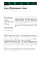
The rhythmic beating of the heart is controlled by an intricate and well-orchestrated flux of ions through a process called excitation–contraction coupling (ECC), where the electrical action potential leads to cellular contraction. Among all the ions involved in ECC, calcium (Ca) plays a critical role and serves as the signal for cardiac contraction. Briefly, upon cardiac excitation a small current of Ca enters the cytoplasm through the sarcolemmal L-type Ca channels (LTCC). This triggers a much larger Ca release from the sarcoplasmic reticulum (SR)—the intracellular Ca store—by opening the type 2 ryanodine receptor (RyR2) channel in a process called Ca-induced-Ca release (CICR) [1]. The resultant elevation of cytoplasmic Ca concentration activates the contractile apparatus, thus leading to myocyte contrac-tion. For relaxation to occur, Ca must be extruded from the cytoplasm. Two main
</div><span class="text_page_counter">Trang 18</span><div class="page_container" data-page="18"><i><small>Cardiac Arrhythmias - Translational Approach from Pathophysiology to Advanced Care</small></i>
mechanisms are involved in removing cytoplasmic Ca: one is by Ca re-sequestration into the SR to replenish the intracellular store, through the action of the SR Ca ATPase (SERCA). The other is by transporting calcium outside of the cell via the
<b>membrane-embedded protein, sodium calcium exchanger (NCX) (Figure 1). Other </b>
avenues for Ca removal do exist (e.g. sarcolemmal Ca-ATPase and mitochondria Ca uniporter), but only play a minor role in this process [1]. The rhythmic rise and fall of cytoplasmic Ca underlies the systolic and diastolic phases of the cardiac cycle.
<b>2. Dysregulated SR Ca release is linked to cardiac pathologies</b>
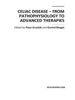
The RyR2 is a large protein with a molecular weight of ~560 kDa that forms
<b>homotetrameric channels in the SR membrane (Figure 2) [2]. Due to its crucial </b>
role in releasing Ca to trigger contraction, it is no surprise that there are a number of auxiliary proteins with likely overlapping/redundant functions acting from both the cytosolic and SR luminal sides to regulate the function of the channel complex. Moreover, the activity of the channel is also subject to regulation by post-translational modifications, including redox modifications, phosphorylation, and nitrosylation [3]. Unfortunately, both genetic and acquired defects due to mutation or posttranslational modification of the channel complex contribute to its dysfunc-tion [3]. These defects typically make the channel hyperactive or leaky, giving rise to dysregulated Ca release (DCR). DCR is implicated in a spectrum of cardiac dysfunctions [3], and in particular, it directly causes a deadly cardiac arrhythmia syndrome, catecholaminergic polymorphic ventricular tachycardia (CPVT) [3, 4].
CPVT is a stress-induced arrhythmia that is triggered by elevated levels of cat-echolamines [5]. Patients do not exhibit the cardiac remodeling typical of structural heart diseases, which makes the diagnosis particularly challenging. Life-threatening cardiac arrhythmias occur following exercise or emotional stress, which elevates circulating catecholamine levels. CPVT mutations have been identified in the genes encoding the RyR2 channel and its several auxiliary proteins. The remainder of this chapter focuses on the regulation of SR Ca release, the molecular mechanisms of CPVT, and the current and future state of therapies targeted towards CPVT.
<i><b><small>Figure 1. </small></b></i>
<i><small>Cardiac excitation-contraction coupling.</small></i>
</div><span class="text_page_counter">Trang 19</span><div class="page_container" data-page="19"><i><small>Molecular Mechanism and Current Therapies for Catecholaminergic Polymorphic Ventricular…DOI: dysregulated SR Ca release has been implicated in multiple cardiac disorders, knowledge obtained from CPVT studies will also shed light on the development of therapeutic approaches for these devastating cardiac dysfunctions as a whole.
<b>3. Modulation of RyR2 Ca release</b>
RyR2s form physically separated/isolated clusters to act as functionally indepen-dent Ca release units [6]. Within dyads, the structural element formed by the close apposition of T-tubules and junctional SR, Ca influx via LTCC in the T-tubule trig-gers more Ca release from RyR2 clusters in the junctional SR to initiate contraction
<b>(Figure 1). Ca release from individually activated RyR2 clusters are known as Ca </b>
sparks and can be experimentally observed during diastole [7]. The systolic Ca tran-sient is the summation of tens of thousands of Ca sparks due to the synchronized Ca release of RyR2 clusters following sarcolemmal depolarization. The positive-feed-back nature of CICR suggests that SR Ca release should terminate upon depletion of SR Ca store. However, only a fraction of the SR Ca store is released during EC coupling [8, 9]. This begs the question of how the Ca release process is terminated. Cytosolic Ca-dependent inactivation of RyR2 has been proposed as a mechanism for the termination of Ca release [10], but lacks widespread support. In contrast, evidence from different research groups collectively points to a mechanism that works to inhibit Ca release from the SR luminal side.
<i>The first piece of evidence comes from an in vitro study conducted with single </i>
RyR2 channels reconstituted into lipid bilayers. This study demonstrated that the opening of RyR2 is significantly reduced at lowered luminal Ca [11]. The chan-nel’s luminal accessory proteins, calsequestrin 2 (CASQ2), junctin, and triadin, are required for this luminal Ca-dependent inhibition of the channel [12]. More
<i><b><small>Figure 2. </small></b></i>
<i><small>SR Ca release channel RyR2 is regulated by both cytosolic and SR luminal proteins.</small></i>
</div><span class="text_page_counter">Trang 20</span><div class="page_container" data-page="20"><i><small>Cardiac Arrhythmias - Translational Approach from Pathophysiology to Advanced Care</small></i>
convincing evidence comes from a subsequent cellular study that manipulated the SR Ca buffering capacity by introducing exogenous Ca chelators into the SR to result in a slower Ca depletion [13]. Enhanced SR Ca buffering drastically increased the amplitude of Ca release (both Ca sparks and global Ca transients) and slowed its termination, hence supporting the role of SR luminal Ca in controlling RyR2 Ca release. Mathematical modelling studies provided further support that luminal Ca-dependent deactivation of RyR2 is involved in termination of Ca release [14].
While the role of luminal Ca-dependent deactivation of RyR2 has been estab-lished, it is unclear what the specific molecular mechanism is. There is evidence supporting either direct activation of RyR2 or through its luminal accessory proteins, i.e. CASQ2, junctin, and triadin, which form the SR Ca release unit with RyR2. Studies performed in human embryonic kidney cells (HEK293) overexpress-ing recombinant RyR2 support a direct activation of RyR2 by luminal Ca. Despite a lack of several Ca handling proteins, HEK293 cells with exogenously expressed RyR2 mutants of CPVT exhibit dysregulated Ca release that is sensitive to SR Ca load in a process called store-overload-induced Ca release (SOICR) [15]. The authors found that CPVT mutations of RyR2 reduce the threshold for SOICR, which is expected to increase the propensity of dysregulated Ca release, hence contribut-ing to cellular arrhythmogenesis. Further, a more recent study from the same group proposed the amino acid E4872 as the luminal Ca sensor for the direct activation of RyR2 [16]. A point mutation of E4872A completely abolishes luminal Ca activation of RyR2 in single channel studies, and markedly reduces dysregulated Ca release in HEK293 and HL-1 cardiac cells. Moreover, mice harboring a heterozygous mutation
<i>of E4872Q are resistant to SOICR and protected from ventricular arrhythmias in </i>
<i>vivo. E4872 is localized in the S6 helix bundle-crossing region, the putative cation </i>
binding pocket of RyR2. Despite recent breakthroughs in resolving the structure of RyR2, the composition of this putative cation binding pocket remains undeter-mined [17], and it is thought that other key amino acids in this region such as E4878 may also play a critical role in luminal Ca activation of RyR2.
On the other hand, independent labs have provided evidence supporting the participation of luminal proteins in regulating RyR2 Ca release. Lipid bilayer single-channel studies found that CASQ2 serves as a sensor to inhibit opening of RyR2 at low luminal [Ca], which notably required the presence of junctin and triadin, thus suggesting these proteins form a regulatory complex to control luminal Ca-dependent deactivation of RyR2 [12]. Additionally, increasing or decreasing the expression of CASQ2 in rat cardiac myocyte not only changes the SR Ca stor-age capacity, consistent with CASQ2’s Ca buffer function, but also affects SR Ca release [18]. In particular, decreased expression of CASQ2 leads to dysregulated arrhythmogenic Ca release, supporting CASQ2 function as an inhibitor of RyR2 [18]. Interestingly, the expression of a competitive peptide in myocytes to disrupt the interaction between CASQ2 and triadin impairs the ability of CASQ2 to stabilize the Ca release channel [19], thus echoing conclusions from earlier bilayer studies that CASQ2 interacts with other luminal proteins to regulate RyR2 activity. The development of a genetic model of CASQ2 KO mouse provided additional evidence supporting CASQ2’s regulation of SR Ca release [20]. CASQ2 KO mice pheno-copied human CPVT by exhibiting catecholamine-induced tachyarrhythmias in vivo. Myocytes isolated from these mice are characterized with β agonist-induced dysregulated Ca release, a hallmark of cellular arrhythmogenesis. However, besides resulting in CPVT, the ablation of major SR Ca buffer protein CASQ2 does not seem to result in more severe cardiac dysfunctions, suggesting the existence of other Ca buffer proteins with similar function at the SR luminal side. Surprisingly, deletion of CASQ2 and another Ca binding protein histidine-rich calcium binding protein (HRC) in a double knockout (DKO) mouse model alleviates arrhythmias as
</div><span class="text_page_counter">Trang 21</span><div class="page_container" data-page="21"><i><small>Molecular Mechanism and Current Therapies for Catecholaminergic Polymorphic Ventricular…DOI: with the CASQ2 KO mouse [21]. HRC binds to RyR2 through the same CASQ2-binding domain on triadin, and results from this DKO mouse study suggest that rather than having redundant roles, CASQ2 and HRC play opposing roles to regulate RyR2 Ca release. Taken together, these studies not only support the notion that luminal accessory proteins of RyR2 participate in controlling SR Ca release, but also highlight the intricate nature of such regulation.
<b>4. Molecular mechanisms of CPVT</b>
CPVT mutations have been identified in 6 genes encoding 4 different proteins
<i>of the Ca release channel complex: RYR2, CASQ2, TRDN, CALM1, CALM2, and </i>
<i>CALM3 [22]. Among them, the 3 genes of calmodulin (CALM1, CALM2, and CALM3) encode the same protein. Of note, these mutations account for up to </i>
60–75% of CPVT cases, with the genetic cause of the remaining clinical cases unknown [23, 24]. It is likely that more disease mutations will be discovered in other proteins of the Ca release channel complex. In this section, we will summarize the proposed molecular mechanisms for different genetic forms of CPVT.
<b>4.1 CPVT linked to RyR2 mutations</b>
Among the genetically confirmed cases of CPVT, over 90% are due to mutations of RyR2 [23]. CPVT linked to RyR2 mutations is autosomal dominant. Up to date, more than 200 gain-of-function mutations in RyR2 have been discovered. RyR2 loss-of-function mutations have also been detected but are less frequent and are associated with arrhythmias distinct from CPVT [25]. Additionally, the loss-of-function mutation appears to cause arrhythmias through an early afterdepolariza-tion (EAD)-based mechanism [26] which is much less studied compared to the classic DAD-based mechanism. Never the less, EADs have been observed in CPVT patient specific induced pluripotent stem cells (iPSC)-derived cardiomyocytes [27] in addition to myocytes isolated from a mouse model harboring a loss-of-function CPVT mutation of RyR2 [26]. The focus of the rest of the chapter will be on the arrhythmias evoked by DADs, rather than EADs.
Three main theories of how gain-of-function RyR2 mutations lead to CPVT have been proposed by different groups. The first comes from the observation that CPVT mutants of RyR2 expressed in HEK293 cells decreased the threshold to induce SOICR [15]. Based on these results, it is proposed that CPVT mutations make RyR2 more sensitive to luminal Ca, thus susceptible to dysregulated arrhythmogenic Ca release. The second theory proposes that CPVT mutations reduce the binding of RyR2 to FKBP12.6, a cytosolic protein thought to stabilize the channel, thus increas-ing RyR2 activity and givincreas-ing rise to arrhythmogenic diastolic calcium release [28]. However, this theory has been challenged by several labs [29–31], a conserved bind-ing between CPVT mutant RyR2 and FKBP has been reported [32]. The majority of RyR2 mutations are found at three “hot spots” which are located in the N-terminal domain (amino acids 1–600), central domain (amino acids ~2100–2500) and C terminal domain (amino acids ~3900–end) of the protein [33, 34]. Structural stud-ies show that many of them are found at the domain-domain interfaces, thus giving rise to the third theory that mutations impair the inter-domain interactions of RyR2 to cause CPVT. Specifically, the interaction between N-terminal and central domains of RyR2 is responsible for the so-called domain “zipping” and is thought to stabilize the channel; the third theory posits that CPVT mutants impair this interac-tion (causing domain unzipping) and causes channel dysfuncinterac-tion. This model of
</div><span class="text_page_counter">Trang 22</span><div class="page_container" data-page="22"><i><small>Cardiac Arrhythmias - Translational Approach from Pathophysiology to Advanced Care</small></i>
<b>4.2 CPVT linked to CASQ2 mutations</b>
The second most common cause of CPVT is mutation of CASQ2, an SR lumi-nal Ca binding protein thought to regulate deactivation of RyR2. CPVT linked to CASQ2 was considered as an autosomal recessive disease until the recent discovery of autosomal dominant disease-causing mutations [38]. CASQ2 is a low-affinity, high-capacity Ca binding protein. It does not contain Ca binding structural domains such as an EF-hand motif, a helix–loop–helix structural domain, found in “typical” Ca binding proteins (troponin C, calmodulin) [39]. Instead, it has multiple (~60–70) negatively charged amino acids, which facilitates electrostatic interactions between the protein and ~ 40–50 Ca ions [40]. CASQ2 monomers change their structure upon Ca binding, and form protein polymers in a Ca-dependent process. Structural studies show that monomeric CASQ2 contains three highly similar tandem domains, resem-bling that of bacterial thioredoxin. However, much less is known about the structure
<i>of the polymers. Based on in vitro biophysical studies by Park et al. [41], the </i>
follow-ing model of CASQ2 polymerization is proposed: CASQ2s exist as monomers at low luminal [Ca]; as [Ca] increases, CASQ2s form dimers, tetramers, and polymers in a [Ca]-dependent process. Thus, CASQ2s polymerize to bind additional Ca at high luminal Ca, but depolymerize to release Ca at low luminal Ca. Considering the Ca and protein concentrations in SR, CASQ2s likely exist as a mixture of monomers, dimers, and polymers of varying sizes [42]. As described above, monomeric CASQ2 is thought to be anchored to RyR2 via junctin and triadin to deactivate Ca release at low luminal Ca. The intriguing question remains whether this Ca-dependent change in the polymerization states of CASQ2 happens on a beat-to-beat basis in response to SR Ca load to regulate RyR2 Ca release. It has been shown that the polymerization state of CASQ2 changes upon depletion of luminal Ca in fibroblast by fluorescent approaches [43]. Additional evidence comes from studies conducted with skeletal muscle fibers demonstrating luminal-Ca dependent changes in polymerization of CASQ1, the skeletal counterpart of CASQ2 [44]. However, whether Ca-dependent changes in CASQ2 polymerization happens in beating cardiomyocytes at a time scale comparable with the cardiac cycle awaits further investigation.
At least 2 molecular mechanisms are proposed to explain how autosomal reces-sive CPVT mutations of CASQ2 cause the disease based on its two primary func-tions, buffering Ca and modulating RyR2 opening [4]. These mutations (nonsense or missense) lead to loss or reduced expression of CASQ2. Subsequently, reduced Ca buffering allows the free Ca to rise faster near the Ca release sites, thereby triggering dysregulated Ca release. Besides reduced Ca buffering power, some missense muta-tions of CASQ2 appear to work through another mechanism: by impairing RyR2 regulation from the luminal side. It’s been shown that the mutation of R33Q leads to abnormal interaction between CASQ2 and the Ca release channel complex [45], and another mutation of D307H reduces the binding between CASQ2 and triadin [46]. These results support the notion that a regulatory complex involving several proteins (CASQ2, triadin, junctin, and potentially others) senses luminal Ca to regulate Ca release, and disruption of interactions between them leads to dysregulation of the channel and disease. Compared with the autosomal recessive mutations, less is known about the autosomal dominant mutations that were more recently identified. Two mutations (K180R and S173I) have been found to interfere the polymerization of CASQ2 [47], likely causing CPVT by reducing the Ca buffering capacity.
<b>4.3 CPVT linked to triadin mutations</b>
CPVT mutations have also been identified in triadin, a trans-SR membrane protein that helps anchor CASQ2 to the RyR2 channel complex. Triadin has a short
</div><span class="text_page_counter">Trang 23</span><div class="page_container" data-page="23"><i><small>Molecular Mechanism and Current Therapies for Catecholaminergic Polymorphic Ventricular…DOI: region located on the cytosolic side of SR, a single membrane spanning domain, and a highly charged C-terminal region that comprises most of the protein and resides in the SR luminal side. The C-terminal tail of the protein contains KEKE motifs formed by stretches of alternating residues with opposite charges. A single KEKE motif consisting of 15 residues (210–224) has been suggested as the CASQ2 binding region [48]. The binding between triadin and CASQ2 is Ca-dependent and they dissociate at high [Ca] (10 mM). In contrast, triadin’s binding to RyR2 is Ca-independent [48]. Due to its role of anchoring CASQ2 to Ca release sites, triadin is thought to facilitate SR Ca release by allowing CASQ2 to buffer Ca near the Ca release channel.
It is also proposed that triadin may play a direct role in regulating RyR2 chan-nel activity. Both overexpression and knockout mouse models of triadin have been created to decipher its function. The overexpression model displayed hypertrophy and altered Ca handling, accompanied by compensatory changes in the expression of several proteins of the RyR2 channel complex, thus masking the functional role of triadin [49]. Similarly, loss of triadin in the KO model also caused compensa-tory changes [50]. Drastic reduction in the interface of junctional SR and T-tubules occurred due to structural remodeling, thus impaired the coupling between RyR2 and LTCC. As a result, inactivation of LTCC is reduced, which increased Ca current, prolonged action potential, and subsequently increased cellular and SR Ca. Due to Ca overload, myocytes from triadin KO model displayed elevated levels of arrhyth-mogenic Ca release, especially when stimulated with catecholamines [50]. While both mouse models support the notion that triadin plays an important role in myo-cyte Ca handling, the massive compensatory changes in these chronic models makes an explanation of the data challenging. Nevertheless, acute overexpression of triadin in cultured myocytes increased RyR2 opening, dysregulated Ca release, and mem-brane depolarization, mimicking the cellular phenotype of CPVT [51]. Relatively few CPVT mutations of triadin have been reported as of yet. In a 2012 study, three autosomal recessive mutations of triadin were discovered, with two of them (one deletion, one nonsense) resulting in loss of the protein. The third one, a missense mutation of T59R, results in a protein that is more susceptible to degradation [52]. Thus, loss or decreased expression of triadin appear to cause CPVT. Another two autosomal recessive triadin mutations were identified in a 2015 study [53], although the underlying disease-causing mechanisms await further investigation.
<b>4.4 CPVT linked to calmodulin mutations</b>
Calmodulin (CaM) is an EF-hand Ca binding protein that binds RyR2 from the cytosolic side to regulate Ca release. CaM has a dumbbell-shaped structure, with its two globular domains connected by a flexible central helix. Each of the two domains contain two EF-hand Ca binding sites. The N-domain of the protein has a lower Ca binding affinity than the C-domain [54, 55]. Upon Ca binding, the hydrophobic pockets in both domains become exposed, thereby allowing CaM to bind its several intracellular targets, including RyR2, LTCC and Na channel (Nav 1.5) [56]. Mutations of CaM have been linked to different types of arrhythmias, such as CPVT, long QT syndrome, and idiopathic ventricular fibrillation, likely due to its impaired regula-tion of various target proteins [22]. Following systolic Ca release and the ensuing increasing in Ca on the cytosolic side of RyR2, CaM binds to the channel and inhibits its opening during the diastole phase of the cardiac cycle [57, 58]. CPVT mutations of CaM appear to have impaired ability to inhibit the channel and promoted the genera-tion of DCR in the form of Ca waves and Ca sparks in a cellular study [59]. They also exhibited higher binding affinity to RyR2 than WT CaM, thereby contributing to the
</div><span class="text_page_counter">Trang 24</span><div class="page_container" data-page="24"><i><small>Cardiac Arrhythmias - Translational Approach from Pathophysiology to Advanced Care</small></i>
<b>5. Impaired Ca signaling refractoriness and generation of DCR</b>
Despite the differences in the molecular details on how the various CPVT mutations of the RyR2 channel complex cause the disease, they all seem to make the channel more susceptible to arrhythmogenic diastolic Ca release. Following systolic Ca release, RyR2 becomes functionally suppressed and remains that way for a brief period, known as Ca signaling refractoriness [60]. Refractoriness of the Ca release channel can be measured by myocyte experiments employing a two-pulse protocol to record the process of Ca transient restitution. It’s been demonstrated that full recovery of Ca transient takes ~1 s (anywhere from ~0.8 − 1.5 s, depending on species) [61–65]. If Ca signaling refractoriness is impaired, the RyR2 chan-nel is expected to recover earlier from the functionally suppressed state, thereby promoting the generation of DCR, thus causing cellular arrhythmogenesis. Indeed, multiple CPVT mutations have been found to shorten refractoriness of RyR2, including mutations of CASQ2 [65], CaM [66], and RyR2 [67]. Moreover, short-ened Ca signaling refractoriness can also occur due to oxidation/hyperphosphoryla-tion of RyR2 as seen in models of acquired heart diseases [63]. Thus, both genetic and acquired defects of RyR2 channel complex seem to converge on shortening Ca signaling refractoriness to cause arrhythmogenic Ca release. Further evidence supporting shortened refractoriness as a unifying mechanism for the generation of DCR comes from a recent study using an engineered therapeutic CaM in an attempt to restore refractoriness and treat CPVT [66], as discussed in a later section on future therapies for CPVT. Taken together, these studies suggest that disease mutations may change the SR Ca dynamics, the modulation of RyR2 by cytosolic or luminal proteins, or conformational changes of the channel protein itself, each of which has been experimentally demonstrated to shorten Ca signaling refractoriness and give rise to arrhythmogenic DCR.
<b>6. Cellular arrhythmogenesis: SR Ca load</b>
At the single myocyte level, DCR is manifested as different forms: Ca sparks, Ca wavelets, and propagating Ca waves. When large enough, DCR activates electro-genic NCX, resulting in an inward current that causes delayed afterdepolarization (DAD) [68–70]. With a large enough amplitude, DADs may surpass the voltage threshold to open Na channels, thus leading to the generation of an ectopic action potential or triggered activity [71, 72]. Both the amplitude and the rate of DCRs are important in determining if it will trigger an ectopic action potential [73]. Localized DCR events in the form of Ca sparks and wavelets are less likely to trigger ectopic action potentials as compared with propagating Ca waves. Ca waves are more likely to occur when SR Ca load is high, such as following activation of β-adrenergic signaling pathways.
β-adrenergic stimulation results in phosphorylation of key EC-coupling proteins and subsequent generation of a larger and faster Ca transient, underlying its posi-tive inotropy (ability to contract) and lusitropy (ability to relax) effect [1]. One of these proteins, phospholamban (PLN), acts as an inhibitor of SERCA. Its inhibition on SERCA is relieved upon β-adrenergic-dependent phosphorylation, thus contrib-uting to a faster Ca transient decay and also higher SR Ca content. This higher SR Ca load facilitates Ca wave generation, and explains the stress-induced arrhythmias that occur in CPVT. Therefore, SERCA’s function to refill the SR with Ca is critical to maintain a certain SR Ca load to stimulate the generation of Ca waves. On the other hand, SERCA directs Ca out of the cytosol while refilling the SR with Ca, which opposes the formation of or “breaks” Ca waves.
</div><span class="text_page_counter">Trang 25</span><div class="page_container" data-page="25"><i><small>Molecular Mechanism and Current Therapies for Catecholaminergic Polymorphic Ventricular…DOI: on these seemingly contradictory effects of SERCA activity on Ca wave generation, an interesting question arises: what will be the consequences of upregulating SERCA activity in the setting of CPVT? Both beneficial and deleteri-ous effects have been reported from studies conducted by different groups. When overexpressing a skeletal isoform of SERCA1a in the CPVT model of CASQ2 KO, the resultant CPVT-SERCA<small>ox</small> mice developed severe Ca-dependent cardiomyopa-thy [74]. These mice suffered from early mortality and contractile dysfunction. Myocytes isolated from the hypertrophied hearts of these animals also displayed enhanced levels of DCR. While these results clearly demonstrate a detrimental effect, the severe cardiomyopathy phenotype due to chronic SERCA overexpression masks the effect of the genetic manipulation on arrhythmias. A follow-up study from the same group conditionally overexpressed SERCA2a in the same CASQ2 KO mice and found that both atrial and ventricular arrhythmias were exacerbated due to acute upregulation of SERCA activity [75]. In contrast, in another study employ-ing a different CPVT model of RyR2 knock in mouse (R4496C<small>+/−</small>), upregulation
<i>of SERCA activity by knocking out its inhibitor PLN suppressed arrhythmias in </i>
<i>vivo. In cellular experiments, Ca waves were also suppressed, due to propagating Ca </i>
waves being converted into non-arrhythmogenic mini waves and Ca sparks [76]. Interestingly, a different study showed that although enhancing SERCA activity by PLB ablation alleviated arrhythmias, it exacerbated myocardial infarction and car-diac damage in a RyR2 model featuring elevated DCR due to a mutation (S2814D) mimicking constitutive CaMKII-mediated phosphorylation of RyR2 [77].
<b>7. Synchronization of DCR in myocardium</b>
It is well established how DCR triggers ectopic action potentials at the cellular level. However, the heart contains billions of cardiomyocytes, and how arrhythmo-genesis at the level of isolated myocytes translates into arrhythmias at the level of a multi-cellular tissue preparation or even the whole heart remains unknown. Within the myocardium, individual myocytes are electrically coupled to their neighboring cells, hence Ca-dependent depolarizing currents generated in any random, isolated cells should be easily absorbed by neighboring cells that act as a current sink (the source-sink mismatch theory) [78]. Therefore, cellular depolarization, if happening randomly in individual cells, cannot generate sufficient current to trigger tissue-level depolarization.
Computational simulation studies suggest a very large number of cells—nearly 7x10<small>5</small>—have to depolarize simultaneously to overcome source-sink mismatch and trigger depolarization to generate an ectopic beat in normal myocardium. This number is reduced by modeling disease conditions such as fibrosis or heart failure related electrical remodeling, but the number of requisite cells still remains quite large [78]. Therefore, it’s been proposed that DCR happens in a synchronous way in multiple cells of the CPVT hearts to cause a tissue-wide ectopic beat. Experimental evidence has been provided in support of the synchronization of DCR in a CPVT model carrying the CASQ2 R33Q mutation [65]. This study quantified the degree of DCR synchronization by measuring the latency, or the time interval to the first DCR, following systolic Ca release. It was found that DCR occurs in a highly synchronized way in both myocytes and cardiac muscle tissue obtained from the R33Q CPVT model. Importantly, two factors are important for the synchronization of DCR: 1) shortened Ca signaling refractoriness that increases the propensity of release sites to fire synchronously by facilitating CICR, and 2) the presence of a preceding systolic action potential acting as a synchronizing event that temporally
</div><span class="text_page_counter">Trang 26</span><div class="page_container" data-page="26"><i><small>Cardiac Arrhythmias - Translational Approach from Pathophysiology to Advanced Care</small></i>
<b>8. The cellular origin of CPVT: Purkinje cells or ventricular myocytes?</b>
Purkinje fibers are a specialized network of electrically excitable cells found in the conduction system of the heart. They radiate throughout the ventricular muscle to ensure a rapid propagation of electrical impulse and a coordinated ventricular contraction. Compared with the myocardium, the Purkinje system has a smaller source-sink mismatch [78]. Based on this and other structural features [79], Purkinje cells have been proposed as the cellular origin of many arrhythmias including CPVT. Experimental evidence obtained from the CPVT model of RyR2 R4496C<small>+/−</small> mouse supports this hypothesis [80]. Optical mapping of R4496C<small>+/−</small>
hearts demonstrates that ventricular tachycardia (VT) may originate from the His-Purkinje system in both ventricles. Cellular studies also found that Purkinje cells had a significantly higher rate of DCR and triggered activity compared to ventricular myocytes [81, 82].
However, a recent study attempting to establish the causal link between Purkinje cells and CPVT did not provide such evidence [83]. In this study, CASQ2 was con-ditionally knocked out in the cardiac conduction system, but not the myocardium, using a conduction system-specific Cre recombinase. Ablation of CASQ2 in the Purkinje fibers failed to produce a CPVT phenotype. Considering CASQ2 ablation is an established molecular cause of CPVT as demonstrated by a global CASQ2 KO model [20], this result argues against Purkinje cells as the origin of CPVT, at least not on their own. On the other hand, in support of myocytes as the origin of CPVT, cells isolated from the myocardium of CPVT mouse models have been shown to exhibit DCR, DAD, and ectopic action potentials in multiple studies [20, 66, 67, 80]. Human iPSC-derived cardiomyocytes generated from biopsies of human CPVT patients also displayed DCR, DAD, and ectopic action potentials characteristic of the above-mentioned CPVT cells [84, 85]. Drug studies based on isolated myocytes also serve as good indicators of drug efficacy in both mouse models and humans [22, 86, 87]. More evidence regarding the cellular origin of CPVT are discussed elsewhere [22].
<b>9. Therapies for CPVT</b>
Symptoms of CPVT vary from palpitations, syncope, or even cardiac arrest. Although a rare disease, the mortality rate of CPVT can reach as high as ~50% in untreated individuals before the age of 40 [23]. In this section, we will first discuss traditional therapies that are currently available to CPVT patients. Next, we will focus on novel therapeutic approaches, based on recent advances in understanding the molecular mechanisms of this life-threatening arrhythmia syndrome.
<b>9.1 Current therapies for CPVT</b>
<i>9.1.1 Beta-blockers</i>
Beta-blockers are the first-line drug therapy to treat CPVT. As discussed above, in CPVT, the β-adrenergic-dependent increase in SR Ca load is important in trigger-ing DCR and subsequent cellular arrhythmogenesis. Thus, blocktrigger-ing the β-adrenergic signaling pathway is expected to decrease DCR and suppress arrhythmias. The most effective beta-blocker at the time of writing is nadolol [88, 89], but it remains unknown why it is more effective than other blockers. Unfortunately, beta-blockers only offer limited protection even with the maximal tolerated dose. It has been reported that more than 30% patients still suffer from arrhythmic events after receiving beta-blockers [90].
</div><span class="text_page_counter">Trang 27</span><div class="page_container" data-page="27"><i><small>Molecular Mechanism and Current Therapies for Catecholaminergic Polymorphic Ventricular…DOI: a beta-blocker that is highly effective in preventing VT in heart failure, has been shown to suppress CPVT through a dual inhibitory action on both β-adrenergic signaling and RyR2 channel activity [91]. Experimentally, an analog of carvedilol with minimal beta-blocking activity still prevented VT in a CPVT mouse model and exhibited improved efficacy when combined with a selective beta-blocker [91]. Nevertheless, further studies are required to assess its effective-ness in CPVT patients. However, this provides a new potential pharmacological approach where a combination of RyR2 channel inhibition and beta-blockade could provide a more effective therapeutic approach than current options based solely on beta-blockers.
<i>9.1.2 Na channel blockers and flecainide</i>
Na channel blockers may serve as anti-arrhythmic drugs due to the critical role of the Nav 1.5 channel in the depolarization phase of action potential. Flecainide, an FDA approved drug to treat arrhythmias, was originally thought to work by blocking the Na channel. Recent studies have found that flecainide prevents CPVT both in mouse models and human patients through a dual inhibition mechanism: inhibiting Na channels as well as RyR2 [86]. Studies show that flecainide appears to be a promising therapy for CPVT patients not responding well to beta-blockers. However, the working mechanism of flecainide was controversial.
A study conducted on the CPVT model of RyR2 R4496C<small>+/−</small> showed that while flecainide was effective in preventing arrhythmias, it did not reduce DCR in the cellular experiments. Instead, it increased the threshold for triggered activity, thus pointing to the other possibility: that the drug works by solely acting as a Na channel blocker [92]. Several follow-up studies attempted to reconcile this dis-crepancy. Evidence has been provided that flecainide is effective in reducing DCR in cells harboring the RyR2 R4496C<small>+/−</small> mutation, but this effect could be masked by experimental conditions such as Ca overload [93]. On the other hand, more convincing evidence comes from a recent study that employed a synthesized analog of flecainide with reduced inhibition on RyR2 activity but unaltered inhibition on Na channel [94]. This analog failed to reduce DCR at cellular level and arrhythmia
<i>burden in vivo, indicating that flecainide acts through inhibition of RyR2 activity. </i>
In support of this, flecainide reduced DCR in permeabilized CPVT cells lacking membrane-residing Na channels, and intact cells pretreated with Na channel blocker. Similar to flecainide, another approved drug propafenone also seems to work through dual inhibition of Na channel and RyR2 [95]. Further studies are required to fully understand its working mechanism.
<i>9.1.3 Other treatment options</i>
Ca channel blockers (LTCC blockers) such as verapamil, have been tested in cellular and animal studies, as well as clinical studies, to examine their efficacy in treating CPVT. Consistently, these studies found Ca channel blockers only confer limited benefits in both cellular preparations and patients already on beta-blockers [96–98]. However, it has been shown to be beneficial for some patients when used in combination with other pharmacological approaches including beta-blockers [98].
Left cardiac sympathetic denervation serves as an alternative treatment. It works by preventing the release of catecholamines from the sympathetic nerve endings. The procedure appears to be effective in reducing major arrhythmic events in clinical studies [99, 100], and thus has been recommended for patients who don’t respond to more conventional pharmacological treatments such as beta-blocker
</div><span class="text_page_counter">Trang 28</span><div class="page_container" data-page="28"><i><small>Cardiac Arrhythmias - Translational Approach from Pathophysiology to Advanced Care</small></i>
stellate ganglion was ablated together with the second and third thoracic ganglia [99]; another case used the thoracoscopic, the transaxillary, and the supraclavicular approaches as the main surgical approaches [100].
Implantable cardiac defibrillators (ICD) have been utilized in patients who still experience symptoms despite drug therapy and/or sympathetic denervation. A recent study systematically analyzed the efficacy of ICDs using existing clini-cal data containing 505 CPVT patients implanted with ICDs [101]. It was found that although effective for ventricular fibrillation, ICDs were not protective for VT. Another study of 136 CPVT patients also suggests ICD implant did not confer survival benefits [102]. Considering the potential complications and psychological burden of implantation, especially for pediatric patients, ICDs are not an optimal treatment for CPVT patients.
<b>9.2 Potential future therapies for CPVT</b>
The molecular mechanisms underlying CPVT have been intensively studied in the past several decades. Several novel therapeutic strategies have been proposed and tested in animal models and even pre-clinical studies. In this section, we will discuss these novel approaches, with a focus on gene therapy.
<i>9.2.1 Gene therapy</i>
With advances in the adeno-associated virus (AAV) vector-based gene transfer technology in the past a few decades, using gene therapy to treat CPVT is starting to become technically feasible. Several proof-of-principle studies have been con-ducted to test the efficacy of different therapeutic strategies. Considering several CPVT mutations, especially the ones identified in CASQ2, cause loss or reduced expression of the associated protein, it would seem that the most straightforward therapeutic approach is to deliver a normal gene encoding the protein. Indeed, AAV9-mediated gene transfer of a WT CASQ2 to both CASQ2 KO and R33Q mouse models restored the normal expression of CASQ2, improved abnormal
<i>electro-physiological properties of cells, and reduced arrhythmia burden in vivo [103, 104]. </i>
However, this gene replacement approach is limited by the size of the AAV vector, thus hindering the delivery of a normal gene for the large RyR2 protein, which accounts for the majority of the CPVT mutations. To solve this problem, AAV9 was instead used to deliver siRNA to silence mutant mRNA of RyR2 in an allele-specific way [105]. This RNA silencing approach increased the ratio of WT-RNA vs. mutant RNA, proving to be effective at normalizing cardiac electrophysiology, alleviating
<i>abnormal ultrastructural remodeling, and inhibiting in vivo VT when tested in the </i>
RyR2 R4496C<small>+/−</small><i> mice. Alternatively, another study attempted in vivo genome </i>
edit-ing usedit-ing the CRISPR/Cas9 system delivered by AAV and also obtained promisedit-ing results in a different CPVT model of RyR2 (R176Q<small>+/−</small>) [106].
While theses gene therapy strategies seem effective, one of the prerequisites for applying this technology is knowing the genetic cause of CPVT. However, the genetic cause of ~30–40% of clinical cases of CPVT remains undetermined. Several groups have developed novel approaches to tackle this problem. It has been found that the Ca binding properties of CaM—in particular, the kinetics of Ca dissocia-tion from CaM—affects RyR2 refractoriness [66]. Based on this, a therapeutic CaM (TCaM) that specifically targets RyR2 and prolongs its refractoriness by slowing Ca dissociation from CaM was engineered. TCaM reduced DCR in CPVT cells and
<i>alleviated arrhythmias in vivo when delivered to a CPVT model of CASQ2 R33Q </i>
mice [66]. Instead of targeting the specific disease-causing gene, TCaM targets the
</div><span class="text_page_counter">Trang 29</span><div class="page_container" data-page="29"><i><small>Molecular Mechanism and Current Therapies for Catecholaminergic Polymorphic Ventricular…DOI: RyR2 refractoriness, and thus it could potentially serve as a therapeutic avenue for distinct forms of CPVT. Another study chose to target CaMKII, an adrenergically activated kinase that is implicated in arrhythmogenesis and patho-logical remodeling in multiple cardiac disorders, including CPVT. Pharmacopatho-logical inhibitors of CaMKII are limited in their efficacy due to their lack of specificity. In contrast, a CaMKII inhibitory peptide was delivered in a cardiomyocyte-specific
<i>way by AAV9 and found to be effective in reducing arrhythmias burden in vivo </i>
in the CPVT model of RyR2 (R176Q<small>+/−</small>) [107]. Collectively, these studies provide strong evidence supporting AAV-based gene therapy as a promising future therapy for CPVT patients.
<b>9.3 Targeting sinus node dysfunction</b>
CPVT patients also present sinus node dysfunction and bradycardia, which are recapitulated in the mouse models of CPVT. The pathophysiological role and under-lying mechanism for sinus node dysfunction are discussed in details elsewhere [22]. Targeting the impaired sinus node dysfunction has been proposed as a therapeutic approach for CPVT. It has been shown that increasing heart rate by (1) pharma-cological intervention (atropine), (2) atrial overdrive pacing, or (3) re-expressing CASQ2 in the CASQ2 KO mouse all appear to reduce arrhythmia burden [83, 108]. Further, atropine has been tested in a small group of 6 CPVT patients and was found to be effective in reducing exercise-induced arrhythmic events [109].
<b>9.4 Other potential therapies</b>
Tremendous effort has been expended on identifying and developing small mol-ecules that specifically target DCR of RyR2, since DCR is implicated in a spectrum of cardiac disorders. Dantrolene, a drug used clinically to treat a skeletal muscle condition of malignant hyperthermia, exhibited partial protection for a subset of CPVT patients [110]. The recently discovered ent-(+)-verticilide, an unnatural verticilide enantiomer, appears to be a potent and selective RyR2 inhibitor [87]. It
<i>reduced DCR, triggered activity in cells, and arrhythmias in vivo when tested with </i>
the CPVT model of CASQ2 KO. It seems to exert a stronger antiarrhythmic effect when compared with dantrolene or flecainide. More details on the current state of therapeutic small molecule development are reviewed elsewhere [111].
<b>10. Conclusion</b>
Great progress has been made in the past few decades to help us better under-stand CPVT and develop therapeutics for this deadly arrhythmia syndrome. These efforts will continue in both basic science and clinical studies and will provide deeper mechanistic insight on the molecular, cellular, and tissue mechanisms of CPVT. Since DCR is implicated in a spectrum of human diseases, knowledge obtained from these studies will also benefit the development of therapies for other cardiac dysfunctions including heart failure and metabolic heart disease.
We want to thank the American Heart Association for providing funding and National Institutes of Health (1R15HL154073).
</div><span class="text_page_counter">Trang 30</span><div class="page_container" data-page="30"><i><small>Cardiac Arrhythmias - Translational Approach from Pathophysiology to Advanced Care</small></i>
<b>Author details</b>
Bin Liu<small>1</small>*, Brian D. Tow<small>1</small> and Ingrid M. Bonilla<small>2</small>
1 Department of Biological Sciences, Mississippi State University, Starkville, Mississippi, USA
2 Department of Pharmacology and Toxicology, University of Puerto Rico Medical School, San Juan, Puerto Rico, USA
*Address all correspondence to:
<b>Abbreviation list</b>
AAV adeno-associated virus Ca calcium
CALM calmodulin (gene) CaM calmodulin (protein) CASQ2 calsequestrin 2
CICR calcium-induced calcium release
CPVT catecholaminergic polymorphic ventricular tachycardia DAD delayed afterdepolarization
DCR dysregulated calcium release DKO double knockout
EAD early afterdepolarization ECC excitation-contraction coupling HEK29 human embryonic kidney cells HRC histidine-rich calcium binding protein ICD implantable cardiac defibrillators iPSC induced pluripotent stem cells LTCC L-type calcium channel PLN phospholamban
RyR2 type 2 ryanodine receptor NCX sodium calcium exchanger
SERCA sarcoplasmic reticulum calcium ATPase SOICR store-overload-induced calcium release SR sarcoplasmic reticulum
TCaM therapeutic calmodulin TRDN triadin
VT ventricular tachycardia
© 2021 The Author(s). Licensee IntechOpen. This chapter is distributed under the terms of the Creative Commons Attribution License ( by/3.0), which permits unrestricted use, distribution, and reproduction in any medium, provided the original work is properly cited.
</div><span class="text_page_counter">Trang 31</span><div class="page_container" data-page="31"><i><small>Molecular Mechanism and Current Therapies for Catecholaminergic Polymorphic Ventricular…DOI: </small>Bers DM. Cardiac excitation-contraction coupling. Nature. 2002;415(6868):198-205.
<small>[2] </small>Gyorke S, Carnes C. Dysregulated sarcoplasmic reticulum calcium release: potential pharmacological target in cardiac disease. Pharmacol Ther. 2008;119(3):340-54.
<small>[3] </small>Belevych AE, Radwanski PB, Carnes CA, Gyorke S. ‘Ryanopathy’: causes and manifestations of RyR2 dysfunction in heart failure.
<small>[5] </small>Priori SG, Chen SR. Inherited dysfunction of sarcoplasmic reticulum Ca2+ handling and arrhythmogenesis. Circulation research. 2011;108(7): 871-83.
<small>[6] </small>Scriven DR, Asghari P, Moore ED. Microarchitecture of the dyad. Cardiovascular research. 2013;98(2): 169-76.
<small>[7] </small>Cheng H, Lederer WJ, Cannell MB. Calcium sparks: elementary events
<small>[9] </small>Shannon TR, Guo T, Bers DM. Ca2+ scraps: local depletions of free [Ca2+] in cardiac sarcoplasmic reticulum during contractions leave substantial Ca2+ reserve. Circulation research.
<small>[10] </small>Fabiato A. Time and calcium dependence of activation and
inactivation of calcium-induced release of calcium from the sarcoplasmic reticulum of a skinned canine cardiac Purkinje cell. The Journal of general physiology. 1985;85(2):247-89.
<small>[11] </small>Gyorke I, Gyorke S. Regulation of the cardiac ryanodine receptor channel by luminal Ca2+ involves luminal Ca2+ sensing sites. Biophysical journal. 1998;75(6):2801-10.
<small>[12] </small>Gyorke I, Hester N, Jones LR, Gyorke S. The role of calsequestrin, triadin, and junctin in conferring cardiac ryanodine receptor responsiveness to luminal calcium. Biophysical journal. 2004;86(4):2121-8.
<small>[13] </small>Terentyev D, Viatchenko-
Karpinski S, Valdivia HH, Escobar AL, Gyorke S. Luminal Ca2+ controls termination and refractory behavior of Ca2+−induced Ca2+ release in cardiac myocytes. Circulation research. 2002;91(5):414-20.
<small>[14] </small>Sobie EA, Dilly KW, dos Santos Cruz J, Lederer WJ, Jafri MS.
Termination of cardiac Ca(2+) sparks: an investigative mathematical model of calcium-induced calcium release. Biophysical journal. 2002;83(1):59-78.
<small>[15] </small>Jiang D, Xiao B, Yang D, Wang R, Choi P, Zhang L, et al. RyR2 mutations linked to ventricular tachycardia and sudden death reduce the threshold for store-overload-induced Ca2+ release (SOICR). Proceedings of the National Academy of Sciences of the United States of America. 2004;101(35): 13062-7.
<small>[16] </small>Chen W, Wang R, Chen B, Zhong X, Kong H, Bai Y, et al. The ryanodine receptor store-sensing gate controls Ca2+ waves and Ca2+−triggered arrhythmias. Nature medicine.
<b>References</b>
</div><span class="text_page_counter">Trang 32</span><div class="page_container" data-page="32"><i><small>Cardiac Arrhythmias - Translational Approach from Pathophysiology to Advanced Care</small></i>
<small>[17] </small>Peng W, Shen H, Wu J, Guo W, Pan X, Wang R, et al. Structural basis for the gating mechanism of the type 2 ryanodine receptor RyR2. Science. 2016;354(6310).
<small>[18] </small>Terentyev D, Viatchenko- Karpinski S, Gyorke I, Volpe P, Williams SC, Gyorke S. Calsequestrin determines the functional size and stability of cardiac intracellular calcium stores: Mechanism for hereditary arrhythmia. Proceedings of the National Academy of Sciences of the United States of America. 2003;100(20): 11759-64.
<small>[19] </small>Terentyev D, Viatchenko- Karpinski S, Vedamoorthyrao S, Oduru S, Gyorke I, Williams SC, et al. Protein protein interactions between triadin and calsequestrin are involved in modulation of sarcoplasmic reticulum calcium release in cardiac myocytes. The Journal of physiology. 2007;583
(Pt 1):71-80.
<small>[20] </small>Knollmann BC, Chopra N, Hlaing T, Akin B, Yang T, Ettensohn K, et al. Casq2 deletion causes sarcoplasmic reticulum volume increase, premature Ca2+ release, and catecholaminergic polymorphic ventricular tachycardia. The Journal of clinical investigation. 2006;116(9):2510-20.
<small>[21] </small>Liu B, Ho HT, Brunello L, Unudurthi SD, Lou Q, Belevych AE, et al. Ablation of HRC alleviates cardiac arrhythmia and improves abnormal Ca handling in CASQ2 knockout mice polymorphic ventricular tachycardia. The Journal of physiology. 2020;598(14): 2817-34.
<small>[23] </small>Perez-Riera AR, Barbosa- Barros R, de Rezende Barbosa MPC,
Daminello-Raimundo R, de Lucca AA, Jr., de Abreu LC. Catecholaminergic polymorphic ventricular tachycardia, an update. Ann Noninvasive Electrocardiol. 2018;23(4):e12512.
<small>[24] </small>Roston TM, Yuchi Z, Kannankeril PJ, Hathaway J, Vinocur JM, Etheridge SP, et al. The clinical and genetic spectrum of catecholaminergic polymorphic ventricular tachycardia: findings from an international multicentre registry. Europace. 2018;20(3):541-7.
<small>[25] </small>Roston TM, Sanatani S, Chen SR. Suppression-of-function mutations in the cardiac ryanodine receptor: Emerging evidence for a novel
arrhythmia syndrome? Heart Rhythm. 2017;14(1):108-9.
<small>[26] </small>Zhao YT, Valdivia CR, Gurrola GB, Powers PP, Willis BC, Moss RL, et al. Arrhythmogenesis in a catecholaminergic polymorphic ventricular tachycardia mutation that depresses ryanodine receptor function. Proceedings of the National Academy of Sciences of the United States of
ventricular tachycardia reveals early and delayed afterdepolarizations. PLoS One. 2012;7(9):e44660.
<small>[28] </small>Wehrens XH, Lehnart SE, Huang F, Vest JA, Reiken SR, Mohler PJ, et al. FKBP12.6 deficiency and defective calcium release channel (ryanodine receptor) function linked to exercise-induced sudden cardiac death. Cell. 2003;113(7):829-40.
<small>[29] </small>Jiang D, Wang R, Xiao B, Kong H, Hunt DJ, Choi P, et al. Enhanced store overload-induced Ca2+ release and channel sensitivity to luminal Ca2+ activation are common defects of RyR2
</div><span class="text_page_counter">Trang 33</span><div class="page_container" data-page="33"><i><small>Molecular Mechanism and Current Therapies for Catecholaminergic Polymorphic Ventricular…DOI: linked to ventricular tachycardia and sudden death. Circulation research. 2005;97(11): 1173-81.
<small>[30] </small>Liu N, Colombi B, Memmi M, Zissimopoulos S, Rizzi N, Negri S, et al. Arrhythmogenesis in catecholaminergic polymorphic ventricular tachycardia: insights from a RyR2 R4496C knock-in mouse model. Circulation research. 2006;99(3):292-8.
<small>[31] </small>George CH, Higgs GV, Lai FA. Ryanodine receptor mutations associated with stress-induced ventricular tachycardia mediate increased calcium release in stimulated cardiomyocytes. Circulation research. 2003;93(6):531-40.
<small>[32] </small>Zhang JZ, Waddell HM, Wu E, Dholakia J, Okolo CA, McLay JC, et al. FKBPs facilitate the termination of spontaneous Ca2+ release in wild-type RyR2 but not CPVT mutant RyR2. Biochem J. 2016;473(14):2049-60.
<small>[33] </small>Yano M, Yamamoto T, Ikemoto N, Matsuzaki M. Abnormal ryanodine receptor function in heart failure. Pharmacol Ther. 2005;107(3):377-91.
<small>[34] </small>George CH, Jundi H, Thomas NL, Fry DL, Lai FA. Ryanodine receptors and ventricular arrhythmias: emerging trends in mutations, mechanisms and therapies. Journal of molecular and cellular cardiology. 2007;42(1):34-50.
<small>[35] </small>Ikemoto N, Yamamoto T. Regulation of calcium release by interdomain interaction within ryanodine receptors. Front Biosci. 2002;7:d671-83.
<small>[36] </small>George CH, Jundi H, Walters N, Thomas NL, West RR, Lai FA. Arrhythmogenic mutation-linked defects in ryanodine receptor autoregulation reveal a novel mechanism of Ca2+ release channel dysfunction. Circulation research.
<small>[37] </small>Sumitomo N. Current topics in catecholaminergic polymorphic ventricular tachycardia. J Arrhythm. 2016;32(5):344-51.
<small>[38] </small>Gray B, Bagnall RD, Lam L, Ingles J, Turner C, Haan E, et al. A novel
heterozygous mutation in cardiac calsequestrin causes autosomal dominant catecholaminergic
polymorphic ventricular tachycardia. Heart Rhythm. 2016;13(8):1652-60.
<small>[39] </small>Scott BT, Simmerman HK, Collins JH, Nadal-Ginard B, Jones LR. Complete amino acid sequence of canine cardiac calsequestrin deduced by cDNA cloning. The Journal of biological chemistry. 1988;263(18):8958-64.
<small>[40] </small>Yano K, Zarain-Herzberg A. Sarcoplasmic reticulum calsequestrins: structural and functional properties. Mol Cell Biochem. 1994;135(1):61-70.
<small>[41] </small>Park H, Park IY, Kim E, Youn B, Fields K, Dunker AK, et al. Comparing skeletal and cardiac calsequestrin structures and their calcium binding: a proposed mechanism for coupled calcium binding and protein polymerization. The Journal of biological chemistry. 2004;279(17): 18026-33.
<small>[42] </small>Gyorke S, Terentyev D. Modulation of ryanodine receptor by luminal calcium and accessory proteins in health and cardiac disease. Cardiovascular research. 2008;77(2):245-55.
<small>[43] </small>Terentyev D, Kubalova Z, Valle G, Nori A, Vedamoorthyrao S,
Terentyeva R, et al. Modulation of SR Ca release by luminal Ca and calsequestrin in cardiac myocytes: effects of CASQ2 mutations linked to sudden cardiac death. Biophysical journal.
<small>[44] </small>Manno C, Figueroa LC, Gillespie D, Fitts R, Kang C, Franzini-Armstrong C,
</div><span class="text_page_counter">Trang 34</span><div class="page_container" data-page="34"><i><small>Cardiac Arrhythmias - Translational Approach from Pathophysiology to Advanced Care</small></i>
calcium is depleted in the sarcoplasmic reticulum of working muscle.
Proceedings of the National Academy of Sciences of the United States of
America. 2017;114(4):E638-E47.
<small>[45] </small>Terentyev D, Nori A, Santoro M, Viatchenko-Karpinski S, Kubalova Z, Gyorke I, et al. Abnormal interactions of calsequestrin with the ryanodine receptor calcium release channel complex linked to exercise-induced sudden cardiac death. Circulation research. 2006;98(9):1151-8.
<small>[46] </small>Dirksen WP, Lacombe VA, Chi M, Kalyanasundaram A,
Viatchenko-Karpinski S, Terentyev D, et al. A mutation in calsequestrin, CASQ2D307H, impairs Sarcoplasmic Reticulum Ca2+ handling and causes complex ventricular arrhythmias in mice. Cardiovascular research. 2007;75(1):69-78.
<small>[47] </small>Titus EW, Deiter FH, Shi C, Wojciak J, Scheinman M, Jura N, et al. The structure of a calsequestrin filament reveals mechanisms of familial
arrhythmia. Nat Struct Mol Biol. 2020;27(12):1142-51.
<small>[48] </small>Kobayashi YM, Alseikhan BA, Jones LR. Localization and
characterization of the calsequestrin-binding domain of triadin 1. Evidence for a charged beta-strand in mediating the protein-protein interaction. The Journal of biological chemistry. 2000;275(23):17639-46.
<small>[49] </small>Kirchhefer U, Neumann J, Baba HA, Begrow F, Kobayashi YM, Reinke U, et al. Cardiac hypertrophy and
impaired relaxation in transgenic mice overexpressing triadin 1. The Journal of biological chemistry. 2001;276(6): 4142-9.
<small>[50] </small>Chopra N, Yang T, Asghari P, Moore ED, Huke S, Akin B, et al. Ablation of triadin causes loss of cardiac Ca2+ release units, impaired
excitation-contraction coupling, and cardiac arrhythmias. Proceedings of the National Academy of Sciences of the United States of America.
<small>[51] </small>Terentyev D, Cala SE, Houle TD, Viatchenko-Karpinski S, Gyorke I, Terentyeva R, et al. Triadin
overexpression stimulates excitation-contraction coupling and increases predisposition to cellular arrhythmia in cardiac myocytes. Circulation research. 2005;96(6):651-8.
<small>[52] </small>Roux-Buisson N, Cacheux M, Fourest-Lieuvin A, Fauconnier J, Brocard J, Denjoy I, et al. Absence of triadin, a protein of the calcium release complex, is responsible for cardiac arrhythmia with sudden death in human. Hum Mol Genet. 2012;21(12): 2759-67.
<small>[53] </small>Rooryck C, Kyndt F, Bozon D, Roux-Buisson N, Sacher F, Probst V, et al. New Family With Catecholaminergic Polymorphic Ventricular Tachycardia Linked to the Triadin Gene. J Cardiovasc Electrophysiol. 2015;26(10):1146-50.
<small>[54] </small>Linse S, Helmersson A, Forsen S. Calcium binding to calmodulin and its globular domains. The Journal of biological chemistry. 1991;266(13): 8050-4.
<small>[55] </small>VanScyoc WS, Sorensen BR, Rusinova E, Laws WR, Ross JB, Shea MA. Calcium binding to calmodulin mutants monitored by domain-specific intrinsic phenylalanine and tyrosine fluorescence. Biophysical journal. 2002;83(5):2767-80.
<small>[56] </small>Yamniuk AP, Vogel HJ. Calmodulin's flexibility allows for promiscuity in its interactions with target proteins and peptides. Mol Biotechnol. 2004;27(1): 33-57.
<small>[57] </small>Yang Y, Guo T, Oda T,
Chakraborty A, Chen L, Uchinoumi H,
</div><span class="text_page_counter">Trang 35</span><div class="page_container" data-page="35"><i><small>Molecular Mechanism and Current Therapies for Catecholaminergic Polymorphic Ventricular…DOI: al. Cardiac myocyte Z-line calmodulin is mainly RyR2-bound, and reduction is arrhythmogenic and occurs in heart failure. Circulation research. 2014;114(2):295-306.
<small>[58] </small>Yamaguchi N, Xu L, Pasek DA, Evans KE, Meissner G. Molecular basis of calmodulin binding to cardiac muscle Ca(2+) release channel (ryanodine receptor). The Journal of biological chemistry. 2003;278(26):23480-6.
<small>[59] </small>Hwang HS, Nitu FR, Yang Y,
Walweel K, Pereira L, Johnson CN, et al. Divergent regulation of ryanodine receptor 2 calcium release channels by arrhythmogenic human calmodulin missense mutants. Circulation research. 2014;114(7):1114-24.
<small>[60] </small>Gyorke S, Belevych AE, Liu B, Kubasov IV, Carnes CA, Radwanski PB. The role of luminal Ca regulation in Ca signaling refractoriness and cardiac arrhythmogenesis. The Journal of general physiology. 2017;149(9):877-88.
<small>[61] </small>DelPrincipe F, Egger M, Niggli E. Calcium signalling in cardiac muscle: refractoriness revealed by coherent activation. Nat Cell Biol. 1999;1(6): 323-9.
<small>[62] </small>Szentesi P, Pignier C, Egger M, Kranias EG, Niggli E. Sarcoplasmic reticulum Ca2+ refilling controls recovery from Ca2+−induced Ca2+ release refractoriness in heart muscle. Circulation research. 2004;95(8):807-13.
<small>[63] </small>Belevych AE, Terentyev D, Terentyeva R, Ho HT, Gyorke I, Bonilla IM, et al. Shortened Ca2+ signaling refractoriness underlies cellular arrhythmogenesis in a
postinfarction model of sudden cardiac death. Circulation research.
<small>[64] </small>Kornyeyev D, Petrosky AD, Zepeda B, Ferreiro M, Knollmann B,
shortens the refractoriness of Ca(2)(+) release and reduces rate-dependent Ca(2)(+)-alternans in intact mouse hearts. Journal of molecular and cellular cardiology. 2012;52(1):21-31.
<small>[65] </small>Brunello L, Slabaugh JL,
Radwanski PB, Ho HT, Belevych AE, Lou Q, et al. Decreased RyR2
refractoriness determines myocardial synchronization of aberrant Ca2+ release in a genetic model of arrhythmia. Proceedings of the National Academy of Sciences of the United States of America. 2013;110(25): 10312-7.
<small>[66] </small>Liu B, Walton SD, Ho HT, Belevych AE, Tikunova SB, Bonilla I, et al. Gene Transfer of Engineered Calmodulin Alleviates Ventricular Arrhythmias in a Calsequestrin-Associated Mouse Model of Catecholaminergic Polymorphic Ventricular Tachycardia. Journal of the American Heart Association. 2018;7(10).
<small>[67] </small>Loaiza R, Benkusky NA, Powers PP, Hacker T, Noujaim S, Ackerman MJ, et al. Heterogeneity of ryanodine receptor dysfunction in a mouse model of catecholaminergic polymorphic ventricular tachycardia. Circulation research. 2013;112(2):298-308.
<small>[68] </small>Lakatta EG. Functional implications of spontaneous sarcoplasmic reticulum Ca2+ release in the heart. Cardiovascular research. 1992;26(3):193-214.
<small>[69] </small>Marban E, Robinson SW, Wier WG. Mechanisms of arrhythmogenic delayed and early afterdepolarizations in ferret ventricular muscle. The Journal of clinical investigation. 1986;78(5): 1185-92.
<small>[70] </small>Orchard CH, Eisner DA, Allen DG. Oscillations of intracellular Ca2+ in mammalian cardiac muscle. Nature.
</div><span class="text_page_counter">Trang 36</span><div class="page_container" data-page="36"><i><small>Cardiac Arrhythmias - Translational Approach from Pathophysiology to Advanced Care</small></i>
<small>[71] </small>Pogwizd SM, Bers DM. Cellular basis of triggered arrhythmias in heart failure. Trends Cardiovasc Med. 2004;14(2):61-6.
<small>[72] </small>Schlotthauer K, Bers DM.
Sarcoplasmic reticulum Ca(2+) release causes myocyte depolarization. Underlying mechanism and threshold for triggered action potentials. Circulation research. 2000;87(9): 774-80.
<small>[73] </small>Hoeker GS, Katra RP, Wilson LD, Plummer BN, Laurita KR. Spontaneous calcium release in tissue from the failing canine heart. Am J Physiol Heart Circ Physiol. 2009;297(4):H1235-42.
<small>[74] </small>Kalyanasundaram A, Lacombe VA, Belevych AE, Brunello L, Carnes CA, Janssen PM, et al. Up-regulation of sarcoplasmic reticulum Ca(2+) uptake leads to cardiac hypertrophy, contractile dysfunction and early mortality in mice deficient in CASQ2. Cardiovascular research. 2013;98(2):297-306.
<small>[75] </small>Liu B, Lou Q, Smith H, Velez-Cortes F, Dillmann WH, Knollmann BC, et al. Conditional Up-Regulation of SERCA2a Exacerbates RyR2-Dependent Ventricular and Atrial Arrhythmias. Int J Mol Sci. 2020;21(7).
<small>[76] </small>Bai Y, Jones PP, Guo J, Zhong X, Clark RB, Zhou Q, et al. Phospholamban knockout breaks arrhythmogenic Ca(2) (+) waves and suppresses catecho laminergic polymorphic ventricular tachycardia in mice. Circulation research. 2013;113(5):517-26.
<small>[77] </small>Valverde CA, Mazzocchi G, Di Carlo MN, Ciocci Pardo A, Salas N, Ragone MI, et al. Ablation of phospholamban rescues reperfusion arrhythmias but exacerbates
myocardium infarction in hearts with Ca2+/calmodulin kinase II constitutive phosphorylation of ryanodine receptors. Cardiovascular research.
<small>[78] </small>Xie Y, Sato D, Garfinkel A, Qu Z, Weiss JN. So little source, so much sink: requirements for afterdepolarizations to propagate in tissue. Biophysical journal. 2010;99(5):1408-15.
<small>[79] </small>Sommer JR, Johnson EA. Cardiac muscle. A comparative study of Purkinje fibers and ventricular fibers. J Cell Biol. 1968;36(3):497-526.
<small>[80] </small>Cerrone M, Noujaim SF, Tolkacheva EG, Talkachou A, O'Connell R, Berenfeld O, et al. Arrhythmogenic mechanisms in a mouse model of catecholaminergic polymorphic ventricular tachycardia. Circulation research. 2007;101(10): 1039-48.
<small>[81] </small>Herron TJ, Milstein ML, Anumonwo J, Priori SG, Jalife J. Purkinje cell calcium dysregulation is the cellular mechanism that underlies catecholaminergic polymorphic ventricular tachycardia. Heart Rhythm. 2010;7(8):1122-8.
<small>[82] </small>Kang G, Giovannone SF, Liu N, Liu FY, Zhang J, Priori SG, et al. Purkinje cells from RyR2 mutant mice are highly arrhythmogenic but responsive to targeted therapy.
Circulation research. 2010;107(4):512-9.
<small>[83] </small>Flores DJ, Duong T, Bran denberger LO, Mitra A, Shirali A, Johnson JC, et al. Conditional ablation and conditional rescue models for Casq2 elucidate the role of development and of cell-type specific expression of Casq2 in the CPVT2 phenotype. Hum Mol Genet. 2018;27(9):1533-44.
<small>[84] </small>Novak A, Barad L, Lorber A, Gherghiceanu M, Reiter I, Eisen B, et al. Functional abnormalities in iPSC-derived cardiomyocytes generated from CPVT1 and CPVT2 patients carrying ryanodine or calsequestrin mutations. J Cell Mol Med. 2015;19(8):2006-18.
<small>[85] </small>Novak A, Barad L, Zeevi-Levin N, Shick R, Shtrichman R, Lorber A, et al.
</div><span class="text_page_counter">Trang 37</span><div class="page_container" data-page="37"><i><small>Molecular Mechanism and Current Therapies for Catecholaminergic Polymorphic Ventricular…DOI: generated from CPVTD307H patients are
arrhythmogenic in response to beta-adrenergic stimulation. J Cell Mol Med. 2012;16(3):468-82.
<small>[86] </small>Watanabe H, Chopra N, Laver D, Hwang HS, Davies SS, Roach DE, et al. Flecainide prevents catecholaminergic polymorphic ventricular tachycardia in mice and humans. Nature medicine. 2009;15(4):380-3.
<small>[87] </small>Batiste SM, Blackwell DJ, Kim K, Kryshtal DO, Gomez-Hurtado N, Rebbeck RT, et al. Unnatural verticilide enantiomer inhibits type 2 ryanodine receptor-mediated calcium leak and is antiarrhythmic. Proceedings of the National Academy of Sciences of the United States of America. 2019;116(11): 4810-5.
<small>[88] </small>Hayashi M, Denjoy I, Extramiana F, Maltret A, Buisson NR, Lupoglazoff JM, et al. Incidence and risk factors of arrhythmic events in catecholaminergic polymorphic ventricular tachycardia. Circulation. 2009;119(18):2426-34.
<small>[89] </small>Leren IS, Saberniak J, Majid E, Haland TF, Edvardsen T, Haugaa KH. Nadolol decreases the incidence and severity of ventricular arrhythmias during exercise stress testing compared with beta1-selective beta-blockers in patients with catecholaminergic polymorphic ventricular tachycardia. Heart Rhythm. 2016;13(2):
<small>[90] </small>van der Werf C, Zwinderman AH, Wilde AA. Therapeutic approach for patients with catecholaminergic polymorphic ventricular tachycardia: state of the art and future developments. Europace. 2012;14(2):175-83.
<small>[91] </small>Zhou Q, Xiao J, Jiang D, Wang R, Vembaiyan K, Wang A, et al. Carvedilol and its new analogs suppress
Ca2+ release. Nature medicine. 2011;17(8):1003-9.
<small>[92] </small>Liu N, Denegri M, Ruan Y, Avelino-Cruz JE, Perissi A, Negri S, et al. Short communication: flecainide exerts an antiarrhythmic effect in a mouse model of catecholaminergic polymorphic ventricular tachycardia by increasing the threshold for triggered activity. Circulation research.
<small>[93] </small>Hwang HS, Baldo MP, Rodriguez JP, Faggioni M, Knollmann BC. Efficacy of Flecainide in Catecholaminergic Polymorphic Ventricular Tachycardia Is Mutation-Independent but Reduced by Calcium Overload. Front Physiol. 2019;10:992.
<small>[94] </small>Kryshtal DO, Blackwell DJ, Egly CL, Smith AN, Batiste SM, Johnston JN, et al. RYR2 Channel Inhibition Is the Principal Mechanism of Flecainide Action in CPVT. Circulation research. 2021;128(3):321-31.
<small>[95] </small>Hwang HS, Hasdemir C, Laver D, Mehra D, Turhan K, Faggioni M, et al. Inhibition of cardiac Ca2+ release channels (RyR2) determines efficacy of class I antiarrhythmic drugs in catecholaminergic polymorphic ventricular tachycardia. Circ
Arrhythm Electrophysiol. 2011;4(2): 128-35.
<small>[96] </small>Swan H, Laitinen P, Kontula K, Toivonen L. Calcium channel
antagonism reduces exercise-induced ventricular arrhythmias in
catecholaminergic polymorphic ventricular tachycardia patients with RyR2 mutations. J Cardiovasc Electrophysiol. 2005;16(2):162-6.
<small>[97] </small>Katz G, Khoury A, Kurtzwald E, Hochhauser E, Porat E, Shainberg A, et al. Optimizing catecholaminergic polymorphic ventricular tachycardia therapy in calsequestrin-mutant mice.
</div><span class="text_page_counter">Trang 38</span><div class="page_container" data-page="38"><i><small>Cardiac Arrhythmias - Translational Approach from Pathophysiology to Advanced Care</small></i>
<small>[98] </small>Rosso R, Kalman JM, Rogowski O, Diamant S, Birger A, Biner S, et al. Calcium channel blockers and beta-blockers versus beta-beta-blockers alone for preventing exercise-induced
arrhythmias in catecholaminergic polymorphic ventricular tachycardia. Heart Rhythm. 2007;4(9):1149-54.
<small>[99] </small>Wilde AA, Bhuiyan ZA, Crotti L, Facchini M, De Ferrari GM, Paul T, et al. Left cardiac sympathetic denervation for catecholaminergic polymorphic ventricular tachycardia. N Engl J Med. 2008;358(19):2024-9.
<small>[100] </small>De Ferrari GM, Dusi V, Spazzolini C, Bos JM, Abrams DJ, Berul CI, et al. Clinical Management of Catecholaminergic Polymorphic Ventricular Tachycardia: The Role of Left Cardiac Sympathetic Denervation. Circulation. 2015;131(25):2185-93.
<small>[101] </small>Roston TM, Jones K, Hawkins NM, Bos JM, Schwartz PJ, Perry F, et al. Implantable cardioverter-defibrillator use in catecholaminergic polymorphic ventricular tachycardia: A systematic review. Heart Rhythm. 2018;15(12): 1791-9.
<small>[102] </small>van der Werf C, Lieve KV, Bos JM, Lane CM, Denjoy I, Roses-Noguer F, et al. Implantable
cardioverter-defibrillators in previously undiagnosed patients with catecholaminergic
polymorphic ventricular tachycardia resuscitated from sudden cardiac arrest. Eur Heart J. 2019;40(35):2953-61.
<small>[103] </small>Denegri M, Avelino-Cruz JE, Boncompagni S, De Simone SA, Auricchio A, Villani L, et al. Viral gene transfer rescues arrhythmogenic phenotype and ultrastructural
abnormalities in adult calsequestrin-null mice with inherited arrhythmias. Circulation research. 2012;110(5):663-8.
<small>[104] </small>Denegri M, Bongianino R,
Lodola F, Boncompagni S, De Giusti VC, Avelino-Cruz JE, et al. Single delivery of
an adeno-associated viral construct to transfer the CASQ2 gene to knock-in mice affected by catecholaminergic polymorphic ventricular tachycardia is able to cure the disease from birth to advanced age. Circulation.
<small>[105] </small>Bongianino R, Denegri M, Mazzanti A, Lodola F, Vollero A, Boncompagni S, et al. Allele-Specific Silencing of Mutant mRNA Rescues Ultrastructural and Arrhythmic Phenotype in Mice Carriers of the R4496C Mutation in the Ryanodine Receptor Gene (RYR2). Circulation research. 2017;121(5):525-36.
<small>[106] </small>Pan X, Philippen L, Lahiri SK, Lee C, Park SH, Word TA, et al. In Vivo Ryr2 Editing Corrects
Catecholaminergic Polymorphic Ventricular Tachycardia. Circulation research. 2018;123(8):953-63.
<small>[107] </small>Bezzerides VJ, Caballero A, Wang S, Ai Y, Hylind RJ, Lu F, et al. Gene Therapy for Catecholaminergic Polymorphic Ventricular Tachycardia by Inhibition of Ca(2+)/Calmodulin-Dependent Kinase II. Circulation. 2019;140(5):405-19.
<small>[108] </small>Faggioni M, Hwang HS, van der Werf C, Nederend I, Kannankeril PJ, Wilde AA, et al. Accelerated sinus rhythm prevents catecholaminergic polymorphic ventricular tachycardia in mice and in patients. Circulation research. 2013;112(4):689-97.
<small>[109] </small>Kannankeril PJ, Shoemaker MB, Gayle KA, Fountain D, Roden DM, Knollmann BC. Atropine-induced sinus tachycardia protects against exercise-induced ventricular arrhythmias in patients with catecholaminergic polymorphic ventricular tachycardia. Europace. 2020;22(4):643-8.
<small>[110] </small>Penttinen K, Swan H, Vanninen S, Paavola J, Lahtinen AM, Kontula K, et al. Antiarrhythmic Effects of
</div><span class="text_page_counter">Trang 39</span><div class="page_container" data-page="39"><i><small>Molecular Mechanism and Current Therapies for Catecholaminergic Polymorphic Ventricular…DOI: in Patients with Catecholaminergic Polymorphic
Ventricular Tachycardia and Replication of the Responses Using iPSC Models. PLoS One. 2015;10(5):e0125366.
<small>[111] </small>Connell P, Word TA, Wehrens XHT. Targeting pathological leak of
ryanodine receptors: preclinical progress and the potential impact on treatments for cardiac arrhythmias and heart failure. Expert Opin Ther Targets. 2020;24(1):25-36.
</div>