Injectable alginate and pluronic-based hydrogels with on-demand bioactive compounds for specific tissue regeneration
Bạn đang xem bản rút gọn của tài liệu. Xem và tải ngay bản đầy đủ của tài liệu tại đây (2.52 MB, 172 trang )
<span class="text_page_counter">Trang 1</span><div class="page_container" data-page="1">
MINISTRY OF EDUCATION
AND TRAINING <sup>VIETNAM ACADEMY OF SCIENCE </sup><sub>AND TECHNOLOGY</sub>
<b>GRADUATE UNIVERSITY OF SCIENCE AND TECHNOLOGY</b>
<b>AUTHOR’S FULL NAME: DANG THI LE HANG</b>
<b>DISSERTATION’S TITLE </b>
<b>INJECTABLE ALGINATE AND PLURONIC-BASEDHYDROGELS WITH ON-DEMAND BIOACTIVE COMPOUNDS FOR SPECIFIC TISSUE REGENERATION</b>
<b>DOCTORAL DISSERTATION ON ORGANIC CHEMISTRY Code: 9.44.01.14</b>
<b>TP. Hồ Chí Minh – 2023</b>
</div><span class="text_page_counter">Trang 2</span><div class="page_container" data-page="2">MINISTRY OF EDUCATION
AND TRAINING <sup>VIETNAM ACADEMY OF SCIENCE </sup><sub>AND TECHNOLOGY</sub>
<b>GRADUATE UNIVERSITY OF SCIENCE AND TECHNOLOGY</b>
<b>Author’s full name: Dang Thi Le Hang</b>
<b>DISSERTATION’S TITLE </b>
<b>INJECTABLE ALGINATE AND PLURONIC-BASED</b>
<b>HYDROGELS WITH ON-DEMAND BIOACTIVE COMPOUNDSFOR SPECIFIC TISSUE REGENERATION</b>
<b>DOCTORAL DISSERTATION ON ORGANIC CHEMISTRY </b>
</div><span class="text_page_counter">Trang 4</span><div class="page_container" data-page="4">The work was conducted at the Department of Pharmaceutical Biomaterials - Institute of Applied Materials Science (IAMS) - Vietnam Academy of Science and Technology (VAST) in Ho Chi MinhCity.
I hereby declare that this is my research work, conducted under the scientific guidance of Associate Professor Dr. Tran Ngoc Quyen. The research content and data obtained are honest and come from my research. The results of this study have not been published in any thesis of the same level.
<b>Ho Chi Minh, September 12<small>th</small>, 2023</b>
</div><span class="text_page_counter">Trang 5</span><div class="page_container" data-page="5">As a member of the Department of Pharmaceutical Biomaterials—IAMS-VAST since 2015, I have made a remarkable academic journey. I have completed my theses from this lab for my bachelor's, master's, and now doctoral research degrees. First, I would like to express my deepest gratitude to my supervisor, Associated Professor Tran Ngoc Quyen, for meaningful work.
I want to thank all the fantastic collaborations with the Department of Pharmaceutical Biomaterials – IAMS-VAST member. I wish to express special thanks to Associated Professor Tien Dung Le and Dr. Thi Phuong Nguyen (now a lecturer at HCM University of Industry and Trade), BSc. Nhat Anh Tong (now a postdoc at Washington University), Professor Cuu Khoa Nguyen (now a lecturer at GUST), BSc. Tuong Van Vo (now PhD candidate at Sydney University), BSc. Thanh Tuyen Nguyen (now working in a Pharmaceutical company), etc., for delightful collaboration on thermal responsive hydrogel for tissue regeneration projects; Dr. Trung Phung (IAMS), Dr. Dinh Phong Tran (IAMS), Dr. Thuy Trang Nguyen (IAMS), Ph.D. candidate Hong Ngoc Pham, MSc. Son Pham (IAMS), MSc. Chieu An Tran (IAMS), etc., for exciting collaboration on this project.
I would also like to express my sincerest gratitude to collaborators outside my institute fortheir help during the enjoyable PhD research. In particular, Dr. Ngoc Hoa Nguyen and MSc. Truc Lam Nguyen at HCM University of Industry and Trade offered an excellent collaboration on material characterization, such as scanning electron microscope (SEM), X-ray diffraction, etc. BSc. Minh Ty Nguyen at ICT-VAST provided the opportunity to use Fluorescence spectra (Fluoromax Plus C (Horiba-Janpan)). Dr. Liem Chau Pham and Assoc. Prof Huu Hieu Nguyen at HCM University of Technology allowed me to use a rheometer to identify the thermal sensitive feature of the hydrogel. Assoc. Prof. Hong Tuoi Do at the University of Medicine and Pharmacy at Ho Chi Minh City and Assoc. Prof. Ha Tran at VNUHCM - University Of Science, as well as the staff in the Department of Pathology at Military Hospital 175, offered an excellent collaboration on diabetic wound healing as well as immunological response. Dr. Tuan Tran at VNUHCM - University Of Science allowed me to explore the ani-bacteria function of the design hydrogel. Dr. Bich Tram Nguyen at the Ho Chi Minh City Biotechnology Center offered a tremendous opportunity to perform cellt e s t s .
My journey in the Ph.D. program would not be possible without generous support fromvarious funding agencies, shared facilities and managers, and services in IAMS and GUST. I wish to express my sincere thankfulness for the financial support from VATS (Support for young scientists of VAST; VAST 7 priority directions Grant Number ĐLTE.07/22-23;Program to
</div><span class="text_page_counter">Trang 6</span><div class="page_container" data-page="6">Develop the excellent research group of VAST Grant Number NCX.01.01/23-25), Ho Chi Minh Department of Science and Technology (number of contract 47/2019/HĐ-QPTKHCN), for shared facility uses at Center for Biological, Chemical and Physical Analysis Instruments; for spiritual encouragement from staff at MSc. Dong Yen Pham, MSc. Cong Truc Nguyen, Hong Thanh Nguyen (chief accountant), Hanh Thai (Treasurer), Thuy Hang Le (accountant), Lan Ngo (archivist) and Dr. Tien Dang. I would also like to give special thanks to MSc. Hoang Hanh Huynh, MSc. Tong Hung Quach and Dr. Minh Nhat Ho helped tremendously by giving me relaxing moments on hard and busy days throughout my graduate studies. Besides, I would like to acknowledge my academic affairs (MSc. Ngan Nguyen and MSc. Khanh Pham) for preparing my educational documents.
Finally, I am deeply grateful for my family's endless love and support: my mother, Chau Trinh, my young brother, Trong Dang, and my older brother, Quan Nguyen. This dissertation is the initial step toward starting the new grand journey in the academy.
Above all, I have a good time that I will never forget. I couldn’t have done it without all of you.
<b>Le Hang Dang</b>
Ho Chi Minh City, Viet Nam
</div><span class="text_page_counter">Trang 7</span><div class="page_container" data-page="7">2.1.1 Tissue regeneration andt i s s u e engineering...4
2.1.2 Hydrogel asbiomimetic ECM...5
2.1.3 Injectable thermal responsive hydrogel – ideal performance for tissue regeneration...7
2.1.4 Emerging trend of injectable hydrogel developing from the hybrid system of polysaccharides andthermo-responsivepolymers...10
<b>2.2Encoding the hydrogel for specifictissueregeneration...20</b>
2.2.1 The stiffness oft h e hydrogel...20
<b>2.4Alginate- A VersatileM a t e r i a l F o r R e g e n e r a t i v e M e d i c i n e Applications 36CHAPTER3:MATERIALS ANDE X P E R I M E N T A L METHODS...41</b>
</div><span class="text_page_counter">Trang 8</span><div class="page_container" data-page="8"><b>3.9Water uptake anddegradationtest...50</b>
<b>3.10 Drug encapsulation andinvitrorelease study...51</b>
<b>3.11 Drug encapsulation...51</b>
3.11.1 Released test...52
<b>3.12 CellCytotoxictest...52</b>
3.12.1 Cytotoxic test with2Dculture...52
3.12.2 Cytotoxic test with 3Dcellculture...52
3.12.3 The function of cell-ladeninhydrogel...52
<b>3.13 Anti-bacteriaassay...53</b>
<b>3.14 Anti-oxidanttest...53</b>
3.14.1 DPPHassay...53
3.14.2 Superoxideanionassay...53
</div><span class="text_page_counter">Trang 9</span><div class="page_container" data-page="9">3.14.3 Monitoring the oxidative stress withB M S C cells...54
3.17.3 Establishing the diabetic mice modelw i t h STZ...56
3.17.4 Establishing the burn wound model on diabeticmice...56
3.17.5 Evaluation of woundh e a l i n g process...56
<b>3.18 Dataanalysis...57</b>
<b>CHAPTER4:CONSTRUCTION OF THERMAL RESPONSIVE HYDROGEL FROM ALGINATE AND PLURONIC VIA GRAFT TECHNIQUES FOR DIABETIC WOUNDH E A L I N G ...58</b>
<b>4.1Characterization ofalginate-Pluroniccopolymerization...58</b>
4.1.1 Characterization of the precursoralginate,alginate-cystamine...58
4.1.2 Characterization of the precursor Pluronic, Pluronic–NPC...60
<b>4.3The bio-adhesive property ofA C P copolymer...66</b>
<b>4.4Morphology oft h e hydrogel...67</b>
<b>4.5Swelling and degradationo f hydrogel...68</b>
<b>4.6The cytotoxicity of ther e s u l t a n t hydrogel...694.7The ability of the ACP hydrogelasa delivery platform for fibroblast cell transplantation</b>
<b>4.8Theabilityoftheresultanthydrogelindualactivecompoundincorporation. .72</b>
</div><span class="text_page_counter">Trang 10</span><div class="page_container" data-page="10">4.8.1 The reasonforselection...72
4.8.2 Optimization the concentration of L-arginineandresversatrol...74
4.8.3 Characterization of thermal behavior ofAR-ACPhydrogel...75
<b>4.9In vivo diabetic wound healing performance of the functional ACP hydrogels 86</b> 4.9.1 Evaluation of thed i a b e t i c model...87
4.9.2 Evaluation of thew o u n d closure...88
4.9.3 Evaluation of the regeneration ofd a m a g e d skin...89
<b>4.10 Conclusion...92</b>
<b>CHAPTER5:CONSTRUCTION OF THERMAL RESPONSIVE HYDROGEL FROM ALGINATE AND PLURONIC VIA CROSS-LINKING TECHNIQUE FOR BONER E G E N E R A T I O N ...95</b>
<b>5.1Critical thinkingf o r development...95</b>
5.2.1 Preparation ofHNPBG...97
5.2.2 Peroxidase-mimicking function ofH N P BG...99
5.2.3 The potential of HNP BGas a catalyst for catechol crosslinking...102
<b>5.3Preparation of catecholprecursor based on alginate and Pluronic...105</b>
5.4.1 The formation of catecholc r o s s l i n k i n g b e t w e e n t w o c a t e c h o l precursors 105 5.4.2 Preparation ofthehydrogel...107
5.4.3 Morphologyofhydrogel...109
5.4.4 Injectable propertyo f hydrogel...111
5.4.5 The cytotoxic of ther e s u l t a n t hydrogel...113
</div><span class="text_page_counter">Trang 12</span><div class="page_container" data-page="12"><b>LIST OF ABBREVIATIONS</b>
<small>1</small>H-NMR: Proton nuclear magnetic resonance AALG: Aminated alginate
AC: alginate-cystamine or Na Alg-cys ACP: Alginate-cys-Pluronic
ADA: Alginate-dopamine
AIC: Akaike Information Criterion Alg= Alginate
AO: Acridine orange
APC 1: Alginate-cys-Pluronic F127 with Pluronic accounting for 87.5% by mass
BJ cells: Fibroblasts established from skin BMSCs: Bone Mesenchymal stem cells CaNT: calcium nitrate tetrahydrate
CGC: Critical gelation concentration DI water: Deionized Water
DLS: Dynamic light scattering
DMAP: 4-(N,N′-dimethylamino)pyridine DMEM: Dulbecco's modified Eagle's medium DOPA: Dopamine = 3,4-dihydroxy phenyl-L-
ELSD: Evaporative light scattering detector eNOS: Endothelial NO synthase
FDA: Food and Drug Administration
HCA: Hydroxycarbonate apatite
HNP BG: Hemin nanoparticles incorporated bioglass
</div><span class="text_page_counter">Trang 13</span><div class="page_container" data-page="13">LCST: Lower critical solution temperature LVR: Linear viscoelastic range
MES: Morpholinoethanesulfonic acid sodium salt
MG63: Human osteosarcoma cells MSCs: Mesenchymal stem cells
Pluronic= poly(ethylene oxide)- Poly(1,2-butylene oxide)– poly(ethylene oxide) triblock copolymers(PEO–PPO–PEO) p-NPC: 4-Nitrophenyl chloroformate PolyNIPAM: poly(N-isopropyl acrylamide) R-ACP: Resveratrol loading ACP hydrogel ROS: Reactive oxygen species
SANS: Small-angle neutron scattering SAO: Small amplitude oscillatory shear SAXS: Small-angle X-ray scattering SEM: Scanning electron microscopes SRB: Sulforhodamine B
STZ: Streptozotocin TBA: tetrabutylammonium
TBNS: 2,4,6-trinitrobenzene sulfonic acid TE: Tissue engineering
TEM: Transmission electron microscopes TEOS: Tetraethyl orthosilicate
TEP: Triethyl phosphate
TNBS: 2,4,6-trinitrobenzenesulfonic acidUCST: Upper critical solution temperature XRD: X-ray diffraction
β-TCP: β-tricalcium phosphate
</div><span class="text_page_counter">Trang 14</span><div class="page_container" data-page="14"><b>LIST OF FIGURE</b>
<b>Figure 2.1:The similar structure between native ECMandhydrogel...7Figure 2.2: The illustration of non-injectable hydrogel andinjectable hydrogel...8Figure 2.3: Graftp o l y m e r i z a t i o n techniques...14Figure 2.4: The illustration of the coupling chemistry for different reactive functional groups </b>
<b>Figure 4.6: Thermal responsive via the sol-gel transition of ACP copolymer solution in </b>
different solvents (DI water, DMEM,a n d PBS)...66
<b>Figure 4.7:The adhesive strength of ACP hydrogel with various grafted Pluronic F127 (from </b>
87.5% to 100%) top o r c i n e tissue...67
<b>Figure 4.8:Characterisation of the ACP hydrogel(20wt%)...69Figure 4.9:The cell biocompatibility of ACP hydrogel with fibroblast cells, BJ (ATCC® </b>
<b>Figure 4.14: Characterisation of the ROS scavenging capacity of ACP hydrogel with biological </b>
cues: DPPH assay ands u p e r o x i d e anion...80
<b>Figure 4.15:The effect of an ACP hydrogel with dual biological cues on the oxidative </b>
stresscells model ofH<small>2</small>O<small>2</small>‐inducedoxidative...82
<b>Figure 4.16:Biosafety (hemocompatibility and irritation) and anti-bacterial activity of result </b>
<b>Figure4.17:TheselectionofSTZdoseinbuildingdiabeticmicemodel...87</b>
</div><span class="text_page_counter">Trang 15</span><div class="page_container" data-page="15"><b>Figure 4.18:In vivo burn wound healing on diabetic mice model with AR20-ACP hydrogel </b>
compared to ACP hydrogel and non-treated group(control)...89
<b>Figure 4.20:The regenerationof the structure of dermal collagen fibers...92</b>
<b>Figure 5.1:Characterisation of heminn a n o p a r t i c l e s , HNPs...98</b>
<b>Figure 5.2:Characterisation ofH N P BG...99</b>
<b>Figure 5.3:The catalyst activity of design systems (HNP, HNP BG) under the oxidative reaction</b> of pyrogallol compared to nativeH R P enzyme...102
<b>Figure 5.4:The oxidative pathway of DA under the catalyst of HRP enzyme or HNP BG in </b> DIwater...104
<b>Figure 5.5:Preparation of catecholic anchorprecursorpolymers...105</b>
<b>Figure 5.6:Characterisation of oxidative reaction of catecholic polymer in the presentation of </b> HNPBG...106
<b>Figure 5.7:Thermal responsive behavior of the resultant hydrogel via the oxidative reaction of </b> catechol under the help ofH N P BG...107
<b>Figure 5.8:Rheological behavior of ther e s u l t a n t hydrogel...109</b>
<b>Figure 5.10: The morphology of the obtained hydrogel from PDA and the mixture </b> betweenPDA and ADA with the help of HNPB G / H<small>2</small>O<small>2...</small>111
Figure 5.11: PDA<small>15</small>-ADA<small>0.5</small>@HNP BG/H<small>2</small>O<small>2</small>hydrogel exposes its rheological behavior <b>Table 2.1:The stiffness ofl i v i n g tissues...21</b>
<b>Table 2.2:A comprehensive review of thermal responsive hydrogel-based pluronic F127 and </b> polysaccharide: materials, synthesis method, key findings,andapplication...33
<b>Table 2.3: Utilization of alginate in tissue regenerative techniques for varioustissues/organs</b> ...37
<b>Table 3.1:List of usedc h e m i c a l agents...41</b>
<b>Table 3.2:List of used reagents in thec e l l studies...42</b>
<b>Table 3.3:List of used reagents forb a c t e r i a test...43</b>
<b>Table 3.4:List of animals involved int h i s study...44</b>
<b>Table 3.5:List ofu s e d equipment...44</b>
<b>Table 4.1:The cohesive energy oft e s t e d materials...62</b>
</div><span class="text_page_counter">Trang 16</span><div class="page_container" data-page="16"><b>LIST OF SCHEMATIC</b>
<b>Schematic 4.1:Schematic illustration of a reasonable design of the AR-ACP hydrogel dressing</b>
<b>Schematic 5.1: A) The illustration of the peroxidase mimicking bioglass based on hemin</b>
nanoparticles (HNP BG) and B) its potential application in making injectable catechol based hydrogel from dopamined e r i v a t i v e polymer...121
</div><span class="text_page_counter">Trang 17</span><div class="page_container" data-page="17"><b>1.1Motivation: the importance of thermal-responsive hydrogel in tissue engineeringand the currentchallenge</b>
Millions of deaths occur globally each year due to injuries and diseases causing tissue damage, with a profound impact on the quality of life and a substantial healthcare burden [1- 4]. Despite the introduction of organ transplantation, conventional reconstruction may not be suitable in many cases [5, 6]. Allograft faces limitations such as the availability of appropriate human grafts, the necessity for immunosuppression, and technical challenges [7, 8]. The immunosuppressive measures for graft recipients carry significant risks, including susceptibility to infections, an elevated riskofcancer, and reduced life expectancy [6, 8]. In response to these challenges, there has been a rapid development of studies in the field oftissue regeneration [1, 3, 5]. The primary objective of these studies is to create functional human tissue equivalents for organ repair and replacement [9,1 0 ] .
A fundamental concept in tissue regeneration involves the utilization of biomaterials that mimic the structure of the extracellular matrix (ECM) to support new cell growth and promote repair [1, 5, 10]. While decellularized techniques initially provided ideal biomaterials (dECM) for tissue regeneration [4, 11-14], concerns regarding relevance, ethical approval, scaling-up, and cost have prompted a shift towards the use of safe and effective tissue- engineered scaffold alternatives [2, 3, 10]. Among them, hydrogels have emerged as prominent and versatile materials in tissue regeneration [1, 3, 5]. Hydrogels are characterized by highly hydrated three-dimensional (3D) networks of water-soluble polymers [1, 3]. Their swollen and hydrated structure closely resembles the ECM in the native tissues, creating a microenvironment favorable for regenerated cells [15, 16]. The porous structure of hydrogels supports the encapsulated process. It can act as the cargo for sensitive biological cues, making it an excellent selection for carrying and releasing drugs/ therapeutic agents [2, 3, 16]. The soft feature of the hydrogel can help reduce the surrounding tissue's inflammatory response, enhancing its biocompatibility [1, 15]. Hydrogels easily adjust their shape following private requirements, making them preferred for implantable materials and devices [2,10].
In recent years, injectable hydrogels have gained more attention than conventional gels due to their advantages in low-invasive surgical procedures and real-time adaptability in shape [17, 18]. Injectable gels, essentially a type of in situ forming hydrogel, simplify cargo incorporation, making them preferable as delivery vehicles [19]. This hydrogel form is particularly suitable for applications where the final form or shape is either unimportant or definedbythevoidorspaceintowhichtheyareinjected[17,18].Apartfromtheinsitugelling
characteristic,theterm"injectable"impliesthatthehydrogeliscreatedthroughaprocessthat
</div><span class="text_page_counter">Trang 18</span><div class="page_container" data-page="18">primarily involves the transport of the sol or the prequel to a targeted site for gelation via an injection device [15, 17]. The need for injectable hydrogels has spurred using smart hydrogels that exhibit a structural response to temperature changes [17, 19-21]. In such hydrogels, at a critical gelation concentration (CGT), the hydrogel displays the behavior of the fluid, transforming from a sol to a gel state above that temperature [17, 22]. Polymers with a low critical solution temperature or phase transition between room and body temperature have been employed to create injectable in situ hydrogels [17]. After injection into the body, the polymer solution undergoes thermally induced self-assembly to form a hydrogel at body temperature [20]. A significant focus has been developing thermosensitive hydrogels by combining thermo-responsive polymers with natural-based polymeric components, such as polysaccharides[20, 23]. This combination allows for the rational design of artificial environments conducive to cell growth and differentiation [24]. Polysaccharides, either conjugated with a thermosensitive polymer or through crosslinking a combination of both polysaccharide and thermosensitive polymers, have been utilized to prepare thermally responsive hydrogels with an optimal critical solution temperature suitable for injection into the body [20, 22, 23]. This combination also offers new possibilities for developing suitable mechanical strength for each tissue application [15,16].
Itisoften hard to gather all essential features in a single hydrogel to fulfill the fundamental requirements for the desired clinical application. Recognizing the limitations of hydrogels has led to the development of hybrid systems with different biological cues [25, 26]. By incorporating bioactive compounds like amino acids [27-29], plant-derived bioactive substances [30-32], and/or bioglass [27, 33] into hydrogels, these systems acquire characteristics that the bare form of the scaffold cannot achieve on their own. Interestingly, strengthening hydrogels through biophysical factors may even outperform or replace the effects of biochemical cues [34], particularly in applications where such factors effectively encourage cultured and/or recruited cells to form functional tissues [29, 31, 32]. As a result, there is a compelling need to explore the application of innovative synthetic designs to address these challenges and create materials that offer greaterp r o g r a m m a b i l i t y .
<b>1.2Aims andobject</b>
The project aimed to identify and develop innovative injectable scaffolds with thermal-responsive properties and on-demand requirements for tissue regeneration. This was achieved using hybrid polymer materials, specifically polysaccharide, alginate, and thermal-responsive polymer pluronic F127. The objective was to create scaffolds that provide a well-defined and suitable microenvironment for guiding cells predictably, thus facilitating tissue regeneration.
Inaddition,thedelicatecombinationofbiologicalcues(plant-drivencompound-Resveratrol,
</div><span class="text_page_counter">Trang 19</span><div class="page_container" data-page="19">amino acids (arginine), or inorganic nanoparticles (bioglass) would be involved as singling regulators in the designed hydrogel systems, resulting in promoted and synergetic efficacy of these materials in tissue regeneration.
Two common strategies for designing thermally responsive hydrogel from Pluronic F127 and alginate, grafting, and crosslinking, were involved in this study. Therefore, this thesis would involve two main detail objects:
The project aims 1: To develop a thermally responsive hydrogel from alginate and Pluronic F127 via grafting techniques. The hypothesis was that after Pluronic grafting on alginate, the biocompatible scaffold would form with the reversible sol-gel transition following the function of temperature. This scaffold would carry the on-demanded biological cues for the specific tissue in a suitable release manner to support this tissue's regenerativep r o c e s s .
The project aims 2: To develop a thermally responsive hydrogel from alginate and Pluronic F127 by employing crosslinking techniques. Inheriting from the oxidative crosslinking of the catechol group, the 3,4-dihydroxyphenyl-L-alanine (DOPA) would be introduced on the alginate and pluronic F127, forming alginate-DOPA (ADA) and Pluronic F127- DOPA (PDA) respectively. Rather than using peroxidase enzyme as the catalytic agent for the oxidative reaction, bioglass would be modified in this study to mimic the catalytic activity of peroxidase. The hypothesis was that the peroxidase-mimicking enzyme based on bioglass would act as a catalysttofacilitatethecatecholoxidation,resultingintheformationofcrosslinkinginalginate and Pluronic with suitable sol-gel transition and provide the biomimetic scaffold for bone regeneration.
</div><span class="text_page_counter">Trang 20</span><div class="page_container" data-page="20"><b>CHAPTER2:STATEOF THE ART ANDLITERATUREREVIEW2.1 The concept of injectable thermal responsive hydrogel in tissuer e g e n e r a t i o n</b>
2.1.1 Tissue regeneration and tissueengineering
A tissue or organ's loss or failure function is a costly problem in the human health care system [3, 7, 9]. Many reasons can induce damage to the body during life, such as injury, trauma, or cancer. Consequently, the tissue can lose its function [8]. Humans have innate regenerative potential in the body [35]. Regeneration is the coordinated migration of cells, differentiation of progenitor cells, and tissue morphogenesis [2, 8, 10]. The concept of tissue regeneration follows the intricate biological phenomena encompassing diverse cell types, growth factors, cytokines, and metabolites [35]. The multiple signaling messages generate billions of new cells to respond to the biological feedback systems to remove the injured cells/ tissues [2, 9, 36]. Although different tissues have different regenerative mechanisms, this process can be categorized into three distinct stages for all types of tissues: inflammation (both acute and chronic), neotissue formation, and tissue remodeling. In the first stage of tissue regeneration, the immune system undertakes various tasks (e.g., damaged tissue debridement, releasing chemokines, enzymes, etc.) as inflammatory cells eliminate damaged cells and infective microorganisms, ultimately diminishing local inflammation reaction and triggering tissue-repair signals. In the next phase, the endogenous tissue stem cells migrate to the injury site and increase, resulting in the replacement of the damaged extracellular matrix (ECM) and the development of vascularized networks. In the final phase, tissue remodeling is focused on the biophysical integrity of the newly formed extracellular matrix (ECM) via reorganization, degradation, and resynthesis of the tissue. However, the degree and duration of this repair response depend on the tissue location and development stage [4, 6, 9, 24, 35, 36]. The regenerative ability is inferior in adults or patients with underlying disorders [35]; consequently, clinical assistance should be applied torestore the body’s regenerative capacity[9]. Through the knowledge abouthow individual cells respond to signals, interact with their environment, and organize into tissues and organisms, researchers have been able to manipulate these processes to mend damaged tissues or even create new ones, leading to the debut of tissue regeneration [2, 3, 8, 10].Tissue regeneration appears tobe one of the most critical methods to improve the tissue of patients suffering from various tissue damage worldwide [37]. Now, tissue regeneration is also known as regenerative medicine [10, 38]. Regenerative medicine is an interdisciplinary field aiming to maintain a stable state, improve the function, or replace the biological function of the tissue [38]. Regenerative medicineisprimarily related to the development of biological substitutes, including scaffolds anddecellularized tissues and/ or their combination with cells and biological cues/signals,t o
</div><span class="text_page_counter">Trang 21</span><div class="page_container" data-page="21">tissue engineering [5]. In tissue engineering,
Support the regeneration of damaged tissue[37].Regenerative medicine is a broad range of cells, scaffolds, and growth factors combine toregenerate or replace damaged or diseased tissues [10, 39]. The terms “tissue engineering” and“regenerative medicine” have become largely interchangeable, as the field hopes to focus oncures instead of treatments for complex, often chronic, diseases[37, 38].
2.1.2 Hydrogel as biomimeticECM
Cells are critical responders in tissue regeneration [35, 37, 39]. As a result, cell-basedtherapies have become a potential strategy for tissue regeneration. Nevertheless, significantchallenges have arisen due to inadequate control over cell delivery and retention at the injurysite [1-3], prompting a shift in focus to another pivotal element in tissue engineering andregenerative medicine strategies: the extracellular matrix (ECM) [15].
ECM assemblies exhibit high dynamism, serving as a three-dimensional supportstructure for cells and actively influencing their behavior [5, 9, 10]. Tissue-specificcharacteristics primarily arise from the dynamic biophysical and biochemical interactionsamong diverse cell types and their microenvironment [40-43]. Mature ECM, responsive toenvironmental cues, can undergo dynamic remodeling through reciprocal interactions betweencells and ECM, facilitating tissue homeostasis and response to stress [44-46]. During theregeneration, the migration and the proliferation of endogenous progenitor and stem cells tothe injury site result in the replacement of the damaged ECM [5, 9, 10]. However, the humanorganism's ability to regenerate and repair damaged tissuesislimited, dependent on age, healthconditions, etc [10, 37, 39]; consequently, endogenous progenitor and stem cells could notwholly reform the ECM [35]. Therefore, it cannot provide instructive cues to direct cellphenotype for tissueregeneration.
In theory, the ideal regenerative scaffold would be the natural extracellular matrix(ECM), leading to the emerging development of decellularized tissue for harvesting ECM(dECM)intissueregeneration[4,7,9].TheprimaryadvantageofusingdECMasascaffoldingmateri al lies in its ability to support and promote the formation of more specific tissue whileminimizing scar tissue [2]. The decellularization process should eliminate all potentiallyimmunogenic components while preserving the original composition and structure of thenative ECM as much as possible [46, 47]. Ineffectual decellularization is often associated withintense inflammatory responses that can impede proper remodeling [48]. Different tissues fromvarious donors, even when subjected to similar decellularization protocols, may exhibitsignificantly different dECM compositions post-process [49]. Despite advancements in thefield, the therapeutic application of dECM still encounters challenges related tostandardization, scaling up, and ethical and regulatory constraints [10, 37, 39, 48].
</div><span class="text_page_counter">Trang 22</span><div class="page_container" data-page="22">Artificial ECMs provide a viable alternative [3-5] to address drawbacks associated withdECM. These structures, typically based on 3D configurations, can be composed of either (i)materials derived from naturally occurring molecules or (ii) synthetic materials withbiomimetic features [10, 17, 19]. In comparison to native ECM, artificial structures are morestraightforward, facilitating large-scale industrial production with lower batch-to-batchvariation [5, 6, 10, 16]. They encounter fewer regulatory issues and are more amenable tomanipulation, including tailoring mechanical and degradation properties, making them easiertoprocess[19,23,24,37].Inaddition,thestructureofdecellularizedECM(ECM)ofallorgansis
heterosporous morphology with a high degree of interconnectivity (figure 1). Inspired bythis discovery, hydrogels have emerged as favored candidates due to their intrinsic ability toclosely mimic several features of the native ECM [15-18]. Hydrogels, which are hydrophilic3D cross-linked polymeric networks with interconnected microscopic pores, can absorbbiological fluid up to 99% of their volume [10, 19, 22]. Hydrogels create a highly hydratedenvironment, resembling the aqueous conditions experienced by cells in the native ECM [10].These hydrogels can be formed from both natural and synthetic polymers, offering highadaptability to chemical modification and process engineering techniques for the controlledmodulation of their properties [16, 41, 44, 50, 51]. The compliant nature of hydrogels allowsfor the presentation of embedded cells in suitable viscoelastic microenvironments, theproperties of which can be adjusted to match those of various native tissues [47, 52-55]. Tofurther tailor their degradation, stimulative bonds (e.g., temperature stimuli bond, pH stimulibond, redox stimuli bond, etc.) can be incorporated into hydrogels based on the requirementsof specific tissues [56-60]. Additionally, these hydrogels may contain specific biochemicalcues to instruct cells and stimulate host cells [51, 61-64]. In comparison to dECM, hydrogelstructures are more straightforward and easy to facilitate large-scale industrial production withlower batch-to-batch variation [1-3, 15, 16]. They encounter fewer regulatory issues and aremore amenable to manipulation, including tailoring mechanical and degradation properties,making them easier to process [16]. Through rational design, hydrogels, serving as artificialmatrices, should offer appropriate mechanical, chemical, and biological support for optimalcell growth and maintenance[65].
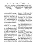
</div><span class="text_page_counter">Trang 23</span><div class="page_container" data-page="23"><b>Figure 2.1:The similar structure between native ECM and hydrogel.</b>
A-I: The morphology of dECM from A) bone [66], B) Skeletal muscle [67], C) Skin[68], D) Lung [14], E) Kidney [13], F) Heart [69], G) Placenta [12], H) Ovary [11], I) testis[70]. J) The illustration of the similarity between native ECM and hydrogel.
2.1.3 Injectable thermal responsive hydrogel – ideal performance for tissuer e g e n e r a t i o n
<i>2.1.3.1 Injectablehydrogel</i>
As an emerging hydrogel in tissue regeneration, hydrogels with injectability have been widely investigated [17, 19, 20, 22]. The concept behind injectable hydrogels is to inject a polymer solution into the treatment site and then allow it to gel (figure 2). Injectable materials can be introduced into the body much faster and have a better affinity for the host tissue [71-75]. Also, injectable formulations can fill a void or defect area with various shapes [10]. These injectable scaffolds typically comprise materials, cells, and biological signals (growth factor,
</div><span class="text_page_counter">Trang 24</span><div class="page_container" data-page="24">protein, drug, or bioactive compounds) to facilitate neo-tissue formation. Various studies with injectable hydrogel have shown advantages in inner tissue regenerative, including cardiovascular, cartilage, and bone, in terms of minimally invasive means [51, 54, 74-77]. Further, injectable hydrogel shows the best suitable performance for the new manufacturing processes [5]. The injectable hydrogel, which can inject through a needle or catheter, has become interested in this field [1, 20-22, 24].
<i><b>Figure 2.2: The illustration of non-injectable hydrogel and injectable hydrogel.</b></i>
The primary requirement of these materials is due to the necessity for polymer design [78]. The initial success of injectable systems is achieved through formulations that can undergo gelation in situ, injecting liquid polymer solutions into tissues, where they solidify [17, 18]. Dual-syringe devices for co-injecting two solutions that react to form a hydrogel upon mixing are commonly used [79]. However, the dual solutions require conditions, such as each component's concentration and the mixing solution's flow rate. The poor mixing can further contribute to heterogeneous gelation [78, 79]. Moreover, as injectable hydrogels form in the body, crosslinking methods are limited to biocompatible mechanisms feasible under physiological conditions [24, 60, 75, 80]. In Tosy for injection, the stimuli-responsive polymers are the strategy to design the single injection [21, 24]. Stimuli-responsive polymers have been engineeredtoundergo sol-gel transitions based on environmental factors such as temperature, pH, and ionic strength [17, 21, 24]. These polymers are designed to remain liquid under nonphysiological conditions (e.g., room temperature, acidic pH, salt-free) but solidify upon introduction into the body (e.g., 37 °C, neutral pH, millimolar salt concentration) [24, 60, 75,80].Whiletheseinjectablesystemshaveshownpromiseinanimalstudies[43,57,58,75,
</div><span class="text_page_counter">Trang 25</span><div class="page_container" data-page="25">80], challenges with gelation kinetics persist [78, 79]. Issues related to gelation kinetics may lead to solidification within the syringe or slow gelation, resulting in premature cargo release in vivo [1]. Despite these limitations, ongoing research aims to enhance the performance of injectablehydrogels.
<i>2.1.3.2 Thermal responsivehydrogel</i>
Temperature-sensitive hydrogels can be divided into positively and negatively thermosensitive, according to their temperature-sensitive structure of polymer [62, 76, 81-85]. Negative thermo-responsive hydrogels are identified by their lower critical solution temperature (LCST) [22, 24]. Below the LCST, the hydrogel structure relaxes, and above this temperature, the polymer in the hydrogel structure is compacted [86]. On the other hand, positive temperature-sensitive hydrogels are identified by their upper critical solution temperature (UCST) [24, 87]. Below UCST, the polymers compact, and above UCST, it relaxes [87]. Following the hydrogel structure, temperature-sensitive hydrogels can be divided into volume and sol-gel phases [59, 85]. The volume phase transition focuses on the hydrogel with the control degree of swelling based on temperature [85, 86, 88]. A sol-gel phase transition is the group of hydrogel that can transition from the solution phase to the gel phase as a function of temperature [89-94]. Among them, a thermosensitive hydrogel with reversible properties and LCST is one of the most widely applied systems for making injectable hydrogel [22, 23, 90, 91], which is the solution state upon injection at a lower temperature and transforms into a gel state immediately at body temperature [94,9 5 ] .
While temperature-responsive and injectable hydrogels exhibit significant promise in various biomedical applications, their preparation and injection processes still pose specific challenges [78, 79]. The polymer network in thermoresponsive hydrogels, and hydrogels in general, is established through polymerization reactions involving crosslinking agents that can be either non-biodegradable or biodegradable [88, 96]. Chemical or physical interactions are employed for crosslinking, impacting the final hydrogel properties and stability crucial for the intended application [97]. Chemical crosslinkers, forming covalent bonds, often yield stiffer hydrogels but may introduce some level of toxicity and may disrupt injectability [23]. Conversely, physical crosslinking through chain entanglement or secondary forces can result in hydrogels with reduced stiffness [22]. Careful consideration of material selection, polymerization reaction, and gelation conditions is imperative to ensure biocompatibility, particularly in injectable systems[15-18].
Thermoresponsive hydrogels can be designed to be biodegradable by incorporating weak bonds susceptible to hydrolysis or other forms of lysis, with cell metabolism and byproduct excretion influencing overall safety [1, 3, 17, 20, 22]. This modified hydrogel class
</div><span class="text_page_counter">Trang 26</span><div class="page_container" data-page="26">should be a freely flowing solution into a target region, undergoing in situ gelation in response to increasing temperatures above ambient values and conforming to the surrounding environment [56, 63, 87, 98]. Parameters such as hydrogel lower critical solution temperature (LCST), gelation rate, viscosity, and mechanical strength are crucial and should be characterized and tuned for biological applications [58, 84, 99]. For instance, adjusting polymer concentration and copolymerization ratio of hydrophobic and hydrophilic monomers can alter LCST and viscosity [43, 87]. Importantly, thermoresponsive hydrogels for tissue regeneration must possess mechanical stiffness matching native tissues to modulate target adhesion, proliferation, differentiation, and proper functioning [100]. Adequate mechanical stiffness is fundamental to withstand in vivo forces, ensuring structural integrity and transmitting physiological forces until the hydrogel is entirely replaced by native tissues, minimizing immune system response [17, 19, 23, 38, 79, 100]. Generally, hydrogel mechanical stiffness can be easily modulated by tuning polymer concentration, modifying polymer, and adjusting crosslinking density to achieve optimal mechanical properties for tissue regeneration applications [2, 3,10].
2.1.4 Emerging trend of injectable hydrogel developing from the hybrid system of polysaccharides and thermo-responsivepolymers.
<i>2.1.4.1 The advance of hybrid hydrogels developing from polysaccharides and thermo-responsivepolymers</i>
In recent years, a notable body of research has emerged discussing stimuli-responsive hydrogels that incorporate polysaccharides [101]. Integrating biopolymers with thermo-responsive materials has garnered significant attention across various fields [56, 63]. Polysaccharides constitute a substantial category of natural polymers featuring a linear or branched arrangement of sugar monomers connected through glycosidic linkages [102]. These polymers have been sourced from various origins, including plants (such as cellulose, starch, Arabic gum, pectin, guar gum), animals (hyaluronan, chitosan, heparin), microbes (pullulan, gellan, levan, dextran, xanthan), and algae (agar, alginate, carrageenan, fucoidan, agarose) [24, 42, 61, 86, 101]. In recent times, naturally derived polysaccharides have gained prominence over synthetic polymers due to their biocompatibility, biodegradability, low or negligible cytotoxicity, extensive diversity, cost-effectiveness, and approval by the US Food and Drug Administration (FDA) for a wide range of applications in pharmaceutical and food industry [61, 101, 103]. Additionally, their numerous active functional groups make them amenable to tailoring and chemical modifications [42, 103]. The intricate structures and high molecular weight of these polymers confer stability in harsh environmental conditions [61]. Furthermore, polysaccharides serve as rheological modifiers, influencing viscosity, gelling, elasticity, and stiffness properties, following their structure and molecular weight [52, 101]. Incorporating
</div><span class="text_page_counter">Trang 27</span><div class="page_container" data-page="27">Polysaccharide with thermal responsive polymer possesses resultant hydrogel, customized thermo-sensitive characteristics, and well-adaptation to the cell environment [24, 51].
For instance, PNIPAMisinherently a thermoresponsive hydrogel [85-87, 98]. However, using unmodified PNIPAM comes with several challenges, including low biodegradability, limited drug loading capacity, slow response to thermal stimuli, and insufficient mechanical strength [85, 98]. The abovementioned limitations have increased the research to adjust the hydrogels' properties and improve their position as an active system for different biomedicine and tissue engineering applications. Additionally, copolymers of PNIPAM and biopolymers generally yield hydrogels with increased strength and improved mechanical properties compared to homopolymer PNIPAM gels [103, 104]. While studies have been conducted to assess the biocompatibility of PNIPAM, most of these studies have focused on specific narrow applications [87]. The NIPAM monomer is toxic to humans, particularly to neural tissue; PNIPAM is generally considered cytocompatible [44]. However, hydrogels based solely on PNIPAM can produce structures unsuitable for tissue regeneration in terms of the resulting scaffold's pore size and mechanical properties [86, 98]. Incorporating natural polysaccharides in combination with PNIPAM aims to address these limitations and create materials that exhibit dual functionality by combining the properties of the thermal responsive feature and the biological/chemical feature of polysaccharides[ 8 6 ] .
Gupta et al. [105] proved the excellent performance of PNIPAM hydrogel after modification with carboxymethyl guar. The hybrid hydrogel exhibited a softer consistency than a pure PNIPAM hydrogel, which was easy to support cell viability and proliferation. Similarly, incorporating carboxymethyl cellulose (CMC) into the PNIPAM hydrogel introduced by Vasile et al. [70] showed an outstanding presentation in reversible thermal trigger gelling systems. They reported that the hybrid system showed a temperature-responsive sol-gel transition, which was not observed in the pure PNIPAM hydrogel. Along with thermal responsive property, the hybrid system exhibited a new function, a sensitive hydrogel system inheritedfromCMC[70].Inotherwords,theviscosityofthehybridsystemcouldbecontrolled by both temperature and pH, while this feature was lacking in the single system. In addition, the authors also noted the excellent biocompatibility of the hybrid hydrogel system rather than a single PNIPAM system. Surprisingly, preliminary enzymatic degradation studies indicated that the hybrid system consisting of PNIPAM and CMC could control the degradation by adding cellulases, while PNIPAM hydrogel couldnot.
The introduction of alginate (Alg) into the PNIPAM structure also achieved inventiveness [47, 88, 106]. An assessment of rheological data of this hybrid hydrogel showed the sol-gel transition as a function of temperature [88]. In particular, the stiffness of the
</div><span class="text_page_counter">Trang 28</span><div class="page_container" data-page="28">PINPAM hydrogel was improved following the addition of Alg. Additionally, Lee et al. [82] discovered that hybrid systems PNIPAM- Alg could self-assemble into micelle structures at temperatures close to physiological conditions by controlling the concentration of Alg. Furthermore, alterations in the molecular weight of the PNIPAM grafted onto the alginate backbone caused variations in the micelle morphology. The authors pointed out that these changes in morphology could have substantial implications for the copolymer's utility in applications like disease targeting, cellular uptake, and drug release.
Tan et al. conducted a study to explore the suitability of injectable Alg-based PNIPAM hydrogel systems for tissue engineering applications [96]. In this study, PNIPAM was grafted onto an aminated alginate (AAlg) backbone, resulting in copolymer AAlg-g-PNIPAM. The introduction of AAlg induced the shift of Critical gelation temperature (CGT) to 35 °C,
<i>makingitsuitableforinvivoapplications.Theviscosityofthehybridgelwassharplyincreased within the</i>
range of 34–36 °C, followed by a plateau, resulting in the easy process of cell incorporation. Human bone mesenchymal stem cells (hBMSCs) laden AAlg-g-PNIPAM gel showed significantly higher viability than native PNIPAM. Based on these findings, Tan et al. concluded that modifying PINIPAM with AAlg provided the potential material for tissue engineering applications [96]. Pentlavalli et al. conducted a similar investigation with a hybrid system, PNIPAM, and Alg [107]. They discovered that the hybrid gel did not exhibit cytotoxic effects on human osteosarcoma cells (MG63) and porcine bone marrow-derived mesenchymal stem cells (pBMSCs), while PNIPAM did. These studies collectively suggest that Alg-g- PNIPAM is a promising candidate for cell encapsulation and may find applications in injectable cell therapies or 3D bioprinting of cells[88, 96,1 0 7 ] .
Another instance of hydrogels incorporating polysaccharides with thermo-responsive polymers involves using Pluronic. Lin et al. [54] devised a hybrid system by combining Pluronic F127 with dextran, resulting in dextran-g-Pluronic F-127. The incorporation of dextran induced the more comprehensive CGT range of pluronic F127. Further, the degradation of pluronic F127 hydrogel was remarkably increased after modification with dextran. Similarly, Park et al. employed a coupling reaction to synthesize Chitosan-g-Pluronic F-127 [76, 81]. Both of these copolymers have demonstrated potential applications in the biomedical field, unlike pure Pluronic F127. Chitosan-g-Pluronic F-127, in particular, has shown promise as a carrier for cartilage regeneration [71, 80]. Chitosan-g-Pluronic F127 exhibited an LCST of approximately 25 °C, which could be adjusted by varying the amount of chitosan. However, using the single Pluronic F127 exhibited quick erosion, increasing viscosity and, consequently, reducing the viability of the encapsulated cells. Interestingly, Chitosan-g-Pluronic F127 was found to be biocompatible and capable of supportingt h e
</div><span class="text_page_counter">Trang 29</span><div class="page_container" data-page="29">proliferation of bovine chondrocytes[56]. The morphology of the Chitosan-g-Pluronic F127 hydrogel had more pore interconnection than pure F127, confirming a suitable environment for cell growth. All these findings suggest that combining polysaccharides into thermo- responsive materials results in the thermo-sensitive hydrogel as a promising technology for tissueregeneration.
<i>2.1.4.2 Technologies adopted in Hydrogels Based on Polysaccharides IncorporatedwithThermo-Responsive Polymers</i>
2.1.4.2.1 Physical strategy vs chemicalstrategy
Three techniques are available for producing hydrogels that combine polysaccharides with thermo-responsive polymers: physical blending, grafting approaches, and cross-linking techniques [24, 44, 51, 77, 81, 108]. In physical blending, ionic crosslinking and thermoregulation methods have created combinations of polysaccharides and thermo-sensitive polymers[65, 97]. However, the non-covalent interactions between polysaccharides and thermo-sensitive polymers result in less stable entities than those bound covalently. This instability can lead to phase separation within the resulting hydrogel[86].
Polysaccharides typically have a high crystalline level due to their abundance of intramolecular bonds[42, 101, 103]. As a result, polysaccharides often have low solubility, presenting a significant challenge when blending with another polymer[103]. The solubility of polysaccharides is frequently pH-dependent; for example, chitosan (CS) dissolves in acidic environments, while hyaluronic acid solubilizes in primary conditions. Their varying solubility complicates the mixing process, particularly in multi-step reaction procedures. To create a stable structure for thermo-sensitive hydrogels, it is necessary to employ two independent cross-linked polymer systems that form a single polymeric network[24, 42, 86, 103]. In the following section, chemical methods, including grafting approaches and chemically induced cross-linking techniques, will be discussed.
2.1.4.2.2 Graftingapproach
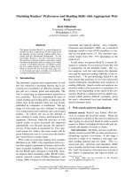
Graft copolymerization is a common strategy for preparing materials for a hybrid hydrogel system[61, 103]. To synthesize polysaccharide-polymer conjugates, three primary routes are available: the "graft-to," "graft-from," and "graft-through" strategies[103] (figure 2.3). In brief, the "graft-to" strategy involves grafting pre-existing polymers onto polysaccharides through a one-step process. The "graft-from" strategy entails the continuous in situ growth of polymer chains from the surfaces of polysaccharides or the utilization of pre-existing polymers as initiators. The "graft-through" strategy involves attaching polymerizable groups to polysaccharides, followed by polymerization to generate polymer-grafted polysaccharides.
</div><span class="text_page_counter">Trang 30</span><div class="page_container" data-page="30"><i><b>Figure 2.3: Graft polymerization techniques.</b></i>
Adapted from the reference [41]
Polymerization techniques play a crucial role in these strategies[53]. Controlled polymerization techniques, such as atom transfer radical polymerization, reversible addition−fragmentation chain-transfer polymerization, nitroxide-mediated polymerization, and ring-opening metathesis polymerization, provide effective means to produce polymer- modified polysaccharides with tailored structures and properties[53, 108]. The reduced viscosity of the hybrid hydrogel barely changed over a 40°C interval, attributed to the uncharged nature of the CMC backbone at low pH values [109]. This method does not guarantee the presence of the desired functional group at the end of every polymer chain. On the other hand, the direct polymerization of functional monomers can yield high functional group densities throughout the polymer backbone, which can be further utilized for preparing polymer brushes or conjugating sensor moieties for susceptible chemical detection[110, 111]. This approach ensures the desired functional group's presence in each polymer backbone's repeat unit. However, employing protective measures for the functional group may be necessary if it hinders the polymerization process. Additionally, the desired functional monomer may not always be commerciallyavailable[108].
To solve these obstacles, a pre-required modification of the polymers using bifunctional reagents is often necessary[110]. These reagents are used as linkers, and they might bring new
</div><span class="text_page_counter">Trang 31</span><div class="page_container" data-page="31">Functionalities into molecules or act just as spacers, improving the spatial availability of functional groups[111, 112]. Functional modification of polymer molecules with high selectivity or specificity is needed. A suitable method for functional modification should be generally applied. This approach involves the attachment of a synthetic polymer, exhibiting a well-defined end-functionality to a complimentary functionality ofp o l y s a c c h a r i d e .
Various coupling techniques have been developed to desire the reactive groups, as illustrated in Figure2.4.
<i><b>Figure 2.4: The illustration of the coupling chemistry for different reactive</b></i>
<i>functionalgroups of thepolymers.</i>
<i>Adapted from the references [10, 84-92]</i>
The carboxylate groups can use carbodiimide derivatives as the coupling agents to make conjugated bonds with nucleophilic functional groups such as primary amino groups, hydroxyl groups,…[113]. The carbodiimide coupling agents include N, N′-dicyclohexylcarbodiimide (DCC), Carbonyldiimidazole (CDI), 1-ethyl-3-(3-dimethylaminopropyl) carbodiimide (EDC) [24, 82, 96, 113]. DCC and CDI can used in organic solvents, while EDC is used in water solvents only. The introduction of a carbodiimide bond would provide a good leaving group; therefore, the compound containing primary Amine/hydroxyl could form the amide, esters,
</div><span class="text_page_counter">Trang 32</span><div class="page_container" data-page="32">ureas, etc, with the carbodiimide-activated groups following the nucleophilic substitution [114-115]. To increase the yield of the reaction, the strong nucleophilic components such aspyridine (in organic solvent), 2-chloro-1-methylpyridinium iodide, 4-(N, N′-dimethylamino)pyridine (DMAP), triethyl amine,.. should be supported. In terms of EDC, the use of N- hydroxysuccinimide (NHS)ispreferred. EDC forms the O-acylisourea intermediate, which is very sensitive to nucleophilic functional groups. The reaction occurs in aqueous physiological pH, while the o-acylisourea intermediate is unstable in this condition. Therefore, N-hydroxysuccinimide (NHS)isrecombined in this case [116]. Another strategy to activate carboxylic groups is the use of the tetrabutylammonium (TBA) salt, specialized in the case of polysaccharides. With the help of TBA, polysaccharides can react in the organic solvent. Also, with the help of TBA, these carboxylate groups can be activated with alkyl halides[114]. Additionally, hydrazination reactions have been utilized for efficiently conjugating the carboxyl groups of polysaccharides to polymer-containing amino or hydroxyl functional groups[115].
There are many methods to improve the nucleophilicity of hydroxyl groups. The introduction of the arylation or acylationisgenerally focused due to the ease of reaction with the amine compounds. However, this strategyisonly available to allyl alcohols or β-amino alcohols. The use of phosgene to increase the nucleophilicity of the hydroxyl group has been introduced in some research [116, 117]. Although phosgene creates chloroformate intermediates sensitive to amino groups, phosgene is very toxic and can make the cyclic [116]. Another strategy is the use of p-nitrophenyl chloroformate. The hydroxyl group formsaromatic carbonate esters with p-nitrophenyl chloroformate. This bond will be cleavage in the presentation of amino groups. Cyanogen halide activation is another method employed for the covalent attachment of amine polymers to the polysaccharide backbone via hydroxyl [118]. The reactive cyanate esters in this reaction may undergo hydrolysis to yield inert carbamates or rearrange to produce intra-/interchain and cyclic imidocarbonate structures. The parallel reactions of these products with the amine polymer can form an isourea derivative[119]. Moreover, dialdehyde polysaccharides obtained after periodate oxidation can form Schiff bases when conjugated with amine polymers[120]. Subsequent reduction using sodium borohydride can enhance the stability of these conjugates[121]. Another approach involves activating polysaccharides using sulfonyl chlorides to create corresponding sulfonyl esters, which can react with polymers containing carboxylic or aminog r o u p s [ 1 2 2 ] .
Rather than direct conjugation, introducing spacers showed some advances [113]. Introducing these spaces helps support the new function of polysaccharides, including the biological functions and/ or solubility [123]. Also, with spacer groups, the reduction in the
</div><span class="text_page_counter">Trang 33</span><div class="page_container" data-page="33">yield of conjugation due to the steric hindrance following the self-assembly of the polysaccharide could be solved [124, 125]. The introduction of spacer groups could use the coupling agent presented in Figure 2.4 [126]. The amino acids and peptides are typically used as spacers[124]. Further, by the introduction of spacers, the degradation could be controlled by external stimuli such as pH, redox,…[127]
Generally, the different routes reported for the grafting approach using coupling chemistry. They can be typically classified into some routes: (i) identify the functional groups at the grafting site, (ii) select the complementary functional groups on the crosslinker, and then (iii) select the activating methods. For this method, copolymer solution creates only physically interconnected, without any chemical bonding, so individual components retain their original properties [103-105, 107, 108, 128].
2.1.4.2.3 Cross-linkingtechnique
Recently, crosslinking technology has gained significant attention as an effective method for developing thermal-responsive hydrogels that incorporate polysaccharides[24, 86]. Like the grafting approach, covalent linkages between polymer chains can be established by reacting functional groups with complementary reactivity[76, 112, 129]. Derived from the fabrication of in situ-forming hydrogels, this approach connects the functional groupsofpolymer chains via supramolecular interactions (such as hydrogen bonding, ionic bonding, etc.) or covalent bonding. Initially, the polysaccharide backbone undergoes chemical pretreatment to introduce active groups, such as catechol or phenol[130]. These treatments create crosslinking regions, initiating the termination phase and resulting in crosslinking between differentpolymers[131].
The chemical crosslinking mediated hydrogel can synthesized under click chemistry, Schiff base, photo-crosslinking, Michael-type addition reaction, and redox reaction[130, 131]. Among them, redox reactions have recently emerged as a promising triggering motif for designing a novel type of responsive polymers[132]. By introducing the redox reactive moieties on the polymer backbone, two polymers are spontaneously cross-linked in thepresentation of the oxidative reagent [131, 132], such as H<small>2</small>O<small>2</small>or O<small>2</small>. Phenolic moieties (e.g.,tyrosine, catechol, and polyphenols) are commonly targeted for thism e t h o d [ 1 3 3 ] .
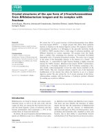
</div><span class="text_page_counter">Trang 34</span><div class="page_container" data-page="34"><i><b>Figure 2.5:Crosslinking mechanisms of catechol‐conjugated polymers for</b></i>
<i>Blue circle: Noncovalent crosslinking, orange circle: Covalent crosslinking.Adaptedfrom the reference [134].</i>
Catechol oxidation chemistry has demonstrated an excellent example of a hybrid network between polysaccharides and thermally responsive polymers [139-140]. The oxidative polymerization of catechol forms the crosslinking between two polymers [76, 112, 129]. This allows for control over the ratios of the two networks and is less time-consuming since diffusion is not required. It provides a straightforward, one-pot method for hydrogel formation (figure 2.5).
Inspired by this strategy, Park and colleagues[135] developed an injectable hybrid hydrogel from polysaccharide (hyaluronic acid) and thermal responsive polymer (Pluronic). This study modified hyaluronic acid with catechol while thiol groups from cysteamine were introduced at the end group of pluronic F127. Via the Michael addition reactions between thiol and catechol, the crosslinking between hyaluronic acid and pluronic F127 was formed,
</div><span class="text_page_counter">Trang 35</span><div class="page_container" data-page="35">resulting in a cross-linked adhesive hydrogel. Taking the same idea, Park et al. [130]developed a hybrid hydrogel-based pluronic with another polysaccharide, chitosan. Both precursors were soluble and could be pre-mixed at 4°C, and the resulting viscous solution rapidly solidified into a gel state when the temperature was raised to 37°C. In addition, chemical and physical crosslinking induced the resultant hydrogel with high mechanical features compared to the thermal responsive hydrogel based on physical crosslinking only[ 1 2 9 - 1 3 0 ] .
The formation of the crosslinking between catechol motifs could be based on their auto-oxidation, catalyzed by native oxygen molecules, to facilitate catechol oxidation without additional supplements [76, 112, 136]. Polymers modified with catechol or tri-hydroxyl phenol residues (e.g., gallon, polyphenols) can be quickly injected into native tissue and form the hydrogel stage without adverse effects[137]. However, the reaction kinetics are relatively slow and highly dependent on environmental factors such as pH. This approach is not suitable for applications requiring rapid crosslinking[136]. Adding oxidative compounds such as sodium periodate could enhance the crosslinking kinetic. The crucial aspect of the crosslinkingstrategy relies on the oxidation of tyrosine and catechol to ortho-quinones[137]. Ortho-quinones possess high electrophilicity, enabling their participation in various reactions, including Schiff- base reactions, Michael-type additions, and coupling reactions[137, 138]. In the Schiff-base reaction, an electron donor group, such as a primary amine, attacks the C=O bond in the ortho- quinone. This results in the formation of ortho-quinone imines containing a C=N bond. When ortho-quinone acts as an electrophilic acceptor, the primary amine and thiol groups can attach to the meta-position (1,4-Michael reaction) and ortho-position (1,6-Michael reaction). Additionally, ortho-quinone can interact with neighboring catechol groups, leading to coupling reactions[137, 138]. In the case of sodium periodate-mediated oxidation, catechol-periodate intermediates break down to form ortho-quinone. However, there are limitations to using sodium periodate-mediated crosslinking in tissue engineering. Firstly, the reaction occurs under alkaline pH conditions. Secondly, sodium periodate causes a ring-opening reaction in polysaccharides (e.g., alginate, hyaluronic acid), producing cytotoxic aldehyde groups. Lastly, sodium periodate itself possesses intrinsic cytotoxicity[137,138].
Biological peroxidase enzymes are known to regulate this process, with various types of peroxidase enzymes demonstrating this function[132, 138, 139]. Horseradish peroxidase (HRP) offers advantages over other peroxidases due to its narrow range of phenol coupling reactions [139]. HRP-mediated coupling is limited to the coupling reaction, as it cannot occur with primary amine and thiol groups[140]. HRP is an enzyme containing heme derived from a plant source[141-143]. The heme group in the HRP structure captures the oxygen moleculesfromt h e h y d r o g e n p e r o x i d a s e ( ( H<small>2</small>O<small>2</small>),f o r m i n g t h e h e m g r o u p s w i t h h i g h o x i d a t i o n s t a t e s .
</div><span class="text_page_counter">Trang 36</span><div class="page_container" data-page="36">This Fe (IV) = O initiates the phenolic oxidative reaction. Indeed, Fe (IV) = O takes the hydrogen atom from the phenolic motif, forming the highly reactive phenolic radical. The complex oxidizes phenolic moieties from the couple via a C-O bond between the ortho-carbon of phenolic groups and the phenolic radical and a C-C bond between the ortho-carbon of phenolic groups[132]. Despite the advantage of HRP in the redox reaction for making hydrogel, there are significant concerns associated with HRP due to an immunological response in some animal studies[143,144].
An alternative to the HRP enzyme is peroxidase-mimicking enzymes[142, 143]. When designing these peroxidase-mimicking catalysts, it's essential to consider the peroxidase-like spatial arrangement of the hemin ligands and activating groups, which serve as the proximal and distal residues of heme[142]. This approach aims to enhance control over the oxidative catechol crosslinking process, addressing issues such as pH sensitivity and immune responses[145]. Ultimately, it enables the broader utilization of catechol oxidative chemistry as a crosslinking strategy to incorporate polysaccharides with thermal-sensitive polymers,
It's important to emphasize that in the context of thermal-responsive hydrogels incorporating polysaccharides, the primary goal of crosslinking reactions is to enhance hydrogel strength without compromising the phase transition due to the presence of polysaccharides[24, 86]. Unlike grafting techniques, crosslinking methods focus on establishing stable polymeric networks, forming the hydrogel structure. In other words, these methods randomly create covalent bonds between adjacent polymer chains. Typically, polysaccharides can engage in chemical crosslinking with each other, while thermal- responsive polymers are part of the mix. However, the heterogeneity within the networks may disrupt the thermal-responsive property. Therefore, the challenge remains to create thermoresponsive hydrogels incorporating polysaccharides using crosslinking techniques with an entirely predictable and controllable phase transition temperature.
<b>2.2 Encoding the hydrogel for specific tissueregeneration</b>
2.2.1 The stiffness of thehydrogel
Understanding the vital role of hydrogel stiffness in guiding tissue regeneration [100], it is evident that initial interactions between cells and the scaffold surface, mediated by focal adhesions with the matrix [146-153], trigger the recruitment and activation of proteins Associated with mechano-signaling pathways [154-157]. This cascade of events leads to adaptive cellular responses, encompassing gene and protein expression changes, influencing cellgrowthanddifferentiation[100,154,156,158].Asrevealedinpriorstudies,the
</div><span class="text_page_counter">Trang 37</span><div class="page_container" data-page="37">interconnection between hydrogel stiffness and cellular response is pivotal in substrate-guided tissue regeneration [155, 156, 158]. For example, in the case of wound healing, the change in the hydrogel stiffness induced the various regenerative capacity [100]. Medium stiff hydrogels (~10<small>3</small>Pa ) have been shown to mitigate scar formation, enhance wound healing, and facilitate myofibroblast transformation, keratinocyte proliferation, extracellular matrix synthesis, and remodeling. The impact of hydrogel stiffness extends to the secretion of crucial factors like TGF-β1 and bFGF, critical players in skin wound healing. This correlation suggests that the therapeutic effects of hydrogels in skin wound healing can be finely tuned by adjusting the stiffness of the hydrogel. Hydrogel stiffness regulates cell behavior and fate, while cells reciprocally contribute to the remodeling of the substrate, influencing the overall tissue regeneration process [100, 156, 158]. In addition, in the human body, different tissues exhibit differences in rigidities, as demonstrated in Table 2.1. Therefore, careful consideration is essential to ensure optimal outcomes when designing hydrogels tailored for specific tissue regeneration applications [155, 157].
<i><b>Table 2.1:The stiffness of living tissues.</b></i>
</div><span class="text_page_counter">Trang 38</span><div class="page_container" data-page="38">2.2.2 The biologicalcues
In tissue engineering, along with stiffness, manipulating the microenvironment with biological cues, such as mitogens, growth factors, and morphogens, is crucial for guiding cells in tissue regeneration [32, 33, 165-168]. These cues can activate specific signaling pathways or sets of genes to direct and control cellular responses [134, 169-173]. Hydrogels have been specifically engineered to deliver various growth factors or chemoattractant signals locally, aiming to recruit stem cells and expedite healing [174-177]. It is essential to highlight that the scaffold's appropriate design is paramount for programmed morphogenesis in the intricate environment where neotissue formation occurs [10, 24, 36, 38] (figure 2.6). Careful consideration of signals' spatial and temporal evolution throughout the regeneration process is necessary [8, 10, 18], involving predicting transport phenomena and monitoring mass transport parameters [15, 19, 21, 23, 35]. The selection of biological cues should align with the pathology of the damaged tissue [168, 172, 178-181], emphasizing the importance of designing systems that can provide these biological cues in a time-controlled manner to mimic thenormal healing process [182] closely. This section will further discuss this concept, focusing on selecting biological cues for diabetic wound healing and boner e g e n e r a t i o n .
<i><b>Figure 2.6:The illustration of the function of biological cues in developing</b></i>
<i>thefunctional scaffold.</i>
<i>To activate resident cells for the regeneration process, the ideal design of thescaffoldfollows the addition of the homing factors to guide cells. These factors would (1)release from the hydrogel scaffold, (2) go into the bloodstream, and (3) make these signals toguide the</i>
</div><span class="text_page_counter">Trang 39</span><div class="page_container" data-page="39"><i>migration of cells and to guide the difference process of cells in support of new tissue growth(4).</i>
<i>2.2.2.1 Diabeticwound</i>
2.2.2.1.4 The wound healing process and the alterations in diabeticc o n d i t i o n s
The wound healing process includes several sequential phases: inflammation, proliferation, granulation, and tissue remodeling [28, 167, 169, 180, 183]. It is a complex cellular response to injury involving the activation of various cell types, including fibroblasts, endothelial cells, macrophages, and platelets [3, 19, 24, 36]. The initial step in wound healing is clot formation, which recruits fibroblasts and immune cells to the injured area [36]. The inflammatory phase, lasting approximately four days, is significantly influenced by macrophages [35, 36]. Macrophages migrate to the wound, explicit necrotic material, and produce factors that induce angiogenesis by endothelial cells, epithelialization by keratinocytes, and matrix deposition by fibroblasts, leading to the reconstruction of ECM [36]. Subsequently, re-epithelialization involves migrating epithelial cells and keratinocytes across the wound barrier and granulation tissue[35].
Nitric oxide, a gaseous molecule, is widely recognized in treatments for wound healing [170, 171, 184-187]. Typically, NO is involved in the natural process of wound healing [184-187]. During hemostasis, NO is produced by endothelial cells to activate the cyclic c-GMP pathway; consequently, it prevents platelet aggregation and then increases vasodilation[169, 171, 187]. The vasodilation helps to increase the blood flow to the wound site and then increases the influx of inflammatory cells[35]. The concentration of NO at the wound siteisincreased due to the activation of iNOS in neutrophils and macrophages[36]. This way, the phagocytosis pathway is increased to clean the wound site[187, 188]. In the inflammatory phase,M1macrophages produce significant amounts of NO (>0.5mM) to combat microbes and prevent wound infection [188]. As the wound transitions to the proliferation stage, NO levels decrease significantly (0.01-0.25mM) [188, 189]. M1 macrophages convert to M2 macrophage cells, which do not produce NO [36]. During this phase, NO production depends on fibroblasts and keratinocytes, stimulating the proliferation and migration of more keratinocytes within the wound [188,189].
In a diabetic state, persistent hyperglycemia triggers the overproduction of superoxide through various pathways, including the polyol pathway, xanthine and NADPH oxidases, cyclooxygenases, uncoupled nitric oxide synthase, glucose auto-oxidation, and the mitochondrial respiratory chain [188-191]. This results in elevated levels of reactive oxygen species (ROS) and diminished antioxidants, defining a state of oxidative stress [192]. The
</div><span class="text_page_counter">Trang 40</span><div class="page_container" data-page="40">excess superoxide activates the NF-κB transcription factor, leadingB transcription factor, leadingtothe upregulation NOS- 2 and subsequent nitric oxide (NO) production [190, 193]. In the presence of high superoxide and low levels of superoxide scavenger molecules, NO rapidly reacts with superoxide to generate peroxynitrite [192], thereby reducing the bioavailability of NO [188, 189]. Additionally, heightened ROS and peroxynitrite levels in hyperglycemia limit the production ofBH4(anessentialco-factorforNOproduction)andinducetheuncouplingofNOS,resulting in superoxide production instead of NO [192]. Another contributing mechanism is the concentration of arginase, which competes with NOS enzymes for the substrate arginine, reducing NO availability and favoring superoxide production [194]. In diabetic wounds, increased arginase activity levels were observed, correlating with delayed healing in various studies[ 2 8 , 1 6 6
-1 6 8 , -1 7 6 , -1 8 -1 , -1 9 4 , -1 9 5 ] . T h e N O d e f i c i e n c y d i s r u p t s e n d o t h e l i a l v a s c u l a r function, impacting vessel functionality [190, 192, 193].
Furthermore, in diabetic patients with elevated cholesterol levels, peroxynitrite promotes lipid peroxidation and triggers an inflammatory response, leading to platelet aggregation [196]. These effects are particularly pronounced in small blood vessels, contributing to the progression of neuropathy [190, 192, 194]. Vascular impairment can result in persistent ischemia and wounds in diabetic individuals are often associated with low oxygen tension [197]. Under physiological conditions, cells express HIF-1α, a pivotal transcription factor enabling cells to adapt to hypoxic conditions by promoting angiogenesis, cell proliferation, survival, and energy metabolism [196]. Nitric oxide (NO) is a crucial regulator in expressing HIF-1α [191, 192]. During the initial phases of wound healing, when M1 macrophages are predominant, the high NO production from cells may facilitate the stabilization of HIF-1α to generate an adaptive response to injury [193, 194, 197]. As the resolution phase begins, lower NO levels from other cells resolve the wound environment and restore its normal state [188, 189]. Fibroblast-produced NO regulates collagen production and may act as the initial response before immune cells arrive at the wound site [190].LowNO levels influence extracellular matrix remodeling/turnover [192, 193]. The dynamic modulation of NO doses at the wound site over the healing process ensures the expected production and regulation of growth factors to maintain cell phenotype and function at each stage [189-191]. Duetoitsreactivityandshortlifespan,NO'soveralleffectoncellsisinfluencedbyaggravating factors, with mechanisms in place to maximize its biological effect for an appropriate host response [171, 184, 187, 198]. Maintaining nitroso-redox balance is crucial, and any persistence of stimuli or inadequate feedback can disrupt the equilibrium of the wound environment, which is characteristic of dysregulated healing in diabetic wounds[199].
</div>