- Trang chủ >>
- Y - Dược >>
- Y học gia đình
case report tắc động mạch cảnh đến muộn qua chụp ct angiography
Bạn đang xem bản rút gọn của tài liệu. Xem và tải ngay bản đầy đủ của tài liệu tại đây (12.28 MB, 33 trang )
<span class="text_page_counter">Trang 1</span><div class="page_container" data-page="1">
CASE REPORT:
TẮC ĐỘNG MẠCH CẢNHĐẾN MUỘN QUA CHỤP
CT ANGIOGRAPHY
BS. PHÙNG TRỌNG KIÊNP.K.T.Q – T.T.Y.K MEDIC
MEDIC 5 (25/04/2024)
</div><span class="text_page_counter">Trang 2</span><div class="page_container" data-page="2">NỘI DUNG
Bài báo cáo trọng tâm vào:
- Trình bày hình ảnh tổn thương não do tắcđm cảnh qua chụp CT angiography.
- Khả năng phát hiện, chỉ định và chống chỉđịnh của CT angiograpy)
TỔNG QUANBỆNH ÁN
BÀN LUẬNKẾT LUẬNTHAM KHẢO
</div><span class="text_page_counter">Trang 3</span><div class="page_container" data-page="3"><small>(1): class="text_page_counter">Trang 4</span><div class="page_container" data-page="4">
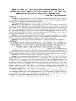
TỔNG QUAN
<small>(3): class="text_page_counter">Trang 5</span><div class="page_container" data-page="5">
TỔNG QUAN
<small>(5): angiography-mra-cerebral-artery-common-338843604.html</small>
</div><span class="text_page_counter">Trang 6</span><div class="page_container" data-page="6">QUAN
Uses of CT Angiography of the Head:
Arteriovenous malformations.Subdural hematoma.
Narrowing or blockage of arteries.
Rupture or tear in the wall of a blood vessel.Brain aneurysm.
Brain tumors.
<small>(6): class="text_page_counter">Trang 7</span><div class="page_container" data-page="7">
TỔNG QUAN
Benefits of CT Angiography of the Head:
Painless and non-invasive procedure.
Produces highly detailed images of the blood vessels of the brain and associated cranial structures.
Less sensitive to patient movement as compared to an MRI.Fast and simple procedure that can detect internal injury or bleeding within the brain quickly.
<small> class="text_page_counter">Trang 8</span><div class="page_container" data-page="8">
TỔNG QUAN
Indications for CT Angiography (CTA) of the Head:
CT Angiography (CTA) of the head may be ordered to investigate symptoms such as:Loss of consciousness.
Double vision or blurry vision.
Altered sensation in the face or scalp.Transient ischemic attacks.
Chronic headaches.Hearing loss.
</div><span class="text_page_counter">Trang 9</span><div class="page_container" data-page="9">TỔNG QUAN
CT scanning has no absolute contraindications:
Repeated X-ray exposure may increase the patient's risk for cancer. However, the risk from any one scan is small, with the benefit of an accurate diagnosis outweighing the risks.
Contrast media is safe in most patients; however, adverse reactions may range from mild to severe.
Caution is indicated in pregnant women, particularly during the first trimester to avoid fetal abnormalities.
It is recommended that nursing mothers should wait 24 hours after contrast administration to resume breastfeeding.
The kidneys may be injured due to the contrast material. In most cases, the kidneys will regain their normal function within five to seven days.
</div><span class="text_page_counter">Trang 10</span><div class="page_container" data-page="10">BỆNH ÁN 1
<small>10</small>
</div><span class="text_page_counter">Trang 11</span><div class="page_container" data-page="11">BỆNH ÁN 1
<small>11</small>
</div><span class="text_page_counter">Trang 12</span><div class="page_container" data-page="12">BỆNH ÁN 1
<small>12</small>
</div><span class="text_page_counter">Trang 13</span><div class="page_container" data-page="13">BỆNH ÁN 1
<small>13</small>
</div><span class="text_page_counter">Trang 14</span><div class="page_container" data-page="14">BỆNH ÁN 1
<small>14</small>
</div><span class="text_page_counter">Trang 15</span><div class="page_container" data-page="15">BỆNH ÁN 1
<small>15</small>
</div><span class="text_page_counter">Trang 16</span><div class="page_container" data-page="16">BỆNH ÁN 1
<small>16</small>
</div><span class="text_page_counter">Trang 17</span><div class="page_container" data-page="17">BỆNH ÁN 1
<small>17</small>
</div><span class="text_page_counter">Trang 18</span><div class="page_container" data-page="18">BỆNH ÁN 1
<small>18</small>
</div><span class="text_page_counter">Trang 19</span><div class="page_container" data-page="19">BỆNH ÁN 1
<small>19</small>
</div><span class="text_page_counter">Trang 20</span><div class="page_container" data-page="20">BỆNH ÁN 2BỆNH ÁN 2
<small>20</small>
</div><span class="text_page_counter">Trang 21</span><div class="page_container" data-page="21">BỆNH ÁN 2
<small>21</small>
</div><span class="text_page_counter">Trang 22</span><div class="page_container" data-page="22">BỆNH ÁN 2
<small>22</small>
</div><span class="text_page_counter">Trang 23</span><div class="page_container" data-page="23">BỆNH ÁN 2
<small>23</small>
</div><span class="text_page_counter">Trang 24</span><div class="page_container" data-page="24">BỆNH ÁN 2
<small>24</small>
</div><span class="text_page_counter">Trang 25</span><div class="page_container" data-page="25">BỆNH ÁN 2
<small>25</small>
</div><span class="text_page_counter">Trang 26</span><div class="page_container" data-page="26">BỆNH ÁN 2
<small>26</small>
</div><span class="text_page_counter">Trang 27</span><div class="page_container" data-page="27">BỆNH ÁN 2
<small>27</small>
</div><span class="text_page_counter">Trang 28</span><div class="page_container" data-page="28">BỆNH ÁN 2
<small>28</small>
</div><span class="text_page_counter">Trang 29</span><div class="page_container" data-page="29">BỆNH ÁN 2
<small>29</small>
</div><span class="text_page_counter">Trang 30</span><div class="page_container" data-page="30">Số lượng bệnh nhân đột quỵ mới trong năm khoảng 200.000 trường hợp.
Trong những nguyên gây đột quỵ, hẹp động mạch cảnh do xơ vữa chiếm khoảng 15%. (7)
Siêu âm doppler sơ bộ phát hiện mức độ tổn thương XVĐM, còn CT angiography cóđánh giá tổn thương động mạch và não chi tiết hơn với hình ảnh rõ nét.
</div><span class="text_page_counter">Trang 31</span><div class="page_container" data-page="31">BÀN LUẬN
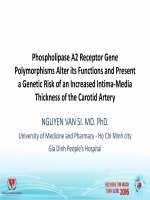
Bệnh nhân 2:- Tắc động mạch cảnh trong bên P sau chỗ chia đôi 10mm đến đến đa giác Willis +giảm tuần hoàn bán cầu não bên P.
- Bn được chuyển đến Bv 115, sau đó xin về điều trị tiếp tục tại Bv SIS Cần ThơCả 2 bn đều đến muộn, đã qua thời gian vàng, chỉ điều trị nội khoa.
Phát hiện tắc động mạch não đến muộn, tuy không giúp bn được can thiệp cấp cứu tuynhiên cũng giúp đánh giá được tổn thương não để có hướng điều trị lâu dài, cải thiệnđược chất lượng sống cho bn.
Các Bs lâm sàng cần nắm chắc các chỉ định chụp CT angiography cổ + não đã nêutrong phần tổng quan để có thể phát hiện sớm tắc đm cổ và não.
Cộng tác truyền thông y học: giúp bn nhận biết sớm các dấu hiệu đe dọa hoặc tai biếnsớm để đến bệnh viện còn trong thời gian vàng can thiệp cấp cứu.
<small>31</small>
</div><span class="text_page_counter">Trang 32</span><div class="page_container" data-page="32">CT angiography chụp mạch vành, mạch não, mạch chi… là thế mạnh củaTTYK Medic với máy MSCT 640 và các Bs hình ảnh có kinh nghiệm./.
<small>32</small>
</div><span class="text_page_counter">Trang 33</span><div class="page_container" data-page="33">THAM KHẢO
<small>(1):